
 IHC (Immunohiostchemistry)
(Figure 2. IHC analysis of TJP1 using anti-TJP1 antibody.TJP1 was detected in paraffin-embedded section of human intestinal cancer tissue. Heat mediated antigen retrieval was performed in citrate buffer (pH6, epitope retrieval solution) for 20 mins. The tissue section was blocked with 10% goat serum. The tissue section was then incubated with 1ug/ml rabbit anti-TJP1 Antibody overnight at 4 degree C. Biotinylated goat anti-rabbit IgG was used as secondary antibody and incubated for 30 minutes at 37 degree C. The tissue section was developed using Strepavidin-Biotin-Complex (SABC) with DAB as the chromogen.)
IHC (Immunohiostchemistry)
(Figure 2. IHC analysis of TJP1 using anti-TJP1 antibody.TJP1 was detected in paraffin-embedded section of human intestinal cancer tissue. Heat mediated antigen retrieval was performed in citrate buffer (pH6, epitope retrieval solution) for 20 mins. The tissue section was blocked with 10% goat serum. The tissue section was then incubated with 1ug/ml rabbit anti-TJP1 Antibody overnight at 4 degree C. Biotinylated goat anti-rabbit IgG was used as secondary antibody and incubated for 30 minutes at 37 degree C. The tissue section was developed using Strepavidin-Biotin-Complex (SABC) with DAB as the chromogen.)
Rabbit Tight junction protein ZO-1 Polyclonal Antibody | anti-TJP1 antibody
Anti-TJP1 Antibody
Each vial contains 5mg BSA, 0.9mg NaCl, 0.2mg Na2HPO4, 0.05mg NaN3.
IHC (Immunohiostchemistry)
(Figure 2. IHC analysis of TJP1 using anti-TJP1 antibody.TJP1 was detected in paraffin-embedded section of human intestinal cancer tissue. Heat mediated antigen retrieval was performed in citrate buffer (pH6, epitope retrieval solution) for 20 mins. The tissue section was blocked with 10% goat serum. The tissue section was then incubated with 1ug/ml rabbit anti-TJP1 Antibody overnight at 4 degree C. Biotinylated goat anti-rabbit IgG was used as secondary antibody and incubated for 30 minutes at 37 degree C. The tissue section was developed using Strepavidin-Biotin-Complex (SABC) with DAB as the chromogen.)
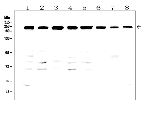
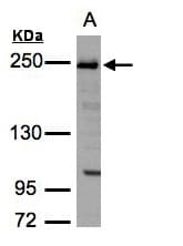
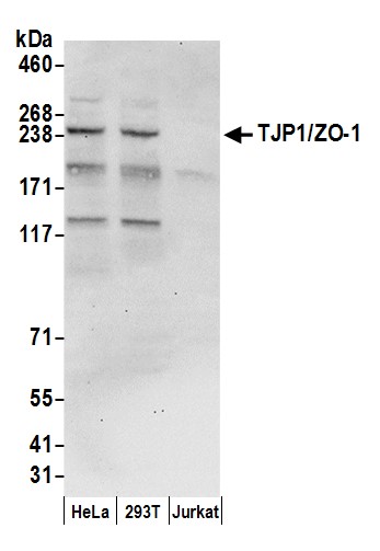
WB (Western Blot)
(Figure 1. Western blot analysis of TJP1 using anti-TJP1 antibodyElectrophoresis was performed on a 5-20% SDS-PAGE gel at 70V (Stacking gel) / 90V (Resolving gel) for 2-3 hours. The sample well of each lane was loaded with 50ug of sample under reducing conditions.Lane 1: human A431 whole cell lysates,Lane 2: human Caco-2 whole cell lysates,Lane 3: human U2OS whole cell lysates,Lane 4: human K562 whole cell lysates.Lane 5: human PC-3 whole cell lysates,Lane 6: human T-47D whole cell lysates,Lane 7: rat ovary tissue lysates,Lane 8: mouse ovary tissue lysates.After Electrophoresis, proteins were transferred to a Nitrocellulose membrane at 150mA for 50-90 minutes. Blocked the membrane with 5% Non-fat Milk/ TBS for 1.5 hour at RT. The membrane was incubated with rabbit anti-TJP1 antigen affinity purified polyclonal antibody at 0.5 ug/mL overnight at 4 degree C, then washed with TBS-0.1%Tween 3 times with 5 minutes each and probed with a goat anti-rabbit IgG-HRP secondary antibody at a dilution of 1:10000 for 1.5 hour at RT. The signal is developed using an Enhanced Chemiluminescent detection (ECL) kit with Tanon 5200 system. A specific band was detected for TJP1 at approximately 220-240KD. The expected band size for TJP1 is at 195KD.)
Background: Tight junction protein ZO-1 is a protein that in humans is encoded by the TJP1 gene. It is mapped to 15q13.1. This gene encodes a protein located on a cytoplasmic membrane surface of intercellular tight junctions. The encoded protein may be involved in signal transduction at cell-cell junctions. It has been found that injected CagA associates with the epithelial tight-junction scaffolding protein TJP1 and the transmembrane protein junctional adhesion molecule, causing an ectopic assembly of tight junction components at sites of bacterial attachment, and altering the composition and function of the apical-junctional complex.
NCBI and Uniprot Product Information
Customer Reviews
Loading reviews...
Share Your Experience
Similar Products


Product Notes
The TJP1 tjp1 (Catalog #AAA46019) is an Antibody produced from Rabbit and is intended for research purposes only. The product is available for immediate purchase. The Anti-TJP1 Antibody reacts with Human, Mouse and Rat. No cross reactivity with other proteins. and may cross-react with other species as described in the data sheet. It is sometimes possible for the material contained within the vial of "Tight junction protein ZO-1, Polyclonal Antibody" to become dispersed throughout the inside of the vial, particularly around the seal of said vial, during shipment and storage. We always suggest centrifuging these vials to consolidate all of the liquid away from the lid and to the bottom of the vial prior to opening. Please be advised that certain products may require dry ice for shipping and that, if this is the case, an additional dry ice fee may also be required.Precautions
All products in the AAA Biotech catalog are strictly for research-use only, and are absolutely not suitable for use in any sort of medical, therapeutic, prophylactic, in-vivo, or diagnostic capacity. By purchasing a product from AAA Biotech, you are explicitly certifying that said products will be properly tested and used in line with industry standard. AAA Biotech and its authorized distribution partners reserve the right to refuse to fulfill any order if we have any indication that a purchaser may be intending to use a product outside of our accepted criteria.Disclaimer
Though we do strive to guarantee the information represented in this datasheet, AAA Biotech cannot be held responsible for any oversights or imprecisions. AAA Biotech reserves the right to adjust any aspect of this datasheet at any time and without notice. It is the responsibility of the customer to inform AAA Biotech of any product performance issues observed or experienced within 30 days of receipt of said product. To see additional details on this or any of our other policies, please see our Terms & Conditions page.Item has been added to Shopping Cart
If you are ready to order, navigate to Shopping Cart and get ready to checkout.