 IHC (Immunohistochemistry)
(AAA31050 at 1/200 staining Rat kidney tissue sections by IHC-P. The tissue was formaldehyde fixed and a heat mediated antigen retrieval step in citrate buffer was performed. The tissue was then blocked and incubated with the antibody for 1.5 hours at 22 degree C. An HRP conjugated goat anti-rabbit antibody was used as the secondary.)
IHC (Immunohistochemistry)
(AAA31050 at 1/200 staining Rat kidney tissue sections by IHC-P. The tissue was formaldehyde fixed and a heat mediated antigen retrieval step in citrate buffer was performed. The tissue was then blocked and incubated with the antibody for 1.5 hours at 22 degree C. An HRP conjugated goat anti-rabbit antibody was used as the secondary.)
Rabbit ZAP-70 Polyclonal Antibody | anti-ZAP-70 antibody
Phospho-ZAP-70 (Tyr319) Antibody
Phosphate buffered saline, pH 7.4, 150mM NaCl, 0.02% sodium azide and 50% glycerol.
IHC: 1:50-1:200
IF/ICC: 1:100-1:500
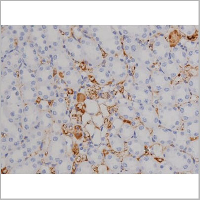
IHC (Immunohistochemistry)
(AAA31050 at 1/200 staining Rat kidney tissue sections by IHC-P. The tissue was formaldehyde fixed and a heat mediated antigen retrieval step in citrate buffer was performed. The tissue was then blocked and incubated with the antibody for 1.5 hours at 22 degree C. An HRP conjugated goat anti-rabbit antibody was used as the secondary.)
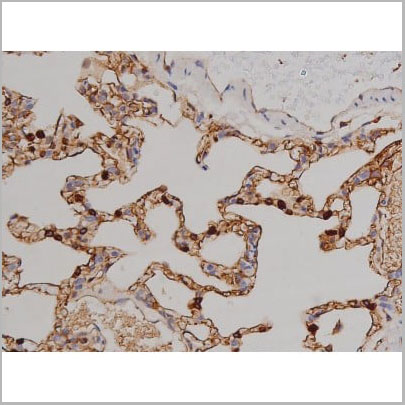
IHC (Immunohistochemistry)
(AAA31050 at 1/200 staining Rat lung tissue sections by IHC-P. The tissue was formaldehyde fixed and a heat mediated antigen retrieval step in citrate buffer was performed. The tissue was then blocked and incubated with the antibody for 1.5 hours at 22 degree C. An HRP conjugated goat anti-rabbit antibody was used as the secondary.)
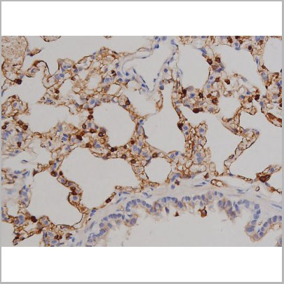
IHC (Immunohistchemistry)
(AAA31050 at 1/200 staining Rat lung tissue sections by IHC-P. The tissue was formaldehyde fixed and a heat mediated antigen retrieval step in citrate buffer was performed. The tissue was then blocked and incubated with the antibody for 1.5 hours at 22 degree C. An HRP conjugated goat anti-rabbit antibody was used as the secondary.)
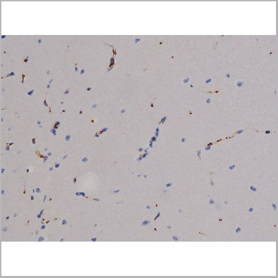
IHC (Immunohistochemistry)
(AAA31050 at 1/200 staining Mouse brain tissue sections by IHC-P. The tissue was formaldehyde fixed and a heat mediated antigen retrieval step in citrate buffer was performed. The tissue was then blocked and incubated with the antibody for 1.5 hours at 22 degree C. An HRP conjugated goat anti-rabbit antibody was used as the secondary.)
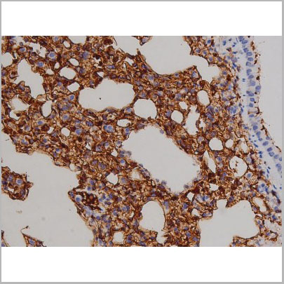
IHC (Immunohistochemistry)
(AAA31050 at 1/200 staining Mouse lung2.JPG tissue sections by IHC-P. The tissue was formaldehyde fixed and a heat mediated antigen retrieval step in citrate buffer was performed. The tissue was then blocked and incubated with the antibody for 1.5 hours at 22 degree C. An HRP conjugated goat anti-rabbit antibody was used as the secondary.)
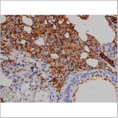
IHC (Immunohistchemistry)
(AAA31050 at 1/200 staining Mouse lung1.JPG tissue sections by IHC-P. The tissue was formaldehyde fixed and a heat mediated antigen retrieval step in citrate buffer was performed. The tissue was then blocked and incubated with the antibody for 1.5 hours at 22 degree C. An HRP conjugated goat anti-rabbit antibody was used as the secondary.)
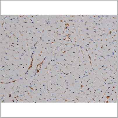
IHC (Immunohistochemistry)
(AAA31050 at 1/200 staining Mouse heart tissue sections by IHC-P. The tissue was formaldehyde fixed and a heat mediated antigen retrieval step in citrate buffer was performed. The tissue was then blocked and incubated with the antibody for 1.5 hours at 22 degree C. An HRP conjugated goat anti-rabbit antibody was used as the secondary.)
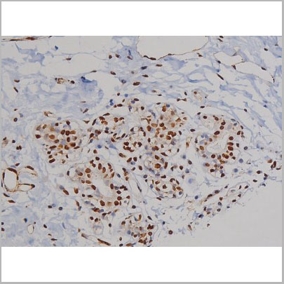
IHC (Immunohistochemistry)
(AAA31050 at 1/200 staining Human heart tissue sections by IHC-P. The tissue was formaldehyde fixed and a heat mediated antigen retrieval step in citrate buffer was performed. The tissue was then blocked and incubated with the antibody for 1.5 hours at 22 degree C. An HRP conjugated goat anti-rabbit antibody was used as the secondary.)
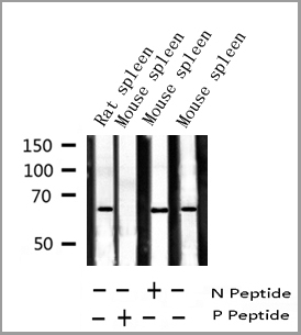
WB (Western Blot)
(Western blot analysis of Phospho-ZAP-70 (Tyr319) expression in various lysates)
WB (Western Blot)
(Western blot analysis of Phospho-ZAP-70 (Tyr319) expression in various lysates)
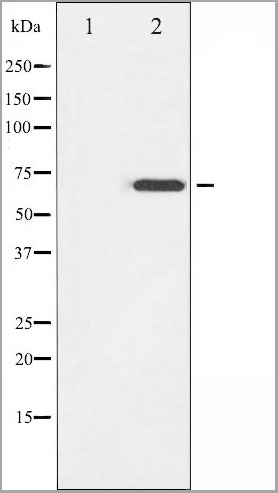
WB (Western Blot)
(Western blot analysis of ZAP-70 phosphorylation expression in Jurkat whole cell lysates, The lane on the left is treated with the antigen-specific peptide.)
WB (Western Blot)
(Western blot analysis of ZAP-70 phosphorylation expression in Jurkat whole cell lysates, The lane on the left is treated with the antigen-specific peptide.)
Function: Tyrosine kinase that plays an essential role in regulation of the adaptive immune response. Regulates motility, adhesion and cytokine expression of mature T-cells, as well as thymocyte development. Contributes also to the development and activation of primary B-lymphocytes. When antigen presenting cells (APC) activate T-cell receptor (TCR), a serie of phosphorylations lead to the recruitment of ZAP70 to the doubly phosphorylated TCR component CD247/CD3Z through ITAM motif at the plasma membrane. This recruitment serves to localization to the stimulated TCR and to relieve its autoinhibited conformation. Release of ZAP70 active conformation is further stabilized by phosphorylation mediated by LCK. Subsequently, ZAP70 phosphorylates at least 2 essential adapter proteins: LAT and LCP2. In turn, a large number of signaling molecules are recruited and ultimately lead to lymphokine production, T-cell proliferation and differentiation. Furthermore, ZAP70 controls cytoskeleton modifications, adhesion and mobility of T-lymphocytes, thus ensuring correct delivery of effectors to the APC. ZAP70 is also required for TCR-CD247/CD3Z internalization and degradation through interaction with the E3 ubiquitin-protein ligase CBL and adapter proteins SLA and SLA2. Thus, ZAP70 regulates both T-cell activation switch on and switch off by modulating TCR expression at the T-cell surface. During thymocyte development, ZAP70 promotes survival and cell-cycle progression of developing thymocytes before positive selection (when cells are still CD4/CD8 double negative). Additionally, ZAP70-dependent signaling pathway may also contribute to primary B-cells formation and activation through B-cell receptor (BCR).
Subunit Structure: Interacts with CD247/CD3Z; this interaction docks ZAP70 at the stimulated TCR (PubMed:1423621, PubMed:7659156, PubMed:26783323). Interacts with NFAM1 (PubMed:15143214). Interacts with adapter protein SLA; this interaction negatively regulates T-cell receptor signaling (PubMed:10449770). Interacts with FCRL3 (PubMed:12051764, PubMed:19843936). Interacts with VAV1 (PubMed:9151714). Interacts with CBL; this interaction promotes ubiquitination, internalization and subsequent degradation of CD247/CD3Z (PubMed:10449770, PubMed:10078535). Identified in a complex with CBL and UBE2L3 (PubMed:10966114). Interacts with SHB (PubMed:12084069). Interacts with adapter protein SLA2; this interaction negatively regulates T-cell receptor signaling. Interacts with CBLB. Interacts (via SH2 domains) with RHOH; this interaction regulates ZAP70 subcellular localization. Interacts with DEF6 (By similarity). Interacts (ubiquitinated form) with OTUD7B and UBASH3B (PubMed:26903241).
Post-translational Modifications: Phosphorylated on tyrosine residues upon T-cell antigen receptor (TCR) stimulation. Phosphorylation of Tyr-315 and Tyr-319 are essential for ZAP70 positive function on T-lymphocyte activation whereas Tyr-292 has a negative regulatory role. Within the C-terminal kinase domain, Tyr-492 and Tyr-493 are phosphorylated after TCR induction, Tyr-492 playing a negative regulatory role and Tyr-493 a positive. Tyr-493 is dephosphorylated by PTN22. Ubiquitinated in response to T cell activation. Deubiquitinated by OTUD7B.
Similarity: Composed of 2 N-terminal SH2 domains and a C-terminal kinase domain. The tandem SH2 domains bind to the doubly phosphorylated tyrosine-based activation motif (ITAM) of CD247/CD3Z and the non-canonical phosphorylated tyrosine-based activation motif (TAM) of RHOH (By similarity). The interdomain B located between the second SH2 and the kinase domain contains 3 tyrosines (Tyr-292, Tyr-315, Tyr-319) that are phosphorylated following TCR activation. These sites have been implicated in binding to other signaling molecules including CBL or VAV1. Thus, ZAP70 can also function as a scaffold by recruiting additional factors to the stimulated TCR complex. Belongs to the protein kinase superfamily. Tyr protein kinase family. SYK/ZAP-70 subfamily.
NCBI and Uniprot Product Information
Predicted: 70 kDa
Similar Products


Product Notes
The ZAP-70 zap70 (Catalog #AAA31050) is an Antibody produced from Rabbit and is intended for research purposes only. The product is available for immediate purchase. The Phospho-ZAP-70 (Tyr319) Antibody reacts with Human, Mouse, Rat and may cross-react with other species as described in the data sheet. AAA Biotech's ZAP-70 can be used in a range of immunoassay formats including, but not limited to, Western Blot (WB), Immunohistochemisty (IHC), Immunofluorescence (IF), Immunocytochemistry (ICC), ELISA (EIA). WB: 1:500-1:2000 IHC: 1:50-1:200 IF/ICC: 1:100-1:500. Researchers should empirically determine the suitability of the ZAP-70 zap70 for an application not listed in the data sheet. Researchers commonly develop new applications and it is an integral, important part of the investigative research process. It is sometimes possible for the material contained within the vial of "ZAP-70, Polyclonal Antibody" to become dispersed throughout the inside of the vial, particularly around the seal of said vial, during shipment and storage. We always suggest centrifuging these vials to consolidate all of the liquid away from the lid and to the bottom of the vial prior to opening. Please be advised that certain products may require dry ice for shipping and that, if this is the case, an additional dry ice fee may also be required.Precautions
All products in the AAA Biotech catalog are strictly for research-use only, and are absolutely not suitable for use in any sort of medical, therapeutic, prophylactic, in-vivo, or diagnostic capacity. By purchasing a product from AAA Biotech, you are explicitly certifying that said products will be properly tested and used in line with industry standard. AAA Biotech and its authorized distribution partners reserve the right to refuse to fulfill any order if we have any indication that a purchaser may be intending to use a product outside of our accepted criteria.Disclaimer
Though we do strive to guarantee the information represented in this datasheet, AAA Biotech cannot be held responsible for any oversights or imprecisions. AAA Biotech reserves the right to adjust any aspect of this datasheet at any time and without notice. It is the responsibility of the customer to inform AAA Biotech of any product performance issues observed or experienced within 30 days of receipt of said product. To see additional details on this or any of our other policies, please see our Terms & Conditions page.Item has been added to Shopping Cart
If you are ready to order, navigate to Shopping Cart and get ready to checkout.