Filters
Clonality
Type
Reactivity
Gene Name
Isotype
Host
Application
Clone
47 results for " Enzyme Activity" - showing 1-47
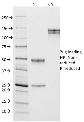
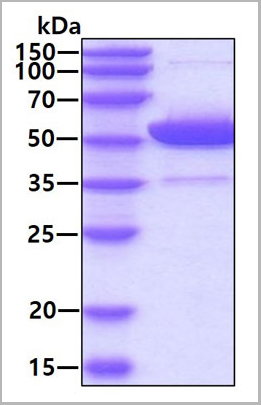
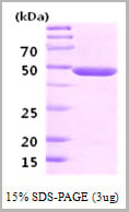
 SDS-PAGE
Aldose reductase, Active Protein (Cat# AAA11762)
Full Name
Aldo-keta reductase family 1, member B1
Gene Names
AKR1B1; AR; ADR; ALR2; ALDR1
Applications
SDS-PAGE
Purity
> 95% by SDS-PAGE
Pricing

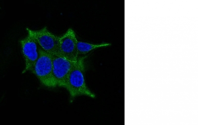
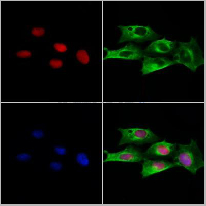
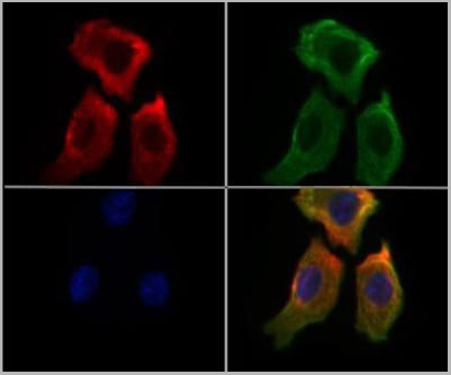
 IF (Immunofluorescence)
(Immunostaining of mixed neuron/glial cultures stained with anti-UCHL1 antibody (AAA14227), green, 1:500) and rabbit anti-GFAP antibody , red, 1:1000. The blue stains nuclear DNA. The anti-UCHL1 stains strongly the cell body and dendrites of neurons, while anti-GFAP specifically labels astrocytes.)
IF (Immunofluorescence)
(Immunostaining of mixed neuron/glial cultures stained with anti-UCHL1 antibody (AAA14227), green, 1:500) and rabbit anti-GFAP antibody , red, 1:1000. The blue stains nuclear DNA. The anti-UCHL1 stains strongly the cell body and dendrites of neurons, while anti-GFAP specifically labels astrocytes.)
Ubiquitin C-terminal Hydrolase 1 (UCHL1) ck, Polyclonal Antibody (Cat# AAA14227)
Full Name
Anti-Ubiquitin C Terminal Hydrolase 1 (UCHL1)
Gene Names
UCHL1; NDGOA; PARK5; PGP95; PGP9.5; Uch-L1; HEL-117; PGP 9.5
Reactivity
Bovine, Human, Mouse, Pig, Rat
Expected Reactivity: Canine, Feline, Goat, Guinea Pig, Hamster, Horse, Non-Human Primate, Rabbit, Sheep, Vole
Expected Reactivity: Canine, Feline, Goat, Guinea Pig, Hamster, Horse, Non-Human Primate, Rabbit, Sheep, Vole
Applications
Western Blot, Immunofluorescence
Purity
Total IgY fraction
Pricing


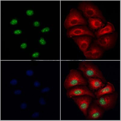
 IF (Immunofluorescence)
(Confocal immunofluorescent analysis of GAPDH Antibody (C-term R248) with Hela cell followed by Alexa Fluor 488-conjugated goat anti-rabbit lgG (green). Actin filaments have been labeled with Alexa Fluor 555 phalloidin (red).DAPI was used to stain the cell nuclear (blue).)
IF (Immunofluorescence)
(Confocal immunofluorescent analysis of GAPDH Antibody (C-term R248) with Hela cell followed by Alexa Fluor 488-conjugated goat anti-rabbit lgG (green). Actin filaments have been labeled with Alexa Fluor 555 phalloidin (red).DAPI was used to stain the cell nuclear (blue).)
GAPDH, Polyclonal Antibody (Cat# AAA28693)
Full Name
GAPDH Antibody (C-term R248)
Gene Names
GAPDH; G3PD; GAPD; HEL-S-162eP
Reactivity
Human (Predicted Reactivity: Chicken, Mouse, Pig, Rat)
Applications
Western Blot, Immunohistochemistry, Flow Cytometry, Immunofluorescence
Purity
Purified Rabbit Polyclonal Antibody (Pab)
Pricing


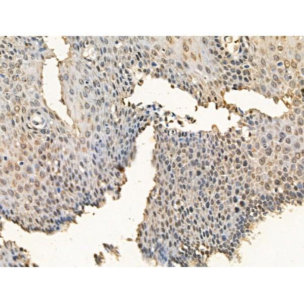
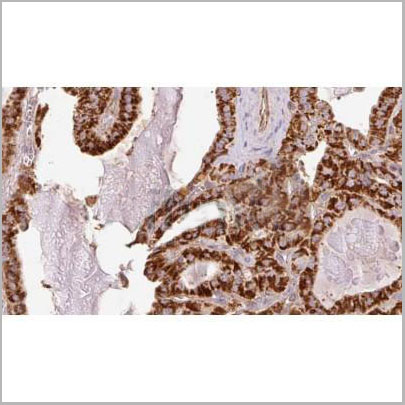
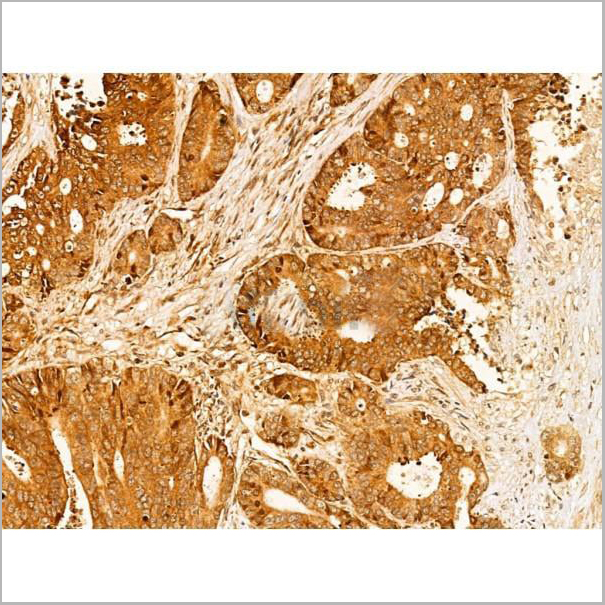
 IHC (Immunohistochemistry)
(At 1/200 staining Human lung cancer tissue sections by IHC-P. The tissue was formaldehyde fixed and a heat mediated antigen retrieval step in citrate buffer was performed. The tissue was then blocked and incubated with the antibody for 1.5 hours at 22 degree C. An HRP conjugated goat anti-rabbit antibody was used as the secondary antibody.)
IHC (Immunohistochemistry)
(At 1/200 staining Human lung cancer tissue sections by IHC-P. The tissue was formaldehyde fixed and a heat mediated antigen retrieval step in citrate buffer was performed. The tissue was then blocked and incubated with the antibody for 1.5 hours at 22 degree C. An HRP conjugated goat anti-rabbit antibody was used as the secondary antibody.)
ERK1/2, Polyclonal Antibody (Cat# AAA31416)
Full Name
Phospho-ERK1/2 (Thr202+Tyr204/Thr185+Tyr187) Antibody
Gene Names
MAPK3; ERK1; ERT2; ERK-1; PRKM3; P44ERK1; P44MAPK; HS44KDAP; HUMKER1A; p44-ERK1; p44-MAPK
Reactivity
Human, Mouse, Rat, Monkey
Predicted Reactivity: Pig (100%), Zebrafish (100%), Bovine (100%), Horse (100%), Sheep (100%), Rabbit (100%)
Predicted Reactivity: Pig (100%), Zebrafish (100%), Bovine (100%), Horse (100%), Sheep (100%), Rabbit (100%)
Applications
Western Blot, Immunohistochemistry, Peptide ELISA
Purity
The antibody is from purified rabbit serum by affinity purification via sequential chromatography on phospho-peptide and non-phospho-peptide affinity columns.
Pricing


 Application Data
(At 25 degree C. Samples were then incubated with primary Ab(At 37 degree C. An AlexaFluor594 conjugated goat anti-rabbit IgG(H+L) Ab(Red) and an AlexaFluor488 conjugated goat anti-mouse IgG(H+L) Ab(Green) were used as the secondary antibody.The nuclear counter stain is DAPI(blue).)
Application Data
(At 25 degree C. Samples were then incubated with primary Ab(At 37 degree C. An AlexaFluor594 conjugated goat anti-rabbit IgG(H+L) Ab(Red) and an AlexaFluor488 conjugated goat anti-mouse IgG(H+L) Ab(Green) were used as the secondary antibody.The nuclear counter stain is DAPI(blue).)
Rac1/cdc42, Polyclonal Antibody (Cat# AAA31342)
Full Name
Rac1/cdc42 Antibody
Gene Names
RAC1; MIG5; Rac-1; TC-25; p21-Rac1
Reactivity
Human, Mouse, Rat
Predicted Reactivity: Pig (100%), Bovine (100%), Sheep (100%), Dog (100%), Xenopus (100%)
Predicted Reactivity: Pig (100%), Bovine (100%), Sheep (100%), Dog (100%), Xenopus (100%)
Applications
Western Blot, Immunohistochemistry, Immunofluorescence, Immunocytochemistry, Peptide ELISA
Purity
The antiserum was purified by peptide affinity chromatography using SulfoLink Coupling Resin
Pricing


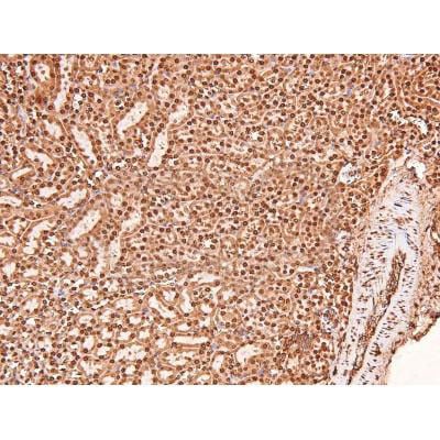
 IHC (Immunohistochemistry)
(At 1/100 staining Mouse kidney tissue by IHC-P. The sample was formaldehyde fixed and a heat mediated antigen retrieval step in citrate buffer was performed. The sample was then blocked and incubated with the primary antibody at 4 degree C overnight. An HRP conjugated anti-Rabbit antibody was used as the secondary antibody.)
IHC (Immunohistochemistry)
(At 1/100 staining Mouse kidney tissue by IHC-P. The sample was formaldehyde fixed and a heat mediated antigen retrieval step in citrate buffer was performed. The sample was then blocked and incubated with the primary antibody at 4 degree C overnight. An HRP conjugated anti-Rabbit antibody was used as the secondary antibody.)
p38 MAPK, Polyclonal Antibody (Cat# AAA31333)
Full Name
Phospho-p38 MAPK (Thr180/Tyr182) Antibody
Gene Names
MAPK14; RK; p38; CSBP; EXIP; Mxi2; CSBP1; CSBP2; CSPB1; PRKM14; PRKM15; SAPK2A; p38ALPHA
Reactivity
Human, Mouse, Rat
Predicted Reactivity: Pig (100%), Bovine (100%), Horse (100%), Sheep (100%), Rabbit (100%), Dog (100%), Xenopus (91%)
Predicted Reactivity: Pig (100%), Bovine (100%), Horse (100%), Sheep (100%), Rabbit (100%), Dog (100%), Xenopus (91%)
Applications
Western Blot, Immunohistochemistry, Immunofluorescence, Immunocytochemistry, Immunoprecipitation, Peptide ELISA
Purity
The antibody is from purified rabbit serum by affinity purification via sequential chromatography on phospho-peptide and non-phospho-peptide affinity columns.
Pricing


 Application Data
(At 25 degree C. Samples were then incubated with primary Ab(At 37 degree C. An AlexaFluor594 conjugated goat anti-rabbit IgG(H+L) Ab(Red) and an AlexaFluor488 conjugated goat anti-mouse IgG(H+L) Ab(Green) were used as the secondary antibody.The nuclear counter stain is DAPI (blue).)
Application Data
(At 25 degree C. Samples were then incubated with primary Ab(At 37 degree C. An AlexaFluor594 conjugated goat anti-rabbit IgG(H+L) Ab(Red) and an AlexaFluor488 conjugated goat anti-mouse IgG(H+L) Ab(Green) were used as the secondary antibody.The nuclear counter stain is DAPI (blue).)
TOP2A, Polyclonal Antibody (Cat# AAA31306)
Full Name
Phospho-TOP2A (Thr1343) Antibody
Gene Names
TOP2A; TOP2; TP2A
Reactivity
Human, Mouse
Applications
Immunohistochemistry, Immunofluorescence, Immunocytochemistry, Peptide ELISA
Purity
The antibody is from purified rabbit serum by affinity purification via sequential chromatography on phospho-peptide and non-phospho-peptide affinity columns.
Pricing


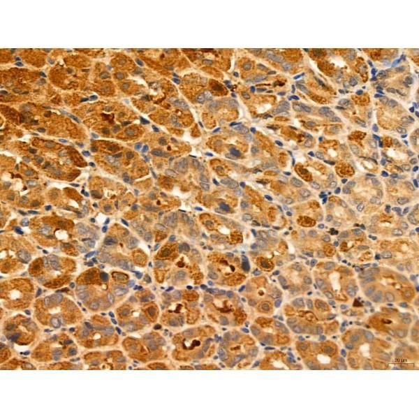
 IHC (Immunohistchemistry)
(Formalin-fixed, paraffin-embedded Rat Pancreas stained with ODC1 Monoclonal Antibody (ODC1/485))
IHC (Immunohistchemistry)
(Formalin-fixed, paraffin-embedded Rat Pancreas stained with ODC1 Monoclonal Antibody (ODC1/485))
Ornithine Decarboxylase-1 (ODC-1), Monoclonal Antibody (Cat# AAA13817)
Full Name
Ornithine Decarboxylase-1 (ODC-1) Mouse Monoclonal Antibody
Gene Names
ODC1; ODC
Reactivity
Human, Rat
Applications
Flow Cytometry, Immunofluorescence, Western Blot, Immunohistochemistry
Pricing


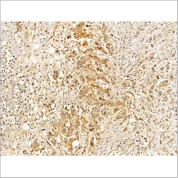
 IHC (Immunohistochemistry)
(At 1/200 staining Human prostate cancer tissue sections by IHC-P. The tissue was formaldehyde fixed and a heat mediated antigen retrieval step in citrate buffer was performed. The tissue was then blocked and incubated with the antibody for 1.5 hours at 22 degree C. An HRP conjugated goat anti-rabbit antibody was used as the secondary antibody.)
IHC (Immunohistochemistry)
(At 1/200 staining Human prostate cancer tissue sections by IHC-P. The tissue was formaldehyde fixed and a heat mediated antigen retrieval step in citrate buffer was performed. The tissue was then blocked and incubated with the antibody for 1.5 hours at 22 degree C. An HRP conjugated goat anti-rabbit antibody was used as the secondary antibody.)
CDK4, Polyclonal Antibody (Cat# AAA31380)
Full Name
Phospho-CDK4 (Thr172) Antibody
Gene Names
CDK4; CMM3; PSK-J3
Reactivity
Human, Mouse, Rat
Predicted Reactivity: Pig (100%), Zebrafish (85%), Bovine (100%), Horse (100%), Sheep (100%), Rabbit (100%), Dog (100%), Xenopus (92%)
Predicted Reactivity: Pig (100%), Zebrafish (85%), Bovine (100%), Horse (100%), Sheep (100%), Rabbit (100%), Dog (100%), Xenopus (92%)
Applications
Western Blot, Immunohistochemistry, Peptide ELISA
Purity
The antibody is from purified rabbit serum by affinity purification via sequential chromatography on phospho-peptide and non-phospho-peptide affinity columns.
Pricing


 Application Data
(At 25 degree C. Samples were then incubated with primary Ab(At 37 degree C. An AlexaFluor594 conjugated goat anti-rabbit IgG(H+L) Ab(Red) and an AlexaFluor488 conjugated goat anti-mouse IgG(H+L) Ab(Green) were used as the secondary antibody.The nuclear counter stain is DAPI (blue).)
Application Data
(At 25 degree C. Samples were then incubated with primary Ab(At 37 degree C. An AlexaFluor594 conjugated goat anti-rabbit IgG(H+L) Ab(Red) and an AlexaFluor488 conjugated goat anti-mouse IgG(H+L) Ab(Green) were used as the secondary antibody.The nuclear counter stain is DAPI (blue).)
ERK1/2, Polyclonal Antibody (Cat# AAA31300)
Full Name
Phospho-ERK1/2 (Thr177/Thr160) Antibody
Gene Names
MAPK3; ERK1; ERT2; ERK-1; PRKM3; P44ERK1; P44MAPK; HS44KDAP; HUMKER1A; p44-ERK1; p44-MAPK
Reactivity
Human, Mouse, Rat
Applications
Immunohistochemistry, Immunofluorescence, Immunocytochemistry, Peptide ELISA
Purity
The antibody is from purified rabbit serum by affinity purification via sequential chromatography on phospho-peptide and non-phospho-peptide affinity columns.
Pricing


 Application Data
(Published customer image: Leukocyte infiltration in COX-2-M/-M and COX-2+/+ mice. MPO enzymatic activity (panel A) was statistically similar in COX-2-M/-M and COX-2+/+ livers at 6 h and 24 h post-IRI. Ly-6G+ neutrophil (panel B) and granulocyte (panel C) infiltration were also comparable in COX-2-M/-M and COX-2+/+ livers after IRI. Mac-1+ (panel D) and CD68 (panel E) infiltrating macrophages were significantly reduced in COX-2-M/-M livers at 24 h post-reperfusion, but were statistically indistinguishable in COX-2-M/-M and COX-2+/+ livers at 6 h after IRI. No statistical differences in MMP-9 expression (panel F) could be demonstrated in livers of COX-2-M/-M and COX-2+/+ mice post-IRI. Representative immunostaining (panel G) of infiltrating Ly-6G+ (a,b,e,f) and Mac-1+ (c,d,g,h) leukocytes in livers of COX-2+/+ (a,c,e,g) and COX-2-M/-M (b,d,f,h) mice at 6 h (a to d) and 24 h (e to h) post IRI; (n = 5 -6/group; * indicates p)
Application Data
(Published customer image: Leukocyte infiltration in COX-2-M/-M and COX-2+/+ mice. MPO enzymatic activity (panel A) was statistically similar in COX-2-M/-M and COX-2+/+ livers at 6 h and 24 h post-IRI. Ly-6G+ neutrophil (panel B) and granulocyte (panel C) infiltration were also comparable in COX-2-M/-M and COX-2+/+ livers after IRI. Mac-1+ (panel D) and CD68 (panel E) infiltrating macrophages were significantly reduced in COX-2-M/-M livers at 24 h post-reperfusion, but were statistically indistinguishable in COX-2-M/-M and COX-2+/+ livers at 6 h after IRI. No statistical differences in MMP-9 expression (panel F) could be demonstrated in livers of COX-2-M/-M and COX-2+/+ mice post-IRI. Representative immunostaining (panel G) of infiltrating Ly-6G+ (a,b,e,f) and Mac-1+ (c,d,g,h) leukocytes in livers of COX-2+/+ (a,c,e,g) and COX-2-M/-M (b,d,f,h) mice at 6 h (a to d) and 24 h (e to h) post IRI; (n = 5 -6/group; * indicates p)
CD68, Monoclonal Antibody (Cat# AAA12105)
Full Name
RAT ANTI MOUSE CD68:FITC
Gene Names
Cd68; Lamp4; gp110; Scard1
Applications
Flow Cytometry
Pricing


 Application Data
(Analysis of Protein Array containing more than 19, 000 full-length human proteins using CD10 Mouse Monoclonal Antibody (MME/1870)Z- and S- Score: The Z-score represents the strength of a signal that a monoclonal antibody (MAb) (in combination with a fluorescently-tagged anti-IgG secondary antibody) produces when binding to a particular protein on the HuProtTM array. Z-scores are described in units of standard deviations (SD's) above the mean value of all signals generated on that array. If targets on HuProtTM are arranged in descending order of the Z-score, the S-score is the difference (also in units of SD's) between the Z-score. S-score therefore represents the relative target specificity of a MAb to its intended target. A MAb is considered to specific to its intended target, if the MAb has an S-score of at least 2.5. For example, if a MAb binds to protein X with a Z-score of 43 and to protein Y with a Z-score of 14, then the S-score for the binding of that MAb to protein X is equal to 29.)
Application Data
(Analysis of Protein Array containing more than 19, 000 full-length human proteins using CD10 Mouse Monoclonal Antibody (MME/1870)Z- and S- Score: The Z-score represents the strength of a signal that a monoclonal antibody (MAb) (in combination with a fluorescently-tagged anti-IgG secondary antibody) produces when binding to a particular protein on the HuProtTM array. Z-scores are described in units of standard deviations (SD's) above the mean value of all signals generated on that array. If targets on HuProtTM are arranged in descending order of the Z-score, the S-score is the difference (also in units of SD's) between the Z-score. S-score therefore represents the relative target specificity of a MAb to its intended target. A MAb is considered to specific to its intended target, if the MAb has an S-score of at least 2.5. For example, if a MAb binds to protein X with a Z-score of 43 and to protein Y with a Z-score of 14, then the S-score for the binding of that MAb to protein X is equal to 29.)
CD10, Monoclonal Antibody (Cat# AAA23901)
Full Name
CD10 (Membrane Metalloendopeptidase)
Gene Names
MME; NEP; SFE; CD10; CALLA; CMT2T; SCA43
Reactivity
Human. Others not tested.
Applications
Immunohistochemistry
Pricing


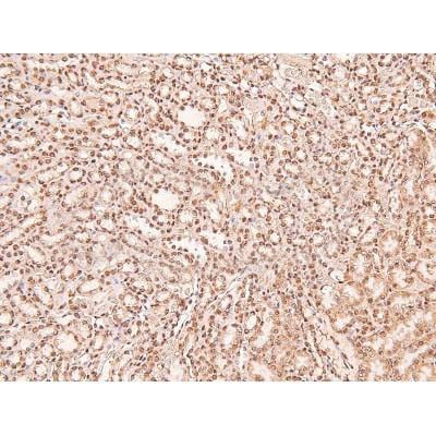
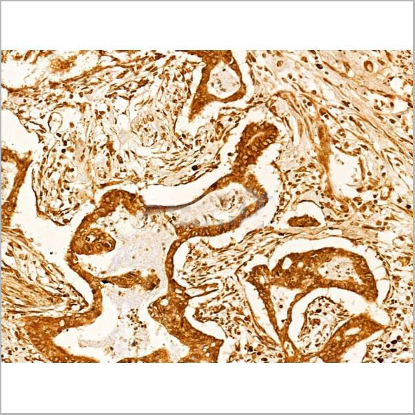
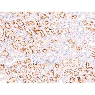 IHC (Immunohistochemistry)
(At 1/200 staining Human kidney tissue sections by IHC-P. The tissue was formaldehyde fixed and a heat mediated antigen retrieval step in citrate buffer was performed. The tissue was then blocked and incubated with the antibody for 1.5 hours at 22 degree C. An HRP conjugated goat anti-rabbit antibody was used as the secondary antibody.)
IHC (Immunohistochemistry)
(At 1/200 staining Human kidney tissue sections by IHC-P. The tissue was formaldehyde fixed and a heat mediated antigen retrieval step in citrate buffer was performed. The tissue was then blocked and incubated with the antibody for 1.5 hours at 22 degree C. An HRP conjugated goat anti-rabbit antibody was used as the secondary antibody.)
RhoA, Polyclonal Antibody (Cat# AAA31383)
Full Name
Phospho-RhoA (Ser188) Antibody
Gene Names
RHOA; ARHA; ARH12; RHO12; RHOH12
Reactivity
Human, Mouse, Rat
Predicted Reactivity: Pig (100%), Bovine (100%), Sheep (100%), Dog (100%)
Predicted Reactivity: Pig (100%), Bovine (100%), Sheep (100%), Dog (100%)
Applications
Western Blot, Immunohistochemistry, Peptide ELISA
Purity
The antibody is from purified rabbit serum by affinity purification via sequential chromatography on phospho-peptide and non-phospho-peptide affinity columns.
Pricing


 Application Data
(Published customer image: Leukocyte infiltration in COX-2-M/-M and COX-2+/+ mice. MPO enzymatic activity (panel A) was statistically similar in COX-2-M/-M and COX-2+/+ livers at 6 h and 24 h post-IRI. Ly-6G+ neutrophil (panel B) and granulocyte (panel C) infiltration were also comparable in COX-2-M/-M and COX-2+/+ livers after IRI. Mac-1+ (panel D) and CD68 (panel E) infiltrating macrophages were significantly reduced in COX-2-M/-M livers at 24 h post-reperfusion, but were statistically indistinguishable in COX-2-M/-M and COX-2+/+ livers at 6 h after IRI. No statistical differences in MMP-9 expression (panel F) could be demonstrated in livers of COX-2-M/-M and COX-2+/+ mice post-IRI. Representative immunostaining (panel G) of infiltrating Ly-6G+ (a,b,e,f) and Mac-1+ (c,d,g,h) leukocytes in livers of COX-2+/+ (a,c,e,g) and COX-2-M/-M (b,d,f,h) mice at 6 h (a to d) and 24 h (e to h) post IRI; (n = 5 -6/group; * indicates p)
Application Data
(Published customer image: Leukocyte infiltration in COX-2-M/-M and COX-2+/+ mice. MPO enzymatic activity (panel A) was statistically similar in COX-2-M/-M and COX-2+/+ livers at 6 h and 24 h post-IRI. Ly-6G+ neutrophil (panel B) and granulocyte (panel C) infiltration were also comparable in COX-2-M/-M and COX-2+/+ livers after IRI. Mac-1+ (panel D) and CD68 (panel E) infiltrating macrophages were significantly reduced in COX-2-M/-M livers at 24 h post-reperfusion, but were statistically indistinguishable in COX-2-M/-M and COX-2+/+ livers at 6 h after IRI. No statistical differences in MMP-9 expression (panel F) could be demonstrated in livers of COX-2-M/-M and COX-2+/+ mice post-IRI. Representative immunostaining (panel G) of infiltrating Ly-6G+ (a,b,e,f) and Mac-1+ (c,d,g,h) leukocytes in livers of COX-2+/+ (a,c,e,g) and COX-2-M/-M (b,d,f,h) mice at 6 h (a to d) and 24 h (e to h) post IRI; (n = 5 -6/group; * indicates p)
CD68, Monoclonal Antibody (Cat# AAA12103)
Full Name
RAT ANTI MOUSE CD68:Biotin
Gene Names
Cd68; Lamp4; gp110; Scard1
Applications
Flow Cytometry
Pricing


 Application Data
(Published customer image: Leukocyte infiltration in COX-2-M/-M and COX-2+/+ mice. MPO enzymatic activity (panel A) was statistically similar in COX-2-M/-M and COX-2+/+ livers at 6 h and 24 h post-IRI. Ly-6G+ neutrophil (panel B) and granulocyte (panel C) infiltration were also comparable in COX-2-M/-M and COX-2+/+ livers after IRI. Mac-1+ (panel D) and CD68 (panel E) infiltrating macrophages were significantly reduced in COX-2-M/-M livers at 24 h post-reperfusion, but were statistically indistinguishable in COX-2-M/-M and COX-2+/+ livers at 6 h after IRI. No statistical differences in MMP-9 expression (panel F) could be demonstrated in livers of COX-2-M/-M and COX-2+/+ mice post-IRI. Representative immunostaining (panel G) of infiltrating Ly-6G+ (a,b,e,f) and Mac-1+ (c,d,g,h) leukocytes in livers of COX-2+/+ (a,c,e,g) and COX-2-M/-M (b,d,f,h) mice at 6 h (a to d) and 24 h (e to h) post IRI; (n = 5 -6/group; * indicates p)
Application Data
(Published customer image: Leukocyte infiltration in COX-2-M/-M and COX-2+/+ mice. MPO enzymatic activity (panel A) was statistically similar in COX-2-M/-M and COX-2+/+ livers at 6 h and 24 h post-IRI. Ly-6G+ neutrophil (panel B) and granulocyte (panel C) infiltration were also comparable in COX-2-M/-M and COX-2+/+ livers after IRI. Mac-1+ (panel D) and CD68 (panel E) infiltrating macrophages were significantly reduced in COX-2-M/-M livers at 24 h post-reperfusion, but were statistically indistinguishable in COX-2-M/-M and COX-2+/+ livers at 6 h after IRI. No statistical differences in MMP-9 expression (panel F) could be demonstrated in livers of COX-2-M/-M and COX-2+/+ mice post-IRI. Representative immunostaining (panel G) of infiltrating Ly-6G+ (a,b,e,f) and Mac-1+ (c,d,g,h) leukocytes in livers of COX-2+/+ (a,c,e,g) and COX-2-M/-M (b,d,f,h) mice at 6 h (a to d) and 24 h (e to h) post IRI; (n = 5 -6/group; * indicates p)
CD68, Monoclonal Antibody (Cat# AAA12110)
Full Name
RAT ANTI MOUSE CD68
Gene Names
Cd68; Lamp4; gp110; Scard1
Applications
Immunohistochemistry, Flow Cytometry, Immunofluorescence, Immunoprecipitation, Immunohistochemistry, Western Blot
Pricing


 IHC (Immunohistochemistry)
(At 1/100 staining Mouse kidney tissue by IHC-P. The sample was formaldehyde fixed and a heat mediated antigen retrieval step in citrate buffer was performed. The sample was then blocked and incubated with the primary antibody at 4 degree C overnight. An HRP conjugated anti-Rabbit antibody was used as the secondary antibody.)
IHC (Immunohistochemistry)
(At 1/100 staining Mouse kidney tissue by IHC-P. The sample was formaldehyde fixed and a heat mediated antigen retrieval step in citrate buffer was performed. The sample was then blocked and incubated with the primary antibody at 4 degree C overnight. An HRP conjugated anti-Rabbit antibody was used as the secondary antibody.)
LC3A, Polyclonal Antibody (Cat# AAA31334)
Full Name
LC3A Antibody
Gene Names
MAP1LC3A; LC3; LC3A; ATG8E; MAP1ALC3; MAP1BLC3
Reactivity
Human, Mouse, Rat
Predicted Reactivity: Pig (100%), Zebrafish (100%), Bovine (100%), Horse (100%), Sheep (100%), Dog (100%), Chicken (100%), Xenopus (100%)
Predicted Reactivity: Pig (100%), Zebrafish (100%), Bovine (100%), Horse (100%), Sheep (100%), Dog (100%), Chicken (100%), Xenopus (100%)
Applications
Western Blot, Immunohistochemistry, Immunofluorescence, Immunocytochemistry, Peptide ELISA
Purity
The antiserum was purified by peptide affinity chromatography using SulfoLink Coupling Resin
Pricing


 Application Data
(At 25 degree C. Samples were then incubated with primary Ab(At 37 degree C. An AlexaFluor594 conjugated goat anti-rabbit IgG(H+L) Ab(Red) and an AlexaFluor488 conjugated goat anti-mouse IgG(H+L) Ab(Green) were used as the secondary antibody.The nuclear counter stain is DAPI(blue).)
Application Data
(At 25 degree C. Samples were then incubated with primary Ab(At 37 degree C. An AlexaFluor594 conjugated goat anti-rabbit IgG(H+L) Ab(Red) and an AlexaFluor488 conjugated goat anti-mouse IgG(H+L) Ab(Green) were used as the secondary antibody.The nuclear counter stain is DAPI(blue).)
MARK2, Polyclonal Antibody (Cat# AAA31392)
Full Name
Phospho-MARK2 (Thr596) Antibody
Gene Names
MARK2; EMK1; EMK-1; PAR-1; Par1b; Par-1b
Reactivity
Human, Mouse, Rat
Predicted Reactivity: Pig (100%), Bovine (100%), Horse (100%), Sheep (100%), Rabbit (100%), Dog (100%), Xenopus (100%)
Predicted Reactivity: Pig (100%), Bovine (100%), Horse (100%), Sheep (100%), Rabbit (100%), Dog (100%), Xenopus (100%)
Applications
Western Blot, Immunohistochemistry, Immunofluorescence, Immunocytochemistry, Peptide ELISA
Purity
The antibody is from purified rabbit serum by affinity purification via sequential chromatography on phospho-peptide and non-phospho-peptide affinity columns.
Pricing


 Application Data
(Published customer image: Leukocyte infiltration in COX-2-M/-M and COX-2+/+ mice. MPO enzymatic activity (panel A) was statistically similar in COX-2-M/-M and COX-2+/+ livers at 6 h and 24 h post-IRI. Ly-6G+ neutrophil (panel B) and granulocyte (panel C) infiltration were also comparable in COX-2-M/-M and COX-2+/+ livers after IRI. Mac-1+ (panel D) and CD68 (panel E) infiltrating macrophages were significantly reduced in COX-2-M/-M livers at 24 h post-reperfusion, but were statistically indistinguishable in COX-2-M/-M and COX-2+/+ livers at 6 h after IRI. No statistical differences in MMP-9 expression (panel F) could be demonstrated in livers of COX-2-M/-M and COX-2+/+ mice post-IRI. Representative immunostaining (panel G) of infiltrating Ly-6G+ (a,b,e,f) and Mac-1+ (c,d,g,h) leukocytes in livers of COX-2+/+ (a,c,e,g) and COX-2-M/-M (b,d,f,h) mice at 6 h (a to d) and 24 h (e to h) post IRI; (n = 5 -6/group; * indicates p)
Application Data
(Published customer image: Leukocyte infiltration in COX-2-M/-M and COX-2+/+ mice. MPO enzymatic activity (panel A) was statistically similar in COX-2-M/-M and COX-2+/+ livers at 6 h and 24 h post-IRI. Ly-6G+ neutrophil (panel B) and granulocyte (panel C) infiltration were also comparable in COX-2-M/-M and COX-2+/+ livers after IRI. Mac-1+ (panel D) and CD68 (panel E) infiltrating macrophages were significantly reduced in COX-2-M/-M livers at 24 h post-reperfusion, but were statistically indistinguishable in COX-2-M/-M and COX-2+/+ livers at 6 h after IRI. No statistical differences in MMP-9 expression (panel F) could be demonstrated in livers of COX-2-M/-M and COX-2+/+ mice post-IRI. Representative immunostaining (panel G) of infiltrating Ly-6G+ (a,b,e,f) and Mac-1+ (c,d,g,h) leukocytes in livers of COX-2+/+ (a,c,e,g) and COX-2-M/-M (b,d,f,h) mice at 6 h (a to d) and 24 h (e to h) post IRI; (n = 5 -6/group; * indicates p)
CD68, Monoclonal Antibody (Cat# AAA12104)
Full Name
RAT ANTI MOUSE CD68:Biotin
Gene Names
Cd68; Lamp4; gp110; Scard1
Applications
Flow Cytometry
Pricing


 Application Data
(Published customer image: Leukocyte infiltration in COX-2-M/-M and COX-2+/+ mice. MPO enzymatic activity (panel A) was statistically similar in COX-2-M/-M and COX-2+/+ livers at 6 h and 24 h post-IRI. Ly-6G+ neutrophil (panel B) and granulocyte (panel C) infiltration were also comparable in COX-2-M/-M and COX-2+/+ livers after IRI. Mac-1+ (panel D) and CD68 (panel E) infiltrating macrophages were significantly reduced in COX-2-M/-M livers at 24 h post-reperfusion, but were statistically indistinguishable in COX-2-M/-M and COX-2+/+ livers at 6 h after IRI. No statistical differences in MMP-9 expression (panel F) could be demonstrated in livers of COX-2-M/-M and COX-2+/+ mice post-IRI. Representative immunostaining (panel G) of infiltrating Ly-6G+ (a,b,e,f) and Mac-1+ (c,d,g,h) leukocytes in livers of COX-2+/+ (a,c,e,g) and COX-2-M/-M (b,d,f,h) mice at 6 h (a to d) and 24 h (e to h) post IRI; (n = 5 -6/group; * indicates p)
Application Data
(Published customer image: Leukocyte infiltration in COX-2-M/-M and COX-2+/+ mice. MPO enzymatic activity (panel A) was statistically similar in COX-2-M/-M and COX-2+/+ livers at 6 h and 24 h post-IRI. Ly-6G+ neutrophil (panel B) and granulocyte (panel C) infiltration were also comparable in COX-2-M/-M and COX-2+/+ livers after IRI. Mac-1+ (panel D) and CD68 (panel E) infiltrating macrophages were significantly reduced in COX-2-M/-M livers at 24 h post-reperfusion, but were statistically indistinguishable in COX-2-M/-M and COX-2+/+ livers at 6 h after IRI. No statistical differences in MMP-9 expression (panel F) could be demonstrated in livers of COX-2-M/-M and COX-2+/+ mice post-IRI. Representative immunostaining (panel G) of infiltrating Ly-6G+ (a,b,e,f) and Mac-1+ (c,d,g,h) leukocytes in livers of COX-2+/+ (a,c,e,g) and COX-2-M/-M (b,d,f,h) mice at 6 h (a to d) and 24 h (e to h) post IRI; (n = 5 -6/group; * indicates p)
CD68, Monoclonal Antibody (Cat# AAA12107)
Full Name
RAT ANTI MOUSE CD68
Gene Names
Cd68; Lamp4; gp110; Scard1
Applications
Immunohistochemistry, Flow Cytometry, Immunofluorescence, Immunoprecipitation, Immunohistochemistry, Western Blot
Purity
Purified
Purified IgG - liquid
Purified IgG - liquid
Pricing


 Application Data
(Published customer image: Leukocyte infiltration in COX-2-M/-M and COX-2+/+ mice. MPO enzymatic activity (panel A) was statistically similar in COX-2-M/-M and COX-2+/+ livers at 6 h and 24 h post-IRI. Ly-6G+ neutrophil (panel B) and granulocyte (panel C) infiltration were also comparable in COX-2-M/-M and COX-2+/+ livers after IRI. Mac-1+ (panel D) and CD68 (panel E) infiltrating macrophages were significantly reduced in COX-2-M/-M livers at 24 h post-reperfusion, but were statistically indistinguishable in COX-2-M/-M and COX-2+/+ livers at 6 h after IRI. No statistical differences in MMP-9 expression (panel F) could be demonstrated in livers of COX-2-M/-M and COX-2+/+ mice post-IRI. Representative immunostaining (panel G) of infiltrating Ly-6G+ (a,b,e,f) and Mac-1+ (c,d,g,h) leukocytes in livers of COX-2+/+ (a,c,e,g) and COX-2-M/-M (b,d,f,h) mice at 6 h (a to d) and 24 h (e to h) post IRI; (n = 5 -6/group; * indicates p)
Application Data
(Published customer image: Leukocyte infiltration in COX-2-M/-M and COX-2+/+ mice. MPO enzymatic activity (panel A) was statistically similar in COX-2-M/-M and COX-2+/+ livers at 6 h and 24 h post-IRI. Ly-6G+ neutrophil (panel B) and granulocyte (panel C) infiltration were also comparable in COX-2-M/-M and COX-2+/+ livers after IRI. Mac-1+ (panel D) and CD68 (panel E) infiltrating macrophages were significantly reduced in COX-2-M/-M livers at 24 h post-reperfusion, but were statistically indistinguishable in COX-2-M/-M and COX-2+/+ livers at 6 h after IRI. No statistical differences in MMP-9 expression (panel F) could be demonstrated in livers of COX-2-M/-M and COX-2+/+ mice post-IRI. Representative immunostaining (panel G) of infiltrating Ly-6G+ (a,b,e,f) and Mac-1+ (c,d,g,h) leukocytes in livers of COX-2+/+ (a,c,e,g) and COX-2-M/-M (b,d,f,h) mice at 6 h (a to d) and 24 h (e to h) post IRI; (n = 5 -6/group; * indicates p)
CD68, Monoclonal Antibody (Cat# AAA12108)
Full Name
RAT ANTI MOUSE CD68:RPE
Gene Names
Cd68; Lamp4; gp110; Scard1
Applications
Flow Cytometry
Pricing
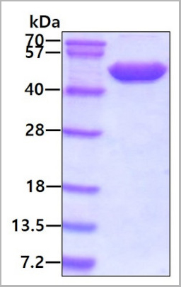
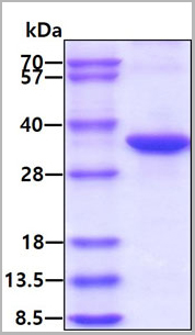
 SDS-PAGE
(3 ug by SDS-PAGE under reducing condition and visualized by coomassie blue stain)
SDS-PAGE
(3 ug by SDS-PAGE under reducing condition and visualized by coomassie blue stain)
NMNAT1, Active Protein (Cat# AAA11834)
Full Name
NMNAT1, 115-270aa, Human, His tag, E Coli (Bioactivity Validated)
Gene Names
NMNAT1; LCA9; NMNAT; PNAT1
Applications
SDS-PAGE, ELISA
Purity
> 95% by SDS-PAGE
Pricing


 Application Data
(Published customer image: Leukocyte infiltration in COX-2-M/-M and COX-2+/+ mice. MPO enzymatic activity (panel A) was statistically similar in COX-2-M/-M and COX-2+/+ livers at 6 h and 24 h post-IRI. Ly-6G+ neutrophil (panel B) and granulocyte (panel C) infiltration were also comparable in COX-2-M/-M and COX-2+/+ livers after IRI. Mac-1+ (panel D) and CD68 (panel E) infiltrating macrophages were significantly reduced in COX-2-M/-M livers at 24 h post-reperfusion, but were statistically indistinguishable in COX-2-M/-M and COX-2+/+ livers at 6 h after IRI. No statistical differences in MMP-9 expression (panel F) could be demonstrated in livers of COX-2-M/-M and COX-2+/+ mice post-IRI. Representative immunostaining (panel G) of infiltrating Ly-6G+ (a,b,e,f) and Mac-1+ (c,d,g,h) leukocytes in livers of COX-2+/+ (a,c,e,g) and COX-2-M/-M (b,d,f,h) mice at 6 h (a to d) and 24 h (e to h) post IRI; (n = 5 -6/group; * indicates p)
Application Data
(Published customer image: Leukocyte infiltration in COX-2-M/-M and COX-2+/+ mice. MPO enzymatic activity (panel A) was statistically similar in COX-2-M/-M and COX-2+/+ livers at 6 h and 24 h post-IRI. Ly-6G+ neutrophil (panel B) and granulocyte (panel C) infiltration were also comparable in COX-2-M/-M and COX-2+/+ livers after IRI. Mac-1+ (panel D) and CD68 (panel E) infiltrating macrophages were significantly reduced in COX-2-M/-M livers at 24 h post-reperfusion, but were statistically indistinguishable in COX-2-M/-M and COX-2+/+ livers at 6 h after IRI. No statistical differences in MMP-9 expression (panel F) could be demonstrated in livers of COX-2-M/-M and COX-2+/+ mice post-IRI. Representative immunostaining (panel G) of infiltrating Ly-6G+ (a,b,e,f) and Mac-1+ (c,d,g,h) leukocytes in livers of COX-2+/+ (a,c,e,g) and COX-2-M/-M (b,d,f,h) mice at 6 h (a to d) and 24 h (e to h) post IRI; (n = 5 -6/group; * indicates p)
CD68, Monoclonal Antibody (Cat# AAA12102)
Full Name
RAT ANTI MOUSE CD68
Gene Names
Cd68; Lamp4; gp110; Scard1
Applications
Immunohistochemistry, Flow Cytometry, Immunofluorescence, Immunoprecipitation, Immunohistochemistry, Western Blot
Pricing


 WB (Western Blot)
(Western Blot Sample: Recombinant LCAT, Mouse;Antibody: Rabbit Anti-Mouse LCAT Ab)
WB (Western Blot)
(Western Blot Sample: Recombinant LCAT, Mouse;Antibody: Rabbit Anti-Mouse LCAT Ab)
Lecithin Cholesterol Acyltransferase (LCAT), Active Protein (Cat# AAA21104)
Full Name
Active Lecithin Cholesterol Acyltransferase (LCAT)
Gene Names
Lcat; AI046659; D8Wsu61e
Reactivity
Mus musculus (Mouse)
Applications
Cell culture; Activity Assays; In vivo assays.
Purity
>98%
Pricing


 FCM (Flow Cytometry)
(Figure 8. Flow Cytometry analysis of HL-60 cells using anti-REA/PHB2 antibody (AAA19273).Overlay histogram showing HL-60 cells stained with AAA19273 (Blue line). The cells were blocked with 10% normal goat serum. And then incubated with rabbit anti-REA/PHB2 Antibody (AAA19273,1μg/1x106 cells) for 30 min at 20 degree C. DyLight®488 conjugated goat anti-rabbit IgG (5-10μg/1x106 cells) was used as secondary antibody for 30 minutes at 20 degree C. Isotype control antibody (Green line) was rabbit IgG (1μg/1x106) used under the same conditions. Unlabelled sample (Red line) was also used as a control.)
FCM (Flow Cytometry)
(Figure 8. Flow Cytometry analysis of HL-60 cells using anti-REA/PHB2 antibody (AAA19273).Overlay histogram showing HL-60 cells stained with AAA19273 (Blue line). The cells were blocked with 10% normal goat serum. And then incubated with rabbit anti-REA/PHB2 Antibody (AAA19273,1μg/1x106 cells) for 30 min at 20 degree C. DyLight®488 conjugated goat anti-rabbit IgG (5-10μg/1x106 cells) was used as secondary antibody for 30 minutes at 20 degree C. Isotype control antibody (Green line) was rabbit IgG (1μg/1x106) used under the same conditions. Unlabelled sample (Red line) was also used as a control.)
REA/PHB2, Polyclonal Antibody (Cat# AAA19273)
Full Name
Anti-REA/PHB2 Antibody
Gene Names
PHB2; BAP; REA; p22; Bap37; BCAP37; PNAS-141
Reactivity
Human, Mouse, Rat
Applications
WB, IHC-P, ICC, IF, FC/FACS/FCM, EIA
Purity
Immunogen affinity purified.
Pricing
Complement C9, Active Protein (Cat# AAA14378)
Full Name
Complement C9 protein
Gene Names
C9; C9D; ARMD15
Purity
> 95% pure
Pricing


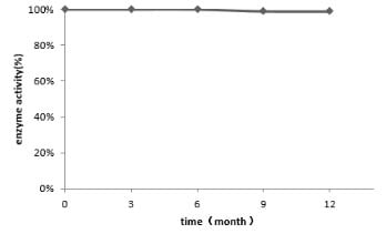 Activity
Proteinase K (PROK), Active Protein (Cat# AAA27052)
Full Name
Recombinant Tritirachium album Proteinase K (PROK) (Active)
Purity
Greater or equal to 85% purity as determined by SDS-PAGE.


 Application Data
(Detect RNase residue by agarose gel electrophores)
Application Data
(Detect RNase residue by agarose gel electrophores)
Proteinase K-lyophilization, Protein (Cat# AAA28067)
Full Name
Proteinase K-lyophilization
Purity
>=95% by SDS-PAGE
Pricing


 Application Data
(Source:Arthrobacter luteus)
Application Data
(Source:Arthrobacter luteus)
Zymolyase 20T, Enzyme (Cat# AAA14844)
Full Name
Zymolyase 20T (Lyticase, Yeast Lytic Enzyme)
Pricing
 Standard Curve (Sample)
Plasma Renin Activity, ELISA Kit (Cat# AAA14209)
Full Name
Plasma Renin Activity (PRA) ELISA
Reactivity
Human
Pricing
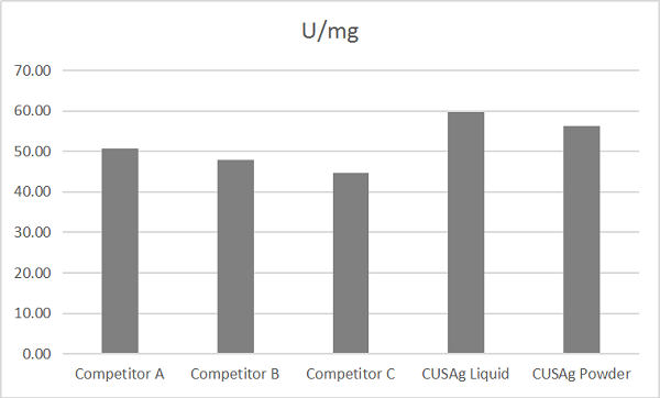
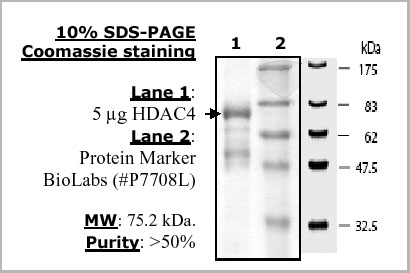 Application Data
HDAC4, Active Protein (Cat# AAA14763)
Full Name
HDAC4, Recombinant, Human (Histone Deacetylase 4)
Purity
> 89%
Pricing
Hypoxanthine-Guanine Phosphoribosyltransferase, Active Protein (Cat# AAA10904)
Full Name
Recombinant Human Hypoxanthine-Guanine Phosphoribosyltransferase, Active
Gene Names
HPRT1; HPRT; HGPRT
Purity
Greater than 95.0% as determined by SDS-PAGE.
Pricing
 Application Data
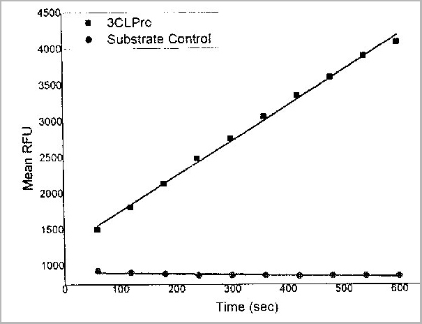
(3CL Proteinase inhibitor screening using 2019-nCoV 3CL MPro Recombinant Protein Provided by customer.)
Application Data
(3CL Proteinase inhibitor screening using 2019-nCoV 3CL MPro Recombinant Protein Provided by customer.)
COVID 19 3CL MPro Coronavirus, Active Protein (Cat# AAA27953)
Full Name
2019-nCoV 3CL MPro Recombinant Protein
Purity
Greater than 95% as determined by reducing SDS-PAGE.
Pricing
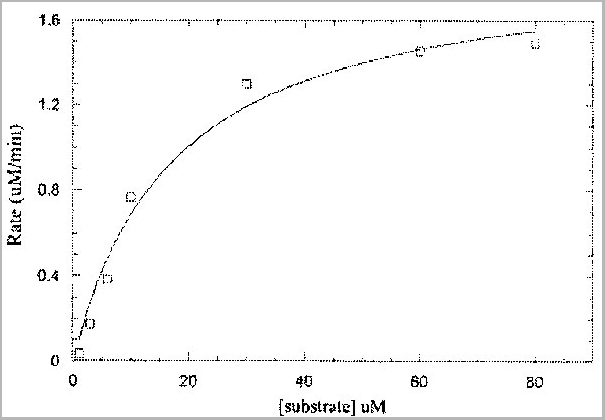 Application Data
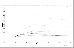
(Measurements of kinetic parameters of Recombinant 2019-nCoV 3C-like Proteinase (CAT# AAA27964) (For reference only).)
Application Data
(Measurements of kinetic parameters of Recombinant 2019-nCoV 3C-like Proteinase (CAT# AAA27964) (For reference only).)
COVID 19 3C-like Proteinase Coronavirus, Active Protein (Cat# AAA27964)
Full Name
Recombinant 2019-nCoV 3C-like Proteinase (N-6His) (CR76)
Purity
> = 95% as determined by reducing SDS-PAGE. Endotoxin: Less than 0.1 ng/ug (1 EU/ug) as determined by LAL test.
Pricing
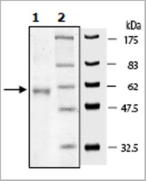 SDS-PAGE
(10% SDS-PAGE Coomassie staining. Lane 1: 3ug F4600-21. Lane 2: Protein Marker.)
SDS-PAGE
(10% SDS-PAGE Coomassie staining. Lane 1: 3ug F4600-21. Lane 2: Protein Marker.)
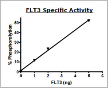
Flt3, active, N-terminal His tag Receptor Tyrosine Kinase, Active Protein (Cat# AAA14760)
Full Name
Flt3, active, Recombinant, Human, N-terminal His tag Receptor Tyrosine Kinase (Fms-like Tyrosine Kinase 3, CD135)
Gene Names
Fiz1; AI790204; mFLJ00416
Reactivity
Human
Purity
> 80% by SDS-PAGE.
Pricing
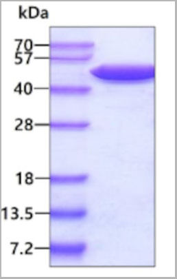 SDS-PAGE
(3ug by SDS-PAGE under reducing condition and visualized by coomassie blue stain.)
SDS-PAGE
(3ug by SDS-PAGE under reducing condition and visualized by coomassie blue stain.)
Alpha-enolase, Active Protein (Cat# AAA11794)
Full Name
Alpha-enolase, 1-434aa, Human, E Coli (Bioactivity Validated)
Gene Names
ENO1; NNE; PPH; MPB1; ENO1L1; HEL-S-17
Applications
Enzyme Activity, SDS-PAGE
Purity
> 95% by SDS-PAGE
Pricing
Human Alanine Aminotransferase, Active Protein (Cat# AAA14927)
Full Name
Recombinant Human Alanine Aminotransferase Glutamie-Pyruvic Transaminase (EC 2.6.1.2)
Pricing
 Application Data
(At 25 degree C. Samples were then incubated with primary Ab(At 37 degree C. An AlexaFluor594 conjugated goat anti-mouse IgG(H+L) Ab(Red) and an AlexaFluor488 conjugated goat anti-rabbit IgG(H+L) Ab(Green) were used as the secondary antibody.The nuclear counter stain is DAPI(blue).)
Application Data
(At 25 degree C. Samples were then incubated with primary Ab(At 37 degree C. An AlexaFluor594 conjugated goat anti-mouse IgG(H+L) Ab(Red) and an AlexaFluor488 conjugated goat anti-rabbit IgG(H+L) Ab(Green) were used as the secondary antibody.The nuclear counter stain is DAPI(blue).)
Cleaved-Caspase 1, Polyclonal Antibody (Cat# AAA31336)
Full Name
Cleaved-Caspase 1 (Ala317), p10 Antibody
Gene Names
CASP1; ICE; P45; IL1BC
Reactivity
Human, Mouse, Rat
Applications
Western Blot, Immunohistochemistry, Immunofluorescence, Immunocytochemistry
Purity
The antiserum was purified by peptide affinity chromatography using SulfoLink Coupling Resin
Pricing
 SDS-PAGE
(3ug by SDS-PAGE under reducing condition and visualized by coomassie blue stain.)
SDS-PAGE
(3ug by SDS-PAGE under reducing condition and visualized by coomassie blue stain.)
ALDH2, Active Protein (Cat# AAA11764)
Full Name
ALDH2,18-517aa, Human, E Coli (Bioactivity Validated)
Gene Names
ALDH2; ALDM; ALDHI; ALDH-E2
Reactivity
Human
Applications
SDS-PAGE, Enzyme Activity
Purity
> 90% by SDS-PAGE
Pricing
Peroxiredoxin-1, Active Protein (Cat# AAA10855)
Full Name
Recombinant Human Peroxiredoxin-1
Gene Names
PRDX1; PAG; PAGA; PAGB; PRX1; PRXI; MSP23; NKEFA; TDPX2; NKEF-A
Purity
Greater than 90.0% as determined by SDS-PAGE.
Pricing
 SDS-PAGE
(3ug by SDS-PAGE under reducing condition and visualized by coomassie blue stain)
SDS-PAGE
(3ug by SDS-PAGE under reducing condition and visualized by coomassie blue stain)
Neuron-Specific Enolase (NSE), Active Protein (Cat# AAA11754)
Full Name
Neuron-Specific Enolase (NSE), 1-434aa, Human, E Coli (Bioactivity Validated)
Gene Names
ENO2; NSE; HEL-S-279
Applications
Enzyme Activity, SDS-PAGE
Purity
> 95% by SDS-PAGE
Pricing
 SDS-PAGE
(3ug by SDS-PAGE under reducing condition and visualized by coomasie blue stain)
SDS-PAGE
(3ug by SDS-PAGE under reducing condition and visualized by coomasie blue stain)
PTP-1B (Protein Tyrosine Phosphatase1B), Active Protein (Cat# AAA11740)
Full Name
PTP-1B (Protein Tyrosine Phosphatase1B), 1-321aa, Human, E Coli (Bioactivity Validated)
Gene Names
PTPN1; PTP1B
Applications
Enzyme Activity, SDS-PAGE
Purity
> 95% by SDS-PAGE
Pricing
 SDS-PAGE
(3ug by SDS-PAGE under reducing condition and visualized by coomassie blue stain.)
SDS-PAGE
(3ug by SDS-PAGE under reducing condition and visualized by coomassie blue stain.)
NQO1, Active Protein (Cat# AAA11735)
Full Name
Recombinant Human NQO1 Protein
Gene Names
NQO1; DTD; QR1; DHQU; DIA4; NMOR1; NMORI
Applications
SDS-PAGE, ELISA
Purity
>95% by SDS-PAGE
Pricing
 IF (Immunofluorescence)
(AAA31127 staining Hela cells by IF/ICC. The samples were fixed with PFA and permeabilized in 0.1% Triton X-100,then blocked in 10% serum for 45 minutes at 25°C. Samples were then incubated with primary Ab(AAA31127 1:200) and mouse anti-beta tubulin Ab( 1:200) for 1 hour at 37°C. An AlexaFluor594 conjugated goat anti-rabbit IgG(H+L) Ab(Red) and an AlexaFluor488 conjugated goat anti-mouse IgG(H+L) Ab(Green) were used as the secondary Ab.The nuclear counter stain is DAPI(blue).)
IF (Immunofluorescence)
(AAA31127 staining Hela cells by IF/ICC. The samples were fixed with PFA and permeabilized in 0.1% Triton X-100,then blocked in 10% serum for 45 minutes at 25°C. Samples were then incubated with primary Ab(AAA31127 1:200) and mouse anti-beta tubulin Ab( 1:200) for 1 hour at 37°C. An AlexaFluor594 conjugated goat anti-rabbit IgG(H+L) Ab(Red) and an AlexaFluor488 conjugated goat anti-mouse IgG(H+L) Ab(Green) were used as the secondary Ab.The nuclear counter stain is DAPI(blue).)
PRDX6, Polyclonal Antibody (Cat# AAA31127)
Full Name
PRDX6 Antibody
Gene Names
PRDX6; PRX; p29; AOP2; 1-Cys; NSGPx; aiPLA2; HEL-S-128m
Reactivity
Human, Mouse, Rat
Applications
Western Blot, Immunohistochemistry
Purity
Peptide affinity purification
Pricing

 Application Data
(The extracellular domain is required for Sln1 kinase activation by wall perturbations. A, representative field showing localization of SLN1-GFP (pSS1881) or sln1ΔECD-GFP (pSS1904). BF, bright field. B, Western analysis showing reduced levels of sln1ΔECD compared with full-length SLN1. SLN1, JF2007 (sln1Δ) carrying full-length SLN1-myc (pCLM994); sln1ΔECD, JF2007 carrying sln1ΔECD-myc (pCLM1774). C, comparison of Hog1 phosphorylation kinetics in SLN1 (JF2008 (sln1Δ sho1Δ) carrying full-length (pCLM994)) and sln1ΔECD (JF2008 (sln1Δ sho1Δ) carrying sln1ΔECD (pCLM1774)) strains following addition of 0.4 M NaCl. D, Northern (RNA) hybridization analysis of RNA prepared from wild type cultures from the sln1Δ strain, JF2007, or sln1Δ ccw12Δ strain, JF2368, transformed with the SLN1 expression plasmids, pCLM994 (SLN1) or pCLM1774 (sln1ΔECD), and treated with 1 unit/ml zymolyase (+) where indicated. NCA3 expression was normalized to expression of the SLN1-SKN7-independent CDC33 gene. The extent of induction by zymolyase is shown relative to the untreated SLN1 or sln1ΔECD strains.)
Application Data
(The extracellular domain is required for Sln1 kinase activation by wall perturbations. A, representative field showing localization of SLN1-GFP (pSS1881) or sln1ΔECD-GFP (pSS1904). BF, bright field. B, Western analysis showing reduced levels of sln1ΔECD compared with full-length SLN1. SLN1, JF2007 (sln1Δ) carrying full-length SLN1-myc (pCLM994); sln1ΔECD, JF2007 carrying sln1ΔECD-myc (pCLM1774). C, comparison of Hog1 phosphorylation kinetics in SLN1 (JF2008 (sln1Δ sho1Δ) carrying full-length (pCLM994)) and sln1ΔECD (JF2008 (sln1Δ sho1Δ) carrying sln1ΔECD (pCLM1774)) strains following addition of 0.4 M NaCl. D, Northern (RNA) hybridization analysis of RNA prepared from wild type cultures from the sln1Δ strain, JF2007, or sln1Δ ccw12Δ strain, JF2368, transformed with the SLN1 expression plasmids, pCLM994 (SLN1) or pCLM1774 (sln1ΔECD), and treated with 1 unit/ml zymolyase (+) where indicated. NCA3 expression was normalized to expression of the SLN1-SKN7-independent CDC33 gene. The extent of induction by zymolyase is shown relative to the untreated SLN1 or sln1ΔECD strains.)
Zymolyase 100T Concentrate 10mg/ml, 0.1M Sorbitol, Enzyme (Cat# AAA14837)
Full Name
Zymolyase 100T Concentrate 10mg/ml, 0.1M Sorbitol (Lyticase, Yeast Lytic Enzyme)
Pricing
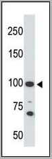
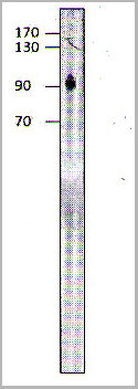 WB (Western Blot)
(WB of with THP on 10% 50S gel. 1:250 antibody dilution in OiluObuffer for 1 hour at room temperature. Apparent MW is 95 kDa .)
WB (Western Blot)
(WB of with THP on 10% 50S gel. 1:250 antibody dilution in OiluObuffer for 1 hour at room temperature. Apparent MW is 95 kDa .)
Progesterone Receptor, Polyclonal Antibody (Cat# AAA14415)
Full Name
Progesterone Receptor Antibody BIOTIN
Gene Names
PGR; PR; NR3C3
Reactivity
Cat, Human, Monkey, Pig
Applications
Immunoprecipitation, Western Blot
Pricing
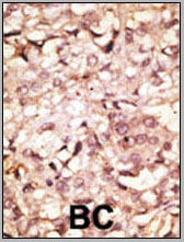
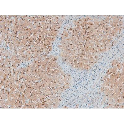
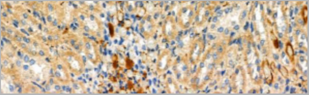
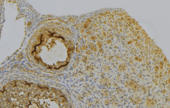
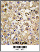 IHC (Immunohistochemistry)
(Formalin-fixed and paraffin-embedded human testis tissue reacted with ACE2 (SARS Receptor) Antibody (C-term) (AAA28762), which was peroxidase-conjugated to the secondary antibody, followed by DAB staining. This data demonstrates the use of this antibody for immunohistochemistry; clinical relevance has not been evaluated.)
IHC (Immunohistochemistry)
(Formalin-fixed and paraffin-embedded human testis tissue reacted with ACE2 (SARS Receptor) Antibody (C-term) (AAA28762), which was peroxidase-conjugated to the secondary antibody, followed by DAB staining. This data demonstrates the use of this antibody for immunohistochemistry; clinical relevance has not been evaluated.)
ACE2 (SARS Receptor), Polyclonal Antibody (Cat# AAA28762)
Full Name
ACE2 (SARS Receptor) Antibody (C-term)
Gene Names
ACE2; ACEH
Reactivity
Human
Applications
Western Blot, Immunohistochemistry
Purity
Purified Rabbit Polyclonal Antibody (Pab)
Pricing
Gamma-Glutamyl Transpeptidase (GGT), Assay Kit (Cat# AAA31531)
Full Name
Gamma-Glutamyl Transpeptidase (GGT) Activity Colorimetric Assay Kit
Pricing