
Filters
Clonality
Type
Reactivity
Gene Name
Isotype
Host
Application
Clone
10000 results for "Recombinant Proteins" - showing 750-800


 WB (Western Blot)
(Western Blot detection against Immunogen (34.21kD).)
WB (Western Blot)
(Western Blot detection against Immunogen (34.21kD).)
SMAD5, Monoclonal Antibody (Cat# AAA25252)
Full Name
SMAD5 (Mothers Against Decapentaplegic Homolog 5, Mothers Against DPP Homolog 5, MAD Homolog 5, Mad5, SMAD Family Member 5, SMAD 5, Smad5, JV5-1, MADH5, DKFZp781C1895, DKFZp781O1323) (FITC)
Gene Names
SMAD5; DWFC; JV5-1; MADH5
Reactivity
Human
Applications
Immunofluorescence, Western Blot
Purity
Purified by Protein A Affinity Chromatography.
Pricing


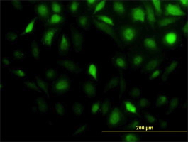


 WB (Western Blot)
(Western Blot detection against Immunogen (85.84kD).)
WB (Western Blot)
(Western Blot detection against Immunogen (85.84kD).)
STIP1, Monoclonal Antibody (Cat# AAA24969)
Full Name
STIP1 (Stress-induced-phosphoprotein 1, STI1, Hsc70/Hsp90-organizing Protein, Hop, P60, Renal Carcinoma Antigen NY-REN-11, STI1L, Transformation-sensitive Protein IEF SSP 3521, IEF-SSP-3521) (Biotin)
Gene Names
STIP1; HOP; P60; STI1; STI1L; HEL-S-94n; IEF-SSP-3521
Reactivity
Human
Applications
Immunofluorescence, Immunohistochemistry, Immunoprecipitation, Western Blot
Purity
Purified by Protein A Affinity Chromatography.
Pricing


 Application Data
(Proximity Ligation Analysis (PLA) of protein-protein interactions between CASP3 and HCLS1. HeLa cells were stained with anti-CASP3 rabbit purified polyclonal 1:1200 and anti-HCLS1 mouse monoclonal antibody 1:50. Signals were detected by Duolink® 30 Detection Kit 613 (red), and nuclei were counterstained with DAPI (blue). Each red dot represents the detection of protein-protein interaction complex.)
Application Data
(Proximity Ligation Analysis (PLA) of protein-protein interactions between CASP3 and HCLS1. HeLa cells were stained with anti-CASP3 rabbit purified polyclonal 1:1200 and anti-HCLS1 mouse monoclonal antibody 1:50. Signals were detected by Duolink® 30 Detection Kit 613 (red), and nuclei were counterstained with DAPI (blue). Each red dot represents the detection of protein-protein interaction complex.)
HCLS1, Monoclonal Antibody (Cat# AAA25416)
Full Name
HCLS1 (Hematopoietic Lineage Cell-specific Protein, Hematopoietic Cell-specific LYN Substrate 1, LckBP1, p75, HS1) (HRP)
Gene Names
HCLS1; HS1; p75; CTTNL; lckBP1
Reactivity
Human
Applications
Immunohistochemistry, Western Blot
Purity
Purified by Protein A Affinity Chromatography.
Pricing


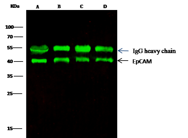 IP (Immunoprecipitation)
(EpCAM was immunoprecipitated using:Lane A:0.5 mg MCF-7 Whole Cell LysateLane B:0.5 mg A431 Whole Cell LysateLane C:0.5 mg HepG2 Whole Cell LysateLane D:0.5 mg Caco-2 Whole Cell Lysate0.5 uL anti-EpCAM rabbit monoclonal antibody and 15 ul of 50 % Protein G agarose.Primary antibody:Anti-EpCAM rabbit monoclonal antibody,at 1:5000 dilution Secondary antibody:Dylight 800-labeled antibody to rabbit IgG (H+L), at 1:5000 dilution Developed using the odssey technique.Performed under reducing conditions.Predicted band size: 40 kDaObserved band size: 40 kDa)
IP (Immunoprecipitation)
(EpCAM was immunoprecipitated using:Lane A:0.5 mg MCF-7 Whole Cell LysateLane B:0.5 mg A431 Whole Cell LysateLane C:0.5 mg HepG2 Whole Cell LysateLane D:0.5 mg Caco-2 Whole Cell Lysate0.5 uL anti-EpCAM rabbit monoclonal antibody and 15 ul of 50 % Protein G agarose.Primary antibody:Anti-EpCAM rabbit monoclonal antibody,at 1:5000 dilution Secondary antibody:Dylight 800-labeled antibody to rabbit IgG (H+L), at 1:5000 dilution Developed using the odssey technique.Performed under reducing conditions.Predicted band size: 40 kDaObserved band size: 40 kDa)
EpCAM, Monoclonal Antibody (Cat# AAA27736)
Full Name
Recombinant Anti-EpCAM Antibody, Rabbit Monoclonal
Gene Names
EPCAM; ESA; KSA; M4S1; MK-1; DIAR5; EGP-2; EGP40; KS1/4; MIC18; TROP1; EGP314; HNPCC8; TACSTD1
Reactivity
Human
Applications
Western Blot, Immunohistochemistry, Flow Cytometry, Immunocytochemistry, Immunofluorescence, Immunoprecipitation
Purity
Protein A
Pricing


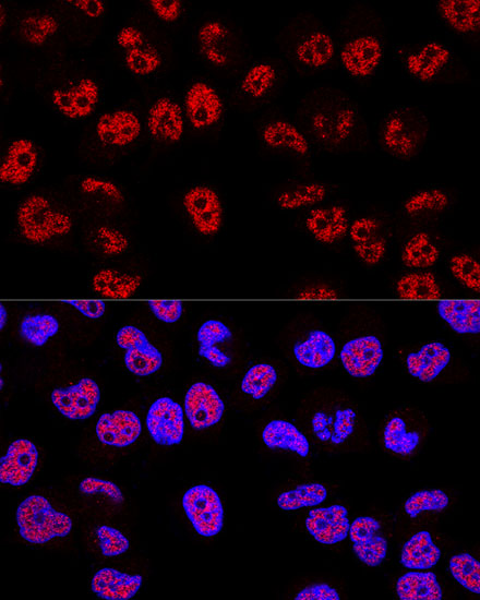


 ChIP (Chromatin Immunoprecipitation)
(Chromatin immunoprecipitation analysis of extracts of 293T cells, using p53 antibody and rabbit IgG. The amount of immunoprecipitated DNA was checked by quantitative PCR. Histogram was constructed by the ratios of the immunoprecipitated DNA to the input.)
ChIP (Chromatin Immunoprecipitation)
(Chromatin immunoprecipitation analysis of extracts of 293T cells, using p53 antibody and rabbit IgG. The amount of immunoprecipitated DNA was checked by quantitative PCR. Histogram was constructed by the ratios of the immunoprecipitated DNA to the input.)
p53, Polyclonal Antibody (Cat# AAA28404)
Full Name
[KO Validated] p53 Rabbit pAb
Gene Names
CDC45; CDC45L; CDC45L2; PORC-PI-1
Reactivity
Human
Applications
Western Blot, Immunohistochemistry, Immunofluorescence, Immunocytochemistry, Immunoprecipitation, Chromatin Immunoprecipitation
Purity
Affinity purification
Pricing


 IHC (Immunohistochemistry)
(Figure 7. IHC analysis of DLST using anti-DLST antibody (AAA19536).DLST was detected in a paraffin-embedded section of rat lymph node tissue. Heat mediated antigen retrieval was performed in EDTA buffer (pH 8.0, epitope retrieval solution). The tissue section was blocked with 10% goat serum. The tissue section was then incubated with 2 ug/ml rabbit anti-DLST Antibody (AAA19536) overnight at 4 degree C. Biotinylated goat anti-rabbit IgG was used as secondary antibody and incubated for 30 minutes at 37 degree C. The tissue section was developed using Strepavidin-Biotin-Complex (SABC) with DAB as the chromogen.)
IHC (Immunohistochemistry)
(Figure 7. IHC analysis of DLST using anti-DLST antibody (AAA19536).DLST was detected in a paraffin-embedded section of rat lymph node tissue. Heat mediated antigen retrieval was performed in EDTA buffer (pH 8.0, epitope retrieval solution). The tissue section was blocked with 10% goat serum. The tissue section was then incubated with 2 ug/ml rabbit anti-DLST Antibody (AAA19536) overnight at 4 degree C. Biotinylated goat anti-rabbit IgG was used as secondary antibody and incubated for 30 minutes at 37 degree C. The tissue section was developed using Strepavidin-Biotin-Complex (SABC) with DAB as the chromogen.)
DLST, Polyclonal Antibody (Cat# AAA19536)
Full Name
Anti-DLST Antibody Picoband
Gene Names
DLST; DLTS
Reactivity
Human, Mouse, Rat
Applications
Western Blot, Immunohistochemistry
Purity
Immunogen affinity purified.
Pricing

 FCM (Flow Cytometry)
(Figure 7. Flow Cytometry analysis of RT4 cells using anti-PTRHD1 antibody (AAA20010).Overlay histogram showing RT4 cells stained with AAA20010 (Blue line). To facilitate intracellular staining, cells were fixed with 4% paraformaldehyde and permeabilized with permeabilization buffer. The cells were blocked with 10% normal goat serum. And then incubated with rabbit anti-PTRHD1 Antibody (AAA20010, 1ug/1x106 cells) for 30 min at 20 degree C. DyLight488 conjugated goat anti-rabbit IgG was used as secondary antibody for 30 minutes at 20 degree C. Isotype control antibody (Green line) was rabbit IgG (1ug/1x106) used under the same conditions. Unlabelled sample (Red line) was also used as a control.)
FCM (Flow Cytometry)
(Figure 7. Flow Cytometry analysis of RT4 cells using anti-PTRHD1 antibody (AAA20010).Overlay histogram showing RT4 cells stained with AAA20010 (Blue line). To facilitate intracellular staining, cells were fixed with 4% paraformaldehyde and permeabilized with permeabilization buffer. The cells were blocked with 10% normal goat serum. And then incubated with rabbit anti-PTRHD1 Antibody (AAA20010, 1ug/1x106 cells) for 30 min at 20 degree C. DyLight488 conjugated goat anti-rabbit IgG was used as secondary antibody for 30 minutes at 20 degree C. Isotype control antibody (Green line) was rabbit IgG (1ug/1x106) used under the same conditions. Unlabelled sample (Red line) was also used as a control.)
PTRHD1, Polyclonal Antibody (Cat# AAA20010)
Full Name
Anti-PTRHD1 Antibody Picoband
Gene Names
PTRHD1; C2orf79
Reactivity
Human, Mouse, Rat
Applications
Western Blot, Immunohistochemistry, Immunocytochemistry, Immunofluorescence, Flow Cytometry
Purity
Immunogen affinity purified.
Pricing
 Standard Curve (Sample)
PCSK9, ELISA Kit (Cat# AAA17859)
Full Name
Mouse PCSK9 ELISA Kit
Gene Names
Pcsk9; FH3; PC9; Narc1; HCHOLA3; AI415265; AI747682
Reactivity
Mouse
Applications
Sandwich ELISA
Pricing


 IHC (Immunohistchemistry)
(Immunohistochemistry of paraffin-embedded human adenomyosis using LSP1 Antibody at dilution of 1:100 (40x lens).)
IHC (Immunohistchemistry)
(Immunohistochemistry of paraffin-embedded human adenomyosis using LSP1 Antibody at dilution of 1:100 (40x lens).)
LSP1, Polyclonal Antibody (Cat# AAA28192)
Full Name
LSP1 Polyclonal Antibody
Gene Names
LSP1; WP34; pp52
Reactivity
Human, Mouse
Applications
Western Blot, Immunohistochemistry
Purity
Affinity Purification
Pricing


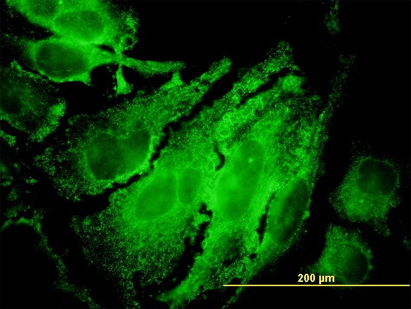

 WB (Western Blot)
(RPS6KB1 monoclonal antibody (M04), clone 4H4. Western Blot analysis of RPS6KB1 expression in PC-12.)
WB (Western Blot)
(RPS6KB1 monoclonal antibody (M04), clone 4H4. Western Blot analysis of RPS6KB1 expression in PC-12.)
RPS6KB1, Monoclonal Antibody (Cat# AAA26636)
Full Name
RPS6KB1 (Ribosomal Protein S6 Kinase, 70kD, Polypeptide 1, PS6K, S6K, S6K1, STK14A, p70(S6K)-alpha, p70-S6K, p70-alpha) (PE)
Gene Names
RPS6KB1; S6K; PS6K; S6K1; STK14A; p70-S6K; p70 S6KA; p70-alpha; S6K-beta-1; p70(S6K)-alpha
Applications
Immunofluorescence, Western Blot
Purity
Purified
Pricing


 Application Data
(Detection limit for recombinant GST tagged RPS7 is ~1ng/ml as a capture antibody.)
Application Data
(Detection limit for recombinant GST tagged RPS7 is ~1ng/ml as a capture antibody.)
RPS7, Monoclonal Antibody (Cat# AAA25233)
Full Name
RPS7 (40S Ribosomal Protein S7) (FITC)
Gene Names
RPS7; S7; DBA8
Reactivity
Human, Mouse, Rat
Applications
Immunofluorescence, Immunohistochemistry, Western Blot
Purity
Purified by Protein A Affinity Chromatography.
Pricing


 WB (Western Blot)
(DDX56 monoclonal antibody. Western Blot analysis of DDX56 expression in PC-12.)
WB (Western Blot)
(DDX56 monoclonal antibody. Western Blot analysis of DDX56 expression in PC-12.)
DDX56, Monoclonal Antibody (Cat# AAA25371)
Full Name
DDX56 (Probable ATP-dependent RNA Helicase DDX56, ATP-dependent 61kD Nucleolar RNA Helicase, DEAD Box Protein 21, DEAD Box Protein 56, DDX21, NOH61) (HRP)
Reactivity
Human, Rat
Applications
Immunohistochemistry, Western Blot
Purity
Purified by Protein A Affinity Chromatography.
Pricing


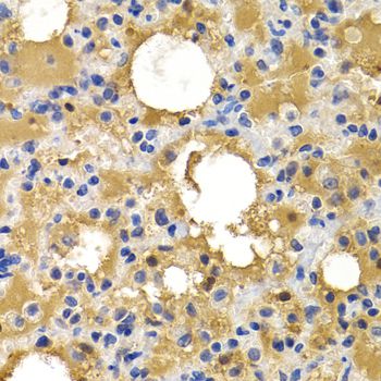

 IF (Immunofluorescence)
(Immunofluorescence analysis of HeLa cells using HSPB8 antibody. Blue: DAPI for nuclear staining.)
IF (Immunofluorescence)
(Immunofluorescence analysis of HeLa cells using HSPB8 antibody. Blue: DAPI for nuclear staining.)
HSPB8, Polyclonal Antibody (Cat# AAA10770)
Full Name
HSPB8 Polyclonal Antibody
Gene Names
HSPB8; H11; HMN2; CMT2L; DHMN2; E2IG1; HMN2A; HSP22
Reactivity
Human, Mouse, Rat
Applications
Western Blot, Immunohistochemistry, Immunofluorescence
Purity
Affinity Purification
Pricing


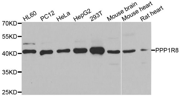
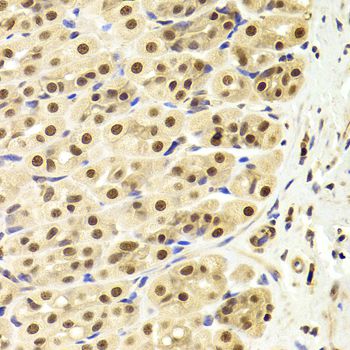 IHC (Immunohistchemistry)
(Immunohistochemistry of paraffin-embedded rat stomach using PPP1R8 Antibody at dilution of 1:100 (40x lens).)
IHC (Immunohistchemistry)
(Immunohistochemistry of paraffin-embedded rat stomach using PPP1R8 Antibody at dilution of 1:100 (40x lens).)
PPP1R8, Polyclonal Antibody (Cat# AAA28105)
Full Name
PPP1R8 Polyclonal Antibody
Gene Names
PPP1R8; ARD1; ARD-1; NIPP1; NIPP-1; PRO2047
Reactivity
Human, Mouse, Rat
Applications
Western Blot, Immunohistochemistry
Purity
Affinity Purification
Pricing


 IHC (Immunohistchemistry)
(Figure 6. IHC analysis of CD13/ANPEP using anti-CD13/ANPEP antibody (AAA19371).CD13/ANPEP was detected in paraffin-embedded section of rat kidney tissue. Heat mediated antigen retrieval was performed in EDTA buffer (pH8. 0, epitope retrieval solution). The tissue section was blocked with 10% goat serum. The tissue section was then incubated with 2μg/ml mouse anti-CD13/ANPEP Antibody (AAA19371) overnight at 4 degree C. Biotinylated goat anti-mouse IgG was used as secondary antibody and incubated for 30 minutes at 37 degree C. The tissue section was developed using Strepavidin-Biotin-Complex (SABC) (Catalog # with DAB as the chromogen.)
IHC (Immunohistchemistry)
(Figure 6. IHC analysis of CD13/ANPEP using anti-CD13/ANPEP antibody (AAA19371).CD13/ANPEP was detected in paraffin-embedded section of rat kidney tissue. Heat mediated antigen retrieval was performed in EDTA buffer (pH8. 0, epitope retrieval solution). The tissue section was blocked with 10% goat serum. The tissue section was then incubated with 2μg/ml mouse anti-CD13/ANPEP Antibody (AAA19371) overnight at 4 degree C. Biotinylated goat anti-mouse IgG was used as secondary antibody and incubated for 30 minutes at 37 degree C. The tissue section was developed using Strepavidin-Biotin-Complex (SABC) (Catalog # with DAB as the chromogen.)
CD13/ANPEP, Monoclonal Antibody (Cat# AAA19371)
Full Name
Anti-CD13/ANPEP Antibody (monoclonal, 5B9)
Gene Names
ANPEP; APN; CD13; LAP1; P150; PEPN; GP150
Reactivity
Human, Rat, Monkey
Applications
Western Blot, Immunohistochemistry
Purity
Immunogen affinity purified.
Pricing


 IHC (Immunohistochemistry)
(Figure 8. IHC analysis of beta Catenin using anti-beta Catenin antibody (AAA19129).beta Catenin was detected in paraffin-embedded section of rat small intestine tissue. Heat mediated antigen retrieval was performed in citrate buffer (pH6, epitope retrieval solution) for 20 mins. The tissue section was blocked with 10% goat serum. The tissue section was then incubated with 1ug/ml rabbit anti-beta Catenin Antibody (AAA19129) overnight at 4 degree C. Biotinylated goat anti-rabbit IgG was used as secondary antibody and incubated for 30 minutes at 37 degree C. The tissue section was developed using Strepavidin-Biotin-Complex (SABC) with DAB as the chromogen.)
IHC (Immunohistochemistry)
(Figure 8. IHC analysis of beta Catenin using anti-beta Catenin antibody (AAA19129).beta Catenin was detected in paraffin-embedded section of rat small intestine tissue. Heat mediated antigen retrieval was performed in citrate buffer (pH6, epitope retrieval solution) for 20 mins. The tissue section was blocked with 10% goat serum. The tissue section was then incubated with 1ug/ml rabbit anti-beta Catenin Antibody (AAA19129) overnight at 4 degree C. Biotinylated goat anti-rabbit IgG was used as secondary antibody and incubated for 30 minutes at 37 degree C. The tissue section was developed using Strepavidin-Biotin-Complex (SABC) with DAB as the chromogen.)
beta Catenin, Polyclonal Antibody (Cat# AAA19129)
Full Name
Anti-beta Catenin Picoband Antibody
Gene Names
CTNNB1; EVR7; CTNNB; MRD19; armadillo
Reactivity
Human, Mouse, Rat
No cross reactivity with other proteins.
No cross reactivity with other proteins.
Applications
Immunohistochemistry, Western Blot
Purity
Immunogen affinity purified
Pricing


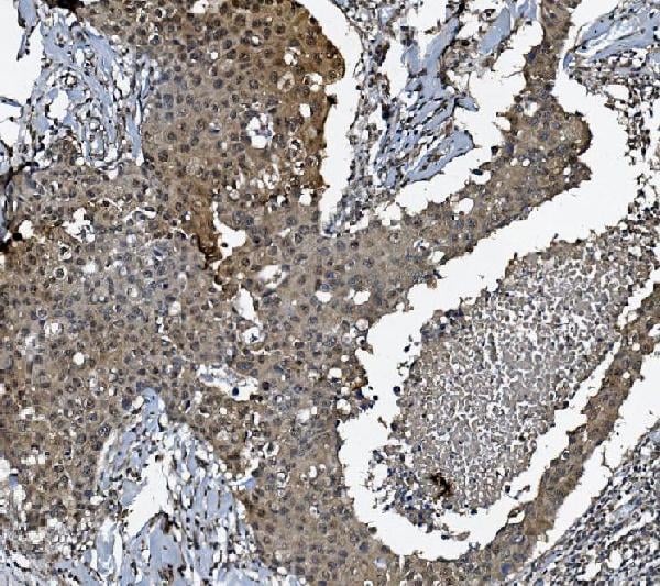


 FCM (Flow Cytometry)
(Figure 9. Flow Cytometry analysis of SiHa cells using anti-DCTN1/p150-glued antibody (AAA19253).Overlay histogram showing SiHa cells stained with AAA19253 (Blue line). The cells were blocked with 10% normal goat serum. And then incubated with rabbit anti-DCTN1/p150-glued Antibody (AAA19253, 1μg/1x106 cells) for 30 min at 20 degree C. DyLight®488 conjugated goat anti-rabbit IgG (5-10μg/1x106 cells) was used as secondary antibody for 30 minutes at 20 degree C. Isotype control antibody (Green line) was rabbit IgG (1μg/1x106) used under the same conditions. Unlabelled sample (Red line) was also used as a control.)
FCM (Flow Cytometry)
(Figure 9. Flow Cytometry analysis of SiHa cells using anti-DCTN1/p150-glued antibody (AAA19253).Overlay histogram showing SiHa cells stained with AAA19253 (Blue line). The cells were blocked with 10% normal goat serum. And then incubated with rabbit anti-DCTN1/p150-glued Antibody (AAA19253, 1μg/1x106 cells) for 30 min at 20 degree C. DyLight®488 conjugated goat anti-rabbit IgG (5-10μg/1x106 cells) was used as secondary antibody for 30 minutes at 20 degree C. Isotype control antibody (Green line) was rabbit IgG (1μg/1x106) used under the same conditions. Unlabelled sample (Red line) was also used as a control.)
DCTN1/p150-glued, Polyclonal Antibody (Cat# AAA19253)
Full Name
Anti-DCTN1/p150-glued Antibody
Gene Names
DCTN1; P135; DP-150; DAP-150
Reactivity
Human, Mouse, Rat
Applications
Western Blot, Immunohistochemistry, Immunocytochemistry, Immunofluorescence, Flow Cytometry, Direct ELISA
Purity
Immunogen affinity purified.
Pricing

 FCM (Flow Cytometry)
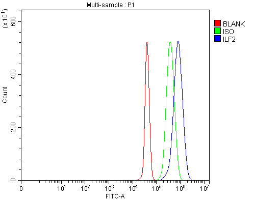
(Figure 10. Flow Cytometry analysis of JK cells using anti-NF45/ILF2 antibody (AAA19816).Overlay histogram showing JK cells stained with AAA19816 (Blue line). To facilitate intracellular staining, cells were fixed with 4% paraformaldehyde and permeabilized with permeabilization buffer. The cells were blocked with 10% normal goat serum. And then incubated with rabbit anti-NF45/ILF2 Antibody (AAA19816, 1ug/1x106 cells) for 30 min at 20 degree C. DyLight488 conjugated goat anti-rabbit IgG was used as secondary antibody for 30 minutes at 20 degree C. Isotype control antibody (Green line) was rabbit IgG (1ug/1x106) used under the same conditions. Unlabelled sample (Red line) was also used as a control.)
FCM (Flow Cytometry)
(Figure 10. Flow Cytometry analysis of JK cells using anti-NF45/ILF2 antibody (AAA19816).Overlay histogram showing JK cells stained with AAA19816 (Blue line). To facilitate intracellular staining, cells were fixed with 4% paraformaldehyde and permeabilized with permeabilization buffer. The cells were blocked with 10% normal goat serum. And then incubated with rabbit anti-NF45/ILF2 Antibody (AAA19816, 1ug/1x106 cells) for 30 min at 20 degree C. DyLight488 conjugated goat anti-rabbit IgG was used as secondary antibody for 30 minutes at 20 degree C. Isotype control antibody (Green line) was rabbit IgG (1ug/1x106) used under the same conditions. Unlabelled sample (Red line) was also used as a control.)
NF45/ILF2, Polyclonal Antibody (Cat# AAA19816)
Full Name
Anti-NF45/ILF2 Antibody Picoband
Gene Names
ILF2; NF45; PRO3063
Reactivity
Human, Mouse, Rat, Monkey
Applications
Immunofluorescence, Immunohistochemistry, Immunocytochemistry, Western Blot, Flow Cytometry
Purity
Immunogen affinity purified.
Pricing


 IHC (Immunohistochemistry)
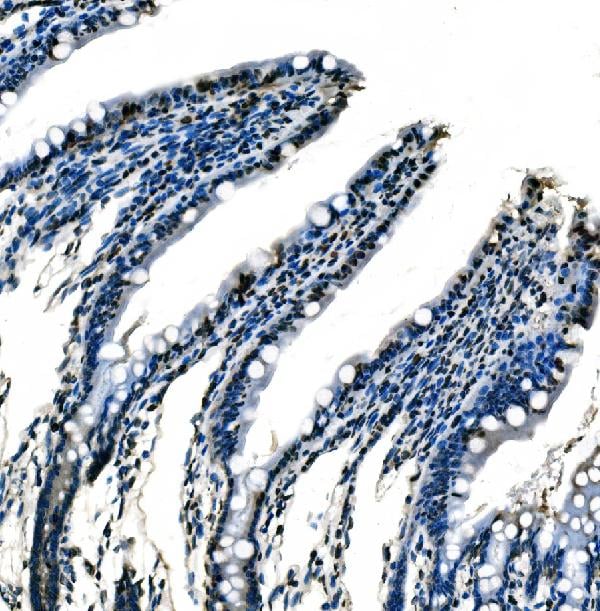
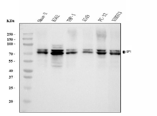
(Figure 14. IHC analysis of splicing factor 1 using anti-splicing factor 1 antibody (AAA19677).splicing factor 1 was detected in a paraffin-embedded section of rat kidney tissue. Heat mediated antigen retrieval was performed in EDTA buffer (pH 8.0, epitope retrieval solution). The tissue section was blocked with 10% goat serum. The tissue section was then incubated with 2 ug/ml mouse anti-splicing factor 1 Antibody (AAA19677) overnight at 4 degree C. Biotinylated goat anti-mouse IgG was used as secondary antibody and incubated for 30 minutes at 37 degree C. The tissue section was developed using Strepavidin-Biotin-Complex (SABC) (#SA1021) with DAB as the chromogen.)
IHC (Immunohistochemistry)
(Figure 14. IHC analysis of splicing factor 1 using anti-splicing factor 1 antibody (AAA19677).splicing factor 1 was detected in a paraffin-embedded section of rat kidney tissue. Heat mediated antigen retrieval was performed in EDTA buffer (pH 8.0, epitope retrieval solution). The tissue section was blocked with 10% goat serum. The tissue section was then incubated with 2 ug/ml mouse anti-splicing factor 1 Antibody (AAA19677) overnight at 4 degree C. Biotinylated goat anti-mouse IgG was used as secondary antibody and incubated for 30 minutes at 37 degree C. The tissue section was developed using Strepavidin-Biotin-Complex (SABC) (#SA1021) with DAB as the chromogen.)
splicing factor 1, Monoclonal Antibody (Cat# AAA19677)
Full Name
Anti-splicing factor 1 Antibody Picoband (monoclonal, 2F5D10)
Gene Names
SF1; BBP; MBBP; ZFM1; ZNF162; D11S636; ZCCHC25
Reactivity
Human, Mouse, Rat
Applications
Western Blot, Immunohistochemistry, Immunocytochemistry, Immunofluorescence, Flow Cytometry
Purity
Immunogen affinity purified.
Pricing


 IF (Immunofluorescence)
(Immunofluorescence analysis of PC-12 cells using METTL14 Rabbit pAb (AAA28216) at dilution of 1:25 (40x lens). Blue: DAPI for nuclear staining.)
IF (Immunofluorescence)
(Immunofluorescence analysis of PC-12 cells using METTL14 Rabbit pAb (AAA28216) at dilution of 1:25 (40x lens). Blue: DAPI for nuclear staining.)
METTL14, Polyclonal Antibody (Cat# AAA28216)
Full Name
METTL14 Polyclonal Antibody
Reactivity
Human
Applications
Western Blot, Immunohistochemistry, Immunofluorescence, Immunoprecipitation
Purity
Affinity purification
Pricing



 IHC (Immunohistchemistry)
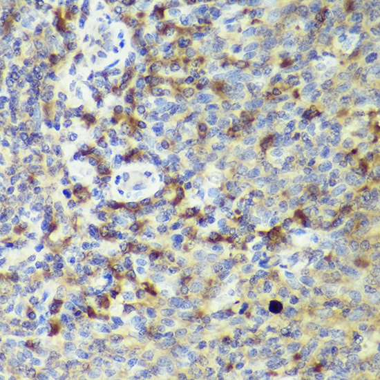
(Immunohistochemistry of paraffin-embedded rat spleen using IL18 antibody at dilution of 1:50 (40x lens).)
IHC (Immunohistchemistry)
(Immunohistochemistry of paraffin-embedded rat spleen using IL18 antibody at dilution of 1:50 (40x lens).)
IL18, Polyclonal Antibody (Cat# AAA28388)
Full Name
IL18 Rabbit pAb
Gene Names
Il18; Igif; Il-18
Reactivity
Human, Mouse, Rat
Applications
Western Blot, Immunohistochemistry
Purity
Affinity purification
Pricing


 WB (Western Blot)
(SMAD3 monoclonal antibody (M01), clone 2C12. Western Blot analysis of SMAD3 expression in Hs 181.Tes.)
WB (Western Blot)
(SMAD3 monoclonal antibody (M01), clone 2C12. Western Blot analysis of SMAD3 expression in Hs 181.Tes.)
SMAD3, Monoclonal Antibody (Cat# AAA26030)
Full Name
SMAD3 (SMAD Family Member 3, DKFZp586N0721, DKFZp686J10186, HSPC193, HsT17436, JV15-2, MADH3, MGC60396) (APC)
Gene Names
SMAD3; LDS3; LDS1C; MADH3; JV15-2; HSPC193; HsT17436
Applications
Immunohistochemistry, Western Blot
Purity
Purified
Pricing


 IF (Immunofluorescence)
(Figure 6. IF analysis of GRSF1 using anti- GRSF1 antibody (AAA19315).GRSF1 was detected in immunocytochemical section of MCF-7 cells. Enzyme antigen retrieval was performed using IHC enzyme antigen retrieval reagent for 15 mins. The cells were blocked with 10% goat serum. And then incubated with 5μg/mL rabbit anti- GRSF1 Antibody (AAA19315) overnight at 4 degree C. DyLight®488 Conjugated Goat Anti-Rabbit IgG was used as secondary antibody at 1:100 dilution and incubated for 30 minutes at 37 degree C. The section was counterstained with DAPI. Visualize using a fluorescence microscope and filter sets appropriate for the label used.)
IF (Immunofluorescence)
(Figure 6. IF analysis of GRSF1 using anti- GRSF1 antibody (AAA19315).GRSF1 was detected in immunocytochemical section of MCF-7 cells. Enzyme antigen retrieval was performed using IHC enzyme antigen retrieval reagent for 15 mins. The cells were blocked with 10% goat serum. And then incubated with 5μg/mL rabbit anti- GRSF1 Antibody (AAA19315) overnight at 4 degree C. DyLight®488 Conjugated Goat Anti-Rabbit IgG was used as secondary antibody at 1:100 dilution and incubated for 30 minutes at 37 degree C. The section was counterstained with DAPI. Visualize using a fluorescence microscope and filter sets appropriate for the label used.)
GRSF1, Polyclonal Antibody (Cat# AAA19315)
Full Name
Anti-GRSF1 Antibody
Reactivity
Human
Applications
Western Blot, Immunohistochemistry, Immunocytochemistry, Immunofluorescence, Flow Cytometry, Direct ELISA
Purity
Immunogen affinity purified.
Pricing

 FCM (Flow Cytometry)
(Figure 10. Flow Cytometry analysis of Raw264.7 cells using anti-Lat antibody (AAA19756).Overlay histogram showing Raw264.7 cells stained with AAA19756 (Blue line). The cells were fixed with 4% paraformaldehyde and blocked with 10% normal goat serum. And then incubated with rabbit anti-Lat Antibody (AAA19756, 1ug/1x106 cells) for 30 min at 20 degree C. DyLight488 conjugated goat anti-rabbit IgG was used as secondary antibody for 30 minutes at 20 degree C. Isotype control antibody (Green line) was rabbit IgG (1ug/1x106) used under the same conditions. Unlabelled sample (Red line) was also used as a control.)
FCM (Flow Cytometry)
(Figure 10. Flow Cytometry analysis of Raw264.7 cells using anti-Lat antibody (AAA19756).Overlay histogram showing Raw264.7 cells stained with AAA19756 (Blue line). The cells were fixed with 4% paraformaldehyde and blocked with 10% normal goat serum. And then incubated with rabbit anti-Lat Antibody (AAA19756, 1ug/1x106 cells) for 30 min at 20 degree C. DyLight488 conjugated goat anti-rabbit IgG was used as secondary antibody for 30 minutes at 20 degree C. Isotype control antibody (Green line) was rabbit IgG (1ug/1x106) used under the same conditions. Unlabelled sample (Red line) was also used as a control.)
Lat, Polyclonal Antibody (Cat# AAA19756)
Full Name
Anti-Lat Antibody Picoband
Gene Names
Lat; pp36; p36-38
Reactivity
Mouse, Rat
Applications
Western Blot, Immunohistochemistry, Flow Cytometry
Purity
Immunogen affinity purified.
Pricing


 IF (Immunofluorescence)
(Figure 6. IF analysis of CBX1/HP1 beta using anti-CBX1/HP1 beta antibody (AAA19508).CBX1/HP1 beta was detected in an immunocytochemical section of CACO-2 cells. Enzyme antigen retrieval was performed using IHC enzyme antigen retrieval reagent (AR0022) for 15 mins. The cells were blocked with 10% goat serum. And then incubated with 5 ug/mL rabbit anti-CBX1/HP1 beta Antibody (AAA19508) overnight at 4 degree C. DyLight594 Conjugated Goat Anti-Rabbit IgG (BA1142) was used as secondary antibody at 1:100 dilution and incubated for 30 minutes at 37 degree C. The section was counterstained with DAPI. Visualize using a fluorescence microscope and filter sets appropriate for the label used.)
IF (Immunofluorescence)
(Figure 6. IF analysis of CBX1/HP1 beta using anti-CBX1/HP1 beta antibody (AAA19508).CBX1/HP1 beta was detected in an immunocytochemical section of CACO-2 cells. Enzyme antigen retrieval was performed using IHC enzyme antigen retrieval reagent (AR0022) for 15 mins. The cells were blocked with 10% goat serum. And then incubated with 5 ug/mL rabbit anti-CBX1/HP1 beta Antibody (AAA19508) overnight at 4 degree C. DyLight594 Conjugated Goat Anti-Rabbit IgG (BA1142) was used as secondary antibody at 1:100 dilution and incubated for 30 minutes at 37 degree C. The section was counterstained with DAPI. Visualize using a fluorescence microscope and filter sets appropriate for the label used.)
CBX1/HP1 beta, Polyclonal Antibody (Cat# AAA19508)
Full Name
Anti-CBX1/HP1 beta Antibody Picoband
Gene Names
CBX1; CBX; M31; MOD1; p25beta; HP1-BETA; HP1Hsbeta; HP1Hs-beta
Reactivity
Human, Mouse, Rat
Applications
Western Blot, Immunohistochemistry, Immunofluorescence
Purity
Immunogen affinity purified.
Pricing
 IHC (Immunohistchemistry)
(Figure 9. IHC analysis of MEP1B using anti-MEP1B antibody (AAA19870).MEP1B was detected in a paraffin-embedded section of rat kidney tissue. Heat mediated antigen retrieval was performed in EDTA buffer (pH 8.0, epitope retrieval solution). The tissue section was blocked with 10% goat serum. The tissue section was then incubated with 2ug/ml rabbit anti-MEP1B Antibody (AAA19870) overnight at 4 degree C. Peroxidase Conjugated Goat Anti-rabbit IgG was used as secondary antibody and incubated for 30 minutes at 37 degree C. The tissue section was developed using HRP Conjugated Rabbit IgG Super Vision Assay Kit ( epitope retrieval solution). The tissue section was blocked with 10% goat serum. The tissue section was then incubated with 2ug/ml rabbit anti-MEP1B Antibody (AAA19870) overnight at 4 degree C. Peroxidase Conjugated Goat Anti-rabbit IgG was used as secondary antibody and incubated for 30 minutes at 37 degree C. The tissue section was developed using HRP Conjugated Rabbit IgG Super Vision Assay Kit ( epitope retrieval solution). The tissue section was blocked with 10% goat serum. The tissue section was then incubated with 2ug/ml rabbit anti-MEP1B Antibody (AAA19870) overnight at 4 degree C. Peroxidase Conjugated Goat Anti-rabbit IgG was used as secondary antibody and incubated for 30 minutes at 37 degree C. The tissue section was developed using HRP Conjugated Rabbit IgG Super Vision Assay Kit ( epitope retrieval solution). The tissue section was blocked with 10% goat serum. The tissue section was then incubated with 2ug/ml rabbit anti-MEP1B Antibody (AAA19870) overnight at 4 degree C. Peroxidase Conjugated Goat Anti-rabbit IgG was used as secondary antibody and incubated for 30 minutes at 37 degree C. The tissue section was developed using HRP Conjugated Rabbit IgG Super Vision Assay Kit ( epitope retrieval solution). The tissue section was blocked with 10% goat serum. The tissue section was then incubated with 2ug/ml rabbit anti-MEP1B Antibody (AAA19870) overnight at 4 degree C. Peroxidase Conjugated Goat Anti-rabbit IgG was used as secondary antibody and incubated for 30 minutes at 37 degree C. The tissue section was developed using HRP Conjugated Rabbit IgG Super Vision Assay Kit ( epitope retrieval solution). The tissue section was blocked with 10% goat serum. The tissue section was then incubated with 2ug/ml rabbit anti-MEP1B Antibody (AAA19870) overnight at 4 degree C. Peroxidase Conjugated Goat Anti-rabbit IgG was used as secondary antibody and incubated for 30 minutes at 37 degree C. The tissue section was developed using HRP Conjugated Rabbit IgG Super Vision Assay Kit ( epitope retrieval solution). The tissue section was blocked with 10% goat serum. The tissue section was then incubated with 2ug/ml rabbit anti-MEP1B Antibody (AAA19870) overnight at 4 degree C. Peroxidase Conjugated Goat Anti-rabbit IgG was used as secondary antibody and incubated for 30 minutes at 37 degree C. The tissue section was developed using HRP Conjugated Rabbit IgG Super Vision Assay Kit ( epitope retrieval solution). The tissue section was blocked with 10% goat serum. The tissue section was then incubated with 2ug/ml rabbit anti-MEP1B Antibody (AAA19870) overnight at 4 degree C. Peroxidase Conjugated Goat Anti-rabbit IgG was used as secondary antibody and incubated for 30 minutes at 37 degree C. The tissue section was developed using HRP Conjugated Rabbit IgG Super Vision Assay Kit (Lane 2: mouse kidney tissue lysates.After electrophoresis, proteins were transferred to a nitrocellulose membrane at 150 mA for 50-90 minutes. Blocked the membrane with 5% non-fat milk/TBS for 1.5 hour at RT. The membrane was incubated with rabbit anti-MEP1B antigen affinity purified polyclonal antibody (#AAA19870) at 0.5ug/mL overnight at 4 degree C, then washed with TBS-0.1%Tween 3 times with 5 minutes each and probed with a goat anti-rabbit IgG-HRP secondary antibody at a dilution of 1:5000 for 1.5 hour at RT. The signal is developed using an Enhanced Chemiluminescent detection (ECL) kit with Tanon 5200 system. A specific band was detected for MEP1B at approximately 97 kDa. The expected band size for MEP1B is at 80 kDa.)
IHC (Immunohistchemistry)
(Figure 9. IHC analysis of MEP1B using anti-MEP1B antibody (AAA19870).MEP1B was detected in a paraffin-embedded section of rat kidney tissue. Heat mediated antigen retrieval was performed in EDTA buffer (pH 8.0, epitope retrieval solution). The tissue section was blocked with 10% goat serum. The tissue section was then incubated with 2ug/ml rabbit anti-MEP1B Antibody (AAA19870) overnight at 4 degree C. Peroxidase Conjugated Goat Anti-rabbit IgG was used as secondary antibody and incubated for 30 minutes at 37 degree C. The tissue section was developed using HRP Conjugated Rabbit IgG Super Vision Assay Kit ( epitope retrieval solution). The tissue section was blocked with 10% goat serum. The tissue section was then incubated with 2ug/ml rabbit anti-MEP1B Antibody (AAA19870) overnight at 4 degree C. Peroxidase Conjugated Goat Anti-rabbit IgG was used as secondary antibody and incubated for 30 minutes at 37 degree C. The tissue section was developed using HRP Conjugated Rabbit IgG Super Vision Assay Kit ( epitope retrieval solution). The tissue section was blocked with 10% goat serum. The tissue section was then incubated with 2ug/ml rabbit anti-MEP1B Antibody (AAA19870) overnight at 4 degree C. Peroxidase Conjugated Goat Anti-rabbit IgG was used as secondary antibody and incubated for 30 minutes at 37 degree C. The tissue section was developed using HRP Conjugated Rabbit IgG Super Vision Assay Kit ( epitope retrieval solution). The tissue section was blocked with 10% goat serum. The tissue section was then incubated with 2ug/ml rabbit anti-MEP1B Antibody (AAA19870) overnight at 4 degree C. Peroxidase Conjugated Goat Anti-rabbit IgG was used as secondary antibody and incubated for 30 minutes at 37 degree C. The tissue section was developed using HRP Conjugated Rabbit IgG Super Vision Assay Kit ( epitope retrieval solution). The tissue section was blocked with 10% goat serum. The tissue section was then incubated with 2ug/ml rabbit anti-MEP1B Antibody (AAA19870) overnight at 4 degree C. Peroxidase Conjugated Goat Anti-rabbit IgG was used as secondary antibody and incubated for 30 minutes at 37 degree C. The tissue section was developed using HRP Conjugated Rabbit IgG Super Vision Assay Kit ( epitope retrieval solution). The tissue section was blocked with 10% goat serum. The tissue section was then incubated with 2ug/ml rabbit anti-MEP1B Antibody (AAA19870) overnight at 4 degree C. Peroxidase Conjugated Goat Anti-rabbit IgG was used as secondary antibody and incubated for 30 minutes at 37 degree C. The tissue section was developed using HRP Conjugated Rabbit IgG Super Vision Assay Kit ( epitope retrieval solution). The tissue section was blocked with 10% goat serum. The tissue section was then incubated with 2ug/ml rabbit anti-MEP1B Antibody (AAA19870) overnight at 4 degree C. Peroxidase Conjugated Goat Anti-rabbit IgG was used as secondary antibody and incubated for 30 minutes at 37 degree C. The tissue section was developed using HRP Conjugated Rabbit IgG Super Vision Assay Kit ( epitope retrieval solution). The tissue section was blocked with 10% goat serum. The tissue section was then incubated with 2ug/ml rabbit anti-MEP1B Antibody (AAA19870) overnight at 4 degree C. Peroxidase Conjugated Goat Anti-rabbit IgG was used as secondary antibody and incubated for 30 minutes at 37 degree C. The tissue section was developed using HRP Conjugated Rabbit IgG Super Vision Assay Kit (Lane 2: mouse kidney tissue lysates.After electrophoresis, proteins were transferred to a nitrocellulose membrane at 150 mA for 50-90 minutes. Blocked the membrane with 5% non-fat milk/TBS for 1.5 hour at RT. The membrane was incubated with rabbit anti-MEP1B antigen affinity purified polyclonal antibody (#AAA19870) at 0.5ug/mL overnight at 4 degree C, then washed with TBS-0.1%Tween 3 times with 5 minutes each and probed with a goat anti-rabbit IgG-HRP secondary antibody at a dilution of 1:5000 for 1.5 hour at RT. The signal is developed using an Enhanced Chemiluminescent detection (ECL) kit with Tanon 5200 system. A specific band was detected for MEP1B at approximately 97 kDa. The expected band size for MEP1B is at 80 kDa.)
MEP1B, Polyclonal Antibody (Cat# AAA19870)
Full Name
Anti-MEP1B Antibody Picoband
Reactivity
Human, Mouse, Rat
Applications
Western Blot, Immunohistochemistry
Purity
Immunogen affinity purified.
Pricing


 WB (Western Blot)
(ACTB monoclonal antibody (M03), clone 1E7. Western Blot analysis of ACTB expression in Raw 264.7.)
WB (Western Blot)
(ACTB monoclonal antibody (M03), clone 1E7. Western Blot analysis of ACTB expression in Raw 264.7.)
ACTB, Monoclonal Antibody (Cat# AAA25992)
Full Name
ACTB (Actin, beta, PS1TP5BP1) (AP)
Gene Names
ACTB; BRWS1; PS1TP5BP1
Reactivity
Human, Mouse, Rat
Applications
Immunohistochemistry, Western Blot
Purity
Purified
Pricing


 IF (Immunofluorescence)
(Immunofluorescence analysis of A549 cells using CAMK1D antibody.)
IF (Immunofluorescence)
(Immunofluorescence analysis of A549 cells using CAMK1D antibody.)
CAMK1D, Polyclonal Antibody (Cat# AAA28183)
Full Name
CAMK1D
Gene Names
CAMK1D; CKLiK; CaM-K1; CaMKID
Reactivity
Human, Mouse, Rat
Applications
Western Blot, Immunohistochemistry
Pricing


 IF (Immunofluorescence)
(Immunofluorescence analysis of MCF-7 cells using CEBPG antibody.)
IF (Immunofluorescence)
(Immunofluorescence analysis of MCF-7 cells using CEBPG antibody.)
CEBPG, Polyclonal Antibody (Cat# AAA28103)
Full Name
CEBPG Polyclonal Antibody
Gene Names
CEBPG; GPE1BP; IG/EBP-1
Reactivity
Human, Mouse, Rat
Applications
Western Blot, Immunohistochemistry, Immunofluorescence
Purity
Affinity Purification
Pricing


 Application Data
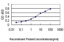
(Detection limit for recombinant GST tagged PCNA is ~0.1ng/ml as a capture antibody.)
Application Data
(Detection limit for recombinant GST tagged PCNA is ~0.1ng/ml as a capture antibody.)
PCNA, Monoclonal Antibody (Cat# AAA24601)
Full Name
PCNA (Proliferating Cell Nuclear Antigen, Cyclin, MGC8367) APC
Gene Names
PCNA; ATLD2
Reactivity
Human
Applications
Immunofluorescence, Immunoprecipitation, Western Blot
Purity
Purified by Protein A Affinity Chromatography.
Pricing


 WB (Western Blot)
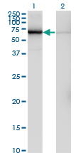
(Western Blot analysis of FTL expression in transfected 293T cell line by FTL monoclonal antibody (M16), clone X1.Lane 1: FTL transfected lysate (20 KDa).Lane 2: Non-transfected lysate.)
WB (Western Blot)
(Western Blot analysis of FTL expression in transfected 293T cell line by FTL monoclonal antibody (M16), clone X1.Lane 1: FTL transfected lysate (20 KDa).Lane 2: Non-transfected lysate.)
FTL, Monoclonal Antibody (Cat# AAA26410)
Full Name
FTL (Ferritin, Light Polypeptide, MGC71996) (FITC)
Gene Names
FTL; LFTD; NBIA3
Applications
Immunofluorescence, Immunoprecipitation, Western Blot
Purity
Purified
Pricing



 WB (Western Blot)
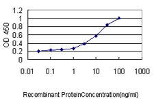
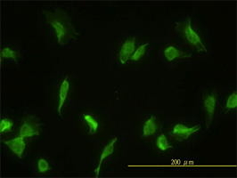
(GLMN monoclonal antibody Western Blot analysis of GLMN expression in HL-60.)
WB (Western Blot)
(GLMN monoclonal antibody Western Blot analysis of GLMN expression in HL-60.)
GLMN, Monoclonal Antibody (Cat# AAA24225)
Full Name
GLMN (FAP48, FAP68, VMGLOM, Glomulin, FK506-binding Protein-associated Protein, FAP, FKBP-associated Protein) (AP)
Gene Names
GLMN; FAP; GVM; GLML; FAP48; FAP68; FKBPAP; VMGLOM
Reactivity
Human
Applications
Immunoprecipitation, Western Blot
Purity
Purified by Protein A Affinity Chromatography.
Pricing



 WB (Western Blot)
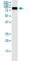
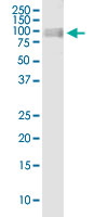
(Western Blot analysis of OPA1 expression in transfected 293T cell line by OPA1 monoclonal antibody (M06), clone 8A32.Lane 1: OPA1 transfected lysate (Predicted MW: 111.6 KDa).Lane 2: Non-transfected lysate.)
WB (Western Blot)
(Western Blot analysis of OPA1 expression in transfected 293T cell line by OPA1 monoclonal antibody (M06), clone 8A32.Lane 1: OPA1 transfected lysate (Predicted MW: 111.6 KDa).Lane 2: Non-transfected lysate.)
OPA1, Monoclonal Antibody (Cat# AAA26684)
Full Name
OPA1 (Pptic Atrophy 1 (Autosomal Dominant), FLJ12460, KIAA0567, MGM1, NPG, NTG, LargeG) (PE)
Gene Names
OPA1; NPG; NTG; MGM1; BERHS; largeG; MTDPS14
Applications
Western Blot
Purity
Purified
Pricing


 WB (Western Blot)
(RRAS2 monoclonal antibody Western Blot analysis of RRAS2 expression in A-431.)
WB (Western Blot)
(RRAS2 monoclonal antibody Western Blot analysis of RRAS2 expression in A-431.)
RRAS2, Monoclonal Antibody (Cat# AAA24938)
Full Name
RRAS2 (Ras-related Protein R-Ras2, Ras-like Protein TC21, Teratocarcinoma Oncogene, TC21) (Biotin)
Reactivity
Human, Mouse, Rat
Applications
Immunofluorescence, Immunohistochemistry, Western Blot
Purity
Purified by Protein A Affinity Chromatography.
Pricing


 FCM (Flow Cytometry)
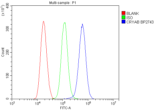
(Figure 13. Flow Cytometry analysis of THP-1 cells using anti-Alpha B Crystallin/CRYAB antibody (AAA19277).Overlay histogram showing THP-1 cells stained with AAA19277 (Blue line). The cells were blocked with 10% normal goat serum. And then incubated with rabbit anti-Alpha B Crystallin/CRYAB Antibody (AAA19277, 1μg/1x106 cells) for 30 min at 20 degree C. DyLight®488 conjugated goat anti-rabbit IgG (5-10μg/1x106 cells) was used as secondary antibody for 30 minutes at 20 degree C. Isotype control antibody (Green line) was rabbit IgG (1μg/1x106) used under the same conditions. Unlabelled sample (Red line) was also used as a control.)
FCM (Flow Cytometry)
(Figure 13. Flow Cytometry analysis of THP-1 cells using anti-Alpha B Crystallin/CRYAB antibody (AAA19277).Overlay histogram showing THP-1 cells stained with AAA19277 (Blue line). The cells were blocked with 10% normal goat serum. And then incubated with rabbit anti-Alpha B Crystallin/CRYAB Antibody (AAA19277, 1μg/1x106 cells) for 30 min at 20 degree C. DyLight®488 conjugated goat anti-rabbit IgG (5-10μg/1x106 cells) was used as secondary antibody for 30 minutes at 20 degree C. Isotype control antibody (Green line) was rabbit IgG (1μg/1x106) used under the same conditions. Unlabelled sample (Red line) was also used as a control.)
Alpha B Crystallin/CRYAB, Polyclonal Antibody (Cat# AAA19277)
Full Name
Anti-Alpha B Crystallin/CRYAB Antibody
Gene Names
CRYAB; CRYA2; CTPP2; HSPB5; CMD1II
Reactivity
Human, Mouse, Rat, Monkey
Applications
Western Blot, Immunohistochemistry, Immunocytochemistry, Immunofluorescence, Flow Cytometry, Direct ELISA
Purity
Immunogen affinity purified.
Pricing
 IF (Immunofluorescence)
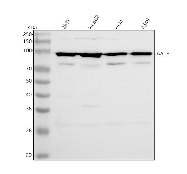
(Figure 7. IF analysis of AATF using anti-AATF antibody (AAA19804).AATF was detected in a paraffin-embedded section of rat brain tissue. Heat mediated antigen retrieval was performed in EDTA buffer (pH 8.0, epitope retrieval solution). The tissue section was blocked with 10% goat serum. The tissue section was then incubated with 5ug/mL rabbit anti-AATF Antibody (AAA19804) overnight at 4 degree C. DyLight594 Conjugated Goat Anti-Rabbit IgG (BA1142) was used as secondary antibody at 1:500 dilution and incubated for 30 minutes at 37 degree C. The section was counterstained with DAPI. Visualize using a fluorescence microscope and filter sets appropriate for the label used.)
IF (Immunofluorescence)
(Figure 7. IF analysis of AATF using anti-AATF antibody (AAA19804).AATF was detected in a paraffin-embedded section of rat brain tissue. Heat mediated antigen retrieval was performed in EDTA buffer (pH 8.0, epitope retrieval solution). The tissue section was blocked with 10% goat serum. The tissue section was then incubated with 5ug/mL rabbit anti-AATF Antibody (AAA19804) overnight at 4 degree C. DyLight594 Conjugated Goat Anti-Rabbit IgG (BA1142) was used as secondary antibody at 1:500 dilution and incubated for 30 minutes at 37 degree C. The section was counterstained with DAPI. Visualize using a fluorescence microscope and filter sets appropriate for the label used.)
AATF, Polyclonal Antibody (Cat# AAA19804)
Full Name
Anti-AATF Antibody Picoband
Gene Names
AATF; DED; CHE1; CHE-1
Reactivity
Human, Mouse, Rat
Applications
Western Blot, Immunohistochemistry, Immunocytochemistry, Immunofluorescence
Purity
Immunogen affinity purified.
Pricing


 IP (Immunoprecipitation)
(Immunoprecipitation analysis of 300ug extracts of Jurkat cells using 3ug FUS antibody. Western blot was performed from the immunoprecipitate using FUS at a dilution of 1:1000.)
IP (Immunoprecipitation)
(Immunoprecipitation analysis of 300ug extracts of Jurkat cells using 3ug FUS antibody. Western blot was performed from the immunoprecipitate using FUS at a dilution of 1:1000.)
FUS, Polyclonal Antibody (Cat# AAA28431)
Full Name
FUS Rabbit pAb
Gene Names
MAFK; P18; NFE2U
Reactivity
Human, Mouse, Rat
Applications
Western Blot, Immunohistochemistry, Immunoprecipitation
Purity
Affinity purification
Pricing


 FCM (Flow Cytometry)
(Figure 9. Flow Cytometry analysis of THP-1 cells using anti-GLUL antibody (AAA19479).Overlay histogram showing THP-1 cells stained with AAA19479 (Blue line). The cells were blocked with 10% normal goat serum. And then incubated with rabbit anti-GLUL Antibody (AAA19479, 1 ug/1x10^6 cells) for 30 min at 20 degree C. DyLight488 conjugated goat anti-rabbit IgG was used as secondary antibody for 30 minutes at 20 degree C. Isotype control antibody (Green line) was rabbit IgG (1 ug/1x10^6) used under the same conditions. Unlabelled sample (Red line) was also used as a control.)
FCM (Flow Cytometry)
(Figure 9. Flow Cytometry analysis of THP-1 cells using anti-GLUL antibody (AAA19479).Overlay histogram showing THP-1 cells stained with AAA19479 (Blue line). The cells were blocked with 10% normal goat serum. And then incubated with rabbit anti-GLUL Antibody (AAA19479, 1 ug/1x10^6 cells) for 30 min at 20 degree C. DyLight488 conjugated goat anti-rabbit IgG was used as secondary antibody for 30 minutes at 20 degree C. Isotype control antibody (Green line) was rabbit IgG (1 ug/1x10^6) used under the same conditions. Unlabelled sample (Red line) was also used as a control.)
GLUL, Polyclonal Antibody (Cat# AAA19479)
Full Name
Anti-GLUL Antibody Picoband
Gene Names
GLUL; GS; GLNS; PIG43; PIG59
Reactivity
Human, Mouse, Rat
Applications
Western Blot, Immunohistochemistry, Immunocytochemistry, Immunofluorescence, Flow Cytometry
Purity
Immunogen affinity purified.
Pricing
 Standard Curve (Sample)
TGF beta 1, ELISA Kit (Cat# AAA17900)
Full Name
Rat TGF beta 1 ELISA Kit
Gene Names
Tgfb1; Tgfb
Reactivity
Rat
Applications
Sandwich ELISA
Pricing


 FCM (Flow Cytometry)
(Figure 7. Flow Cytometry analysis of HELA cells using anti-MCU antibody (AAA19226).Overlay histogram showing HELA cells stained with AAA19226 (Blue line). The cells were blocked with 10% normal goat serum. And then incubated with rabbit anti-MCU Antibody (AAA19226, 1μg/1x106 cells) for 30 min at 20 degree C. DyLight®488 conjugated goat anti-rabbit IgG (5-10μg/1x106 cells) was used as secondary antibody for 30 minutes at 20 degree C. Isotype control antibody (Green line) was rabbit IgG (1μg/1x106) used under the same conditions. Unlabelled sample (Red line) was also used as a control.)
FCM (Flow Cytometry)
(Figure 7. Flow Cytometry analysis of HELA cells using anti-MCU antibody (AAA19226).Overlay histogram showing HELA cells stained with AAA19226 (Blue line). The cells were blocked with 10% normal goat serum. And then incubated with rabbit anti-MCU Antibody (AAA19226, 1μg/1x106 cells) for 30 min at 20 degree C. DyLight®488 conjugated goat anti-rabbit IgG (5-10μg/1x106 cells) was used as secondary antibody for 30 minutes at 20 degree C. Isotype control antibody (Green line) was rabbit IgG (1μg/1x106) used under the same conditions. Unlabelled sample (Red line) was also used as a control.)
MCU, Polyclonal Antibody (Cat# AAA19226)
Full Name
Anti-MCU Antibody
Gene Names
MCU; C10orf42; CCDC109A
Reactivity
Human, Mouse, Rat, Monkey
Applications
Western Blot, Immunohistochemistry, Flow Cytometry, Direct ELISA
Purity
Immunogen affinity purified.
Pricing


 IF (Immunofluorescence)
(Figure 9. IF analysis of P4HB using anti- P4HB antibody (AAA19258).P4HB was detected in immunocytochemical section of T-47D cells. Enzyme antigen retrieval was performed using IHC enzyme antigen retrieval reagent for 15 mins. The cells were blocked with 10% goat serum. And then incubated with 5μg/mL rabbit anti- P4HB Antibody (AAA19258) overnight at 4 degree C. DyLight®488 Conjugated Goat Anti-Rabbit IgG was used as secondary antibody at 1:100 dilution and incubated for 30 minutes at 37 degree C. The section was counterstained with DAPI. Visualize using a fluorescence microscope and filter sets appropriate for the label used.)
IF (Immunofluorescence)
(Figure 9. IF analysis of P4HB using anti- P4HB antibody (AAA19258).P4HB was detected in immunocytochemical section of T-47D cells. Enzyme antigen retrieval was performed using IHC enzyme antigen retrieval reagent for 15 mins. The cells were blocked with 10% goat serum. And then incubated with 5μg/mL rabbit anti- P4HB Antibody (AAA19258) overnight at 4 degree C. DyLight®488 Conjugated Goat Anti-Rabbit IgG was used as secondary antibody at 1:100 dilution and incubated for 30 minutes at 37 degree C. The section was counterstained with DAPI. Visualize using a fluorescence microscope and filter sets appropriate for the label used.)
P4HB, Polyclonal Antibody (Cat# AAA19258)
Full Name
Anti-P4HB Antibody
Gene Names
P4HB; DSI; GIT; PDI; PHDB; PDIA1; PO4DB; PO4HB; PROHB; ERBA2L; P4Hbeta
Reactivity
Human, Monkey, Mouse, Rat
Applications
Western Blot, Immunohistochemistry, Immunocytochemistry, Immunofluorescence, Flow Cytometry, Direct ELISA
Purity
Immunogen affinity purified.
Pricing
 IHC (Immunohistchemistry)
(Figure 9. IHC analysis of ERBB4 using anti-ERBB4 antibody (AAA19729).ERBB4 was detected in a paraffin-embedded section of human urothelial carcinoma tissue. Heat mediated antigen retrieval was performed in EDTA buffer (pH 8.0, epitope retrieval solution). The tissue section was blocked with 10% goat serum. The tissue section was then incubated with 2ug/ml rabbit anti-ERBB4 Antibody (AAA19729) overnight at 4 degree C. Peroxidase Conjugated Goat Anti-rabbit IgG was used as secondary antibody and incubated for 30 minutes at 37 degree C. The tissue section was developed using HRP Conjugated Rabbit IgG Super Vision Assay Kit ( epitope retrieval solution). The tissue section was blocked with 10% goat serum. The tissue section was then incubated with 2ug/ml rabbit anti-ERBB4 Antibody (AAA19729) overnight at 4 degree C. Peroxidase Conjugated Goat Anti-rabbit IgG was used as secondary antibody and incubated for 30 minutes at 37 degree C. The tissue section was developed using HRP Conjugated Rabbit IgG Super Vision Assay Kit ( epitope retrieval solution). The tissue section was blocked with 10% goat serum. The tissue section was then incubated with 2ug/ml rabbit anti-ERBB4 Antibody (AAA19729) overnight at 4 degree C. Peroxidase Conjugated Goat Anti-rabbit IgG was used as secondary antibody and incubated for 30 minutes at 37 degree C. The tissue section was developed using HRP Conjugated Rabbit IgG Super Vision Assay Kit ( epitope retrieval solution). The tissue section was blocked with 10% goat serum. The tissue section was then incubated with 2ug/ml rabbit anti-ERBB4 Antibody (AAA19729) overnight at 4 degree C. Peroxidase Conjugated Goat Anti-rabbit IgG was used as secondary antibody and incubated for 30 minutes at 37 degree C. The tissue section was developed using HRP Conjugated Rabbit IgG Super Vision Assay Kit ( epitope retrieval solution). The tissue section was blocked with 10% goat serum. The tissue section was then incubated with 2ug/ml rabbit anti-ERBB4 Antibody (AAA19729) overnight at 4 degree C. Peroxidase Conjugated Goat Anti-rabbit IgG was used as secondary antibody and incubated for 30 minutes at 37 degree C. The tissue section was developed using HRP Conjugated Rabbit IgG Super Vision Assay Kit ( epitope retrieval solution). The tissue section was blocked with 10% goat serum. The tissue section was then incubated with 2ug/ml rabbit anti-ERBB4 Antibody (AAA19729) overnight at 4 degree C. Peroxidase Conjugated Goat Anti-rabbit IgG was used as secondary antibody and incubated for 30 minutes at 37 degree C. The tissue section was developed using HRP Conjugated Rabbit IgG Super Vision Assay Kit ( epitope retrieval solution). The tissue section was blocked with 10% goat serum. The tissue section was then incubated with 2ug/ml rabbit anti-ERBB4 Antibody (AAA19729) overnight at 4 degree C. Peroxidase Conjugated Goat Anti-rabbit IgG was used as secondary antibody and incubated for 30 minutes at 37 degree C. The tissue section was developed using HRP Conjugated Rabbit IgG Super Vision Assay Kit ( epitope retrieval solution). The tissue section was blocked with 10% goat serum. The tissue section was then incubated with 2ug/ml rabbit anti-ERBB4 Antibody (AAA19729) overnight at 4 degree C. Peroxidase Conjugated Goat Anti-rabbit IgG was used as secondary antibody and incubated for 30 minutes at 37 degree C. The tissue section was developed using HRP Conjugated Rabbit IgG Super Vision Assay Kit (Lane 2: rat brain tissue lysates,Lane 3: rat PC-12 whole cell lysates,Lane 4: mouse brain tissue lysates.After electrophoresis, proteins were transferred to a nitrocellulose membrane at 150 mA for 50-90 minutes. Blocked the membrane with 5% non-fat milk/TBS for 1.5 hour at RT. The membrane was incubated with rabbit anti-ERBB4 antigen affinity purified polyclonal antibody (#AAA19729) at 0.5ug/mL overnight at 4 degree C, then washed with TBS-0.1%Tween 3 times with 5 minutes each and probed with a goat anti-rabbit IgG-HRP secondary antibody at a dilution of 1:5000 for 1.5 hour at RT. The signal is developed using an Enhanced Chemiluminescent detection (ECL) kit with Tanon 5200 system. A specific band was detected for ERBB4 at approximately 180 kDa. The expected band size for ERBB4 is at 147 kDa.)
IHC (Immunohistchemistry)
(Figure 9. IHC analysis of ERBB4 using anti-ERBB4 antibody (AAA19729).ERBB4 was detected in a paraffin-embedded section of human urothelial carcinoma tissue. Heat mediated antigen retrieval was performed in EDTA buffer (pH 8.0, epitope retrieval solution). The tissue section was blocked with 10% goat serum. The tissue section was then incubated with 2ug/ml rabbit anti-ERBB4 Antibody (AAA19729) overnight at 4 degree C. Peroxidase Conjugated Goat Anti-rabbit IgG was used as secondary antibody and incubated for 30 minutes at 37 degree C. The tissue section was developed using HRP Conjugated Rabbit IgG Super Vision Assay Kit ( epitope retrieval solution). The tissue section was blocked with 10% goat serum. The tissue section was then incubated with 2ug/ml rabbit anti-ERBB4 Antibody (AAA19729) overnight at 4 degree C. Peroxidase Conjugated Goat Anti-rabbit IgG was used as secondary antibody and incubated for 30 minutes at 37 degree C. The tissue section was developed using HRP Conjugated Rabbit IgG Super Vision Assay Kit ( epitope retrieval solution). The tissue section was blocked with 10% goat serum. The tissue section was then incubated with 2ug/ml rabbit anti-ERBB4 Antibody (AAA19729) overnight at 4 degree C. Peroxidase Conjugated Goat Anti-rabbit IgG was used as secondary antibody and incubated for 30 minutes at 37 degree C. The tissue section was developed using HRP Conjugated Rabbit IgG Super Vision Assay Kit ( epitope retrieval solution). The tissue section was blocked with 10% goat serum. The tissue section was then incubated with 2ug/ml rabbit anti-ERBB4 Antibody (AAA19729) overnight at 4 degree C. Peroxidase Conjugated Goat Anti-rabbit IgG was used as secondary antibody and incubated for 30 minutes at 37 degree C. The tissue section was developed using HRP Conjugated Rabbit IgG Super Vision Assay Kit ( epitope retrieval solution). The tissue section was blocked with 10% goat serum. The tissue section was then incubated with 2ug/ml rabbit anti-ERBB4 Antibody (AAA19729) overnight at 4 degree C. Peroxidase Conjugated Goat Anti-rabbit IgG was used as secondary antibody and incubated for 30 minutes at 37 degree C. The tissue section was developed using HRP Conjugated Rabbit IgG Super Vision Assay Kit ( epitope retrieval solution). The tissue section was blocked with 10% goat serum. The tissue section was then incubated with 2ug/ml rabbit anti-ERBB4 Antibody (AAA19729) overnight at 4 degree C. Peroxidase Conjugated Goat Anti-rabbit IgG was used as secondary antibody and incubated for 30 minutes at 37 degree C. The tissue section was developed using HRP Conjugated Rabbit IgG Super Vision Assay Kit ( epitope retrieval solution). The tissue section was blocked with 10% goat serum. The tissue section was then incubated with 2ug/ml rabbit anti-ERBB4 Antibody (AAA19729) overnight at 4 degree C. Peroxidase Conjugated Goat Anti-rabbit IgG was used as secondary antibody and incubated for 30 minutes at 37 degree C. The tissue section was developed using HRP Conjugated Rabbit IgG Super Vision Assay Kit ( epitope retrieval solution). The tissue section was blocked with 10% goat serum. The tissue section was then incubated with 2ug/ml rabbit anti-ERBB4 Antibody (AAA19729) overnight at 4 degree C. Peroxidase Conjugated Goat Anti-rabbit IgG was used as secondary antibody and incubated for 30 minutes at 37 degree C. The tissue section was developed using HRP Conjugated Rabbit IgG Super Vision Assay Kit (Lane 2: rat brain tissue lysates,Lane 3: rat PC-12 whole cell lysates,Lane 4: mouse brain tissue lysates.After electrophoresis, proteins were transferred to a nitrocellulose membrane at 150 mA for 50-90 minutes. Blocked the membrane with 5% non-fat milk/TBS for 1.5 hour at RT. The membrane was incubated with rabbit anti-ERBB4 antigen affinity purified polyclonal antibody (#AAA19729) at 0.5ug/mL overnight at 4 degree C, then washed with TBS-0.1%Tween 3 times with 5 minutes each and probed with a goat anti-rabbit IgG-HRP secondary antibody at a dilution of 1:5000 for 1.5 hour at RT. The signal is developed using an Enhanced Chemiluminescent detection (ECL) kit with Tanon 5200 system. A specific band was detected for ERBB4 at approximately 180 kDa. The expected band size for ERBB4 is at 147 kDa.)
ERBB4, Polyclonal Antibody (Cat# AAA19729)
Full Name
Anti-ERBB4 Antibody Picoband
Gene Names
ERBB4; HER4; p180erbB4
Reactivity
Human, Mouse, Rat
Applications
Western Blot, Immunohistochemistry
Purity
Immunogen affinity purified.
Pricing

 IF (Immunofluorescence)
(Figure 7. IF analysis of OSGIN1 using anti-OSGIN1 antibody (AAA19937).OSGIN1 was detected in a paraffin-embedded section of human lung cancer tissue. Heat mediated antigen retrieval was performed in EDTA buffer (pH 8.0, epitope retrieval solution). The tissue section was blocked with 10% goat serum. The tissue section was then incubated with 5ug/mL rabbit anti-OSGIN1 Antibody (AAA19937) overnight at 4 degree C. Cy3 Conjugated Goat Anti-Rabbit IgG (BA1032) was used as secondary antibody at 1:500 dilution and incubated for 30 minutes at 37 degree C. Visualize using a fluorescence microscope and filter sets appropriate for the label used.)
IF (Immunofluorescence)
(Figure 7. IF analysis of OSGIN1 using anti-OSGIN1 antibody (AAA19937).OSGIN1 was detected in a paraffin-embedded section of human lung cancer tissue. Heat mediated antigen retrieval was performed in EDTA buffer (pH 8.0, epitope retrieval solution). The tissue section was blocked with 10% goat serum. The tissue section was then incubated with 5ug/mL rabbit anti-OSGIN1 Antibody (AAA19937) overnight at 4 degree C. Cy3 Conjugated Goat Anti-Rabbit IgG (BA1032) was used as secondary antibody at 1:500 dilution and incubated for 30 minutes at 37 degree C. Visualize using a fluorescence microscope and filter sets appropriate for the label used.)
OSGIN1, Polyclonal Antibody (Cat# AAA19937)
Full Name
Anti-OSGIN1 Antibody Picoband
Gene Names
OSGIN1; BDGI; OKL38
Reactivity
Human
Applications
Western Blot, Immunohistochemistry, Immunocytochemistry, Immunofluorescence
Purity
Immunogen affinity purified.
Pricing


 FCM (Flow Cytometry)
(Figure 7. Flow Cytometry analysis of HL-60 cells using anti-NLRX1 antibody (AAA19829).Overlay histogram showing HL-60 cells stained with AAA19829 (Blue line). To facilitate intracellular staining, cells were fixed with 4% paraformaldehyde and permeabilized with permeabilization buffer. The cells were blocked with 10% normal goat serum. And then incubated with rabbit anti-NLRX1 Antibody (AAA19829, 1ug/1x106 cells) for 30 min at 20 degree C. DyLight488 conjugated goat anti-rabbit IgG was used as secondary antibody for 30 minutes at 20 degree C. Isotype control antibody (Green line) was rabbit IgG (1ug/1x106) used under the same conditions. Unlabelled sample without incubation with primary antibody and secondary antibody (Red line) was used as a blank control.)
FCM (Flow Cytometry)
(Figure 7. Flow Cytometry analysis of HL-60 cells using anti-NLRX1 antibody (AAA19829).Overlay histogram showing HL-60 cells stained with AAA19829 (Blue line). To facilitate intracellular staining, cells were fixed with 4% paraformaldehyde and permeabilized with permeabilization buffer. The cells were blocked with 10% normal goat serum. And then incubated with rabbit anti-NLRX1 Antibody (AAA19829, 1ug/1x106 cells) for 30 min at 20 degree C. DyLight488 conjugated goat anti-rabbit IgG was used as secondary antibody for 30 minutes at 20 degree C. Isotype control antibody (Green line) was rabbit IgG (1ug/1x106) used under the same conditions. Unlabelled sample without incubation with primary antibody and secondary antibody (Red line) was used as a blank control.)
NLRX1, Polyclonal Antibody (Cat# AAA19829)
Full Name
Anti-NLRX1 Antibody Picoband
Gene Names
NLRX1; NOD5; NOD9; NOD26; DLNB26; CLR11.3
Reactivity
Human, Mouse, Rat
Applications
Western Blot, Immunohistochemistry, Immunocytochemistry, Immunofluorescence, Flow Cytometry
Purity
Immunogen affinity purified.
Pricing


 FCM (Flow Cytometry)
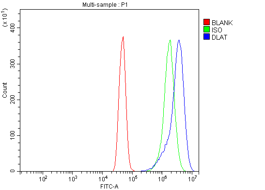
(Figure 9. Flow Cytometry analysis of A431 cells using anti-DLAT antibody (AAA19518).Overlay histogram showing A431 cells stained with AAA19518 (Blue line). The cells were blocked with 10% normal goat serum. And then incubated with rabbit anti-DLAT Antibody (AAA19518, 1 ug/1x10^6 cells) for 30 min at 20 degree C. DyLight488 conjugated goat anti-rabbit IgG was used as secondary antibody for 30 minutes at 20 degree C. Isotype control antibody (Green line) was rabbit IgG (1 ug/1x10^6) used under the same conditions. Unlabelled sample (Red line) was also used as a control.)
FCM (Flow Cytometry)
(Figure 9. Flow Cytometry analysis of A431 cells using anti-DLAT antibody (AAA19518).Overlay histogram showing A431 cells stained with AAA19518 (Blue line). The cells were blocked with 10% normal goat serum. And then incubated with rabbit anti-DLAT Antibody (AAA19518, 1 ug/1x10^6 cells) for 30 min at 20 degree C. DyLight488 conjugated goat anti-rabbit IgG was used as secondary antibody for 30 minutes at 20 degree C. Isotype control antibody (Green line) was rabbit IgG (1 ug/1x10^6) used under the same conditions. Unlabelled sample (Red line) was also used as a control.)
DLAT, Polyclonal Antibody (Cat# AAA19518)
Full Name
Anti-DLAT Antibody Picoband
Gene Names
DLAT; DLTA; PDCE2; PDC-E2
Reactivity
Human, Mouse, Rat
Applications
Western Blot, Immunohistochemistry, Immunocytochemistry, Immunofluorescence, Flow Cytometry
Purity
Immunogen affinity purified.
Pricing


 IHC (Immunohistochemistry)
(Figure 12. IHC analysis of splicing factor 1 using anti-splicing factor 1 antibody (AAA19676).splicing factor 1 was detected in a paraffin-embedded section of rat colon tissue. Heat mediated antigen retrieval was performed in EDTA buffer (pH 8.0, epitope retrieval solution). The tissue section was blocked with 10% goat serum. The tissue section was then incubated with 2 ug/ml mouse anti-splicing factor 1 Antibody (M00179-1) overnight at 4 degree C. Biotinylated goat anti-mouse IgG was used as secondary antibody and incubated for 30 minutes at 37 degree C. The tissue section was developed using Strepavidin-Biotin-Complex (SABC) (#SA1021) with DAB as the chromogen.)
IHC (Immunohistochemistry)
(Figure 12. IHC analysis of splicing factor 1 using anti-splicing factor 1 antibody (AAA19676).splicing factor 1 was detected in a paraffin-embedded section of rat colon tissue. Heat mediated antigen retrieval was performed in EDTA buffer (pH 8.0, epitope retrieval solution). The tissue section was blocked with 10% goat serum. The tissue section was then incubated with 2 ug/ml mouse anti-splicing factor 1 Antibody (M00179-1) overnight at 4 degree C. Biotinylated goat anti-mouse IgG was used as secondary antibody and incubated for 30 minutes at 37 degree C. The tissue section was developed using Strepavidin-Biotin-Complex (SABC) (#SA1021) with DAB as the chromogen.)
splicing factor 1, Monoclonal Antibody (Cat# AAA19676)
Full Name
Anti-splicing factor 1 Antibody Picoband (monoclonal, 7D9E3)
Gene Names
SF1; BBP; MBBP; ZFM1; ZNF162; D11S636; ZCCHC25
Reactivity
Human, Mouse, Rat
Applications
Western Blot, Immunohistochemistry, Immunocytochemistry, Immunofluorescence, Flow Cytometry
Purity
Immunogen affinity purified.
Pricing

 IHC (Immunohistochemistry)
(Immunohistochemistry of paraffin-embedded rat liver using PLA2R1 antibody at dilution of 1:100 (40x lens).)
IHC (Immunohistochemistry)
(Immunohistochemistry of paraffin-embedded rat liver using PLA2R1 antibody at dilution of 1:100 (40x lens).)
PLA2R1, Polyclonal Antibody (Cat# AAA28218)
Full Name
PLA2R1 Polyclonal Antibody
Gene Names
PLA2R1; PLA2R; PLA2-R; PLA2IR; CLEC13C; PLA2G1R
Reactivity
Human, Mouse, Rat
Applications
Western Blot
Purity
Affinity purification
Pricing



 IF (Immunofluorescence)
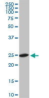
(Immunofluorescence of monoclonal antibody to RPL19 on HeLa cell. [antibody concentration 10ug/ml])
IF (Immunofluorescence)
(Immunofluorescence of monoclonal antibody to RPL19 on HeLa cell. [antibody concentration 10ug/ml])
RPL19, Monoclonal Antibody (Cat# AAA24637)
Full Name
RPL19 (60S Ribosomal Protein L19, DKFZp779D216, FLJ27452, HGNC:10312, MGC71997) APC
Gene Names
RPL19; L19
Reactivity
Human, Mouse, Rat
Applications
Immunofluorescence, Immunohistochemistry, Western Blot
Purity
Purified by Protein A Affinity Chromatography.
Pricing


 Application Data
(Detection limit for recombinant GST tagged PHB is ~0.03ng/ml as a capture antibody.)
Application Data
(Detection limit for recombinant GST tagged PHB is ~0.03ng/ml as a capture antibody.)
PHB, Monoclonal Antibody (Cat# AAA24904)
Full Name
PHB (Prohibitin) (Biotin)
Gene Names
PHB; PHB1; HEL-215; HEL-S-54e
Reactivity
Human
Applications
Immunofluorescence, Immunohistochemistry, Western Blot
Purity
Purified by Protein A Affinity Chromatography.
Pricing


 Application Data
(Detection limit for recombinant GST tagged S100A4 is ~0.1ng/ml as a capture antibody.)
Application Data
(Detection limit for recombinant GST tagged S100A4 is ~0.1ng/ml as a capture antibody.)
S100A4, Monoclonal Antibody (Cat# AAA24646)
Full Name
S100A4 (Protein S100-A4, Calvasculin, Metastasin, Placental Calcium-binding Protein, Protein Mts1, S100 Calcium-binding Protein A4, CAPL, MTS1) APC
Gene Names
S100A4; 42A; 18A2; CAPL; FSP1; MTS1; P9KA; PEL98
Reactivity
Human, Mouse
Applications
Immunofluorescence, Western Blot
Purity
Purified by Protein A Affinity Chromatography.
Pricing