Filters
Clonality
Type
Reactivity
Gene Name
Isotype
Host
Application
Clone
9510 results for " Cell " - showing 9500-9510


 Application Data
(Published customer image Infiltration of GFP+ BM-cells in infarct and peri-infarct regions. (A-B) Dot plots of viable macrophages/granulocytes (CD11b+CD45high, top right quadrants) and microglia (CD11b+CD45dim, bottom right quadrants) in cortex from BM-chimeric unmanipulated mice and mice exposed to pMCAO. (C) Bar graph showing mean numbers of CD11b+CD45dim microglia and CD11b+CD45high macrophages/granulocytes in BM-chimeric mice 24 hours after pMCAO, subdivided based on expression of GFP (n = 5). Approximately 92% of of the CD45high population were GFP+. (D) Estimation and comparison of mean numbers of CD11b+CD45dim microglia in non-chimeric (n = 10) versus BM-chimeric mice (n = 5) 24 hours after of pMCAO shows significantly fewer CD11b+CD45dim microglial cells in irradiated mice. (E) Overview, showing distribution of infiltrating GFP+ BM-derived cells into infarct (IF) and peri-infarct (P-IF) regions 24 hours after pMCAO. (E-G) By 24 hours, GFP+ single cells (F) and vessel-associated aggregates of GFP+ cells (arrows in G) were observed in infarct and peri-infarct regions. Some of the vessel-associated cells were round, leukocyte-like cells (arrows) while others were elongated cells lining the vasculature (arrow heads in G and in insert). (H) Bar graph showing mean numbers of single GFP+ cells and vessel-associated aggregates of GFP+ cells in ipsi- and contralateral cortex 24 hours after surgery (n = 10). (I-P) Immunohistochemical staining of CD45.1 (I, K), CD45.2 (J, L), IgG2a (M, O) and CD45 (N, P) in ischemic tissue in BM-chimeric (I, J, M, N) and non-chimeric mice (K, L, O, P) 24 hours after pMCAO. N.D, none detected. Scale bars: 200 um (A), 10 um (B, C). 50 um (I-P) *P < 0.05, **P < 0.01, and ***P < 0.001.From: Clausen BH, Lambertsen KL, Babcock AA, Holm TH, Dagnaes-Hansen F, Finsen B. Interleukin-1beta and tumor necrosis factor-alpha are expressed by different subsets of microglia and macrophages after ischemic stroke in mice. J Neuroinflammation. 2008 Oct 23;5:46.)
Application Data
(Published customer image Infiltration of GFP+ BM-cells in infarct and peri-infarct regions. (A-B) Dot plots of viable macrophages/granulocytes (CD11b+CD45high, top right quadrants) and microglia (CD11b+CD45dim, bottom right quadrants) in cortex from BM-chimeric unmanipulated mice and mice exposed to pMCAO. (C) Bar graph showing mean numbers of CD11b+CD45dim microglia and CD11b+CD45high macrophages/granulocytes in BM-chimeric mice 24 hours after pMCAO, subdivided based on expression of GFP (n = 5). Approximately 92% of of the CD45high population were GFP+. (D) Estimation and comparison of mean numbers of CD11b+CD45dim microglia in non-chimeric (n = 10) versus BM-chimeric mice (n = 5) 24 hours after of pMCAO shows significantly fewer CD11b+CD45dim microglial cells in irradiated mice. (E) Overview, showing distribution of infiltrating GFP+ BM-derived cells into infarct (IF) and peri-infarct (P-IF) regions 24 hours after pMCAO. (E-G) By 24 hours, GFP+ single cells (F) and vessel-associated aggregates of GFP+ cells (arrows in G) were observed in infarct and peri-infarct regions. Some of the vessel-associated cells were round, leukocyte-like cells (arrows) while others were elongated cells lining the vasculature (arrow heads in G and in insert). (H) Bar graph showing mean numbers of single GFP+ cells and vessel-associated aggregates of GFP+ cells in ipsi- and contralateral cortex 24 hours after surgery (n = 10). (I-P) Immunohistochemical staining of CD45.1 (I, K), CD45.2 (J, L), IgG2a (M, O) and CD45 (N, P) in ischemic tissue in BM-chimeric (I, J, M, N) and non-chimeric mice (K, L, O, P) 24 hours after pMCAO. N.D, none detected. Scale bars: 200 um (A), 10 um (B, C). 50 um (I-P) *P < 0.05, **P < 0.01, and ***P < 0.001.From: Clausen BH, Lambertsen KL, Babcock AA, Holm TH, Dagnaes-Hansen F, Finsen B. Interleukin-1beta and tumor necrosis factor-alpha are expressed by different subsets of microglia and macrophages after ischemic stroke in mice. J Neuroinflammation. 2008 Oct 23;5:46.)
CD11b, Monoclonal Antibody (Cat# AAA12186)
Full Name
RAT ANTI MOUSE CD11b:RPE
Gene Names
Itgam; CR3; CR3A; MAC1; Cd11b; Ly-40; Mac-1; Mac-1a; CD11b/CD18; F730045J24Rik
Applications
FC/FACS
Pricing


 IHC (Immunohistochemistry)
(At 1/100 staining Human colorectal cancer and adjacent normal tissues by IHC-P. The sample was formaldehyde fixed and a heat mediated antigen retrieval step in citrate buffer was performed. The sample was then blocked and incubated with the primary antibody at 4 degree C overnight. An HRP conjugated anti-Rabbit antibody was used as the secondary antibody.)
IHC (Immunohistochemistry)
(At 1/100 staining Human colorectal cancer and adjacent normal tissues by IHC-P. The sample was formaldehyde fixed and a heat mediated antigen retrieval step in citrate buffer was performed. The sample was then blocked and incubated with the primary antibody at 4 degree C overnight. An HRP conjugated anti-Rabbit antibody was used as the secondary antibody.)
Histone H3, Polyclonal Antibody (Cat# AAA31356)
Full Name
Acetyl-Histone H3 (Lys9/14/18/23/27) Antibody
Gene Names
HIST1H3A; H3/A; H3FA
Reactivity
Human, Mouse, Rat
Predicted Reactivity: Bovine (100%)
Predicted Reactivity: Bovine (100%)
Applications
WB, IHC, IF, ICC, EIA
Purity
The antiserum was purified by peptide affinity chromatography using SulfoLink Coupling Resin
Pricing


 Application Data
(At 25 degree C. Samples were then incubated with primary Ab(At 37 degree C. An AlexaFluor594 conjugated goat anti-rabbit IgG(H+L) Ab(Red) and an AlexaFluor488 conjugated goat anti-mouse IgG(H+L) Ab(Green) were used as the secondary antibody.The nuclear counter stain is DAPI(blue).)
Application Data
(At 25 degree C. Samples were then incubated with primary Ab(At 37 degree C. An AlexaFluor594 conjugated goat anti-rabbit IgG(H+L) Ab(Red) and an AlexaFluor488 conjugated goat anti-mouse IgG(H+L) Ab(Green) were used as the secondary antibody.The nuclear counter stain is DAPI(blue).)
Histone H3, Polyclonal Antibody (Cat# AAA31345)
Full Name
Acetyl-Histone H3 (Lys14) Antibody
Gene Names
HIST1H3A; H3/A; H3FA
Reactivity
Human, Mouse, Rat
Predicted Reactivity: Bovine (100%)
Predicted Reactivity: Bovine (100%)
Applications
WB, IHC, IF, ICC, EIA
Purity
The antiserum was purified by peptide affinity chromatography using SulfoLink Coupling Resin
Pricing


 WB (Western Blot)
(FG Pancreatic Carcinoma Cell Lines stably expressing vector along (FG-V) the b3 integrin subunit (FG-b3) or a b3 truncation mutant (FG-759x). Src Mab (AAA28639) was diluted 1:500 in 1% BSA/TBST and incubated Overnight at 4 degree C. After washing 3x 5 min. with TBST the blots were incubated with 1:5000 Goat anti-mouse or Goat anti-rabbit secondary antibody for 1 hr at Room temperature. The blots were again washed 3x 5 min. with TBST and developed using ECL reagent.Data and protocol kindly provided by Dr. Weis of Cheresh Lab, UCSD.)
WB (Western Blot)
(FG Pancreatic Carcinoma Cell Lines stably expressing vector along (FG-V) the b3 integrin subunit (FG-b3) or a b3 truncation mutant (FG-759x). Src Mab (AAA28639) was diluted 1:500 in 1% BSA/TBST and incubated Overnight at 4 degree C. After washing 3x 5 min. with TBST the blots were incubated with 1:5000 Goat anti-mouse or Goat anti-rabbit secondary antibody for 1 hr at Room temperature. The blots were again washed 3x 5 min. with TBST and developed using ECL reagent.Data and protocol kindly provided by Dr. Weis of Cheresh Lab, UCSD.)
SRC, Monoclonal Antibody (Cat# AAA28639)
Full Name
SRC Antibody
Gene Names
SRC; ASV; SRC1; c-SRC; p60-Src
Reactivity
Human, mouse
Applications
WB, EIA, IF
Purity
This antibody is purified through a protein G column, followed by dialysis against PBS.
Pricing


 Application Data
(Immunoperoxidase staining of a human tonsil cryosection with Mouse anti Human CD163 antibody, clone EDHu-1 followed by the Histar detection system . Low power)
Application Data
(Immunoperoxidase staining of a human tonsil cryosection with Mouse anti Human CD163 antibody, clone EDHu-1 followed by the Histar detection system . Low power)
CD163, Monoclonal Antibody (Cat# AAA12100)
Full Name
MOUSE ANTI HUMAN CD163
Gene Names
CD163; M130; MM130
Applications
EIA, FC/FACS, IF, IY, WB
Pricing


 Application Data
(At 25 degree C. Samples were then incubated with primary Ab(At 37 degree C. An AlexaFluor594 conjugated goat anti-rabbit IgG(H+L) Ab(Red) and an AlexaFluor488 conjugated goat anti-mouse IgG(H+L) Ab(Green) were used as the secondary antibody.The nuclear counter stain is DAPI(blue).)
Application Data
(At 25 degree C. Samples were then incubated with primary Ab(At 37 degree C. An AlexaFluor594 conjugated goat anti-rabbit IgG(H+L) Ab(Red) and an AlexaFluor488 conjugated goat anti-mouse IgG(H+L) Ab(Green) were used as the secondary antibody.The nuclear counter stain is DAPI(blue).)
HSF1, Polyclonal Antibody (Cat# AAA31391)
Full Name
Phospho-HSF1 (Ser307) Antibody
Gene Names
HSF1; HSTF1
Reactivity
Human, Mouse, Rat
Applications
WB, IHC, IF, ICC, EIA
Purity
The antibody is from purified rabbit serum by affinity purification via sequential chromatography on phospho-peptide and non-phospho-peptide affinity columns.
Pricing


 Application Data
(At 25 degree C. The primary antibody was diluted at 1/200 and incubated with the sample for 1 hour at 37 degree C. An Alexa Fluor 594 conjugated goat anti-rabbit IgG (H+L) Ab, diluted at 1/600, was used as the secondary antibody.)
Application Data
(At 25 degree C. The primary antibody was diluted at 1/200 and incubated with the sample for 1 hour at 37 degree C. An Alexa Fluor 594 conjugated goat anti-rabbit IgG (H+L) Ab, diluted at 1/600, was used as the secondary antibody.)
p53, Polyclonal Antibody (Cat# AAA31429)
Full Name
Phospho-p53 (Ser392) Antibody
Gene Names
TP53; P53; BCC7; LFS1; TRP53
Reactivity
Human, Mouse
Predicted Reactivity: Pig (88%), Bovine (88%), Sheep (88%), Rabbit (88%)
Predicted Reactivity: Pig (88%), Bovine (88%), Sheep (88%), Rabbit (88%)
Applications
WB, IHC, IF, ICC, EIA
Purity
The antibody is from purified rabbit serum by affinity purification via sequential chromatography on phospho-peptide and non-phospho-peptide affinity columns.
Pricing


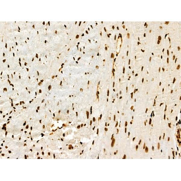
 IHC (Immunohistochemistry)
(At 1/100 staining Human esophageal cancer by IHC-P. The sample was formaldehyde fixed and a heat mediated antigen retrieval step in citrate buffer was performed. The sample was then blocked and incubated with the primary antibody at 4 degree C overnight. An HRP conjugated anti-Rabbit antibody was used as the secondary antibody.)
IHC (Immunohistochemistry)
(At 1/100 staining Human esophageal cancer by IHC-P. The sample was formaldehyde fixed and a heat mediated antigen retrieval step in citrate buffer was performed. The sample was then blocked and incubated with the primary antibody at 4 degree C overnight. An HRP conjugated anti-Rabbit antibody was used as the secondary antibody.)
p53, Polyclonal Antibody (Cat# AAA31354)
Full Name
Acetyl-p53 (Lys373) Antibody
Gene Names
TP53; P53; BCC7; LFS1; TRP53
Reactivity
Human, Mouse, Rat
Predicted Reactivity: Pig (91%), Sheep (80%), Rabbit (91%), Dog (91%)
Predicted Reactivity: Pig (91%), Sheep (80%), Rabbit (91%), Dog (91%)
Applications
WB, IHC, EIA
Purity
The antiserum was purified by peptide affinity chromatography using SulfoLink Coupling Resin
Pricing


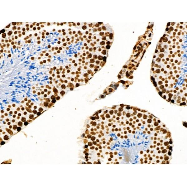
 IHC (Immunohistochemistry)
(At 1/100 staining Human esophageal cancer by IHC-P. The sample was formaldehyde fixed and a heat mediated antigen retrieval step in citrate buffer was performed. The sample was then blocked and incubated with the primary antibody at 4 degree C overnight. An HRP conjugated anti-Rabbit antibody was used as the secondary antibody.)
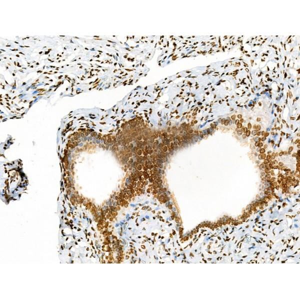
IHC (Immunohistochemistry)
(At 1/100 staining Human esophageal cancer by IHC-P. The sample was formaldehyde fixed and a heat mediated antigen retrieval step in citrate buffer was performed. The sample was then blocked and incubated with the primary antibody at 4 degree C overnight. An HRP conjugated anti-Rabbit antibody was used as the secondary antibody.)
p53, Polyclonal Antibody (Cat# AAA31377)
Full Name
p53 Antibody
Gene Names
TP53; P53; BCC7; LFS1; TRP53
Reactivity
Human, Mouse, Rat
Predicted Reactivity: Pig (91%), Sheep (80%), Rabbit (91%), Dog (91%)
Predicted Reactivity: Pig (91%), Sheep (80%), Rabbit (91%), Dog (91%)
Applications
WB, IHC, EIA
Purity
The antiserum was purified by peptide affinity chromatography using SulfoLink Coupling Resin
Pricing


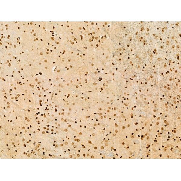
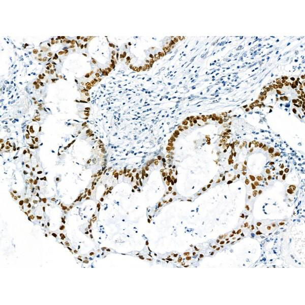
 IHC (Immunohistochemistry)
(At 1/100 staining Human pancreatic cancer by IHC-P. The sample was formaldehyde fixed and a heat mediated antigen retrieval step in citrate buffer was performed. The sample was then blocked and incubated with the primary antibody at 4 degree C overnight. An HRP conjugated anti-Rabbit antibody was used as the secondary antibody.)
IHC (Immunohistochemistry)
(At 1/100 staining Human pancreatic cancer by IHC-P. The sample was formaldehyde fixed and a heat mediated antigen retrieval step in citrate buffer was performed. The sample was then blocked and incubated with the primary antibody at 4 degree C overnight. An HRP conjugated anti-Rabbit antibody was used as the secondary antibody.)
p53, Polyclonal Antibody (Cat# AAA31355)
Full Name
Acetyl-p53 (Lys381) Antibody
Gene Names
TP53; P53; BCC7; LFS1; TRP53
Reactivity
Human, Mouse, Rat
Predicted Reactivity: Rabbit (90%), Dog (100%)
Predicted Reactivity: Rabbit (90%), Dog (100%)
Applications
WB, IHC, EIA
Purity
The antiserum was purified by peptide affinity chromatography using SulfoLink Coupling Resin
Pricing