Filters
Clonality
Type
Reactivity
Gene Name
Isotype
Host
Application
Clone
321 results for " Conjugated Proteins" - showing 50-100


 WB (Western Blot)
(Western blot analysis of COX1 on different lysates using anti-COX1 antibody at 1/1, 000 dilution. Positive control: Lane 1: C2C12 Lane 2: A431)
WB (Western Blot)
(Western blot analysis of COX1 on different lysates using anti-COX1 antibody at 1/1, 000 dilution. Positive control: Lane 1: C2C12 Lane 2: A431)
COX1/Cyclooxygenase 1, Monoclonal Antibody (Cat# AAA30153)
Full Name
COX1/Cyclooxygenase 1 Antibody
Gene Names
PTGS1; COX1; COX3; PHS1; PCOX1; PES-1; PGHS1; PTGHS; PGG/HS; PGHS-1
Reactivity
Human, Mouse, Rat
Applications
WB, ICC, IHC, FC/FACS, IP
Purity
ProA affinity purified
Pricing


 IHC (Immunohistchemistry)
(Immunochemical staining PTBP1 in rat kidney with rabbit polyclonal antibody at 1:2000 dilution, formalin-fixed paraffin embedded sections.)
IHC (Immunohistchemistry)
(Immunochemical staining PTBP1 in rat kidney with rabbit polyclonal antibody at 1:2000 dilution, formalin-fixed paraffin embedded sections.)
PTBP1, Polyclonal Antibody (Cat# AAA27723)
Full Name
Anti-PTBP1 Antibody, Rabbit Polyclonal
Gene Names
PTBP1; PTB; PTB2; PTB3; PTB4; pPTB; HNRPI; PTB-1; PTB-T; HNRNPI; HNRNP-I
Reactivity
Human, Mouse, Rat
Applications
WB, IHC-P, ICC, IF, IP
Purity
Protein A & Antigen Affinity


 WB (Western Blot)
(Anti-HA Tag mouse monoclonal antibody at 1:1000 dilutionLane A: HA-GST (Recombinant protein)(30ng)Lane B: GST-HA (Recombinant protein)(10ng)Lane C: HA-ARG1-myc transfected 293 cell lysate (2ug)Lane D: HA-mFABP4-myc transfected 293 cell lysate (2ug)Lane E: myc-mFABP4-HA transfected 293 cell lysate (0.5ug)Lane F: myc-ARG1-HA transfected 293 cell lysate (2ug)SecondaryGoat Anti-Mouse IgG H&L (Dylight800) at 1/15000 dilution.Developed using the Odyssey technique. Performed under reducing conditions.)
WB (Western Blot)
(Anti-HA Tag mouse monoclonal antibody at 1:1000 dilutionLane A: HA-GST (Recombinant protein)(30ng)Lane B: GST-HA (Recombinant protein)(10ng)Lane C: HA-ARG1-myc transfected 293 cell lysate (2ug)Lane D: HA-mFABP4-myc transfected 293 cell lysate (2ug)Lane E: myc-mFABP4-HA transfected 293 cell lysate (0.5ug)Lane F: myc-ARG1-HA transfected 293 cell lysate (2ug)SecondaryGoat Anti-Mouse IgG H&L (Dylight800) at 1/15000 dilution.Developed using the Odyssey technique. Performed under reducing conditions.)
HA, Monoclonal Antibody (Cat# AAA27712)
Full Name
Anti-HA tag Antibody, Mouse Monoclonal
Reactivity
HA tag
Applications
WB, EIA, FC/FACS/FCM, ICC, IF, IP
Purity
Protein A
Pricing


 WB (Western Blot)
(Western blot analysis of lysates from MDA-MB-468, SW620, T47D cell line, mouse spleen, mouse testis tissue (from left to right), using EZH2 Antibody. AAA28665 was diluted at 1:1000 at each lane. A goat anti-rabbit IgG H&L(HRP) at 1:10000 dilution was used as the secondary antibody. Lysates at 20ug per lane.)
WB (Western Blot)
(Western blot analysis of lysates from MDA-MB-468, SW620, T47D cell line, mouse spleen, mouse testis tissue (from left to right), using EZH2 Antibody. AAA28665 was diluted at 1:1000 at each lane. A goat anti-rabbit IgG H&L(HRP) at 1:10000 dilution was used as the secondary antibody. Lysates at 20ug per lane.)
EZH2, Polyclonal Antibody (Cat# AAA28665)
Full Name
EZH2 Antibody
Gene Names
EZH2; WVS; ENX1; EZH1; KMT6; WVS2; ENX-1; EZH2b; KMT6A
Reactivity
Human, mouse
Applications
WB, EIA, IHC, IF, FC/FACS
Purity
Peptide Affinity Purified Rabbit Polyclonal Antibody (Pab)
Pricing


 FCM (Flow Cytometry)
(Flow cytometry analysis of Hsp105alpha in Hep3B cell line, staining at 2-5ug for 1x10⁶cells (red line). The secondary antibody used goat anti-mouse IgG Alexa fluor 488 conjugate. Isotype control antibody was mouse IgG (black line).)
FCM (Flow Cytometry)
(Flow cytometry analysis of Hsp105alpha in Hep3B cell line, staining at 2-5ug for 1x10⁶cells (red line). The secondary antibody used goat anti-mouse IgG Alexa fluor 488 conjugate. Isotype control antibody was mouse IgG (black line).)
HSP105alpha, Monoclonal Antibody (Cat# AAA11727)
Full Name
HSP105alpha antibody
Gene Names
HSPH1; HSP105; HSP105A; HSP105B; NY-CO-25
Reactivity
Human
Applications
ELISA, WB, Flow cytometry, ICC/IF
Purity
By protein-G affinity chromatography
Pricing


 IP (Immunoprecipitation)
(HDAC2 was immunoprecipitated using:Lane A:0.5 mg Jurkat Whole Cell LysateLane B:0.5 mg NIH-3T3 Whole Cell LysateLane C:0.5 mg Hela Whole Cell Lysate4 uL anti-HDAC2 rabbit polyclonal antibody and 15 ul of 50 % Protein G agarose.Primary antibody:Anti-HDAC2 rabbit polyclonal antibody,at 1:100 dilution Secondary antibody:Dylight 800-labeled antibody to rabbit IgG (H+L), at 1:5000 dilution Developed using the odssey technique.Performed under reducing conditions.Predicted band size: 60 kDaObserved band size: 60 kDa)
IP (Immunoprecipitation)
(HDAC2 was immunoprecipitated using:Lane A:0.5 mg Jurkat Whole Cell LysateLane B:0.5 mg NIH-3T3 Whole Cell LysateLane C:0.5 mg Hela Whole Cell Lysate4 uL anti-HDAC2 rabbit polyclonal antibody and 15 ul of 50 % Protein G agarose.Primary antibody:Anti-HDAC2 rabbit polyclonal antibody,at 1:100 dilution Secondary antibody:Dylight 800-labeled antibody to rabbit IgG (H+L), at 1:5000 dilution Developed using the odssey technique.Performed under reducing conditions.Predicted band size: 60 kDaObserved band size: 60 kDa)
HDAC2, Polyclonal Antibody (Cat# AAA27718)
Full Name
Anti-HDAC2 Antibody, Rabbit Polyclonal
Gene Names
HDAC2; HD2; RPD3; YAF1
Reactivity
Human
Applications
WB, IHC-P, ICC, IF, IP
Purity
Protein A & Antigen Affinity
Pricing


 IP (Immunoprecipitation)
(CBX4 was immunoprecipitated using: Lane A:0.5 mg K562 Whole Cell Lysate 4 uL anti-CBX4 rabbit polyclonal antibody and 60 ug of Immunomagnetic beads Protein A/G. Primary antibody: Anti-CBX4 rabbit polyclonal antibody,at 1:100 dilution Secondary antibody: Clean-Blot IP Detection Reagent (HRP) at 1:1000dilution Developed using the ECL technique. Performed under reducing conditions. Predicted band size: 44 kDa Observed band size :45 kDa)
IP (Immunoprecipitation)
(CBX4 was immunoprecipitated using: Lane A:0.5 mg K562 Whole Cell Lysate 4 uL anti-CBX4 rabbit polyclonal antibody and 60 ug of Immunomagnetic beads Protein A/G. Primary antibody: Anti-CBX4 rabbit polyclonal antibody,at 1:100 dilution Secondary antibody: Clean-Blot IP Detection Reagent (HRP) at 1:1000dilution Developed using the ECL technique. Performed under reducing conditions. Predicted band size: 44 kDa Observed band size :45 kDa)
PAX2, Polyclonal Antibody (Cat# AAA27725)
Full Name
Anti-PAX2 Antibody, Rabbit Polyclonal
Gene Names
PAX2; FSGS7; PAPRS
Reactivity
Human, Mouse
Applications
WB, IHC-P, ICC, IF, IP
Purity
Protein A & Antigen Affinity
Pricing


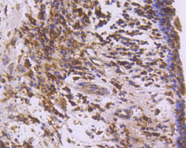
 IHC (Immunohistchemistry)
(At 1/200 staining Human lung cancer tissue sections by IHC-P. The tissue was formaldehyde fixed and a heat mediated antigen retrieval step in citrate buffer was performed. The tissue was then blocked and incubated with the antibody for 1.5 hours at 22 degree C. An HRP conjugated goat anti-rabbit antibody was used as the secondary antibody.)
IHC (Immunohistchemistry)
(At 1/200 staining Human lung cancer tissue sections by IHC-P. The tissue was formaldehyde fixed and a heat mediated antigen retrieval step in citrate buffer was performed. The tissue was then blocked and incubated with the antibody for 1.5 hours at 22 degree C. An HRP conjugated goat anti-rabbit antibody was used as the secondary antibody.)
CUTL1, Polyclonal Antibody (Cat# AAA31424)
Full Name
Phospho-CUTL1 (Ser1237) Antibody
Gene Names
CUX1; CDP; CUX; p75; CASP; CDP1; COY1; Clox; p100; p110; p200; CUTL1; GOLIM6; CDP/Cut; Cux/CDP; Nbla10317
Reactivity
Human, Mouse, Rat
Predicted Reactivity: Pig (100%), Sheep (100%), Dog (100%), Xenopus (100%)
Predicted Reactivity: Pig (100%), Sheep (100%), Dog (100%), Xenopus (100%)
Applications
WB, IHC, EIA
Purity
The antibody is from purified rabbit serum by affinity purification via sequential chromatography on phospho-peptide and non-phospho-peptide affinity columns.
Pricing


 IHC (Immunohistchemistry)
(At 1/200 staining Human lung cancer tissue sections by IHC-P. The tissue was formaldehyde fixed and a heat mediated antigen retrieval step in citrate buffer was performed. The tissue was then blocked and incubated with the antibody for 1.5 hours at 22 degree C. An HRP conjugated goat anti-rabbit antibody was used as the secondary antibody.)
IHC (Immunohistchemistry)
(At 1/200 staining Human lung cancer tissue sections by IHC-P. The tissue was formaldehyde fixed and a heat mediated antigen retrieval step in citrate buffer was performed. The tissue was then blocked and incubated with the antibody for 1.5 hours at 22 degree C. An HRP conjugated goat anti-rabbit antibody was used as the secondary antibody.)
BCLAF1, Polyclonal Antibody (Cat# AAA31422)
Full Name
Phospho-BCLAF1 (Ser531) Antibody
Gene Names
BCLAF1; BTF; bK211L9.1
Reactivity
Human, Mouse, Rat
Predicted Reactivity: Pig (100%), Bovine (100%), Horse (100%), Sheep (100%), Rabbit (100%), Dog (100%), Chicken (100%)
Predicted Reactivity: Pig (100%), Bovine (100%), Horse (100%), Sheep (100%), Rabbit (100%), Dog (100%), Chicken (100%)
Applications
WB, IHC, EIA
Purity
The antibody is from purified rabbit serum by affinity purification via sequential chromatography on phospho-peptide and non-phospho-peptide affinity columns.
Pricing


 IF (Immunofluorescence)
(Immunofluorescent analysis of 4% paraformaldehyde-fixed, 0.1% Triton X-100 permeabilized MCF-7 (human breast cancer cell line) cells labeling Pdx1 with AAA14796 at 1:25 dilution, followed by Dylight® 488-conjugated IgG goat anti-rabbit secondary antibody at 1:200 dilution (green). Immunofluorescence image showing cytoplasm staining on MCF-7 cell line. Cytoplasmic actin is detected with Dylight® 554 Phalloidin (PD18466410) at 1:100 dilution (red). The nuclear counter stain is DAPI (blue).)
IF (Immunofluorescence)
(Immunofluorescent analysis of 4% paraformaldehyde-fixed, 0.1% Triton X-100 permeabilized MCF-7 (human breast cancer cell line) cells labeling Pdx1 with AAA14796 at 1:25 dilution, followed by Dylight® 488-conjugated IgG goat anti-rabbit secondary antibody at 1:200 dilution (green). Immunofluorescence image showing cytoplasm staining on MCF-7 cell line. Cytoplasmic actin is detected with Dylight® 554 Phalloidin (PD18466410) at 1:100 dilution (red). The nuclear counter stain is DAPI (blue).)
OPN-a/b, Polyclonal Antibody (Cat# AAA14796)
Full Name
OPN-a/b, NT (SPP1, BNSP, OPN, Osteopontin, Bone sialoprotein 1, Nephropontin, Secreted phosphoprotein 1, Urinary stone protein, Uropontin)
Reactivity
Human
Applications
EL/EIA, WB, IHC, IF
Purity
Affinity Purified
Purified by Protein A affinity chromatography.
Purified by Protein A affinity chromatography.
Pricing


 WB (Western Blot)
(Anti-Beta-Tubulin mouse monoclonal antibody at 1:2000 dilution Lane A: HepG2 Whole Cell Lysate Lane B: Daudi Whole Cell Lysate Lane C: MOLT-4 Whole Cell Lysate Lane D: A549 Whole Cell Lysate Lane E: 293T Whole Cell Lysate Lane F: HelaS3 Whole Cell Lysate Lysates/proteins at 30 ug per lane. Secondary Goat Anti-Mouse IgG H&L (Dylight800) at 1/15000 dilution. Developed using the Odyssey technique. Performed under reducing conditions. Predicted band size:50 kDa Observed band size:54 kDa)
WB (Western Blot)
(Anti-Beta-Tubulin mouse monoclonal antibody at 1:2000 dilution Lane A: HepG2 Whole Cell Lysate Lane B: Daudi Whole Cell Lysate Lane C: MOLT-4 Whole Cell Lysate Lane D: A549 Whole Cell Lysate Lane E: 293T Whole Cell Lysate Lane F: HelaS3 Whole Cell Lysate Lysates/proteins at 30 ug per lane. Secondary Goat Anti-Mouse IgG H&L (Dylight800) at 1/15000 dilution. Developed using the Odyssey technique. Performed under reducing conditions. Predicted band size:50 kDa Observed band size:54 kDa)
Beta-Tubulin, Monoclonal Antibody (Cat# AAA27713)
Full Name
Beta-Tubulin Loading Control Antibody, Mouse MAb
Reactivity
Human
Applications
WB, IHC-P, ICC, IF, IP
Purity
Protein A
Pricing


 IP (Immunoprecipitation)
(MAP2K3 was immunoprecipitated using: Lane A:0.5 mg Jurkat Whole Cell Lysate Lane B:0.5 mg HepG2 Whole Cell Lysate Lane C:0.5 mg HeLa Whole Cell Lysate 2 uL anti-MAP2K3 rabbit polyclonal antibody and 60 ug of Immunomagnetic beads Protein A/G. Primary antibody: Anti-MAP2K3 rabbit polyclonal antibody,at 1:100 dilution Secondary antibody: Clean-Blot IP Detection Reagent (HRP) at 1:1000dilution Developed using the ECL technique. Performed under reducing conditions. Predicted band size: 39 kDa Observed band size :39 kDa)
IP (Immunoprecipitation)
(MAP2K3 was immunoprecipitated using: Lane A:0.5 mg Jurkat Whole Cell Lysate Lane B:0.5 mg HepG2 Whole Cell Lysate Lane C:0.5 mg HeLa Whole Cell Lysate 2 uL anti-MAP2K3 rabbit polyclonal antibody and 60 ug of Immunomagnetic beads Protein A/G. Primary antibody: Anti-MAP2K3 rabbit polyclonal antibody,at 1:100 dilution Secondary antibody: Clean-Blot IP Detection Reagent (HRP) at 1:1000dilution Developed using the ECL technique. Performed under reducing conditions. Predicted band size: 39 kDa Observed band size :39 kDa)
MEK3/MKK3, Polyclonal Antibody (Cat# AAA27720)
Full Name
Anti-MEK3/MKK3 Antibody, Rabbit Polyclonal
Gene Names
MAP2K3; MEK3; MKK3; MAPKK3; PRKMK3; SAPKK2; SAPKK-2
Reactivity
Human, Mouse
Applications
WB, IHC-P, ICC, IF, IP
Purity
Protein A & Antigen Affinity
Pricing


 IHC (Immunohistochemistry)
(Immunochemical staining HMGB1 in rat liver with rabbit polyclonal antibody (1:1000, formalin-fixed paraffin embedded sections).)
IHC (Immunohistochemistry)
(Immunochemical staining HMGB1 in rat liver with rabbit polyclonal antibody (1:1000, formalin-fixed paraffin embedded sections).)
HMGB1, Polyclonal Antibody (Cat# AAA27719)
Full Name
Anti-HMGB1 Antibody, Rabbit Polyclonal
Gene Names
HMGB1; HMG1; HMG3; SBP-1
Reactivity
Human, Mouse, Rat
Applications
WB, IHC-P, ICC, IF, IP
Purity
Protein A & Antigen Affinity
Pricing


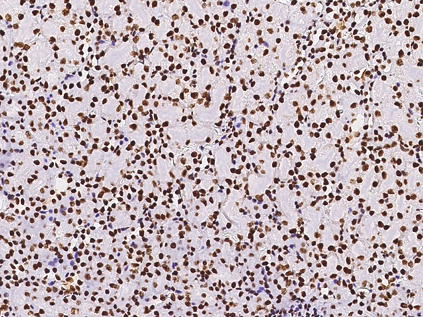
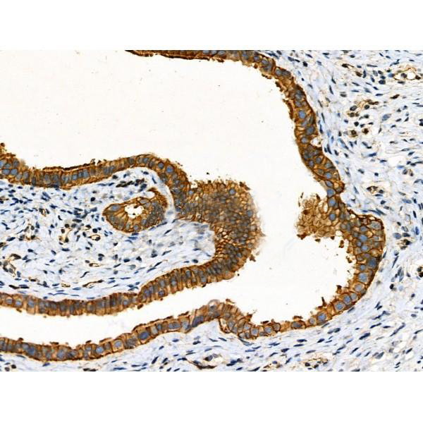
 IHC (Immunohistchemistry)
(At 1/200 staining Human kidney tissue sections by IHC-P. The tissue was formaldehyde fixed and a heat mediated antigen retrieval step in citrate buffer was performed. The tissue was then blocked and incubated with the antibody for 1.5 hours at 22 degree C. An HRP conjugated goat anti-rabbit antibody was used as the secondary antibody.)
IHC (Immunohistchemistry)
(At 1/200 staining Human kidney tissue sections by IHC-P. The tissue was formaldehyde fixed and a heat mediated antigen retrieval step in citrate buffer was performed. The tissue was then blocked and incubated with the antibody for 1.5 hours at 22 degree C. An HRP conjugated goat anti-rabbit antibody was used as the secondary antibody.)
BAG3, Polyclonal Antibody (Cat# AAA31421)
Full Name
Phospho-BAG3 (Tyr457) Antibody
Gene Names
BAG3; BIS; BAG-3; CAIR-1
Reactivity
Human, Mouse, Rat
Predicted Reactivity: Pig (100%), Bovine (100%), Horse (100%), Sheep (100%), Rabbit (100%), Dog (100%)
Predicted Reactivity: Pig (100%), Bovine (100%), Horse (100%), Sheep (100%), Rabbit (100%), Dog (100%)
Applications
WB, IHC, EIA
Purity
The antibody is from purified rabbit serum by affinity purification via sequential chromatography on phospho-peptide and non-phospho-peptide affinity columns.
Pricing


 WB (Western Blot)
(Western Blot Analysis of Panc-1 cell lysate using S100A4 Recombinant Mouse Monoclonal Antibody (rS100A4/1481).)
WB (Western Blot)
(Western Blot Analysis of Panc-1 cell lysate using S100A4 Recombinant Mouse Monoclonal Antibody (rS100A4/1481).)
S100A4/Metastasin/Calvasculin, Antibody (Cat# AAA23934)
Full Name
S100A4/Metastasin/Calvasculin (Marker of Tumor Metastasis)
Gene Names
S100A4; 42A; 18A2; CAPL; FSP1; MTS1; P9KA; PEL98
Reactivity
Human, Mouse
Applications
FC/FACS, WB, IF, IHC
Purity
Purified Ab with BSA and Azide at 200ug/ml OR Purified Ab WITHOUT BSA and Azide at 1.0mg/ml
Pricing


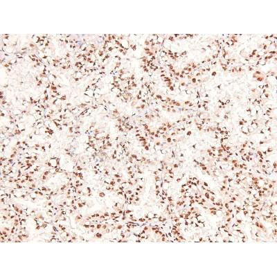
 IHC (Immunohistochemistry)
(At 1/100 staining Human kidney cancer and adjacent normal tissues by IHC-P. The sample was formaldehyde fixed and a heat mediated antigen retrieval step in citrate buffer was performed. The sample was then blocked and incubated with the primary antibody at 4 degree C overnight. An HRP conjugated anti-Rabbit antibody was used as the secondary antibody.)
IHC (Immunohistochemistry)
(At 1/100 staining Human kidney cancer and adjacent normal tissues by IHC-P. The sample was formaldehyde fixed and a heat mediated antigen retrieval step in citrate buffer was performed. The sample was then blocked and incubated with the primary antibody at 4 degree C overnight. An HRP conjugated anti-Rabbit antibody was used as the secondary antibody.)
Smad5, Polyclonal Antibody (Cat# AAA31331)
Full Name
Phospho-Smad5 (Ser463/Ser465) Antibody
Gene Names
SMAD5; DWFC; JV5-1; MADH5
Reactivity
Human, Mouse, Rat
Applications
WB, IHC, EIA
Purity
The antibody is from purified rabbit serum by affinity purification via sequential chromatography on phospho-peptide and non-phospho-peptide affinity columns.
Pricing


 WB (Western Blot)
(Anti-GP73 rabbit polyclonal antibody at 1:500 dilution Lane A: GOLM1 konckout Hela Whole Cell Lysate Lane B: Hela Whole Cell Lysate Lysates/proteins at 10 ug per lane. Secondary Goat Anti-Rabbit IgG (H+L)/HRP at 1/10000 dilution. Developed using the ECL technique. Performed under reducing conditions. Predicted band size:45 kDa Observed band size: kDa)
WB (Western Blot)
(Anti-GP73 rabbit polyclonal antibody at 1:500 dilution Lane A: GOLM1 konckout Hela Whole Cell Lysate Lane B: Hela Whole Cell Lysate Lysates/proteins at 10 ug per lane. Secondary Goat Anti-Rabbit IgG (H+L)/HRP at 1/10000 dilution. Developed using the ECL technique. Performed under reducing conditions. Predicted band size:45 kDa Observed band size: kDa)
GOLPH2/GOLM1, Polyclonal Antibody (Cat# AAA27751)
Full Name
Anti-GOLPH2/GOLM1 Antibody, Rabbit Polyclonal
Gene Names
GOLM1; GP73; HEL46; GOLPH2; C9orf155; PSEC0257; bA379P1.3
Reactivity
Human
Applications
WB, EIA, IHC-P, ICC, IF, IP
Purity
Protein A & Antigen Affinity
Pricing


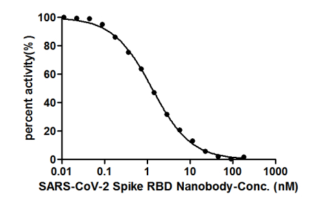


 Application Data
(ELISA: Immobilize various types of SARS proteins at concentration of 2mg/ml on solid substrate, then react with SARS-CoV-2 Spike RBD Nanobody at concentration of 100mg/ml, 10mg/ml and 1mg/ml. It shows the SARS-CoV-2 Spike RBD Nanobody (AAA27051) is specific for SARS-CoV-2-S1-RBD protein, without any cross-reactivity with MERS-CoV, SARS-CoV, HCoV-OC43 or HCoV-229E.)
Application Data
(ELISA: Immobilize various types of SARS proteins at concentration of 2mg/ml on solid substrate, then react with SARS-CoV-2 Spike RBD Nanobody at concentration of 100mg/ml, 10mg/ml and 1mg/ml. It shows the SARS-CoV-2 Spike RBD Nanobody (AAA27051) is specific for SARS-CoV-2-S1-RBD protein, without any cross-reactivity with MERS-CoV, SARS-CoV, HCoV-OC43 or HCoV-229E.)
COVID 19 Spike RBD Coronavirus, Monoclonal Recombinant Antibody (Cat# AAA27051)
Full Name
SARS-CoV-2 Spike RBD Nanobody
Reactivity
Human Novel Coronavirus (SARS-CoV-2/ 2019-nCoV)
Applications
EIA, GICA
Purity
Affinity-chromatography
Pricing


 WB (Western Blot)
(Western Blot Analysis: Representative lot data. Lysate from HEK-293 cells was resolved by electrophoresis, transferred to PVDF and probed with anti-PI3 Kinase, p110beta (0.05 ug/mL). Proteins were visualized using donkey anti-rabbit secondary antibody conjugated to HRP and chemiluminescence detection. Arrow indicates PI3 Kinase, p110beta (~110kD).)
WB (Western Blot)
(Western Blot Analysis: Representative lot data. Lysate from HEK-293 cells was resolved by electrophoresis, transferred to PVDF and probed with anti-PI3 Kinase, p110beta (0.05 ug/mL). Proteins were visualized using donkey anti-rabbit secondary antibody conjugated to HRP and chemiluminescence detection. Arrow indicates PI3 Kinase, p110beta (~110kD).)
Phosphoinositide 3 Kinase, p110 beta, Polyclonal Antibody (Cat# AAA26895)
Full Name
Phosphoinositide 3 Kinase, p110 beta (DKFZp779K1237, MGC133043, p110beta, Phosphatidylinositol 3 Kinase Catalytic beta Polypeptide, Phosphatidylinositol-4,5-bisphosphate 3-kinase Catalytic Subunit beta Isoform, Phosphoinositide 3 Kinase Catalytic beta Pol
Gene Names
PIK3CA; MCM; CWS5; MCAP; PI3K; CLOVE; MCMTC; p110-alpha
Reactivity
Human
Applications
ICC, EIA, WB, IP, IHC
Purity
Purified by Immunoaffinity chromatography.
Pricing


 WB (Western Blot)
(Western Blot Analysis: Representative lot data. Lysate from HEK-293 cells was resolved by electrophoresis, transferred to PVDF and probed with anti-PI3 Kinase, p110beta (0.05 ug/mL). Proteins were visualized using donkey anti-rabbit secondary antibody conjugated to HRP and chemiluminescence detection. Arrow indicates PI3 Kinase, p110beta (~110kD).)
WB (Western Blot)
(Western Blot Analysis: Representative lot data. Lysate from HEK-293 cells was resolved by electrophoresis, transferred to PVDF and probed with anti-PI3 Kinase, p110beta (0.05 ug/mL). Proteins were visualized using donkey anti-rabbit secondary antibody conjugated to HRP and chemiluminescence detection. Arrow indicates PI3 Kinase, p110beta (~110kD).)
Phosphoinositide 3 Kinase, p110 beta, Polyclonal Antibody (Cat# AAA26901)
Full Name
Phosphoinositide 3 Kinase, p110 beta (DKFZp779K1237, MGC133043, p110beta, Phosphatidylinositol 3 Kinase Catalytic beta Polypeptide, Phosphatidylinositol-4,5-bisphosphate 3-kinase Catalytic Subunit beta Isoform, Phosphoinositide 3 Kinase Catalytic beta Pol
Gene Names
PIK3CA; MCM; CWS5; MCAP; PI3K; CLOVE; MCMTC; p110-alpha
Reactivity
Human
Applications
ICC, WB, IP, IHC
Purity
Purified by Immunoaffinity chromatography.
Pricing


 WB (Western Blot)
(Western Blot Analysis: Representative lot data. Lysate from HEK-293 cells was resolved by electrophoresis, transferred to PVDF and probed with anti-PI3 Kinase, p110beta (0.05 ug/mL). Proteins were visualized using donkey anti-rabbit secondary antibody conjugated to HRP and chemiluminescence detection. Arrow indicates PI3 Kinase, p110beta (~110kD).)
WB (Western Blot)
(Western Blot Analysis: Representative lot data. Lysate from HEK-293 cells was resolved by electrophoresis, transferred to PVDF and probed with anti-PI3 Kinase, p110beta (0.05 ug/mL). Proteins were visualized using donkey anti-rabbit secondary antibody conjugated to HRP and chemiluminescence detection. Arrow indicates PI3 Kinase, p110beta (~110kD).)
Phosphoinositide 3 Kinase, p110 beta, Polyclonal Antibody (Cat# AAA26899)
Full Name
Phosphoinositide 3 Kinase, p110 beta (DKFZp779K1237, MGC133043, p110beta, Phosphatidylinositol 3 Kinase Catalytic beta Polypeptide, Phosphatidylinositol-4,5-bisphosphate 3-kinase Catalytic Subunit beta Isoform, Phosphoinositide 3 Kinase Catalytic beta Pol
Gene Names
PIK3CA; MCM; CWS5; MCAP; PI3K; CLOVE; MCMTC; p110-alpha
Reactivity
Human
Applications
ICC, WB, IP, IHC
Purity
Purified by Immunoaffinity chromatography.
Pricing


 WB (Western Blot)
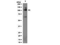
(Western blot analysis of RYK (arrow) using rabbit polyclonal RYK Antibody. 293 cell lysates (2 ug/lane) either nontransfected (Lane 1) or transiently transfected with the RYK gene (Lane 2) (Origene Technologies).)
WB (Western Blot)
(Western blot analysis of RYK (arrow) using rabbit polyclonal RYK Antibody. 293 cell lysates (2 ug/lane) either nontransfected (Lane 1) or transiently transfected with the RYK gene (Lane 2) (Origene Technologies).)
RYK, Polyclonal Antibody (Cat# AAA28767)
Full Name
RYK Antibody
Gene Names
RYK; JTK5; RYK1; JTK5A; D3S3195
Reactivity
Human, mouse, rat
Applications
WB, EIA, IHC-P
Purity
Purified Rabbit Polyclonal Antibody (Pab)
Pricing


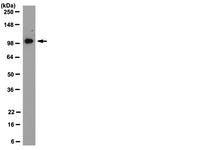
 IP (Immunoprecipitation)
(LAMP1 was immunoprecipitated using:Lane A:0.5 mg Jurkat Whole Cell Lysate1 uL anti-LAMP1 rabbit monoclonal antibody and 15 ul of 50 % Protein G agarose.Primary antibody:Anti-LAMP1 rabbit monoclonal antibody,at 1:500 dilution Secondary antibody:Dylight 800-labeled antibody to rabbit IgG (H+L), at 1:5000 dilution Developed using the odssey technique.Performed under reducing conditions.Predicted band size: 45 kDaObserved band size: 113 kDa)
IP (Immunoprecipitation)
(LAMP1 was immunoprecipitated using:Lane A:0.5 mg Jurkat Whole Cell Lysate1 uL anti-LAMP1 rabbit monoclonal antibody and 15 ul of 50 % Protein G agarose.Primary antibody:Anti-LAMP1 rabbit monoclonal antibody,at 1:500 dilution Secondary antibody:Dylight 800-labeled antibody to rabbit IgG (H+L), at 1:5000 dilution Developed using the odssey technique.Performed under reducing conditions.Predicted band size: 45 kDaObserved band size: 113 kDa)
LAMP1, Monoclonal Antibody (Cat# AAA27745)
Full Name
Recombinant Anti-LAMP1 Antibody, Rabbit Monoclonal
Gene Names
LAMP1; LAMPA; CD107a; LGP120
Reactivity
Human
Applications
WB, EIA, IHC-P, FC/FACS/FCM, ICC, IF, IP
Purity
Protein A
Pricing


 IHC (Immunohistochemistry)
(At 1/100 staining Human gastric cancer by IHC-P. The sample was formaldehyde fixed and a heat mediated antigen retrieval step in citrate buffer was performed. The sample was then blocked and incubated with the primary antibody at 4 degree C overnight. An HRP conjugated anti-Rabbit antibody was used as the secondary antibody.)
IHC (Immunohistochemistry)
(At 1/100 staining Human gastric cancer by IHC-P. The sample was formaldehyde fixed and a heat mediated antigen retrieval step in citrate buffer was performed. The sample was then blocked and incubated with the primary antibody at 4 degree C overnight. An HRP conjugated anti-Rabbit antibody was used as the secondary antibody.)
Smad1/9, Polyclonal Antibody (Cat# AAA31453)
Full Name
Phospho-Smad1/9 (Ser463/Ser465) Antibody
Gene Names
SMAD1; BSP1; JV41; BSP-1; JV4-1; MADH1; MADR1
Reactivity
Human, Mouse, Rat
Predicted Reactivity: Pig (100%), Zebrafish (100%), Bovine (100%), Sheep (100%), Rabbit (100%), Dog (100%), Chicken (100%), Xenopus (100%)
Predicted Reactivity: Pig (100%), Zebrafish (100%), Bovine (100%), Sheep (100%), Rabbit (100%), Dog (100%), Chicken (100%), Xenopus (100%)
Applications
WB, IHC, EIA
Purity
The antibody is from purified rabbit serum by affinity purification via sequential chromatography on phospho-peptide and non-phospho-peptide affinity columns.
Pricing


 Application Data
(Staining of KG1 lymphocytes with Mouse anti Human CD59:FITC)
Application Data
(Staining of KG1 lymphocytes with Mouse anti Human CD59:FITC)
CD59, Monoclonal Antibody (Cat# AAA12022)
Full Name
MOUSE ANTI HUMAN CD59:RPE
Gene Names
CD59; 1F5; EJ16; EJ30; EL32; G344; MIN1; MIN2; MIN3; MIRL; HRF20; MACIF; MEM43; MIC11; MSK21; 16.3A5; HRF-20; MAC-IP; p18-20
Applications
FC/FACS
Pricing


 WB (Western Blot)
(Western Blot Analysis: Representative lot data. Lysate from HEK-293 cells was resolved by electrophoresis, transferred to PVDF and probed with anti-PI3 Kinase, p110beta (0.05 ug/mL). Proteins were visualized using donkey anti-rabbit secondary antibody conjugated to HRP and chemiluminescence detection. Arrow indicates PI3 Kinase, p110beta (~110kD).)
WB (Western Blot)
(Western Blot Analysis: Representative lot data. Lysate from HEK-293 cells was resolved by electrophoresis, transferred to PVDF and probed with anti-PI3 Kinase, p110beta (0.05 ug/mL). Proteins were visualized using donkey anti-rabbit secondary antibody conjugated to HRP and chemiluminescence detection. Arrow indicates PI3 Kinase, p110beta (~110kD).)
Phosphoinositide 3 Kinase, p110 beta, Polyclonal Antibody (Cat# AAA26902)
Full Name
Phosphoinositide 3 Kinase, p110 beta (DKFZp779K1237, MGC133043, p110beta, Phosphatidylinositol 3 Kinase Catalytic beta Polypeptide, Phosphatidylinositol-4,5-bisphosphate 3-kinase Catalytic Subunit beta Isoform, Phosphoinositide 3 Kinase Catalytic beta Pol
Gene Names
PIK3CA; MCM; CWS5; MCAP; PI3K; CLOVE; MCMTC; p110-alpha
Reactivity
Human
Applications
ICC, WB, IP, IHC
Purity
Purified by Immunoaffinity chromatography.
Pricing


 WB (Western Blot)
(Western Blot Analysis: Representative lot data. Lysate from HEK-293 cells was resolved by electrophoresis, transferred to PVDF and probed with anti-PI3 Kinase, p110beta (0.05 ug/mL). Proteins were visualized using donkey anti-rabbit secondary antibody conjugated to HRP and chemiluminescence detection. Arrow indicates PI3 Kinase, p110beta (~110kD).)
WB (Western Blot)
(Western Blot Analysis: Representative lot data. Lysate from HEK-293 cells was resolved by electrophoresis, transferred to PVDF and probed with anti-PI3 Kinase, p110beta (0.05 ug/mL). Proteins were visualized using donkey anti-rabbit secondary antibody conjugated to HRP and chemiluminescence detection. Arrow indicates PI3 Kinase, p110beta (~110kD).)
Phosphoinositide 3 Kinase, p110 beta, Polyclonal Antibody (Cat# AAA26894)
Full Name
Phosphoinositide 3 Kinase, p110 beta (DKFZp779K1237, MGC133043, p110beta, Phosphatidylinositol 3 Kinase Catalytic beta Polypeptide, Phosphatidylinositol-4,5-bisphosphate 3-kinase Catalytic Subunit beta Isoform, Phosphoinositide 3 Kinase Catalytic beta Pol
Gene Names
PIK3CA; MCM; CWS5; MCAP; PI3K; CLOVE; MCMTC; p110-alpha
Reactivity
Human
Applications
ICC, EIA, WB, IP, IHC
Purity
Purified by Immunoaffinity chromatography.
Pricing


 WB (Western Blot)
(Western Blot Analysis: Representative lot data. Lysate from HEK-293 cells was resolved by electrophoresis, transferred to PVDF and probed with anti-PI3 Kinase, p110beta (0.05 ug/mL). Proteins were visualized using donkey anti-rabbit secondary antibody conjugated to HRP and chemiluminescence detection. Arrow indicates PI3 Kinase, p110beta (~110kD).)
WB (Western Blot)
(Western Blot Analysis: Representative lot data. Lysate from HEK-293 cells was resolved by electrophoresis, transferred to PVDF and probed with anti-PI3 Kinase, p110beta (0.05 ug/mL). Proteins were visualized using donkey anti-rabbit secondary antibody conjugated to HRP and chemiluminescence detection. Arrow indicates PI3 Kinase, p110beta (~110kD).)
Phosphoinositide 3 Kinase, p110 beta, Polyclonal Antibody (Cat# AAA26903)
Full Name
Phosphoinositide 3 Kinase, p110 beta (DKFZp779K1237, MGC133043, p110beta, Phosphatidylinositol 3 Kinase Catalytic beta Polypeptide, Phosphatidylinositol-4,5-bisphosphate 3-kinase Catalytic Subunit beta Isoform, Phosphoinositide 3 Kinase Catalytic beta Pol
Gene Names
PIK3CA; MCM; CWS5; MCAP; PI3K; CLOVE; MCMTC; p110-alpha
Reactivity
Human
Applications
ICC, WB, IP, IHC
Purity
Purified by Immunoaffinity chromatography.
Pricing


 WB (Western Blot)
(Western Blot Analysis: Representative lot data. Lysate from HEK-293 cells was resolved by electrophoresis, transferred to PVDF and probed with anti-PI3 Kinase, p110beta (0.05 ug/mL). Proteins were visualized using donkey anti-rabbit secondary antibody conjugated to HRP and chemiluminescence detection. Arrow indicates PI3 Kinase, p110beta (~110kD).)
WB (Western Blot)
(Western Blot Analysis: Representative lot data. Lysate from HEK-293 cells was resolved by electrophoresis, transferred to PVDF and probed with anti-PI3 Kinase, p110beta (0.05 ug/mL). Proteins were visualized using donkey anti-rabbit secondary antibody conjugated to HRP and chemiluminescence detection. Arrow indicates PI3 Kinase, p110beta (~110kD).)
Phosphoinositide 3 Kinase, p110 beta, Polyclonal Antibody (Cat# AAA26898)
Full Name
Phosphoinositide 3 Kinase, p110 beta (DKFZp779K1237, MGC133043, p110beta, Phosphatidylinositol 3 Kinase Catalytic beta Polypeptide, Phosphatidylinositol-4,5-bisphosphate 3-kinase Catalytic Subunit beta Isoform, Phosphoinositide 3 Kinase Catalytic beta Pol
Gene Names
PIK3CA; MCM; CWS5; MCAP; PI3K; CLOVE; MCMTC; p110-alpha
Reactivity
Human
Applications
ICC, EIA, WB, IP, IHC
Purity
Purified by Immunoaffinity chromatography.
Pricing


 WB (Western Blot)
(Western Blot Analysis: Representative lot data. Lysate from HEK-293 cells was resolved by electrophoresis, transferred to PVDF and probed with anti-PI3 Kinase, p110beta (0.05 ug/mL). Proteins were visualized using donkey anti-rabbit secondary antibody conjugated to HRP and chemiluminescence detection. Arrow indicates PI3 Kinase, p110beta (~110kD).)
WB (Western Blot)
(Western Blot Analysis: Representative lot data. Lysate from HEK-293 cells was resolved by electrophoresis, transferred to PVDF and probed with anti-PI3 Kinase, p110beta (0.05 ug/mL). Proteins were visualized using donkey anti-rabbit secondary antibody conjugated to HRP and chemiluminescence detection. Arrow indicates PI3 Kinase, p110beta (~110kD).)
Phosphoinositide 3 Kinase, p110 beta, Polyclonal Antibody (Cat# AAA26900)
Full Name
Phosphoinositide 3 Kinase, p110 beta (DKFZp779K1237, MGC133043, p110beta, Phosphatidylinositol 3 Kinase Catalytic beta Polypeptide, Phosphatidylinositol-4,5-bisphosphate 3-kinase Catalytic Subunit beta Isoform, Phosphoinositide 3 Kinase Catalytic beta Pol
Gene Names
PIK3CA; MCM; CWS5; MCAP; PI3K; CLOVE; MCMTC; p110-alpha
Reactivity
Human
Applications
ICC, WB, IP, IHC
Purity
Purified by Immunoaffinity chromatography.
Pricing


 WB (Western Blot)
(Western Blot Analysis: Representative lot data. Lysate from HEK-293 cells was resolved by electrophoresis, transferred to PVDF and probed with anti-PI3 Kinase, p110beta (0.05 ug/mL). Proteins were visualized using donkey anti-rabbit secondary antibody conjugated to HRP and chemiluminescence detection. Arrow indicates PI3 Kinase, p110beta (~110kD).)
WB (Western Blot)
(Western Blot Analysis: Representative lot data. Lysate from HEK-293 cells was resolved by electrophoresis, transferred to PVDF and probed with anti-PI3 Kinase, p110beta (0.05 ug/mL). Proteins were visualized using donkey anti-rabbit secondary antibody conjugated to HRP and chemiluminescence detection. Arrow indicates PI3 Kinase, p110beta (~110kD).)
Phosphoinositide 3 Kinase, p110 beta, Polyclonal Antibody (Cat# AAA26896)
Full Name
Phosphoinositide 3 Kinase, p110 beta (DKFZp779K1237, MGC133043, p110beta, Phosphatidylinositol 3 Kinase Catalytic beta Polypeptide, Phosphatidylinositol-4,5-bisphosphate 3-kinase Catalytic Subunit beta Isoform, Phosphoinositide 3 Kinase Catalytic beta Pol
Gene Names
PIK3CA; MCM; CWS5; MCAP; PI3K; CLOVE; MCMTC; p110-alpha
Reactivity
Human
Applications
ICC, EIA, WB, IP, IHC
Purity
Purified by Immunoaffinity chromatography.
Pricing


 WB (Western Blot)
(Western Blot Analysis: Representative lot data. Lysate from HEK-293 cells was resolved by electrophoresis, transferred to PVDF and probed with anti-PI3 Kinase, p110beta (0.05 ug/mL). Proteins were visualized using donkey anti-rabbit secondary antibody conjugated to HRP and chemiluminescence detection. Arrow indicates PI3 Kinase, p110beta (~110kD).)
WB (Western Blot)
(Western Blot Analysis: Representative lot data. Lysate from HEK-293 cells was resolved by electrophoresis, transferred to PVDF and probed with anti-PI3 Kinase, p110beta (0.05 ug/mL). Proteins were visualized using donkey anti-rabbit secondary antibody conjugated to HRP and chemiluminescence detection. Arrow indicates PI3 Kinase, p110beta (~110kD).)
Phosphoinositide 3 Kinase, p110 beta, Polyclonal Antibody (Cat# AAA26897)
Full Name
Phosphoinositide 3 Kinase, p110 beta (DKFZp779K1237, MGC133043, p110beta, Phosphatidylinositol 3 Kinase Catalytic beta Polypeptide, Phosphatidylinositol-4,5-bisphosphate 3-kinase Catalytic Subunit beta Isoform, Phosphoinositide 3 Kinase Catalytic beta Pol
Gene Names
PIK3CA; MCM; CWS5; MCAP; PI3K; CLOVE; MCMTC; p110-alpha
Reactivity
Human
Applications
ICC, EIA, WB, IP, IHC
Purity
Purified by Immunoaffinity chromatography.
Pricing


 WB (Western Blot)
(Western Blot Analysis: Representative lot data. Lysate from HEK-293 cells was resolved by electrophoresis, transferred to PVDF and probed with anti-PI3 Kinase, p110beta (0.05 ug/mL). Proteins were visualized using donkey anti-rabbit secondary antibody conjugated to HRP and chemiluminescence detection. Arrow indicates PI3 Kinase, p110beta (~110kD).)
WB (Western Blot)
(Western Blot Analysis: Representative lot data. Lysate from HEK-293 cells was resolved by electrophoresis, transferred to PVDF and probed with anti-PI3 Kinase, p110beta (0.05 ug/mL). Proteins were visualized using donkey anti-rabbit secondary antibody conjugated to HRP and chemiluminescence detection. Arrow indicates PI3 Kinase, p110beta (~110kD).)
Phosphoinositide 3 Kinase, p110 beta, Polyclonal Antibody (Cat# AAA26904)
Full Name
Phosphoinositide 3 Kinase, p110 beta (DKFZp779K1237, MGC133043, p110beta, Phosphatidylinositol 3 Kinase Catalytic beta Polypeptide, Phosphatidylinositol-4,5-bisphosphate 3-kinase Catalytic Subunit beta Isoform, Phosphoinositide 3 Kinase Catalytic beta Pol
Gene Names
PIK3CA; MCM; CWS5; MCAP; PI3K; CLOVE; MCMTC; p110-alpha
Reactivity
Human
Applications
ICC, EIA, WB, IP, IHC
Purity
Purified by Immunoaffinity chromatography.
Pricing


 Application Data
(Staining of KG1 lymphocytes with Mouse anti Human CD59:FITC)
Application Data
(Staining of KG1 lymphocytes with Mouse anti Human CD59:FITC)
CD59, Monoclonal Antibody (Cat# AAA12068)
Full Name
MOUSE ANTI HUMAN CD59:FITC
Gene Names
CD59; 1F5; EJ16; EJ30; EL32; G344; MIN1; MIN2; MIN3; MIRL; HRF20; MACIF; MEM43; MIC11; MSK21; 16.3A5; HRF-20; MAC-IP; p18-20
Applications
FC/FACS
Pricing


 Application Data
(Staining of KG1 lymphocytes with Mouse anti Human CD59:FITC)
Application Data
(Staining of KG1 lymphocytes with Mouse anti Human CD59:FITC)
CD59, Monoclonal Antibody (Cat# AAA11857)
Full Name
MOUSE ANTI HUMAN CD59:FITC
Gene Names
CD59; 1F5; EJ16; EJ30; EL32; G344; MIN1; MIN2; MIN3; MIRL; HRF20; MACIF; MEM43; MIC11; MSK21; 16.3A5; HRF-20; MAC-IP; p18-20
Applications
FC/FACS
Pricing


 WB (Western Blot)
(Anti-ANP32A rabbit polyclonal antibody at 1:500 dilution Lane A: ANP32A konckout Hela Whole Cell Lysate Lane B: Hela Whole Cell Lysate Lysates/proteins at 20 ug per lane. Secondary Goat Anti-Rabbit IgG (H+L)/HRP at 1/10000 dilution. Developed using the ECL technique. Performed under reducing conditions. Predicted band size:28 kDa Observed band size:28 kDa)
WB (Western Blot)
(Anti-ANP32A rabbit polyclonal antibody at 1:500 dilution Lane A: ANP32A konckout Hela Whole Cell Lysate Lane B: Hela Whole Cell Lysate Lysates/proteins at 20 ug per lane. Secondary Goat Anti-Rabbit IgG (H+L)/HRP at 1/10000 dilution. Developed using the ECL technique. Performed under reducing conditions. Predicted band size:28 kDa Observed band size:28 kDa)
ANP32A, Polyclonal Antibody (Cat# AAA27748)
Full Name
Anti-ANP32A Antibody, Rabbit Polyclonal
Gene Names
ANP32A; LANP; MAPM; PP32; HPPCn; PHAP1; PHAPI; I1PP2A; C15orf1
Reactivity
Human
Applications
WB, EIA, IHC-P, ICC, IF, IP
Purity
Protein A & Antigen Affinity
Pricing


 WB (Western Blot)
(Western Blot Analysis: Representative lot data. Lysate from HEK-293 cells was resolved by electrophoresis, transferred to PVDF and probed with anti-PI3 Kinase, p110β (0.05 ug/mL). Proteins were visualized using donkey anti-rabbit secondary antibody conjugated to HRP and chemiluminescence detection. Arrow indicates PI3 Kinase, p110β (~110kD).)
WB (Western Blot)
(Western Blot Analysis: Representative lot data. Lysate from HEK-293 cells was resolved by electrophoresis, transferred to PVDF and probed with anti-PI3 Kinase, p110β (0.05 ug/mL). Proteins were visualized using donkey anti-rabbit secondary antibody conjugated to HRP and chemiluminescence detection. Arrow indicates PI3 Kinase, p110β (~110kD).)
Phosphoinositide 3 Kinase, p110 beta, Polyclonal Antibody (Cat# AAA14720)
Full Name
Phosphoinositide 3 Kinase, p110 beta (DKFZp779K1237, MGC133043, p110beta, Phosphatidylinositol 3 Kinase Catalytic beta Polypeptide, Phosphatidylinositol-4,5-bisphosphate 3-kinase Catalytic Subunit beta Isoform, Phosphoinositide 3 Kinase Catalytic beta Pol
Gene Names
PIK3CB; PI3K; PIK3C1; P110BETA; PI3KBETA; MGC133043; DKFZp779K1237
Reactivity
Human
Applications
EL/EIA, WB, IP, IHC, ICC
Purity
Affinity Purified
Purified by immunoaffinity chromatography.
Purified by immunoaffinity chromatography.
Pricing


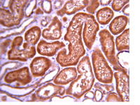
 IHC (Immunohistchemistry)
(Figure 6. IHC analysis of CD13/ANPEP using anti-CD13/ANPEP antibody (AAA19371).CD13/ANPEP was detected in paraffin-embedded section of rat kidney tissue. Heat mediated antigen retrieval was performed in EDTA buffer (pH8. 0, epitope retrieval solution). The tissue section was blocked with 10% goat serum. The tissue section was then incubated with 2μg/ml mouse anti-CD13/ANPEP Antibody (AAA19371) overnight at 4 degree C. Biotinylated goat anti-mouse IgG was used as secondary antibody and incubated for 30 minutes at 37 degree C. The tissue section was developed using Strepavidin-Biotin-Complex (SABC) (Catalog # with DAB as the chromogen.)
IHC (Immunohistchemistry)
(Figure 6. IHC analysis of CD13/ANPEP using anti-CD13/ANPEP antibody (AAA19371).CD13/ANPEP was detected in paraffin-embedded section of rat kidney tissue. Heat mediated antigen retrieval was performed in EDTA buffer (pH8. 0, epitope retrieval solution). The tissue section was blocked with 10% goat serum. The tissue section was then incubated with 2μg/ml mouse anti-CD13/ANPEP Antibody (AAA19371) overnight at 4 degree C. Biotinylated goat anti-mouse IgG was used as secondary antibody and incubated for 30 minutes at 37 degree C. The tissue section was developed using Strepavidin-Biotin-Complex (SABC) (Catalog # with DAB as the chromogen.)
CD13/ANPEP, Monoclonal Antibody (Cat# AAA19371)
Full Name
Anti-CD13/ANPEP Antibody (monoclonal, 5B9)
Gene Names
ANPEP; APN; CD13; LAP1; P150; PEPN; GP150
Reactivity
Human, Rat, Monkey
Applications
WB, IHC-P
Purity
Immunogen affinity purified.
Pricing


 IF (Immunofluorescence)
(Immunofluorescent analysis of 4% paraformaldehyde-fixed, 0.1% Triton X-100 permeabilized MCF-7 (human breast cancer cell line) cells labeling Pdx1 with at 1:25 dilution, followed by DyLight 488-conjugated IgG goat anti-rabbit secondary antibody at 1:200 dilution (green). Immunofluorescence image showing cytoplasm staining on MCF-7 cell line. Cytoplasmic actin is detected with DyLight 554 Phalloidin (PD18466410) at 1:100 dilution (red). The nuclear counter stain is DAPI (blue).)
IF (Immunofluorescence)
(Immunofluorescent analysis of 4% paraformaldehyde-fixed, 0.1% Triton X-100 permeabilized MCF-7 (human breast cancer cell line) cells labeling Pdx1 with at 1:25 dilution, followed by DyLight 488-conjugated IgG goat anti-rabbit secondary antibody at 1:200 dilution (green). Immunofluorescence image showing cytoplasm staining on MCF-7 cell line. Cytoplasmic actin is detected with DyLight 554 Phalloidin (PD18466410) at 1:100 dilution (red). The nuclear counter stain is DAPI (blue).)
OPN-a/b, Polyclonal Antibody (Cat# AAA26857)
Full Name
OPN-a/b, NT (SPP1, BNSP, OPN, Osteopontin, Bone sialoprotein 1, Nephropontin, Secreted phosphoprotein 1, Urinary stone protein, Uropontin) (APC)
Gene Names
SPP1; OPN; BNSP; BSPI; ETA-1
Reactivity
Human
Applications
WB, IHC, IF
Purity
Purified by Protein A and Peptide Affinity Chromatography.
Pricing


 IF (Immunofluorescence)
(Immunofluorescent analysis of 4% paraformaldehyde-fixed, 0.1% Triton X-100 permeabilized MCF-7 (human breast cancer cell line) cells labeling Pdx1 with at 1:25 dilution, followed by DyLight 488-conjugated IgG goat anti-rabbit secondary antibody at 1:200 dilution (green). Immunofluorescence image showing cytoplasm staining on MCF-7 cell line. Cytoplasmic actin is detected with DyLight 554 Phalloidin (PD18466410) at 1:100 dilution (red). The nuclear counter stain is DAPI (blue).)
IF (Immunofluorescence)
(Immunofluorescent analysis of 4% paraformaldehyde-fixed, 0.1% Triton X-100 permeabilized MCF-7 (human breast cancer cell line) cells labeling Pdx1 with at 1:25 dilution, followed by DyLight 488-conjugated IgG goat anti-rabbit secondary antibody at 1:200 dilution (green). Immunofluorescence image showing cytoplasm staining on MCF-7 cell line. Cytoplasmic actin is detected with DyLight 554 Phalloidin (PD18466410) at 1:100 dilution (red). The nuclear counter stain is DAPI (blue).)
OPN-a/b, Polyclonal Antibody (Cat# AAA26860)
Full Name
OPN-a/b, NT (SPP1, BNSP, OPN, Osteopontin, Bone sialoprotein 1, Nephropontin, Secreted phosphoprotein 1, Urinary stone protein, Uropontin) (MaxLight 405)
Gene Names
SPP1; OPN; BNSP; BSPI; ETA-1
Reactivity
Human
Applications
WB, IHC, IF
Purity
Purified by Protein A and Peptide Affinity Chromatography.
Pricing


 IF (Immunofluorescence)
(Immunofluorescent analysis of 4% paraformaldehyde-fixed, 0.1% Triton X-100 permeabilized MCF-7 (human breast cancer cell line) cells labeling Pdx1 with at 1:25 dilution, followed by DyLight 488-conjugated IgG goat anti-rabbit secondary antibody at 1:200 dilution (green). Immunofluorescence image showing cytoplasm staining on MCF-7 cell line. Cytoplasmic actin is detected with DyLight 554 Phalloidin (PD18466410) at 1:100 dilution (red). The nuclear counter stain is DAPI (blue).)
IF (Immunofluorescence)
(Immunofluorescent analysis of 4% paraformaldehyde-fixed, 0.1% Triton X-100 permeabilized MCF-7 (human breast cancer cell line) cells labeling Pdx1 with at 1:25 dilution, followed by DyLight 488-conjugated IgG goat anti-rabbit secondary antibody at 1:200 dilution (green). Immunofluorescence image showing cytoplasm staining on MCF-7 cell line. Cytoplasmic actin is detected with DyLight 554 Phalloidin (PD18466410) at 1:100 dilution (red). The nuclear counter stain is DAPI (blue).)
OPN-a/b, Polyclonal Antibody (Cat# AAA26859)
Full Name
OPN-a/b, NT (SPP1, BNSP, OPN, Osteopontin, Bone sialoprotein 1, Nephropontin, Secreted phosphoprotein 1, Urinary stone protein, Uropontin) (FITC)
Gene Names
SPP1; OPN; BNSP; BSPI; ETA-1
Reactivity
Human
Applications
WB, IHC, IF
Purity
Purified by Protein A and Peptide Affinity Chromatography.
Pricing


 IF (Immunofluorescence)
(Immunofluorescent analysis of 4% paraformaldehyde-fixed, 0.1% Triton X-100 permeabilized MCF-7 (human breast cancer cell line) cells labeling Pdx1 with at 1:25 dilution, followed by DyLight 488-conjugated IgG goat anti-rabbit secondary antibody at 1:200 dilution (green). Immunofluorescence image showing cytoplasm staining on MCF-7 cell line. Cytoplasmic actin is detected with DyLight 554 Phalloidin (PD18466410) at 1:100 dilution (red). The nuclear counter stain is DAPI (blue).)
IF (Immunofluorescence)
(Immunofluorescent analysis of 4% paraformaldehyde-fixed, 0.1% Triton X-100 permeabilized MCF-7 (human breast cancer cell line) cells labeling Pdx1 with at 1:25 dilution, followed by DyLight 488-conjugated IgG goat anti-rabbit secondary antibody at 1:200 dilution (green). Immunofluorescence image showing cytoplasm staining on MCF-7 cell line. Cytoplasmic actin is detected with DyLight 554 Phalloidin (PD18466410) at 1:100 dilution (red). The nuclear counter stain is DAPI (blue).)
OPN-a/b, Polyclonal Antibody (Cat# AAA26864)
Full Name
OPN-a/b, NT (SPP1, BNSP, OPN, Osteopontin, Bone sialoprotein 1, Nephropontin, Secreted phosphoprotein 1, Urinary stone protein, Uropontin) (MaxLight 750)
Gene Names
SPP1; OPN; BNSP; BSPI; ETA-1
Reactivity
Human
Applications
WB, IHC, IF
Purity
Purified by Protein A and Peptide Affinity Chromatography.
Pricing


 IF (Immunofluorescence)
(Immunofluorescent analysis of 4% paraformaldehyde-fixed, 0.1% Triton X-100 permeabilized MCF-7 (human breast cancer cell line) cells labeling Pdx1 with at 1:25 dilution, followed by DyLight 488-conjugated IgG goat anti-rabbit secondary antibody at 1:200 dilution (green). Immunofluorescence image showing cytoplasm staining on MCF-7 cell line. Cytoplasmic actin is detected with DyLight 554 Phalloidin (PD18466410) at 1:100 dilution (red). The nuclear counter stain is DAPI (blue).)
IF (Immunofluorescence)
(Immunofluorescent analysis of 4% paraformaldehyde-fixed, 0.1% Triton X-100 permeabilized MCF-7 (human breast cancer cell line) cells labeling Pdx1 with at 1:25 dilution, followed by DyLight 488-conjugated IgG goat anti-rabbit secondary antibody at 1:200 dilution (green). Immunofluorescence image showing cytoplasm staining on MCF-7 cell line. Cytoplasmic actin is detected with DyLight 554 Phalloidin (PD18466410) at 1:100 dilution (red). The nuclear counter stain is DAPI (blue).)
OPN-a/b, Polyclonal Antibody (Cat# AAA26863)
Full Name
OPN-a/b, NT (SPP1, BNSP, OPN, Osteopontin, Bone sialoprotein 1, Nephropontin, Secreted phosphoprotein 1, Urinary stone protein, Uropontin) (MaxLight 650)
Gene Names
SPP1; OPN; BNSP; BSPI; ETA-1
Reactivity
Human
Applications
WB, IHC, IF
Purity
Purified by Protein A and Peptide Affinity Chromatography.
Pricing


 IF (Immunofluorescence)
(Immunofluorescent analysis of 4% paraformaldehyde-fixed, 0.1% Triton X-100 permeabilized MCF-7 (human breast cancer cell line) cells labeling Pdx1 with at 1:25 dilution, followed by DyLight 488-conjugated IgG goat anti-rabbit secondary antibody at 1:200 dilution (green). Immunofluorescence image showing cytoplasm staining on MCF-7 cell line. Cytoplasmic actin is detected with DyLight 554 Phalloidin (PD18466410) at 1:100 dilution (red). The nuclear counter stain is DAPI (blue).)
IF (Immunofluorescence)
(Immunofluorescent analysis of 4% paraformaldehyde-fixed, 0.1% Triton X-100 permeabilized MCF-7 (human breast cancer cell line) cells labeling Pdx1 with at 1:25 dilution, followed by DyLight 488-conjugated IgG goat anti-rabbit secondary antibody at 1:200 dilution (green). Immunofluorescence image showing cytoplasm staining on MCF-7 cell line. Cytoplasmic actin is detected with DyLight 554 Phalloidin (PD18466410) at 1:100 dilution (red). The nuclear counter stain is DAPI (blue).)
OPN-a/b, Polyclonal Antibody (Cat# AAA26862)
Full Name
OPN-a/b, NT (SPP1, BNSP, OPN, Osteopontin, Bone sialoprotein 1, Nephropontin, Secreted phosphoprotein 1, Urinary stone protein, Uropontin) (MaxLight 550)
Gene Names
SPP1; OPN; BNSP; BSPI; ETA-1
Reactivity
Human
Applications
WB, IHC, IF
Purity
Purified by Protein A and Peptide Affinity Chromatography.
Pricing


 IHC (Immunohistchemistry)
(At 1/100 staining Human ovarian cancer and adjacent normal tissues by IHC-P. The sample was formaldehyde fixed and a heat mediated antigen retrieval step in citrate buffer was performed. The sample was then blocked and incubated with the primary antibody at 4 degree C overnight. An HRP conjugated anti-Rabbit antibody was used as the secondary antibody.)
IHC (Immunohistchemistry)
(At 1/100 staining Human ovarian cancer and adjacent normal tissues by IHC-P. The sample was formaldehyde fixed and a heat mediated antigen retrieval step in citrate buffer was performed. The sample was then blocked and incubated with the primary antibody at 4 degree C overnight. An HRP conjugated anti-Rabbit antibody was used as the secondary antibody.)
alpha 1 Catenin, Polyclonal Antibody (Cat# AAA31319)
Full Name
Phospho-alpha 1 Catenin (Ser655/Thr658) Antibody
Gene Names
CTNNA1; CAP102
Reactivity
Human, Mouse, Rat
Applications
WB, IHC, EIA
Purity
The antibody is from purified rabbit serum by affinity purification via sequential chromatography on phospho-peptide and non-phospho-peptide affinity columns.
Pricing


 IF (Immunofluorescence)
(Immunofluorescent analysis of 4% paraformaldehyde-fixed, 0.1% Triton X-100 permeabilized MCF-7 (human breast cancer cell line) cells labeling Pdx1 with at 1:25 dilution, followed by DyLight 488-conjugated IgG goat anti-rabbit secondary antibody at 1:200 dilution (green). Immunofluorescence image showing cytoplasm staining on MCF-7 cell line. Cytoplasmic actin is detected with DyLight 554 Phalloidin (PD18466410) at 1:100 dilution (red). The nuclear counter stain is DAPI (blue).)
IF (Immunofluorescence)
(Immunofluorescent analysis of 4% paraformaldehyde-fixed, 0.1% Triton X-100 permeabilized MCF-7 (human breast cancer cell line) cells labeling Pdx1 with at 1:25 dilution, followed by DyLight 488-conjugated IgG goat anti-rabbit secondary antibody at 1:200 dilution (green). Immunofluorescence image showing cytoplasm staining on MCF-7 cell line. Cytoplasmic actin is detected with DyLight 554 Phalloidin (PD18466410) at 1:100 dilution (red). The nuclear counter stain is DAPI (blue).)
OPN-a/b, Polyclonal Antibody (Cat# AAA26861)
Full Name
OPN-a/b, NT (SPP1, BNSP, OPN, Osteopontin, Bone sialoprotein 1, Nephropontin, Secreted phosphoprotein 1, Urinary stone protein, Uropontin) (MaxLight 490)
Gene Names
SPP1; OPN; BNSP; BSPI; ETA-1
Reactivity
Human
Applications
WB, IHC, IF
Purity
Purified by Protein A and Peptide Affinity Chromatography.
Pricing


 IF (Immunofluorescence)
(Immunofluorescent analysis of 4% paraformaldehyde-fixed, 0.1% Triton X-100 permeabilized MCF-7 (human breast cancer cell line) cells labeling Pdx1 with at 1:25 dilution, followed by DyLight 488-conjugated IgG goat anti-rabbit secondary antibody at 1:200 dilution (green). Immunofluorescence image showing cytoplasm staining on MCF-7 cell line. Cytoplasmic actin is detected with DyLight 554 Phalloidin (PD18466410) at 1:100 dilution (red). The nuclear counter stain is DAPI (blue).)
IF (Immunofluorescence)
(Immunofluorescent analysis of 4% paraformaldehyde-fixed, 0.1% Triton X-100 permeabilized MCF-7 (human breast cancer cell line) cells labeling Pdx1 with at 1:25 dilution, followed by DyLight 488-conjugated IgG goat anti-rabbit secondary antibody at 1:200 dilution (green). Immunofluorescence image showing cytoplasm staining on MCF-7 cell line. Cytoplasmic actin is detected with DyLight 554 Phalloidin (PD18466410) at 1:100 dilution (red). The nuclear counter stain is DAPI (blue).)
OPN-a/b, Polyclonal Antibody (Cat# AAA26865)
Full Name
OPN-a/b, NT (SPP1, BNSP, OPN, Osteopontin, Bone sialoprotein 1, Nephropontin, Secreted phosphoprotein 1, Urinary stone protein, Uropontin) (PE)
Gene Names
SPP1; OPN; BNSP; BSPI; ETA-1
Reactivity
Human
Applications
WB, IHC, IF
Purity
Purified by Protein A and Peptide Affinity Chromatography.
Pricing


 IHC (Immunohistochemistry)
(Figure 8. IHC analysis of FOXP1 using anti-FOXP1 antibody (AAA19227).FOXP1 was detected in paraffin-embedded section of rat brain tissue. Heat mediated antigen retrieval was performed in EDTA buffer (pH8. 0, epitope retrieval solution). The tissue section was blocked with 10% goat serum. The tissue section was then incubated with 2μg/ml rabbit anti-FOXP1 Antibody (AAA19227) overnight at 4 degree C. Biotinylated goat anti-rabbit IgG was used as secondary antibody and incubated for 30 minutes at 37 degree C. The tissue section was developed using Strepavidin-Biotin-Complex (SABC) (Catalog # with DAB as the chromogen.)
IHC (Immunohistochemistry)
(Figure 8. IHC analysis of FOXP1 using anti-FOXP1 antibody (AAA19227).FOXP1 was detected in paraffin-embedded section of rat brain tissue. Heat mediated antigen retrieval was performed in EDTA buffer (pH8. 0, epitope retrieval solution). The tissue section was blocked with 10% goat serum. The tissue section was then incubated with 2μg/ml rabbit anti-FOXP1 Antibody (AAA19227) overnight at 4 degree C. Biotinylated goat anti-rabbit IgG was used as secondary antibody and incubated for 30 minutes at 37 degree C. The tissue section was developed using Strepavidin-Biotin-Complex (SABC) (Catalog # with DAB as the chromogen.)
FOXP1, Polyclonal Antibody (Cat# AAA19227)
Full Name
Anti-FOXP1 Antibody
Gene Names
FOXP1; QRF1; 12CC4; hFKH1B; HSPC215
Reactivity
Human, Mouse, Rat, Monkey
Applications
WB, IHC-P
Purity
Immunogen affinity purified.
Pricing


 IHC (Immunohistochemistry)
(At 1/100 staining Human gastric cancer by IHC-P. The sample was formaldehyde fixed and a heat mediated antigen retrieval step in citrate buffer was performed. The sample was then blocked and incubated with the primary antibody at 4 degree C overnight. An HRP conjugated anti-Rabbit antibody was used as the secondary antibody.)
IHC (Immunohistochemistry)
(At 1/100 staining Human gastric cancer by IHC-P. The sample was formaldehyde fixed and a heat mediated antigen retrieval step in citrate buffer was performed. The sample was then blocked and incubated with the primary antibody at 4 degree C overnight. An HRP conjugated anti-Rabbit antibody was used as the secondary antibody.)
alpha 1 Catenin, Polyclonal Antibody (Cat# AAA31379)
Full Name
alpha 1 Catenin Antibody
Gene Names
CTNNA1; CAP102
Reactivity
Human, Mouse, Rat
Applications
WB, IHC, EIA
Purity
The antiserum was purified by peptide affinity chromatography using SulfoLink Coupling Resin
Pricing


 FCM (Flow Cytometry)
(Figure 8. Flow Cytometry analysis of LLC cells using anti-Synaptotagmin 1 antibody (AAA19158).Overlay histogram showing LLC cells stained with AAA19158 (Blue line).The cells were blocked with 10% normal goat serum. And then incubated with rabbit anti-Synaptotagmin 1 Antibody (AAA19158,1ug/1x10^6 cells) for 30 min at 20 degree C. DyLight®488 conjugated goat anti-rabbit IgG (5-10ug/1x10^6 cells) was used as secondary antibody for 30 minutes at 20 degree C. Isotype control antibody (Green line) was rabbit IgG (1ug/1x106) used under the same conditions. Unlabelled sample (Red line) was also used as a control.)
FCM (Flow Cytometry)
(Figure 8. Flow Cytometry analysis of LLC cells using anti-Synaptotagmin 1 antibody (AAA19158).Overlay histogram showing LLC cells stained with AAA19158 (Blue line).The cells were blocked with 10% normal goat serum. And then incubated with rabbit anti-Synaptotagmin 1 Antibody (AAA19158,1ug/1x10^6 cells) for 30 min at 20 degree C. DyLight®488 conjugated goat anti-rabbit IgG (5-10ug/1x10^6 cells) was used as secondary antibody for 30 minutes at 20 degree C. Isotype control antibody (Green line) was rabbit IgG (1ug/1x106) used under the same conditions. Unlabelled sample (Red line) was also used as a control.)
Synaptotagmin 1, Polyclonal Antibody (Cat# AAA19158)
Full Name
Anti-Synaptotagmin 1 Picoband antibody
Gene Names
SYT1; P65; SYT; SVP65
Reactivity
Human, Mouse, Rat
No cross reactivity with other proteins.
No cross reactivity with other proteins.
Applications
IHC, WB
Pricing