
Filters
Clonality
Type
Reactivity
Gene Name
Isotype
Host
Application
Clone
1075 results for " Inflammation" - showing 100-150
 Application Data
(Staining of New Zealand Black mouse peripheral blood granulocytes with Rat anti Mouse Ly-6B.2 conjugated to FITC Data)
Application Data
(Staining of New Zealand Black mouse peripheral blood granulocytes with Rat anti Mouse Ly-6B.2 conjugated to FITC Data)
Ly-6B.2 ALLOANTIGEN, Monoclonal Antibody (Cat# AAA12198)
Full Name
RAT ANTI MOUSE Ly-6B.2 ALLOANTIGEN:RPE
Applications
Flow Cytometry
Pricing
 Application Data
(Staining of New Zealand Black mouse peripheral blood granulocytes with Rat anti Mouse Ly-6B.2 conjugated to FITC Data)
Application Data
(Staining of New Zealand Black mouse peripheral blood granulocytes with Rat anti Mouse Ly-6B.2 conjugated to FITC Data)
Ly-6B.2 ALLOANTIGEN, Monoclonal Antibody (Cat# AAA12193)
Full Name
RAT ANTI MOUSE Ly-6B.2 ALLOANTIGEN:FITC
Applications
Flow Cytometry
Pricing
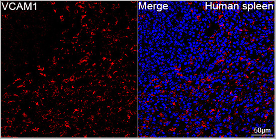
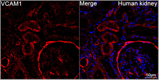
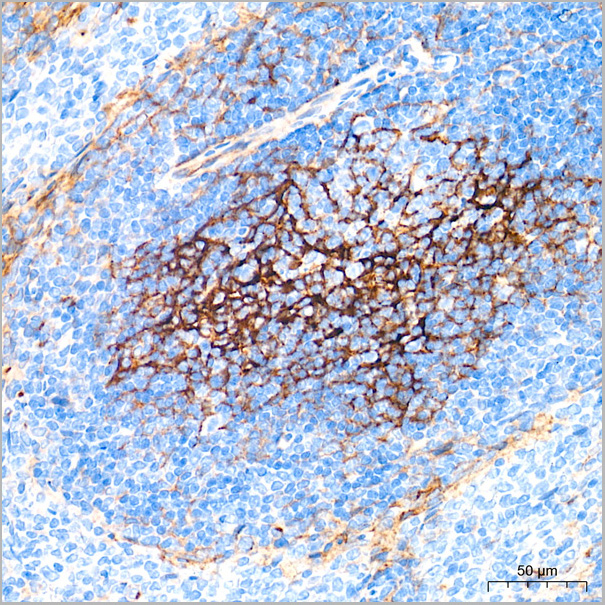
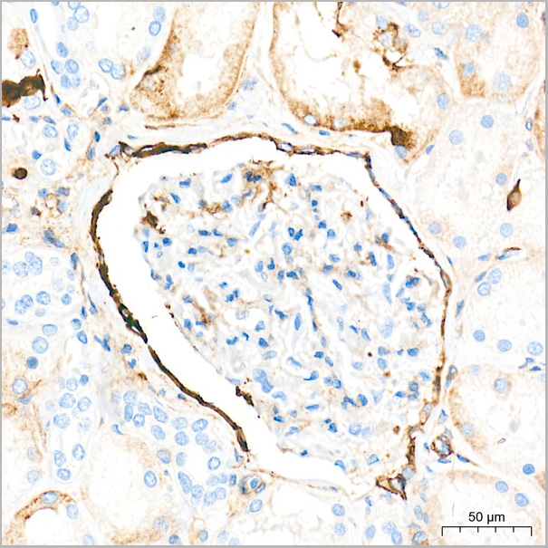
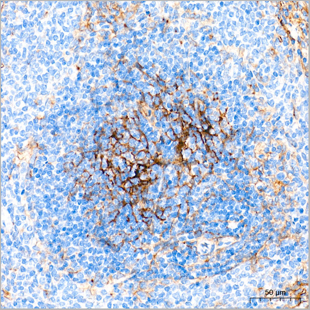
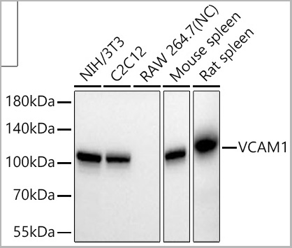
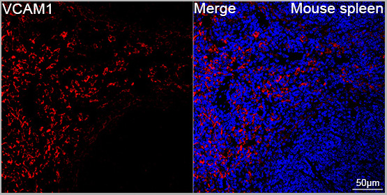 ICC (Immunocytochemistry)
(Confocal imaging of paraffin-embedded Mouse spleen tissue using VCAM1 Rabbit PolymAb® (A19131-PM, dilution 1:200) followed by a further incubation with Cy3 Goat Anti-Rabbit IgG (H+L) (AS007, dilution 1:500) (Red). DAPI was used for nuclear staining (Blue). High pressure antigen retrieval performed with 0.01M Citrate Buffer (pH 6.0) prior to IF staining. Objective: 40x.)
ICC (Immunocytochemistry)
(Confocal imaging of paraffin-embedded Mouse spleen tissue using VCAM1 Rabbit PolymAb® (A19131-PM, dilution 1:200) followed by a further incubation with Cy3 Goat Anti-Rabbit IgG (H+L) (AS007, dilution 1:500) (Red). DAPI was used for nuclear staining (Blue). High pressure antigen retrieval performed with 0.01M Citrate Buffer (pH 6.0) prior to IF staining. Objective: 40x.)
VCAM1, Monoclonal Antibody (Cat# AAA28494)
Full Name
VCAM1 Rabbit PolymAb
Reactivity
Human, Mouse
Applications
Western Blot, Immunohistochemistry, Immunofluorescence, Immunocytochemistry
Purity
Affinity purification
Pricing
 ICC (Immunocytochemistry)
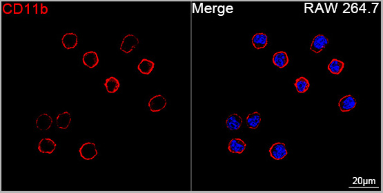
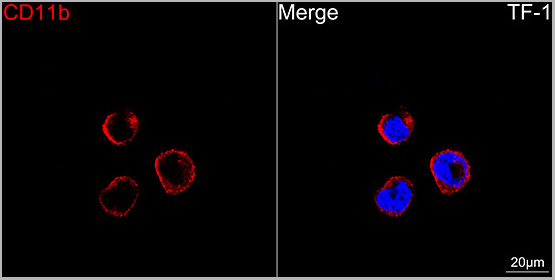
(Confocal imaging of TF-1 cells using CD11b Rabbit PolymAb® (AAA28592, dilution 1:200) followed by a further incubation with Cy3 Goat Anti-Rabbit IgG (H+L) (AS007, dilution 1:500) (Red). DAPI was used for nuclear staining (Blue). Objective: 100x.)
ICC (Immunocytochemistry)
(Confocal imaging of TF-1 cells using CD11b Rabbit PolymAb® (AAA28592, dilution 1:200) followed by a further incubation with Cy3 Goat Anti-Rabbit IgG (H+L) (AS007, dilution 1:500) (Red). DAPI was used for nuclear staining (Blue). Objective: 100x.)
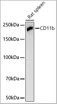
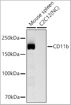
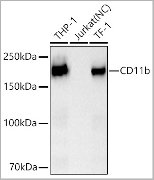
CD11b, Monoclonal Antibody (Cat# AAA28592)
Full Name
CD11b Rabbit PolymAb
Reactivity
Human
Applications
Western Blot, Immunohistochemistry, Immunofluorescence, Immunocytochemistry
Purity
Affinity purification
Pricing
 IF (Immunofluorescence)
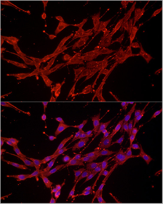
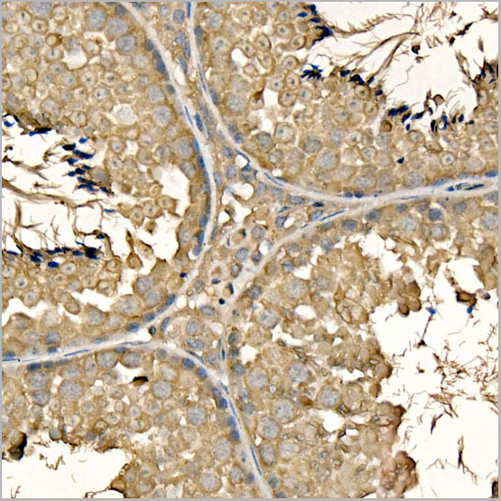
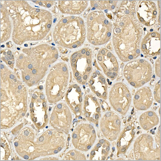
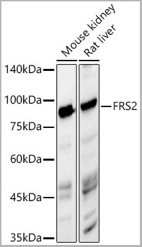
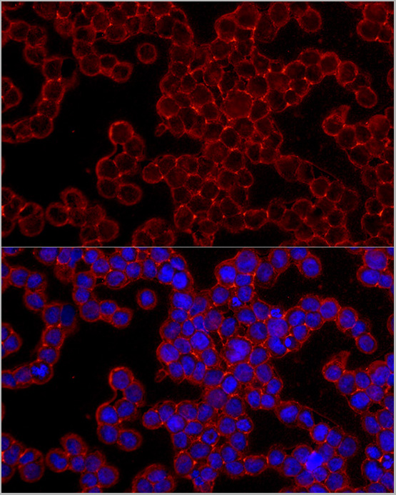
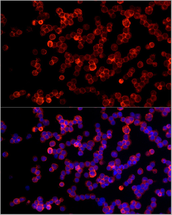
(Immunofluorescence analysis of THP-1 cells using FRS2 Rabbit PolymAb® (AAA28559) at dilution of 1:300 (40x lens). Secondary antibody: Cy3-conjugated Goat anti-Rabbit IgG (H+L) (AS007) at 1:500 dilution. Blue: DAPI for nuclear staining.)
IF (Immunofluorescence)
(Immunofluorescence analysis of THP-1 cells using FRS2 Rabbit PolymAb® (AAA28559) at dilution of 1:300 (40x lens). Secondary antibody: Cy3-conjugated Goat anti-Rabbit IgG (H+L) (AS007) at 1:500 dilution. Blue: DAPI for nuclear staining.)
FRS2, Monoclonal Antibody (Cat# AAA28559)
Full Name
FRS2 Rabbit PolymAb
Reactivity
Human
Applications
Western Blot, Immunohistochemistry, Immunofluorescence, Immunocytochemistry
Purity
Affinity purification
Pricing
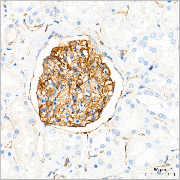
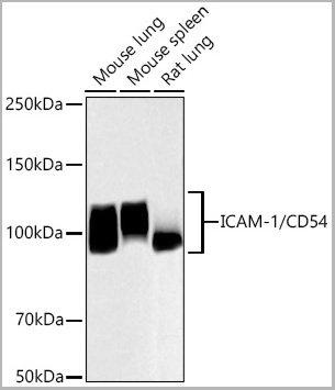
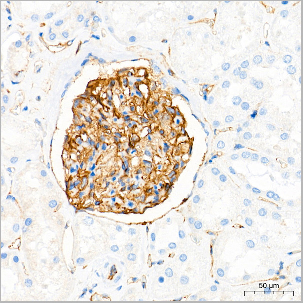
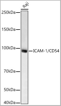
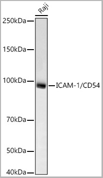
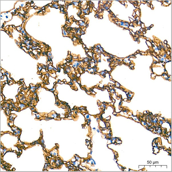 ICC (Immunocytochemistry)
(Confocal imaging of paraffin-embedded Mouse spleen tissue using ICAM-1/CD54 Rabbit PolymAb® (AAA28578, dilution 1:4000) followed by a further incubation with Cy3 Goat Anti-Rabbit IgG (H+L) (AS007, dilution 1:500) (Red). DAPI was used for nuclear staining (Blue). High pressure antigen retrieval performed with 0.01M Citrate Buffer (pH 6.0) prior to IF staining. Objective: 40x.)
ICC (Immunocytochemistry)
(Confocal imaging of paraffin-embedded Mouse spleen tissue using ICAM-1/CD54 Rabbit PolymAb® (AAA28578, dilution 1:4000) followed by a further incubation with Cy3 Goat Anti-Rabbit IgG (H+L) (AS007, dilution 1:500) (Red). DAPI was used for nuclear staining (Blue). High pressure antigen retrieval performed with 0.01M Citrate Buffer (pH 6.0) prior to IF staining. Objective: 40x.)
ICAM-1/CD54, Monoclonal Antibody (Cat# AAA28578)
Full Name
ICAM-1/CD54 Rabbit PolymAb
Reactivity
Human, Mouse
Applications
Western Blot, Immunohistochemistry, Immunofluorescence, Immunocytochemistry
Purity
Affinity purification
Pricing
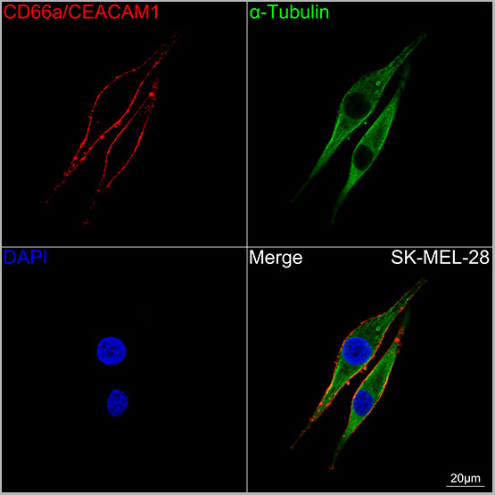
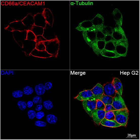
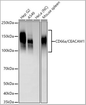
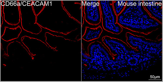 ICC (Immunocytochemistry)
(Confocal imaging of paraffin-embedded Mouse intestine tissue using CD66a/CEACAM1 Rabbit PolymAb® (AAA28579, dilution 1:4000) followed by a further incubation with Cy3 Goat Anti-Rabbit IgG (H+L) (AS007, dilution 1:500) (Red). DAPI was used for nuclear staining (Blue). High pressure antigen retrieval performed with 0.01M Citrate Buffer (pH 6.0) prior to IF staining. Objective: 40x.)
ICC (Immunocytochemistry)
(Confocal imaging of paraffin-embedded Mouse intestine tissue using CD66a/CEACAM1 Rabbit PolymAb® (AAA28579, dilution 1:4000) followed by a further incubation with Cy3 Goat Anti-Rabbit IgG (H+L) (AS007, dilution 1:500) (Red). DAPI was used for nuclear staining (Blue). High pressure antigen retrieval performed with 0.01M Citrate Buffer (pH 6.0) prior to IF staining. Objective: 40x.)
CD66a/CEACAM1, Monoclonal Antibody (Cat# AAA28579)
Full Name
CD66a/CEACAM1 Rabbit PolymAb
Reactivity
Human, Mouse
Applications
Western Blot, Immunohistochemistry, Immunofluorescence, Immunocytochemistry
Purity
Affinity purification
Pricing
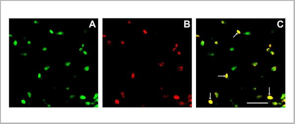
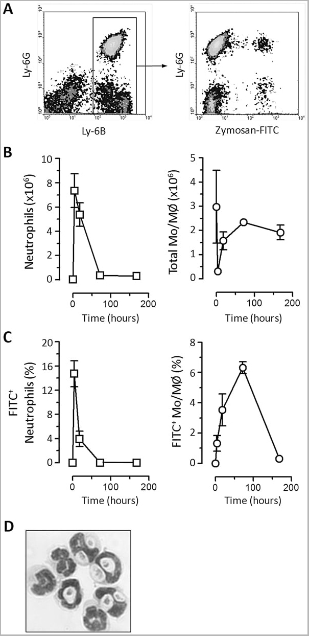 Application Data
(Staining of New Zealand Black mouse peripheral blood granulocytes with Rat anti Mouse Ly-6B.2 conjugated to FITC Data)
Application Data
(Staining of New Zealand Black mouse peripheral blood granulocytes with Rat anti Mouse Ly-6B.2 conjugated to FITC Data)
Ly-6B.2 ALLOANTIGEN, Monoclonal Antibody (Cat# AAA12189)
Full Name
RAT ANTI MOUSE Ly-6B.2 ALLOANTIGEN:Low Endotoxin
Applications
Immunohistochemistry, Flow Cytometry, Immunofluorescence, Immunohistochemistry, Western Blot
Pricing
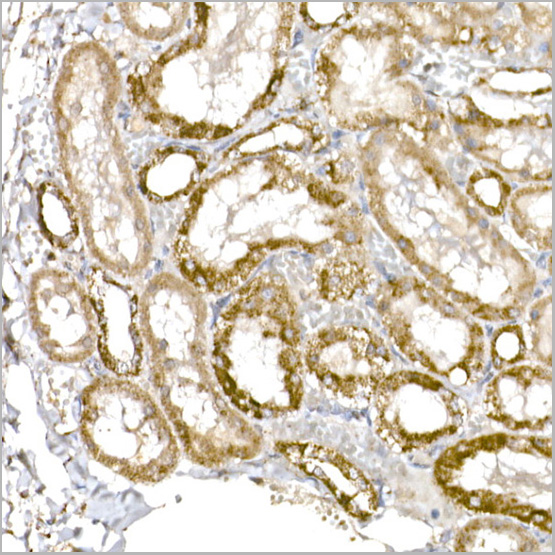
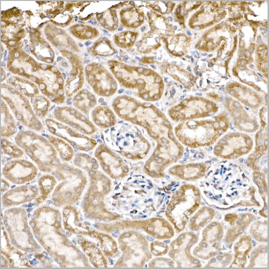
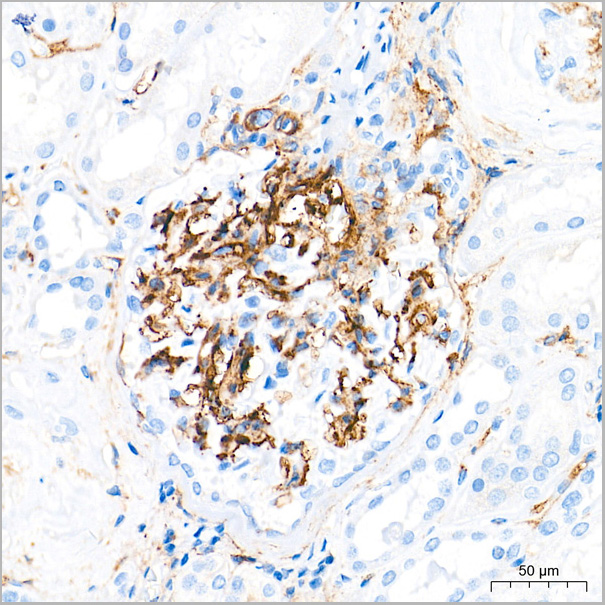
 IHC (Immunohistochemistry)
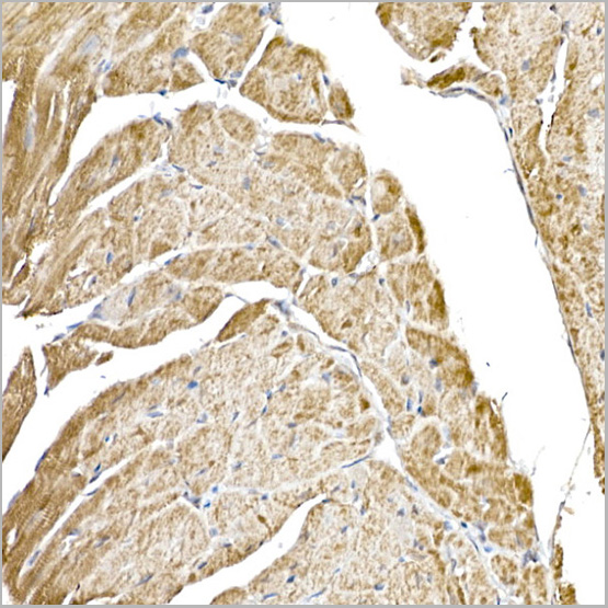
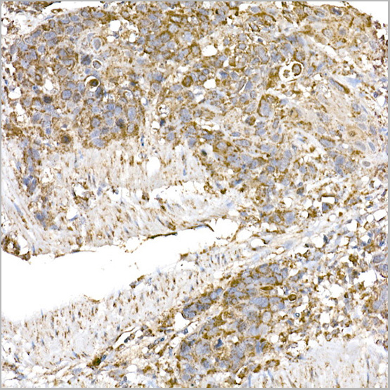
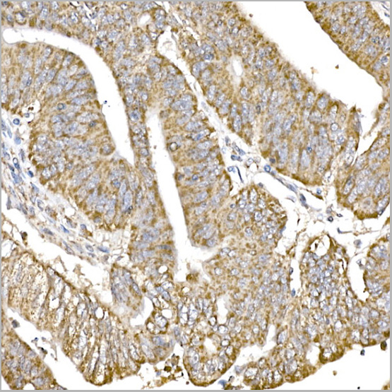
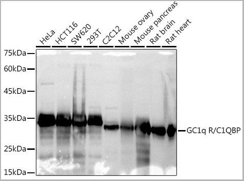
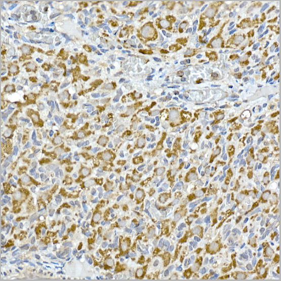
(Immunohistochemistry analysis of paraffin-embedded Mouse kidney tissue using GC1q R/C1QBP Rabbit mAb (AAA28455) at a dilution of 1:200 (40x lens). High pressure antigen retrieval was performed with 0.01 M citrate buffer (pH 6.0) prior to IHC staining.)
IHC (Immunohistochemistry)
(Immunohistochemistry analysis of paraffin-embedded Mouse kidney tissue using GC1q R/C1QBP Rabbit mAb (AAA28455) at a dilution of 1:200 (40x lens). High pressure antigen retrieval was performed with 0.01 M citrate buffer (pH 6.0) prior to IHC staining.)
GC1q R/C1QBP, Monoclonal Antibody (Cat# AAA28455)
Full Name
GC1q R/C1QBP Rabbit mAb
Reactivity
Human
Applications
Western Blot, Immunohistochemistry
Purity
Affinity purification
Pricing
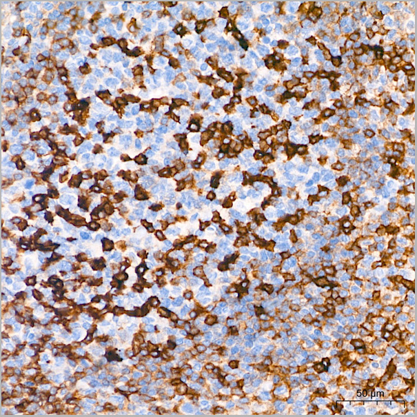
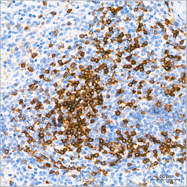
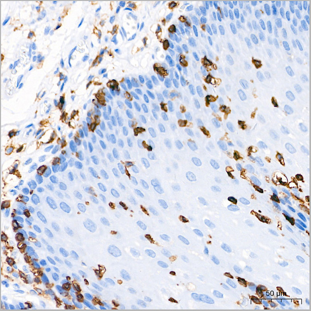
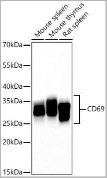
 ICC (Immunocytochemistry)
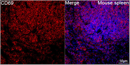
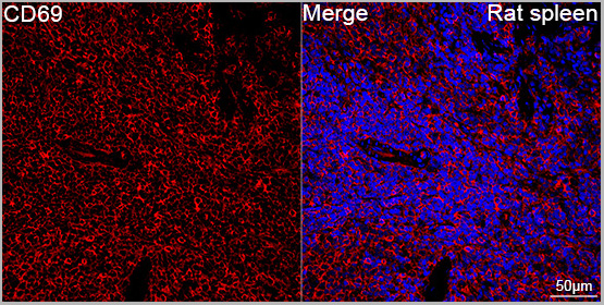
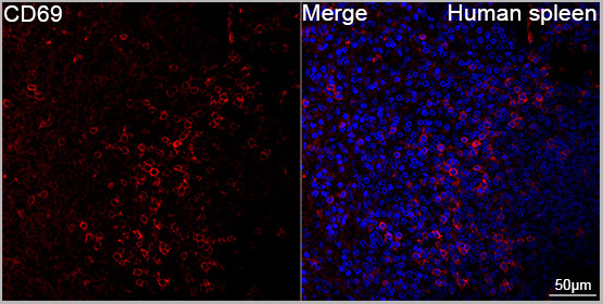
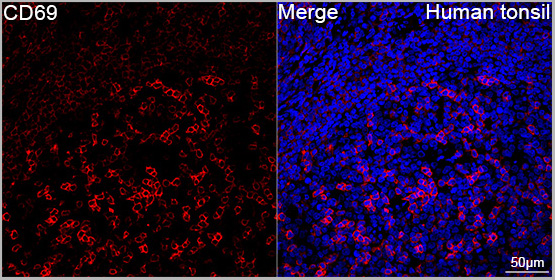
(Confocal imaging of paraffin-embedded Human tonsil tissue using CD69 Rabbit PolymAb® (AAA28586, dilution 1:200) followed by a further incubation with Cy3 Goat Anti-Rabbit IgG (H+L) (AS007, dilution 1:500) (Red). DAPI was used for nuclear staining (Blue). High pressure antigen retrieval performed with 0.01M Citrate Buffer(pH 6.0) prior to IF staining. Objective: 40x.)
ICC (Immunocytochemistry)
(Confocal imaging of paraffin-embedded Human tonsil tissue using CD69 Rabbit PolymAb® (AAA28586, dilution 1:200) followed by a further incubation with Cy3 Goat Anti-Rabbit IgG (H+L) (AS007, dilution 1:500) (Red). DAPI was used for nuclear staining (Blue). High pressure antigen retrieval performed with 0.01M Citrate Buffer(pH 6.0) prior to IF staining. Objective: 40x.)
CD69, Monoclonal Antibody (Cat# AAA28586)
Full Name
CD69 Rabbit PolymAb
Reactivity
Human
Applications
Western Blot, Immunohistochemistry, Immunofluorescence, Immunocytochemistry
Purity
Affinity purification
Pricing
 Application Data
(Staining of New Zealand Black mouse peripheral blood granulocytes with Rat anti Mouse Ly-6B.2 conjugated to FITC Data)
Application Data
(Staining of New Zealand Black mouse peripheral blood granulocytes with Rat anti Mouse Ly-6B.2 conjugated to FITC Data)
Ly-6B.2 ALLOANTIGEN, Monoclonal Antibody (Cat# AAA12192)
Full Name
RAT ANTI MOUSE Ly-6B.2 ALLOANTIGEN:FITC
Applications
Flow Cytometry
Pricing
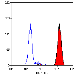 Application Data
(Staining of New Zealand Black mouse peripheral blood granulocytes with Rat anti Mouse Ly-6B.2 conjugated to FITC Data)
Application Data
(Staining of New Zealand Black mouse peripheral blood granulocytes with Rat anti Mouse Ly-6B.2 conjugated to FITC Data)
Ly-6B.2 ALLOANTIGEN, Monoclonal Antibody (Cat# AAA12195)
Full Name
RAT ANTI MOUSE Ly-6B.2 ALLOANTIGEN
Applications
Immunohistochemistry, Flow Cytometry, Immunofluorescence, Immunohistochemistry, Western Blot
Pricing
 Application Data
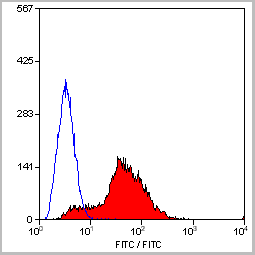
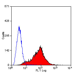
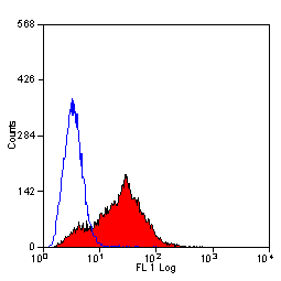
(Staining of mouse spleen with Hamster anti Mouse CD81: Alexa Fluor 488)
Application Data
(Staining of mouse spleen with Hamster anti Mouse CD81: Alexa Fluor 488)
CD81, Monoclonal Antibody (Cat# AAA11869)
Full Name
HAMSTER ANTI MOUSE CD81:FITC
Gene Names
Cd81; Tapa1; Tapa-1; Tspan28
Applications
Flow Cytometry
Pricing
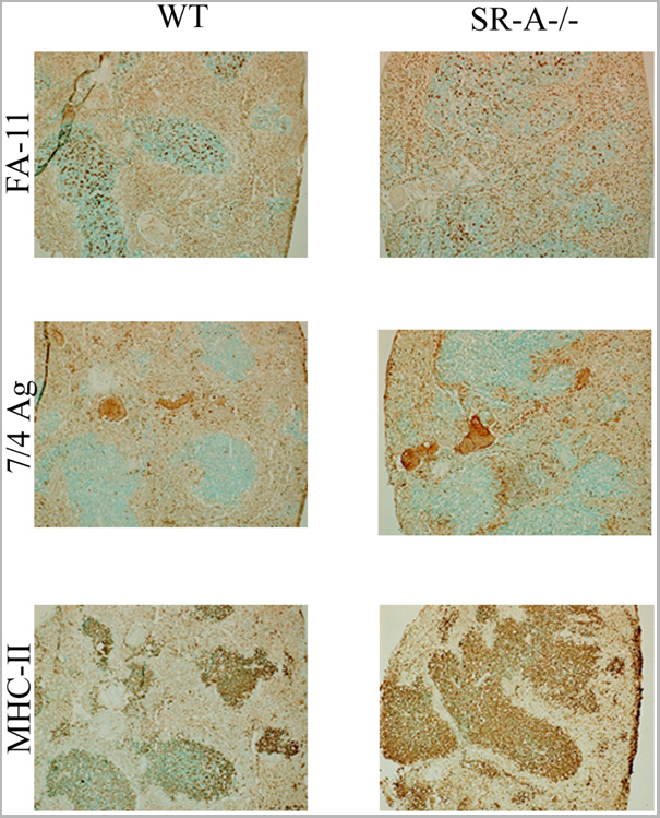 Application Data
(Staining of New Zealand Black mouse peripheral blood granulocytes with Rat anti Mouse Ly-6B.2 conjugated to FITC Data)
Application Data
(Staining of New Zealand Black mouse peripheral blood granulocytes with Rat anti Mouse Ly-6B.2 conjugated to FITC Data)
Ly-6B.2 ALLOANTIGEN, Monoclonal Antibody (Cat# AAA12190)
Full Name
RAT ANTI MOUSE Ly-6B.2 ALLOANTIGEN:FITC
Applications
Flow Cytometry
Pricing
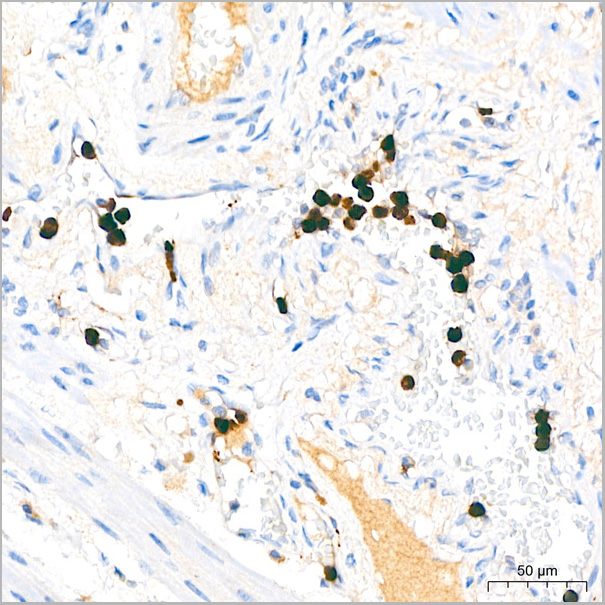
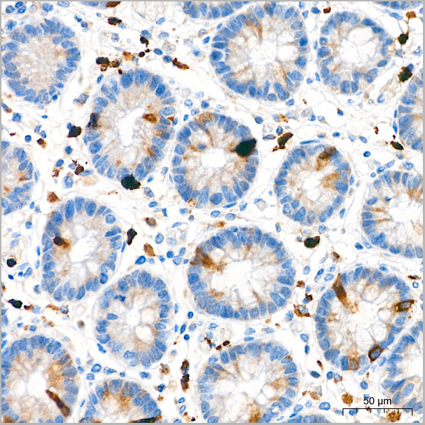
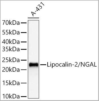
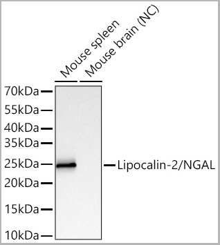
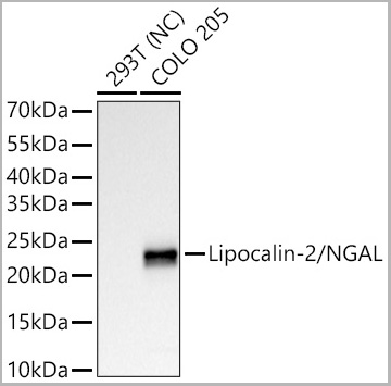
 ICC (Immunocytochemistry)
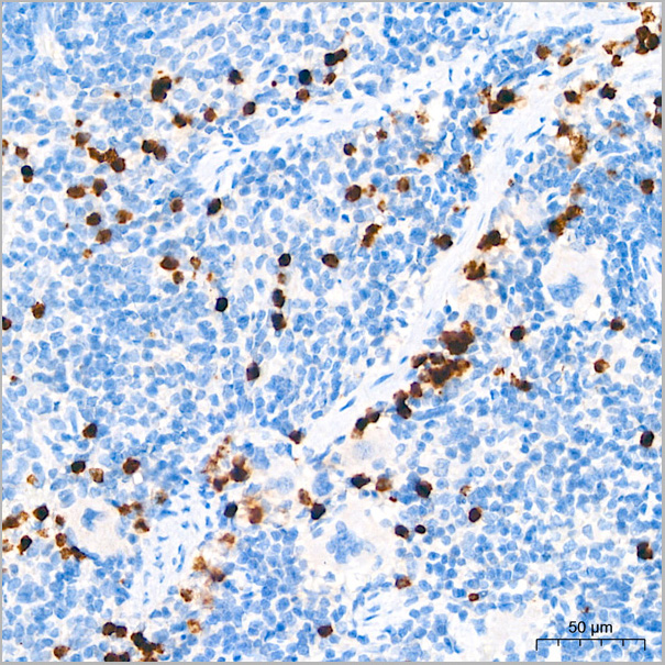
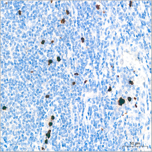
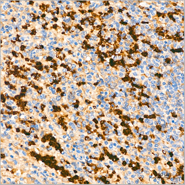
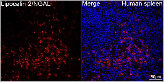
(Confocal imaging of paraffin-embedded Human spleen tissue using Lipocalin-2/NGAL Rabbit PolymAb® (AAA28588, dilution 1:200) followed by a further incubation with Cy3 Goat Anti-Rabbit IgG (H+L) (AS007, dilution 1:500) (Red). DAPI was used for nuclear staining (Blue). High pressure antigen retrieval performed with 0.01M Citrate Buffer(pH 6.0) prior to IF staining. Objective: 40x.)
ICC (Immunocytochemistry)
(Confocal imaging of paraffin-embedded Human spleen tissue using Lipocalin-2/NGAL Rabbit PolymAb® (AAA28588, dilution 1:200) followed by a further incubation with Cy3 Goat Anti-Rabbit IgG (H+L) (AS007, dilution 1:500) (Red). DAPI was used for nuclear staining (Blue). High pressure antigen retrieval performed with 0.01M Citrate Buffer(pH 6.0) prior to IF staining. Objective: 40x.)
Lipocalin-2/NGAL, Monoclonal Antibody (Cat# AAA28588)
Full Name
Lipocalin-2/NGAL Rabbit PolymAb
Reactivity
Human, Mouse
Applications
Western Blot, Immunohistochemistry, Immunofluorescence, Immunocytochemistry
Purity
Affinity purification
Pricing
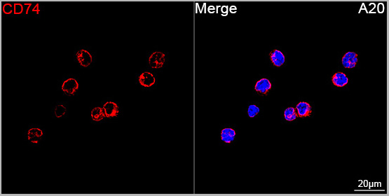
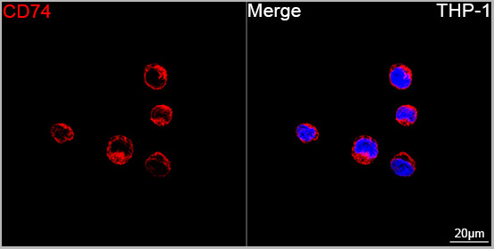
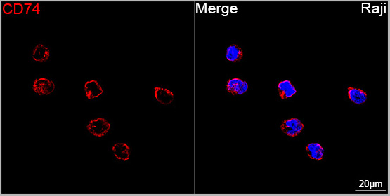
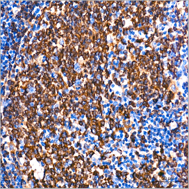
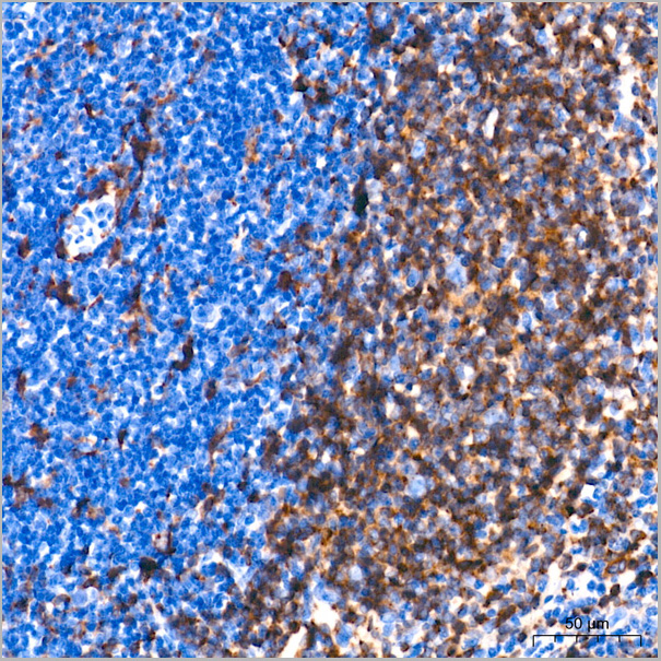
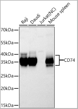
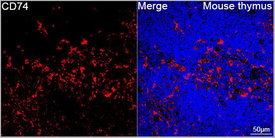 ICC (Immunocytochemistry)
(Confocal imaging of paraffin-embedded Mouse thymus tissue using CD74 Rabbit PolymAb® (A26945-PM, dilution 1:200) followed by a further incubation with Cy3 Goat Anti-Rabbit IgG (H+L) (AS007, dilution 1:500) (Red). DAPI was used for nuclear staining (Blue). High pressure antigen retrieval performed with 0.01M Citrate Buffer (pH 6.0) prior to IF staining. Objective: 40x.)
ICC (Immunocytochemistry)
(Confocal imaging of paraffin-embedded Mouse thymus tissue using CD74 Rabbit PolymAb® (A26945-PM, dilution 1:200) followed by a further incubation with Cy3 Goat Anti-Rabbit IgG (H+L) (AS007, dilution 1:500) (Red). DAPI was used for nuclear staining (Blue). High pressure antigen retrieval performed with 0.01M Citrate Buffer (pH 6.0) prior to IF staining. Objective: 40x.)
CD74, Monoclonal Antibody (Cat# AAA28597)
Full Name
CD74 Rabbit PolymAb
Reactivity
Human, Mouse
Applications
Western Blot, Immunohistochemistry, Immunofluorescence, Immunocytochemistry
Purity
Affinity purification
Pricing
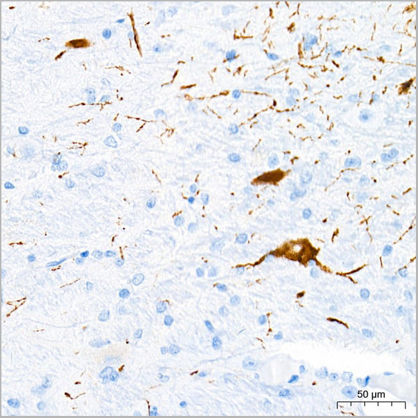
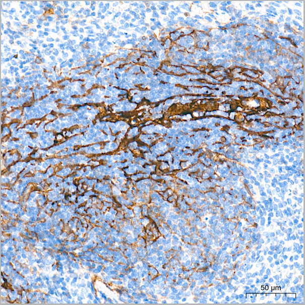
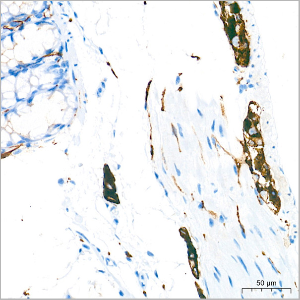
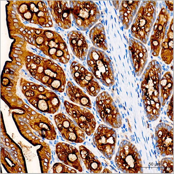
 IHC (Immunohistochemistry)
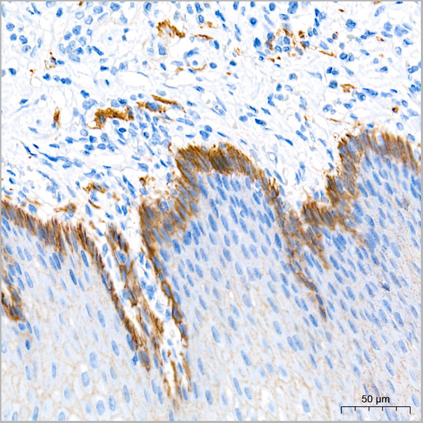
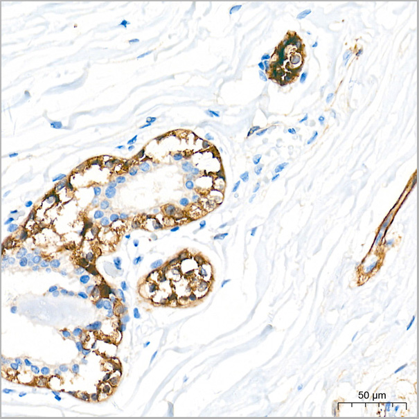
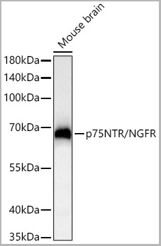
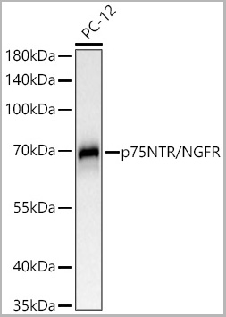
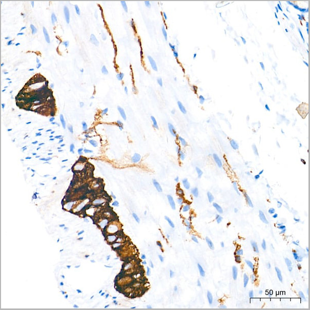
(Immunohistochemistry analysis of paraffin-embedded Rat colon tissue using p75NTR/NGFR Rabbit PolymAb® (AAA28585) at a dilution of 1:2000 (40x lens). High pressure antigen retrieval performed with 0.01M Tris-EDTA Buffer(pH 9.0) prior to IHC staining.)
IHC (Immunohistochemistry)
(Immunohistochemistry analysis of paraffin-embedded Rat colon tissue using p75NTR/NGFR Rabbit PolymAb® (AAA28585) at a dilution of 1:2000 (40x lens). High pressure antigen retrieval performed with 0.01M Tris-EDTA Buffer(pH 9.0) prior to IHC staining.)
p75NTR/NGFR, Monoclonal Antibody (Cat# AAA28585)
Full Name
p75NTR/NGFR Rabbit PolymAb
Reactivity
Human, Mouse
Applications
Western Blot, Immunohistochemistry
Purity
Affinity purification
Pricing
 Application Data
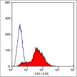
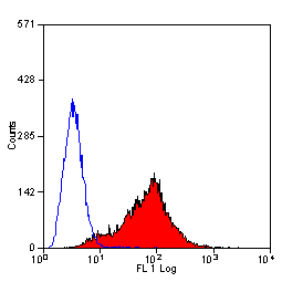
(Staining of mouse spleen with Hamster anti Mouse CD81: Alexa Fluor 488)
Application Data
(Staining of mouse spleen with Hamster anti Mouse CD81: Alexa Fluor 488)
CD81, Monoclonal Antibody (Cat# AAA12033)
Full Name
HAMSTER ANTI MOUSE CD81:RPE
Gene Names
Cd81; Tapa1; Tapa-1; Tspan28
Applications
Flow Cytometry
Pricing
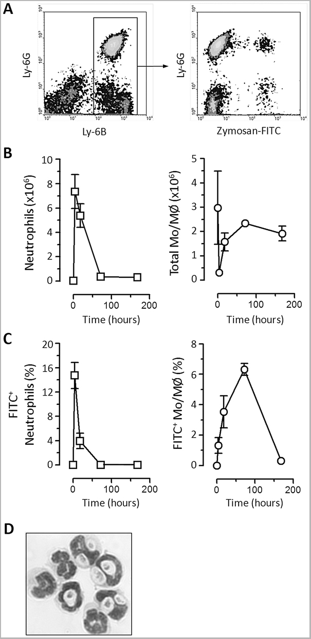 Application Data
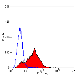
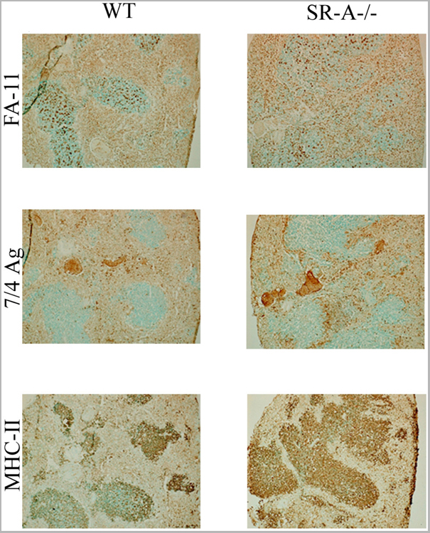
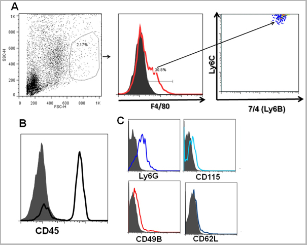
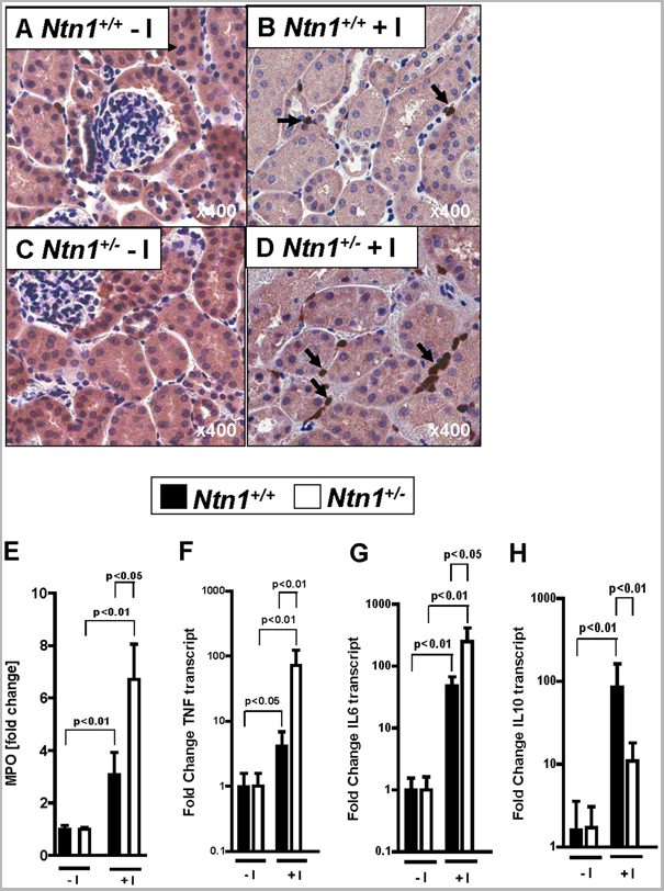
(Staining of New Zealand Black mouse peripheral blood granulocytes with Rat anti Mouse Ly-6B.2 conjugated to FITC Data)
Application Data
(Staining of New Zealand Black mouse peripheral blood granulocytes with Rat anti Mouse Ly-6B.2 conjugated to FITC Data)
Ly-6B.2 ALLOANTIGEN, Monoclonal Antibody (Cat# AAA12199)
Full Name
RAT ANTI MOUSE Ly-6B.2 ALLOANTIGEN:RPE
Applications
Flow Cytometry
Pricing
 Application Data
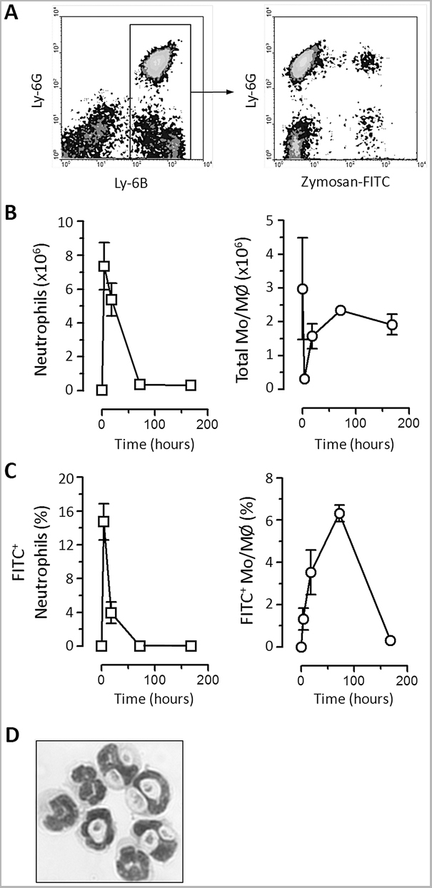
(Staining of New Zealand Black mouse peripheral blood granulocytes with Rat anti Mouse Ly-6B.2 conjugated to FITC Data)
Application Data
(Staining of New Zealand Black mouse peripheral blood granulocytes with Rat anti Mouse Ly-6B.2 conjugated to FITC Data)
Ly-6B.2 ALLOANTIGEN, Monoclonal Antibody (Cat# AAA12196)
Full Name
RAT ANTI MOUSE Ly-6B.2 ALLOANTIGEN
Applications
Immunohistochemistry, Flow Cytometry, Immunofluorescence, Immunohistochemistry, Western Blot
Pricing
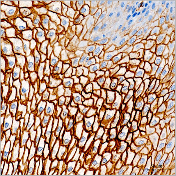
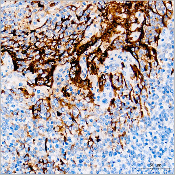
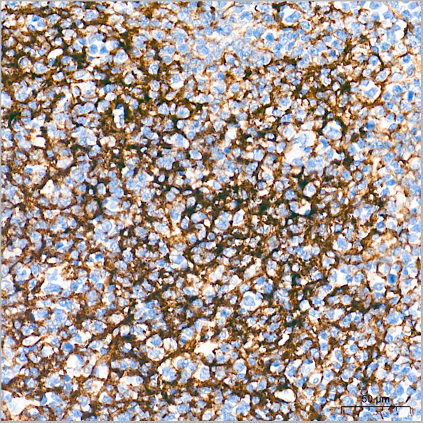
 IHC (Immunohistchemistry)
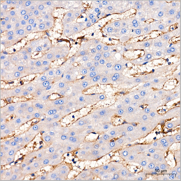
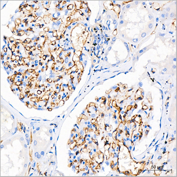
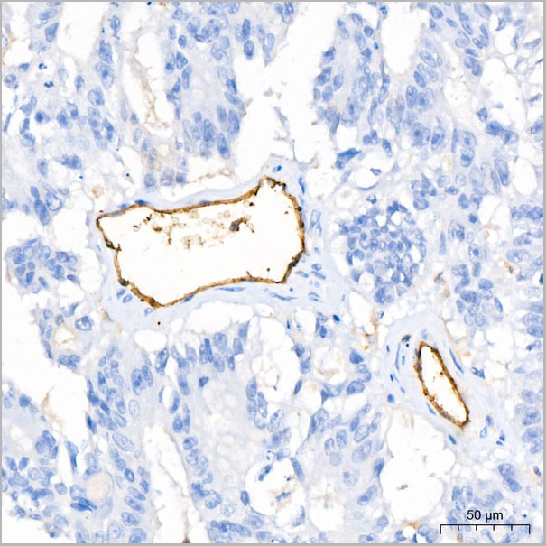
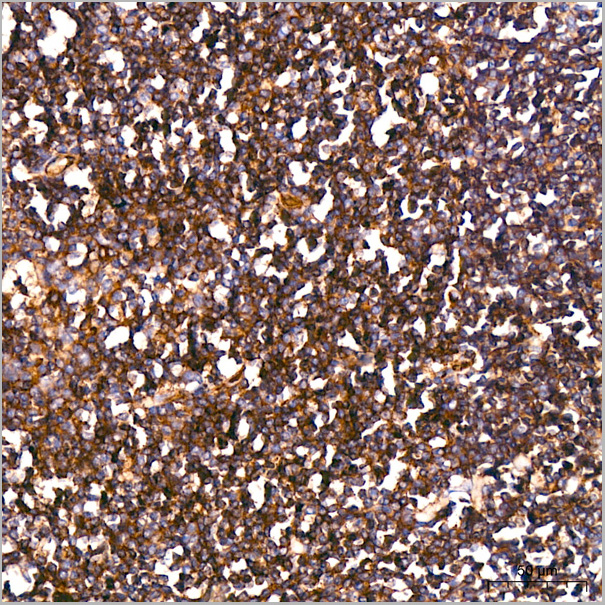
(Immunohistochemistry analysis of paraffin-embedded Human tonsil using CD102 Rabbit PolymAb® (AAA28564) at dilution of 1:200 (2:3) (40x lens). High pressure antigen retrieval performed with 0.01M Citrate Bufferr (pH 6.0) prior to IHC staining.)
IHC (Immunohistchemistry)
(Immunohistochemistry analysis of paraffin-embedded Human tonsil using CD102 Rabbit PolymAb® (AAA28564) at dilution of 1:200 (2:3) (40x lens). High pressure antigen retrieval performed with 0.01M Citrate Bufferr (pH 6.0) prior to IHC staining.)
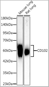
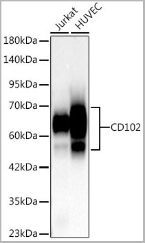
CD102, Monoclonal Antibody (Cat# AAA28564)
Full Name
CD102 Rabbit PolymAb
Reactivity
Human, Mouse
Applications
Western Blot, Immunohistochemistry
Purity
Affinity purification
Pricing
 Structure
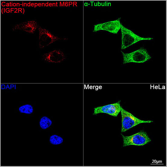 ICC (Immunocytochemistry)
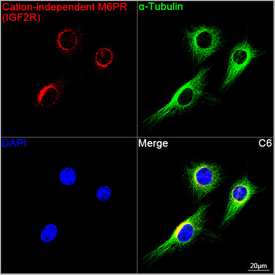
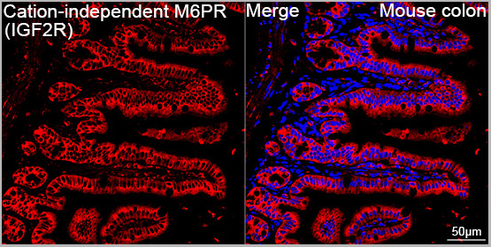
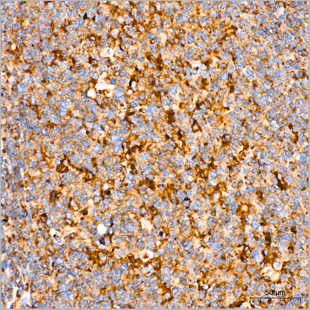
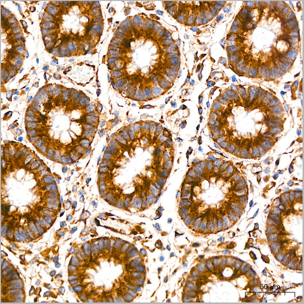
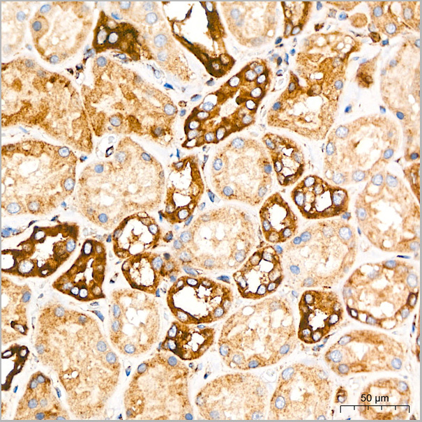
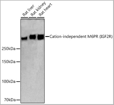
(Confocal imaging of HeLa cells using Cation-independent M6PR (IGF2R) Rabbit PolymAb® (A22421-PM, dilution 1:200) followed by a further incubation with Cy3 Goat Anti-Rabbit IgG (H+L) (AS007, dilution 1:500) (Red). The cells were counterstained with ?-Tubulin Mouse mAb (AC012, dilution 1:400) followed by incubation with ABflo® 488-conjugated Goat Anti-Mouse IgG (H+L) Ab (AS076, dilution 1:500) (Green). DAPI was used for nuclear staining (Blue). Objective: 100x.)
ICC (Immunocytochemistry)
(Confocal imaging of HeLa cells using Cation-independent M6PR (IGF2R) Rabbit PolymAb® (A22421-PM, dilution 1:200) followed by a further incubation with Cy3 Goat Anti-Rabbit IgG (H+L) (AS007, dilution 1:500) (Red). The cells were counterstained with ?-Tubulin Mouse mAb (AC012, dilution 1:400) followed by incubation with ABflo® 488-conjugated Goat Anti-Mouse IgG (H+L) Ab (AS076, dilution 1:500) (Green). DAPI was used for nuclear staining (Blue). Objective: 100x.)
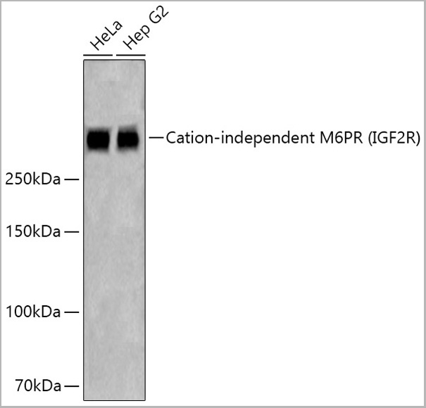
Cation-independent M6PR (IGF2R), Monoclonal Antibody (Cat# AAA28549)
Full Name
Cation-independent M6PR (IGF2R) Rabbit PolymAb
Reactivity
Human
Applications
Western Blot, Immunohistochemistry, Immunofluorescence, Immunocytochemistry
Purity
Affinity purification
Pricing
Helicobacter pylori, Native Protein (Cat# AAA14355)
Full Name
Helicobacter pylori protein
Purity
Highly purified
Pricing
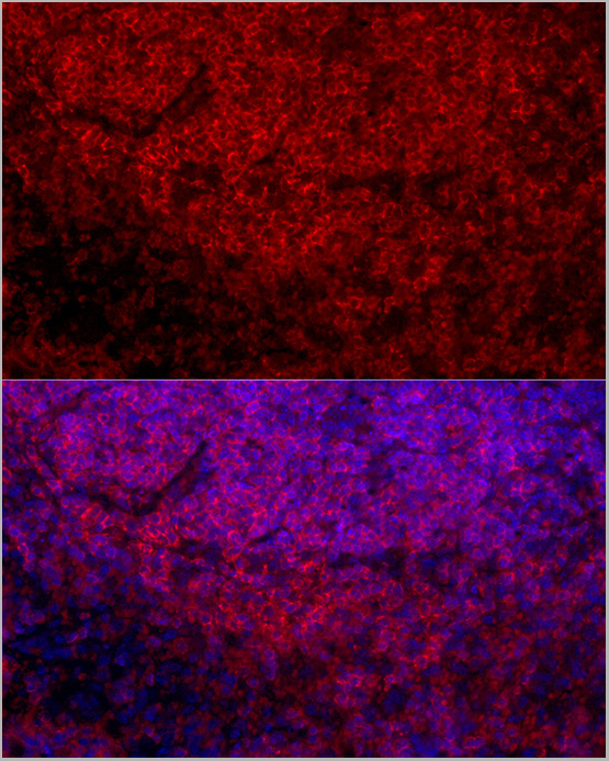
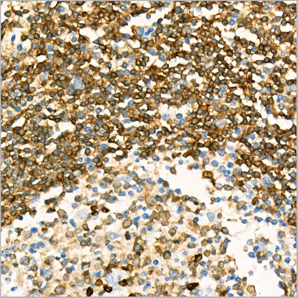
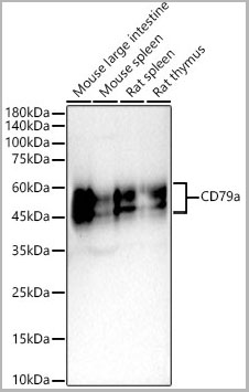
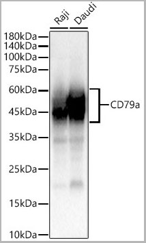
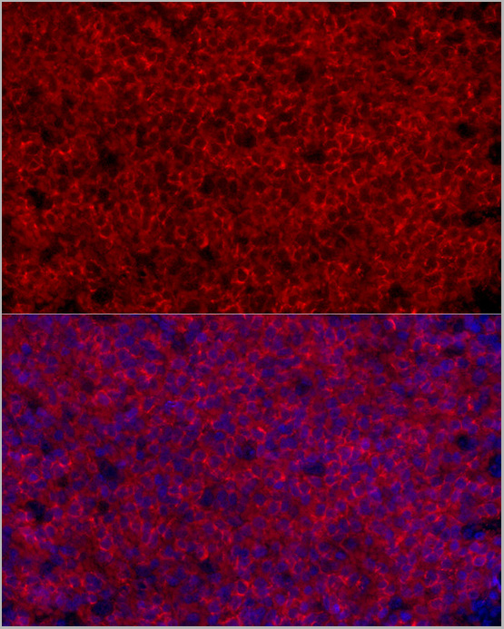 IF (Immunofluorescence)
(Immunofluorescence analysis of paraffin-embedded rat spleen using CD79a Rabbit PolymAb® (AAA28555) at dilution of 1:50 (40x lens). Secondary antibody: Cy3-conjugated Goat anti-Rabbit IgG (H+L) (AS007) at 1:500 dilution. Blue: DAPI for nuclear staining.)
IF (Immunofluorescence)
(Immunofluorescence analysis of paraffin-embedded rat spleen using CD79a Rabbit PolymAb® (AAA28555) at dilution of 1:50 (40x lens). Secondary antibody: Cy3-conjugated Goat anti-Rabbit IgG (H+L) (AS007) at 1:500 dilution. Blue: DAPI for nuclear staining.)
CD79a, Monoclonal Antibody (Cat# AAA28555)
Full Name
CD79a Rabbit PolymAb
Reactivity
Human
Applications
Western Blot, Immunohistochemistry, Immunofluorescence, Immunocytochemistry
Purity
Affinity purification
Pricing
Serum amyloid A, Antigen (Cat# AAA26946)
Full Name
Human Serum amyloid A protein
Gene Names
SAA1; SAA
Applications
Latex Enhanced Turbidimetric Immunoassay, Chemiluminescence Immunoassay
Purity
>95% by SDS-PAGE
Pricing
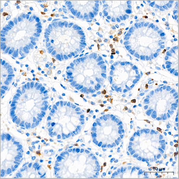
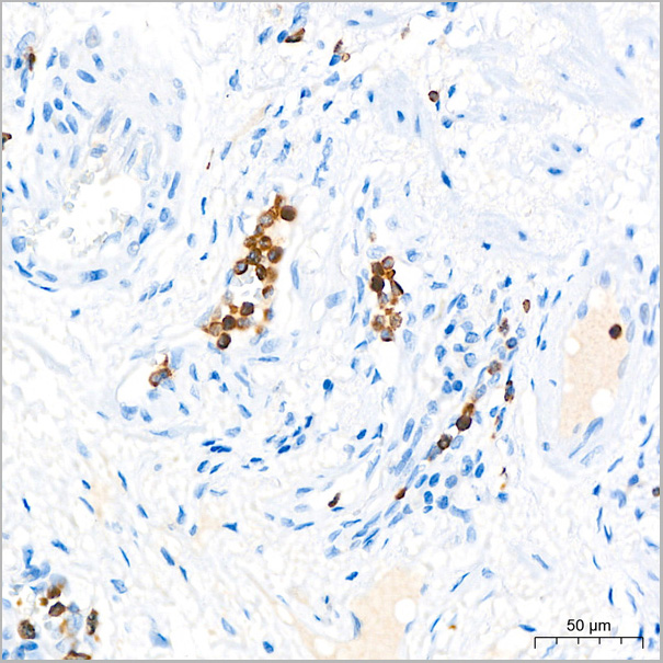
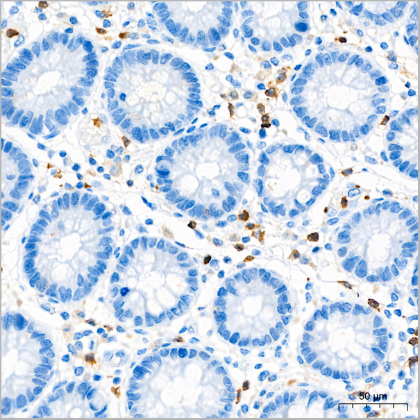
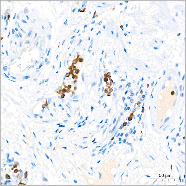
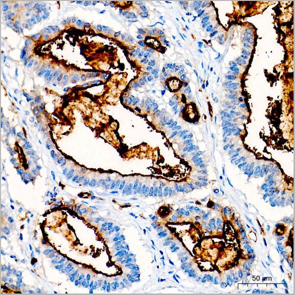
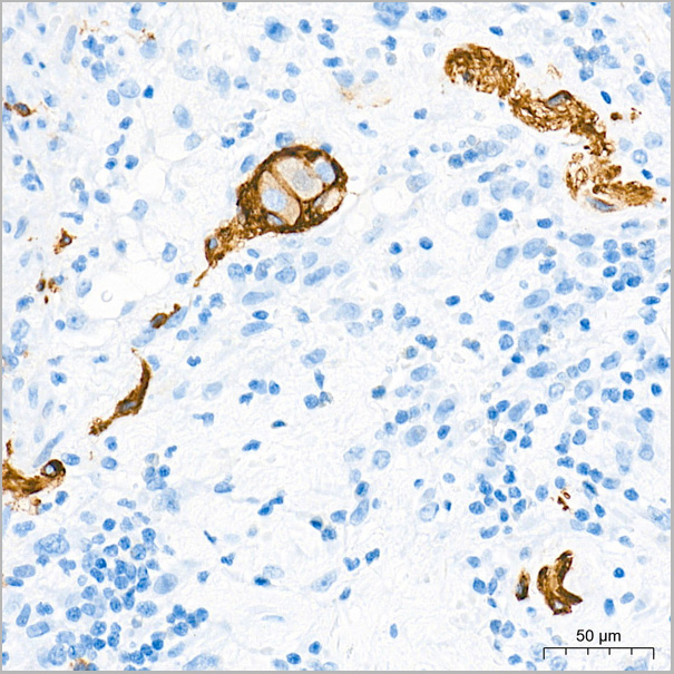
 IHC (Immunohistchemistry)
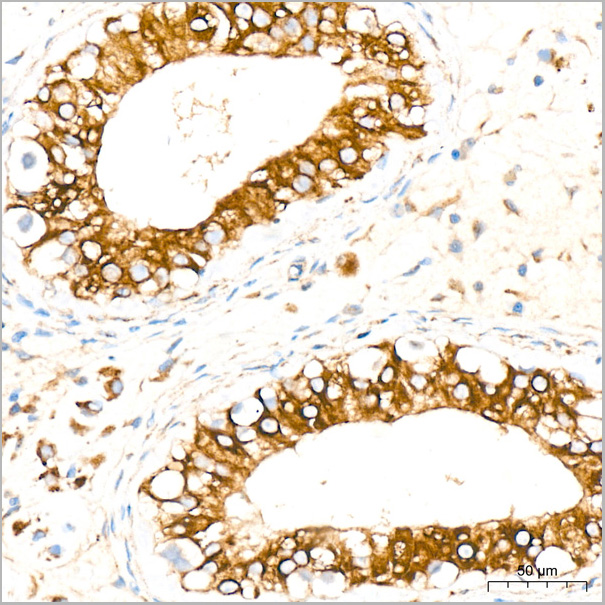
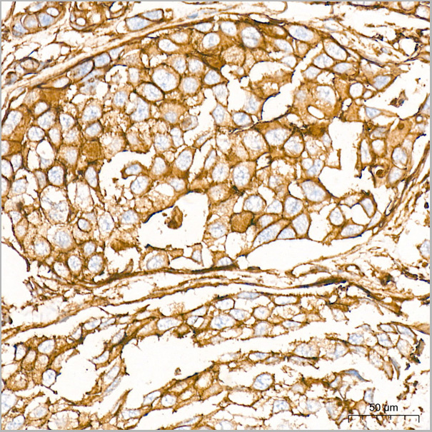
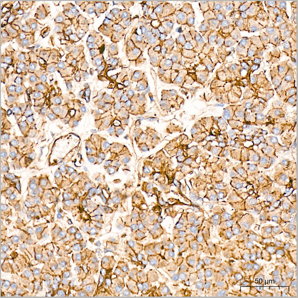
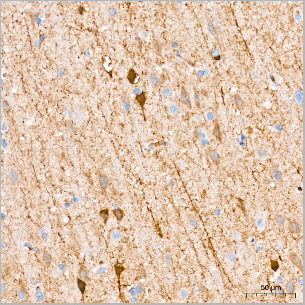
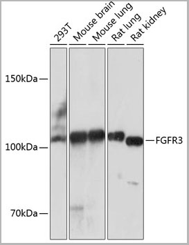
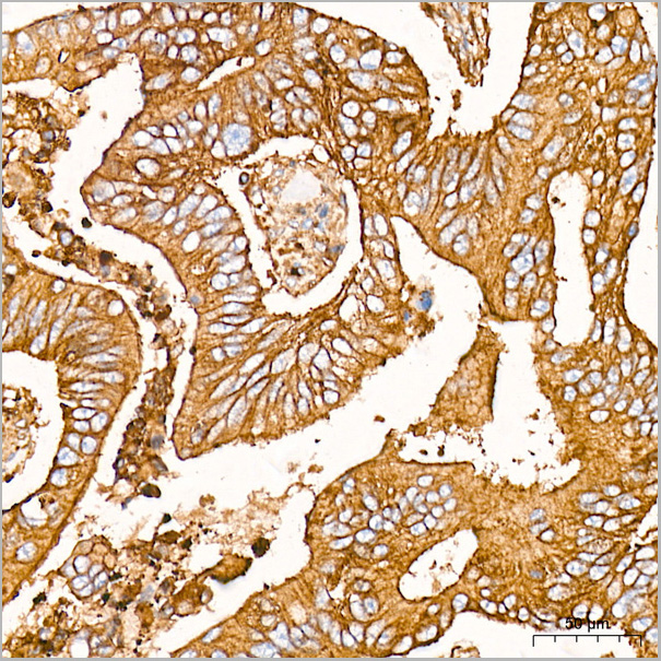
(Immunohistochemistry analysis of paraffin-embedded Human colon carcinoma tissue using FGFR3 Rabbit mAb (AAA28474) at a dilution of 1:2000 (40x lens). High pressure antigen retrieval was performed with 0.01 M citrate buffer (pH 6.0) prior to IHC staining.)
IHC (Immunohistchemistry)
(Immunohistochemistry analysis of paraffin-embedded Human colon carcinoma tissue using FGFR3 Rabbit mAb (AAA28474) at a dilution of 1:2000 (40x lens). High pressure antigen retrieval was performed with 0.01 M citrate buffer (pH 6.0) prior to IHC staining.)
FGFR3, Monoclonal Antibody (Cat# AAA28474)
Full Name
FGFR3 Rabbit mAb
Reactivity
Human
Applications
Western Blot, Immunohistochemistry
Purity
Affinity purification
Pricing
 Bioactivity
(C-reactive protein (CRP) is an annular (ring-shaped), pentameric protein found in blood plasma, whose levels rise in response to inflammation. It is an acute-phase protein of hepatic origin that increases following interleukin-6 secretion by macrophages and T cells. Its physiological role is to bind to lysophosphatidylcholine expressed on the surface of dead or dying cells (and some types of bacteria) in order to activate the complement system via C1q. Besides, Ficolin 2 (FCN2) has been identified as an interactor of CRP, thus a functional binding ELISA assay was conducted to detect the interaction of recombinant human CRP and recombinant mouse FCN2. Briefly, CRP was diluted serially in PBS with 0.01% BSA (pH 7.4). Duplicate samples of 100 ul were then transferred to FCN2-coated microtiter wells and incubated for 1h at 37 degree C. Wells were washed with PBST and incubated for 1h with anti-CRP pAb, then aspirated and washed 3 times. After incubation with HRP labelled secondary antibody for 1h at 37 degree C, wells were aspirated and washed 5 times. With the addition of substrate solution, wells were incubated 15-25 minutes at 37 degree C. Finally, add 50 uL stop solution to the wells and read at 450/630nm immediately. The binding activity of recombinant human CRP and recombinant mouse FCN2 was shown in Figure 1, the EC50 for this effect is 0.008 ug/mL.)
Bioactivity
(C-reactive protein (CRP) is an annular (ring-shaped), pentameric protein found in blood plasma, whose levels rise in response to inflammation. It is an acute-phase protein of hepatic origin that increases following interleukin-6 secretion by macrophages and T cells. Its physiological role is to bind to lysophosphatidylcholine expressed on the surface of dead or dying cells (and some types of bacteria) in order to activate the complement system via C1q. Besides, Ficolin 2 (FCN2) has been identified as an interactor of CRP, thus a functional binding ELISA assay was conducted to detect the interaction of recombinant human CRP and recombinant mouse FCN2. Briefly, CRP was diluted serially in PBS with 0.01% BSA (pH 7.4). Duplicate samples of 100 ul were then transferred to FCN2-coated microtiter wells and incubated for 1h at 37 degree C. Wells were washed with PBST and incubated for 1h with anti-CRP pAb, then aspirated and washed 3 times. After incubation with HRP labelled secondary antibody for 1h at 37 degree C, wells were aspirated and washed 5 times. With the addition of substrate solution, wells were incubated 15-25 minutes at 37 degree C. Finally, add 50 uL stop solution to the wells and read at 450/630nm immediately. The binding activity of recombinant human CRP and recombinant mouse FCN2 was shown in Figure 1, the EC50 for this effect is 0.008 ug/mL.)
C ReProtein (CRP), Active Protein (Cat# AAA161782)
Full Name
Active C Reactive Protein (CRP)
Gene Names
CRP; PTX1
Applications
Activity Assay, Cell Culture
Purity
Greater than 90% by SDS-PAGE
Pricing
 Application Data
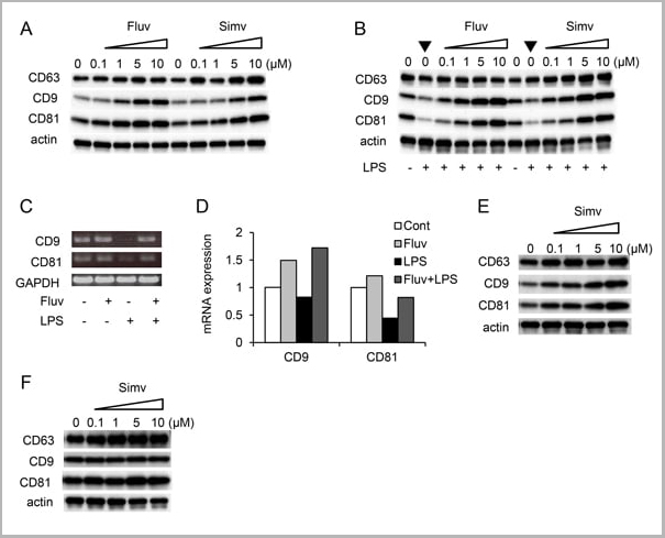
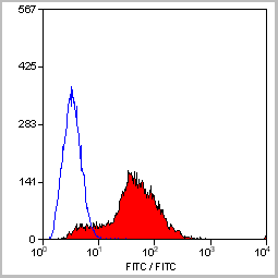
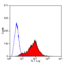
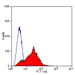
(Staining of mouse spleen with Hamster anti Mouse CD81: Alexa Fluor 488)
Application Data
(Staining of mouse spleen with Hamster anti Mouse CD81: Alexa Fluor 488)
CD81, Monoclonal Antibody (Cat# AAA11942)
Full Name
HAMSTER ANTI MOUSE CD81
Gene Names
Cd81; Tapa1; Tapa-1; Tspan28
Reactivity
Rat
Applications
Immunohistochemistry, Flow Cytometry, Immunoprecipitation, Western Blot
Pricing
HBsAg, Polyclonal Antibody (Cat# AAA14317)
Full Name
HBsAg antibody
Reactivity
Specific for all 4 subtypes of Hepatitis B surface Antigens
Purity
HBsAg antibody was purified by affinity chromatography.
Pricing
 WB (Western Blot)
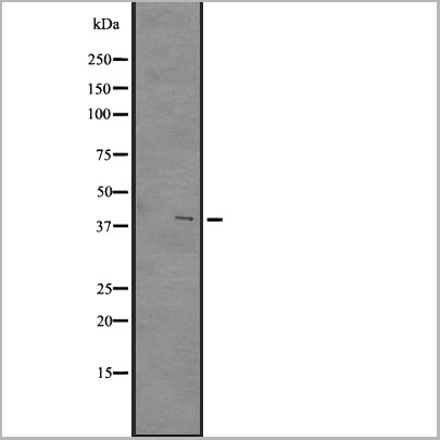
(Western blot analysis of GPR84 expression in A431 whole cell lysates)
WB (Western Blot)
(Western blot analysis of GPR84 expression in A431 whole cell lysates)
GPR84, Polyclonal Antibody (Cat# AAA31105)
Full Name
GPR84 Antibody
Gene Names
GPR84; EX33; GPCR4
Reactivity
Human, Mouse
Applications
Western Blot, Immunohistochemistry
Purity
The antiserum was purified by peptide affinity chromatography using SulfoLinkTM Coupling Resin (Thermo Fisher Scientific).
Pricing

 Application Data
Tumor Necrosis Factor Alpha (TNFa), Active Protein (Cat# AAA146615)
Full Name
Active Tumor Necrosis Factor Alpha (TNFa)
Gene Names
Tnf; DIF; Tnfa; TNF-a; TNFSF2; Tnlg1f; Tnfsf1a; TNFalpha; TNF-alpha
Applications
Activity Assay, Cell Culture
Purity
> 95%
Pricing
S100A9, Monoclonal Antibody (Cat# AAA13837)
Full Name
S100A9 (Macrophage Marker) Mouse Monoclonal Antibody
Gene Names
S100A9; MIF; NIF; P14; CAGB; CFAG; CGLB; L1AG; LIAG; MRP14; 60B8AG; MAC387
Reactivity
Human and Rat. Others not tested.
Applications
Flow Cytometry, Immunofluorescence, Immunohistochemistry
Pricing
 Bioactivity
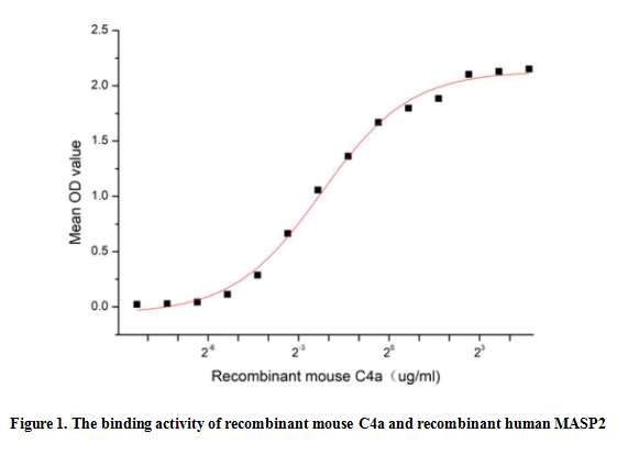
(Complement Component 4a (C4a) is a component of the complement system, which is a cleavage product of the complement C4 protein. C4a has been implicated in various inflammatory and immune responses. It acts as a chemoattractant, recruiting immune cells such as neutrophils and macrophages to the site of inflammation. Additionally, C4a can stimulate the release of pro-inflammatory cytokines and chemokines, further amplifying the immune response. Besides, the binding of MASP2 to C4a is an important step in the lectin pathway of the complement system, thus a functional binding ELISA assay was conducted to detect the interaction of recombinant mouse C4a and recombinant human MASP2. Briefly, C4a was diluted serially in PBS with 0.01% BSA (pH 7.4). Duplicate samples of 100 ul were then transferred to MASP2-coated microtiter wells and incubated for 1h at 37 degree C. Wells were washed with PBST and incubated for 1h with anti-C4a pAb, then aspirated and washed 3 times. After incubation with HRP labelled secondary antibody for 1h at 37 degree C, wells were aspirated and washed 5 times. With the addition of substrate solution, wells were incubated 15-25 minutes at 37 degree C. Finally, add 50 uL stop solution to the wells and read at 450/630 nm immediately. The binding activity of recombinant mouse C4a and recombinant human MASP2 was shown in Figure 1, the EC50 for this effect is 0.21 ug/mL.)
Bioactivity
(Complement Component 4a (C4a) is a component of the complement system, which is a cleavage product of the complement C4 protein. C4a has been implicated in various inflammatory and immune responses. It acts as a chemoattractant, recruiting immune cells such as neutrophils and macrophages to the site of inflammation. Additionally, C4a can stimulate the release of pro-inflammatory cytokines and chemokines, further amplifying the immune response. Besides, the binding of MASP2 to C4a is an important step in the lectin pathway of the complement system, thus a functional binding ELISA assay was conducted to detect the interaction of recombinant mouse C4a and recombinant human MASP2. Briefly, C4a was diluted serially in PBS with 0.01% BSA (pH 7.4). Duplicate samples of 100 ul were then transferred to MASP2-coated microtiter wells and incubated for 1h at 37 degree C. Wells were washed with PBST and incubated for 1h with anti-C4a pAb, then aspirated and washed 3 times. After incubation with HRP labelled secondary antibody for 1h at 37 degree C, wells were aspirated and washed 5 times. With the addition of substrate solution, wells were incubated 15-25 minutes at 37 degree C. Finally, add 50 uL stop solution to the wells and read at 450/630 nm immediately. The binding activity of recombinant mouse C4a and recombinant human MASP2 was shown in Figure 1, the EC50 for this effect is 0.21 ug/mL.)
Complement Component 4a (C4a), Active Protein (Cat# AAA161729)
Full Name
Active Complement Component 4a (C4a)
Applications
Activity Assay, Cell Culture
Purity
Greater than 90% by SDS-PAGE
Pricing
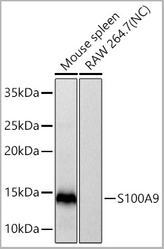
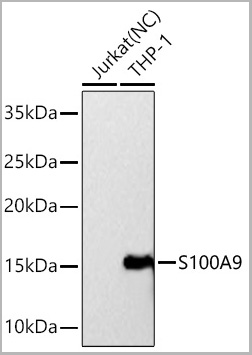
 ICC (Immunocytochemistry)
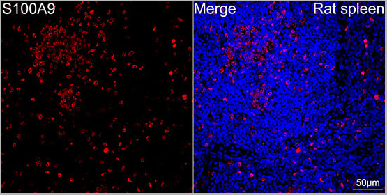
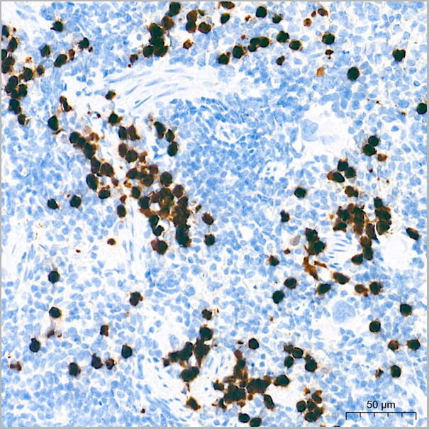
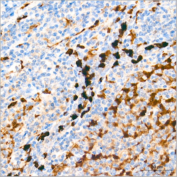
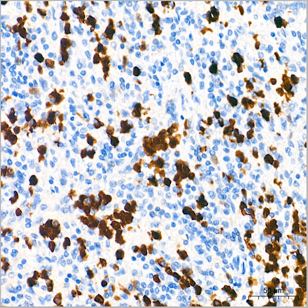
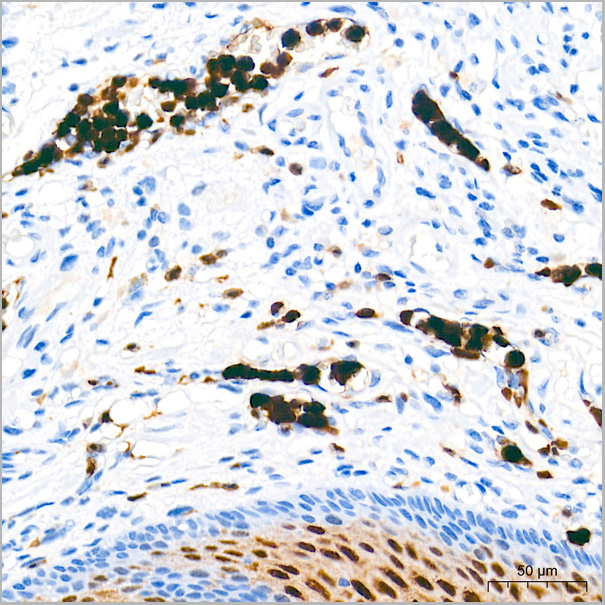
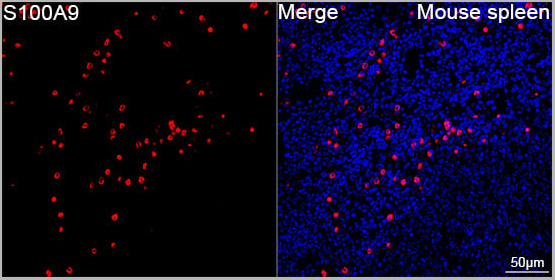
(Confocal imaging of paraffin-embedded Mouse spleen tissue using S100A9 Rabbit PolymAb® (AAA28595, dilution 1:200) followed by a further incubation with Cy3 Goat Anti-Rabbit IgG (H+L) (AS007, dilution 1:500) (Red). DAPI was used for nuclear staining (Blue). High pressure antigen retrieval performed with 0.01M Citrate Buffer (pH 6.0) prior to IF staining. Objective: 40x.)
ICC (Immunocytochemistry)
(Confocal imaging of paraffin-embedded Mouse spleen tissue using S100A9 Rabbit PolymAb® (AAA28595, dilution 1:200) followed by a further incubation with Cy3 Goat Anti-Rabbit IgG (H+L) (AS007, dilution 1:500) (Red). DAPI was used for nuclear staining (Blue). High pressure antigen retrieval performed with 0.01M Citrate Buffer (pH 6.0) prior to IF staining. Objective: 40x.)
S100A9, Monoclonal Antibody (Cat# AAA28595)
Full Name
S100A9 Rabbit PolymAb
Reactivity
Human, Mouse
Applications
Western Blot, Immunohistochemistry, Immunofluorescence, Immunocytochemistry
Purity
Affinity purification
Pricing
 Bioactivity
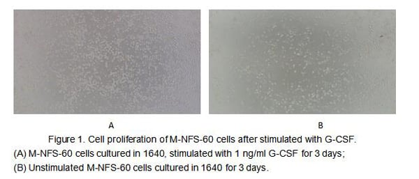
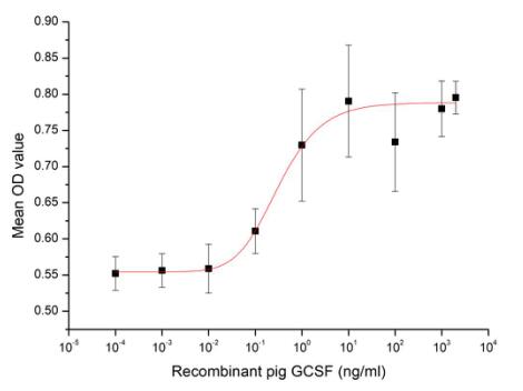
(Figure 2. Cell proliferation of M-NFS-60 cells after stimulated with G-CSF.)
Bioactivity
(Figure 2. Cell proliferation of M-NFS-60 cells after stimulated with G-CSF.)
Colony Stimulating Factor 3, Granulocyte (GCSF), Active Protein (Cat# AAA161665)
Full Name
Active Colony Stimulating Factor 3, Granulocyte (GCSF)
Gene Names
CSF3; G-CSF
Applications
Activity Assay, Cell Culture
Purity
Greater than 80% by SDS-PAGE
Pricing
HBsAg, Monoclonal Antibody (Cat# AAA14308)
Full Name
HBsAg antibody
Reactivity
To be determined by end-user
Applications
ELISA
Pricing
 Bioactivity
(Neutrophil elastase (NE), also known as polymorphonuclear leukocyte elastase, is a major protease in the primary granules of neutrophils, is involved in microbicidal activity. It is located primarily in the azurophil granules of polymorphonuclear leukocytes. NE is an important factor promoting inflammation, has bactericidal effects, and shortens the inflammatory process. NE also regulates tumor growth by promoting metastasis and tumor microenvironment remodeling. However, NE plays a role in killing tumors under certain conditions and promotes other diseases such as pulmonary ventilation dysfunction. Additionally, it plays a complex role in various physiological processes and mediates several diseases. Myeloperoxidase (MPO) has been identified as an interactor of NE, thus a functional binding ELISA assay was conducted to detect the interaction of recombinant human NE and recombinant rat MPO. Briefly, biotin-linked NE were diluted serially in PBS, with 0.01% BSA (pH 7.4). Duplicate samples of 100 ul were then transferred to MPO-coated microtiter wells and incubated for 1h at 37 degree C. Wells were washed with PBST 3 times and incubation with Streptavidin-HRP for 30min, then wells were aspirated and washed 5 times. With the addition of substrate solution, wells were incubated 15-25 minutes at 37 degree C. Finally, add 50 ul stop solution to the wells and read at 450 nm immediately. The binding activity of recombinant human NE and recombinant rat MPO was shown in Figure 1, the EC50 for this effect is 0.66 ug/mL.)
Bioactivity
(Neutrophil elastase (NE), also known as polymorphonuclear leukocyte elastase, is a major protease in the primary granules of neutrophils, is involved in microbicidal activity. It is located primarily in the azurophil granules of polymorphonuclear leukocytes. NE is an important factor promoting inflammation, has bactericidal effects, and shortens the inflammatory process. NE also regulates tumor growth by promoting metastasis and tumor microenvironment remodeling. However, NE plays a role in killing tumors under certain conditions and promotes other diseases such as pulmonary ventilation dysfunction. Additionally, it plays a complex role in various physiological processes and mediates several diseases. Myeloperoxidase (MPO) has been identified as an interactor of NE, thus a functional binding ELISA assay was conducted to detect the interaction of recombinant human NE and recombinant rat MPO. Briefly, biotin-linked NE were diluted serially in PBS, with 0.01% BSA (pH 7.4). Duplicate samples of 100 ul were then transferred to MPO-coated microtiter wells and incubated for 1h at 37 degree C. Wells were washed with PBST 3 times and incubation with Streptavidin-HRP for 30min, then wells were aspirated and washed 5 times. With the addition of substrate solution, wells were incubated 15-25 minutes at 37 degree C. Finally, add 50 ul stop solution to the wells and read at 450 nm immediately. The binding activity of recombinant human NE and recombinant rat MPO was shown in Figure 1, the EC50 for this effect is 0.66 ug/mL.)
Neutrophil Elastase (NE), Active Protein (Cat# AAA161712)
Full Name
Active Neutrophil Elastase (NE)
Gene Names
ELANE; GE; NE; HLE; HNE; ELA2; SCN1; PMN-E
Applications
Activity Assay, Cell Culture
Purity
Greater than 95% by SDS-PAGE
Pricing
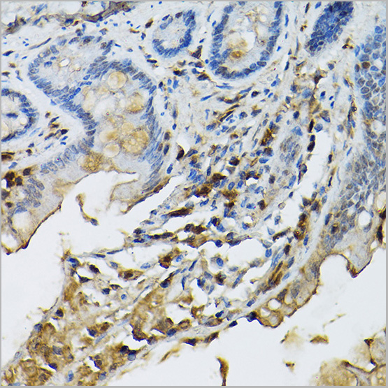
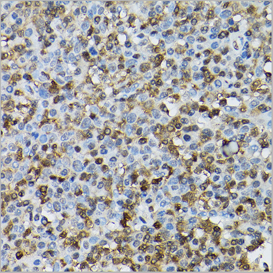
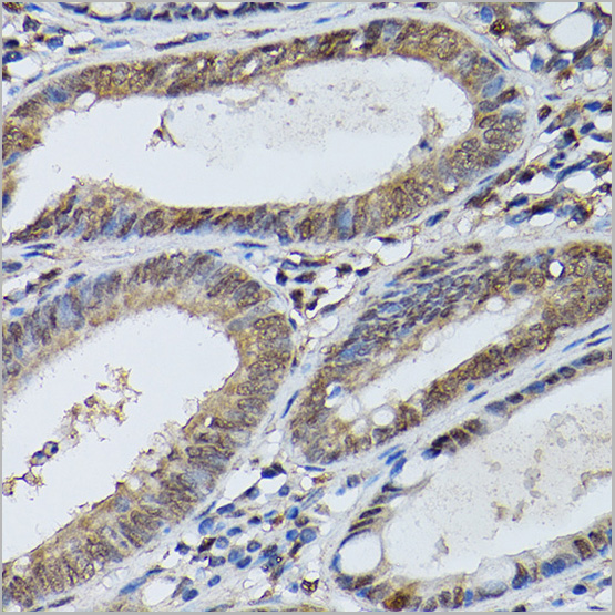
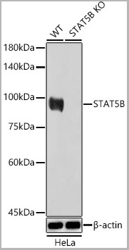
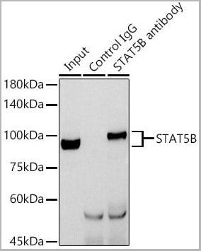 IP (Immunoprecipitation)
(Immunoprecipitation analysis of 600ug extracts of Mouse thymus cells using 3ug STAT5B antibody. Western blot was performed from the immunoprecipitate using STAT5B antibody at a dilution of 1:1000.)
IP (Immunoprecipitation)
(Immunoprecipitation analysis of 600ug extracts of Mouse thymus cells using 3ug STAT5B antibody. Western blot was performed from the immunoprecipitate using STAT5B antibody at a dilution of 1:1000.)
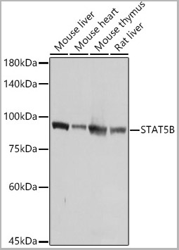
STAT5B, Polyclonal Antibody (Cat# AAA28416)
Full Name
[KO Validated] STAT5B Rabbit pAb
Reactivity
Human, Mouse, Rat
Applications
Western Blot, Immunohistochemistry, Immunoprecipitation, Immunofluorescence, Immunocytochemistry
Purity
Affinity purification
Pricing

 Bioactivity
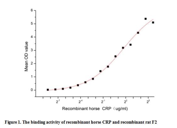
(C reactive protein (CRP) is an annular (ring-shaped), pentameric protein found in blood plasma, whose levels rise in response to inflammation. It is an acute-phase protein of hepatic origin that increases following interleukin-6 secretion by macrophages and T cells. Its physiological role is to bind to lysophosphatidylcholine expressed on the surface of dead or dying cells (and some types of bacteria) in order to activate the complement system via C1q. Besides, Coagulation Factor II (F2) has been identified as an interactor of CRP, thus a functional binding ELISA assay was conducted to detect the interaction of recombinant horse CRP and recombinant rat F2. Briefly, CRP was diluted serially in PBS with 0.01% BSA (pH 7.4). Duplicate samples of 100 ul were then transferred to F2-coated microtiter wells and incubated for 1h at 37 degree C. Wells were washed with PBST and incubated for 1h with anti-CRP pAb, then aspirated and washed 3 times. After incubation with HRP labelled secondary antibody for 1h at 37 degree C, wells were aspirated and washed 5 times. With the addition of substrate solution, wells were incubated 15-25 minutes at 37 degree C. Finally, add 50 uL stop solution to the wells and read at 450/630 nm immediately. The binding activity of recombinant horse CRP and recombinant rat F2 was shown in Figure 1, the EC50 for this effect is 3.29 ug/mL.)
Bioactivity
(C reactive protein (CRP) is an annular (ring-shaped), pentameric protein found in blood plasma, whose levels rise in response to inflammation. It is an acute-phase protein of hepatic origin that increases following interleukin-6 secretion by macrophages and T cells. Its physiological role is to bind to lysophosphatidylcholine expressed on the surface of dead or dying cells (and some types of bacteria) in order to activate the complement system via C1q. Besides, Coagulation Factor II (F2) has been identified as an interactor of CRP, thus a functional binding ELISA assay was conducted to detect the interaction of recombinant horse CRP and recombinant rat F2. Briefly, CRP was diluted serially in PBS with 0.01% BSA (pH 7.4). Duplicate samples of 100 ul were then transferred to F2-coated microtiter wells and incubated for 1h at 37 degree C. Wells were washed with PBST and incubated for 1h with anti-CRP pAb, then aspirated and washed 3 times. After incubation with HRP labelled secondary antibody for 1h at 37 degree C, wells were aspirated and washed 5 times. With the addition of substrate solution, wells were incubated 15-25 minutes at 37 degree C. Finally, add 50 uL stop solution to the wells and read at 450/630 nm immediately. The binding activity of recombinant horse CRP and recombinant rat F2 was shown in Figure 1, the EC50 for this effect is 3.29 ug/mL.)
C ReProtein (CRP), Active Protein (Cat# AAA161779)
Full Name
Active C Reactive Protein (CRP)
Applications
Activity Assay, Cell Culture
Purity
Greater than 90% by SDS-PAGE
Pricing
 Bioactivity
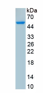
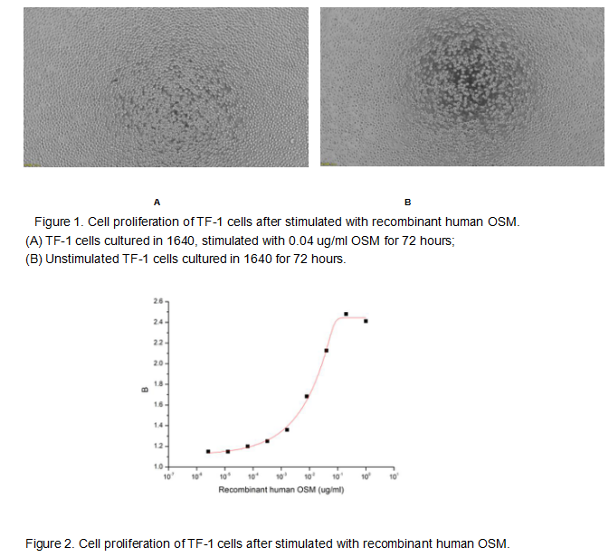
(Oncostatin M (OSM) is an approximately 30 KDa secreted cytokine belonging to the Interleukin-6 family. Like other members of the IL-6 family such as IL-11, CNTF, and Cardiotrophin-1, OSM plays crucial roles in inflammation, neuroprotection, hematopoiesis, metabolism and development. To test the effect of OSM on cell proliferation, TF-1 cells were seeded into triplicate wells of 96-well plates with various concentrations of recombinant human OSM. After incubated for 72 hours, cells were observed by inverted microscope and cell proliferation was measured by Cell Counting Kit-8 (CCK-8). Briefly, 10 ul of CCK-8 solution was added to each well of the plate, then the absorbance at 450 nm was measured using a microplate reader after incubating the plate for 1-4 hours at 37 degree C. Proliferation of TF-1 cells after incubation with OSM for 72 hours observed by inverted microscope was shown in Figure 1. Cell viability was assessed by CCK-8 (Cell Counting Kit-8) assay after incubation with recombinant human OSM for 72 hours. The result was shown in Figure 2. It was obvious that recombinant human OSM significantly increased cell viability of TF-1 cells, the EC50 was 0.014 ug/ml.)
Bioactivity
(Oncostatin M (OSM) is an approximately 30 KDa secreted cytokine belonging to the Interleukin-6 family. Like other members of the IL-6 family such as IL-11, CNTF, and Cardiotrophin-1, OSM plays crucial roles in inflammation, neuroprotection, hematopoiesis, metabolism and development. To test the effect of OSM on cell proliferation, TF-1 cells were seeded into triplicate wells of 96-well plates with various concentrations of recombinant human OSM. After incubated for 72 hours, cells were observed by inverted microscope and cell proliferation was measured by Cell Counting Kit-8 (CCK-8). Briefly, 10 ul of CCK-8 solution was added to each well of the plate, then the absorbance at 450 nm was measured using a microplate reader after incubating the plate for 1-4 hours at 37 degree C. Proliferation of TF-1 cells after incubation with OSM for 72 hours observed by inverted microscope was shown in Figure 1. Cell viability was assessed by CCK-8 (Cell Counting Kit-8) assay after incubation with recombinant human OSM for 72 hours. The result was shown in Figure 2. It was obvious that recombinant human OSM significantly increased cell viability of TF-1 cells, the EC50 was 0.014 ug/ml.)
Oncostatin M (OSM), Active Protein (Cat# AAA161689)
Full Name
Active Oncostatin M (OSM)
Applications
Activity Assay, Cell Culture
Purity
Greater than 90% by SDS-PAGE
Pricing
 Bioactivity
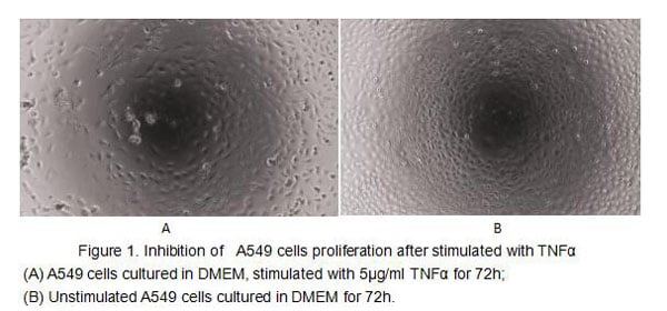
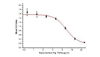
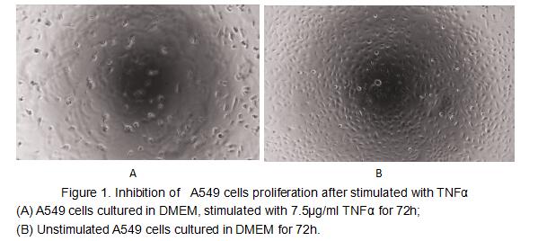
(Figure 2. Inhibition of A549 cells proliferation after stimulated with TNFalpha.)
Bioactivity
(Figure 2. Inhibition of A549 cells proliferation after stimulated with TNFalpha.)
Tumor Necrosis Factor Alpha (TNFa), Active Protein (Cat# AAA152990)
Full Name
Active Tumor Necrosis Factor Alpha (TNFa)
Gene Names
TNF; TNFa
Applications
Activity Assay, Cell Culture
Purity
Greater than 90% by SDS-PAGE
Pricing

 Bioactivity
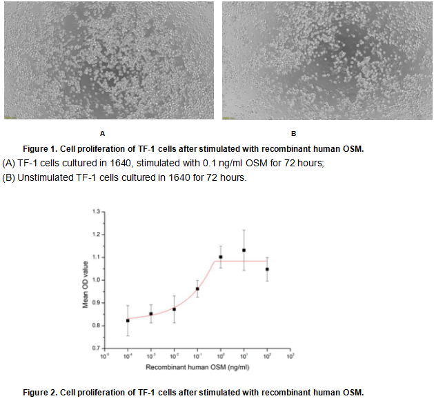
(Oncostatin M (OSM) is an approximately 30 KDa secreted cytokine belonging to the Interleukin-6 family. Like other members of the IL-6 family such as IL-11, CNTF, and Cardiotrophin-1, OSM plays crucial roles in inflammation, neuroprotection, hematopoiesis, metabolism and development. To test the effect of OSM on cell proliferation, TF-1 cells were seeded into triplicate wells of 96-well plates at a density of 20,000 cells/well with various concentrations of recombinant human OSM. After incubated for 72 hours, cells were observed by inverted microscope and cell proliferation was measured by Cell Counting Kit-8(CCK-8). Briefly, 10 ul of CCK-8 solution was added to each well of the plate, then the absorbance at 450 nm was measured using a microplate reader after incubating the plate for 1-4 hours at 37 degree C. Proliferation of TF-1 cells after incubation with OSM for 72 hours observed by inverted microscope was shown in Figure 1. Cell viability was assessed by CCK-8 (Cell Counting Kit-8) assay after incubation with recombinant human OSM for 72 hours. The result was shown in Figure 2. It was obvious that recombinant human OSM significantly increased cell viability of TF-1 cells, the EC50 was 0.08 ng/ml.)
Bioactivity
(Oncostatin M (OSM) is an approximately 30 KDa secreted cytokine belonging to the Interleukin-6 family. Like other members of the IL-6 family such as IL-11, CNTF, and Cardiotrophin-1, OSM plays crucial roles in inflammation, neuroprotection, hematopoiesis, metabolism and development. To test the effect of OSM on cell proliferation, TF-1 cells were seeded into triplicate wells of 96-well plates at a density of 20,000 cells/well with various concentrations of recombinant human OSM. After incubated for 72 hours, cells were observed by inverted microscope and cell proliferation was measured by Cell Counting Kit-8(CCK-8). Briefly, 10 ul of CCK-8 solution was added to each well of the plate, then the absorbance at 450 nm was measured using a microplate reader after incubating the plate for 1-4 hours at 37 degree C. Proliferation of TF-1 cells after incubation with OSM for 72 hours observed by inverted microscope was shown in Figure 1. Cell viability was assessed by CCK-8 (Cell Counting Kit-8) assay after incubation with recombinant human OSM for 72 hours. The result was shown in Figure 2. It was obvious that recombinant human OSM significantly increased cell viability of TF-1 cells, the EC50 was 0.08 ng/ml.)
Oncostatin M (OSM), Active Protein (Cat# AAA161690)
Full Name
Active Oncostatin M (OSM)
Applications
Activity Assay, Cell Culture
Purity
Greater than 95% by SDS-PAGE
Pricing
HBsAg, Native Protein (Cat# AAA14343)
Full Name
HBsAg protein (Subtype ad)
Purity
> 95% by SDS-PAGE
Pricing
 Bioactivity
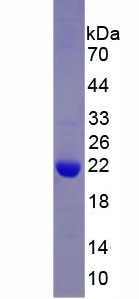
(Tumor necrosis factor (TNF, tumor necrosis factor alpha, TNFalpha, cachexin, or cachectin) is a cell signaling protein (cytokine) involved in systemic inflammation and is one of the cytokines that make up the acute phase reaction. The primary role of TNF)
Bioactivity
(Tumor necrosis factor (TNF, tumor necrosis factor alpha, TNFalpha, cachexin, or cachectin) is a cell signaling protein (cytokine) involved in systemic inflammation and is one of the cytokines that make up the acute phase reaction. The primary role of TNF)
Tumor Necrosis Factor Alpha (TNFa), Active Protein (Cat# AAA152994)
Full Name
Active Tumor Necrosis Factor Alpha (TNFa)
Gene Names
TNF; TNF-ALPHA
Applications
Activity Assay, Cell Culture
Purity
Greater than 90% by SDS-PAGE
Pricing
 Application Data
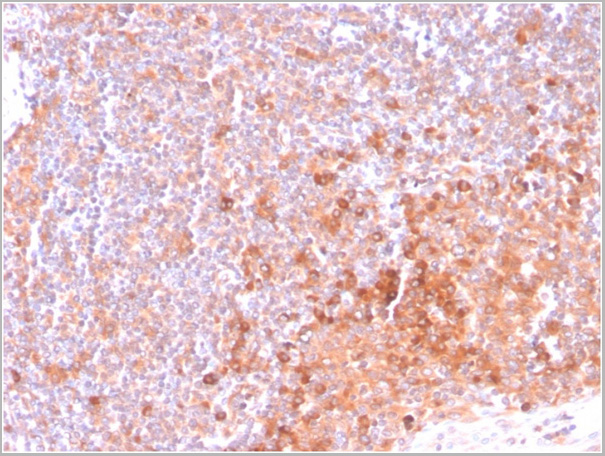
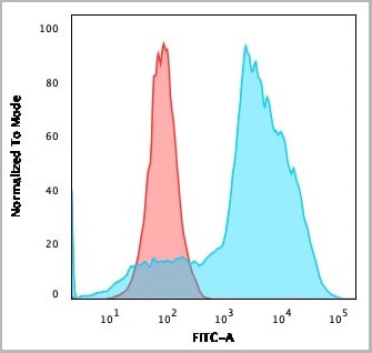
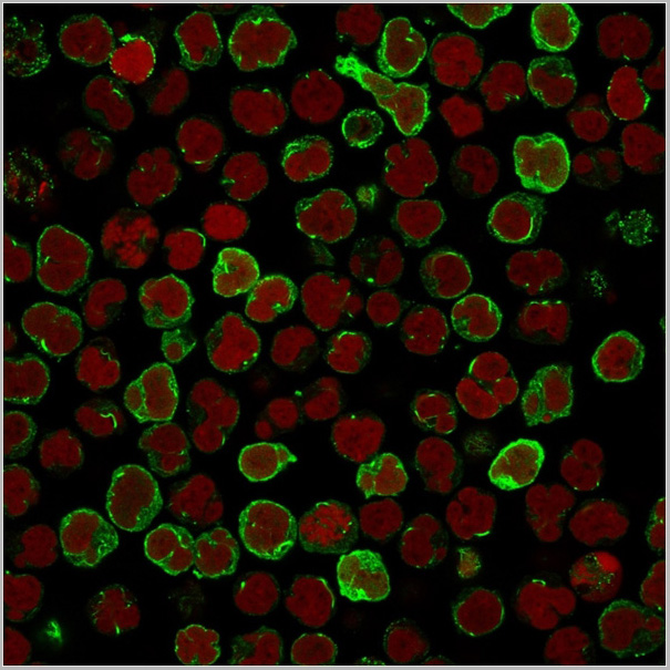
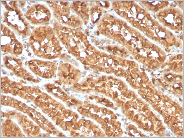
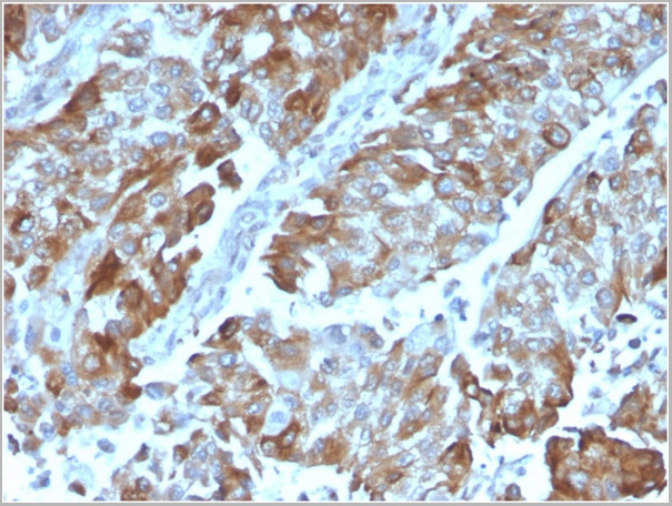
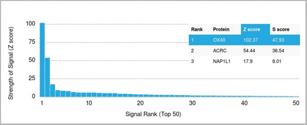
(Analysis of Protein Array containing more than 19,000 full-length human proteins using OX40 Mouse Monoclonal Antibody (OX40/3108). Z- and S- Score: The Z-score represents the strength of a signal that a monoclonal antibody (MAb) (in combination with a fluorescently-tagged anti-IgG secondary antibody) produces when binding to a particular protein on the HuProtTM array. Z-scores are described in units of standard deviations (SD’s) above the mean value of all signals generated on that array. If targets on HuProtTM are arranged in descending order of the Z-score, the S-score is the difference (also in units of SD’s) between the Z-score. S-score therefore represents the relative target specificity of a MAb to its intended target. A MAb is considered to specific to its intended target, if the MAb has an S-score of at least 2.5. For example, if a MAb binds to protein X with a Z-score of 43 and to protein Y with a Z-score of 14, then the S-score for the binding of that MAb to protein X is equal to 29.)
Application Data
(Analysis of Protein Array containing more than 19,000 full-length human proteins using OX40 Mouse Monoclonal Antibody (OX40/3108). Z- and S- Score: The Z-score represents the strength of a signal that a monoclonal antibody (MAb) (in combination with a fluorescently-tagged anti-IgG secondary antibody) produces when binding to a particular protein on the HuProtTM array. Z-scores are described in units of standard deviations (SD’s) above the mean value of all signals generated on that array. If targets on HuProtTM are arranged in descending order of the Z-score, the S-score is the difference (also in units of SD’s) between the Z-score. S-score therefore represents the relative target specificity of a MAb to its intended target. A MAb is considered to specific to its intended target, if the MAb has an S-score of at least 2.5. For example, if a MAb binds to protein X with a Z-score of 43 and to protein Y with a Z-score of 14, then the S-score for the binding of that MAb to protein X is equal to 29.)
OX40/CD134/ TNFRSF4, Monoclonal Antibody (Cat# AAA23936)
Full Name
OX40/CD134/ TNFRSF4 (Immuno-Oncology Target)
Gene Names
TNFRSF4; OX40; ACT35; CD134; IMD16; TXGP1L
Reactivity
Human, Chimpanzee
Applications
Flow Cytometry, Immunofluorescence, Immunohistochemistry
Purity
Purified Ab with BSA and Azide at 200ug/ml OR Purified Ab WITHOUT BSA and Azide at 1.0mg/ml
Pricing
 Bioactivity
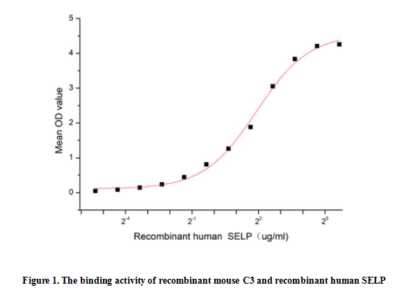
(Complement Component 3 (C3) is a vital component of the complement system that plays a central role in immune response and inflammation. C3 is a b2 globulin synthesized by the liver, whose structure consists of two polypeptide chains, a and b, and is the most abundant complement component in serum. It has reported that P-Selectin (SELP) can interact with C3, thus a functional binding ELISA assay was conducted to detect the interaction of recombinant mouse C3 and recombinant human SELP. Briefly, biotin-linked SELP were diluted serially in PBS, with 0.01% BSA (pH 7.4). Duplicate samples of 100 ul were then transferred to C3-coated microtiter wells and incubated for 1h at 37 degree C. Wells were washed with PBST 3 times and incubation with Streptavidin-HRP for 30min, then wells were aspirated and washed 5 times. With the addition of substrate solution, wells were incubated 15-25 minutes at 37 degree C. Finally, add 50 ul stop solution to the wells and read at 450 nm immediately. When Recombinant mouse C3 is lmmobilized at 2 ug/mL (100 uL/well), the concentration of human SELP that produces 50% optimal binding response is found to be approximately 3.75 ug/mL.)
Bioactivity
(Complement Component 3 (C3) is a vital component of the complement system that plays a central role in immune response and inflammation. C3 is a b2 globulin synthesized by the liver, whose structure consists of two polypeptide chains, a and b, and is the most abundant complement component in serum. It has reported that P-Selectin (SELP) can interact with C3, thus a functional binding ELISA assay was conducted to detect the interaction of recombinant mouse C3 and recombinant human SELP. Briefly, biotin-linked SELP were diluted serially in PBS, with 0.01% BSA (pH 7.4). Duplicate samples of 100 ul were then transferred to C3-coated microtiter wells and incubated for 1h at 37 degree C. Wells were washed with PBST 3 times and incubation with Streptavidin-HRP for 30min, then wells were aspirated and washed 5 times. With the addition of substrate solution, wells were incubated 15-25 minutes at 37 degree C. Finally, add 50 ul stop solution to the wells and read at 450 nm immediately. When Recombinant mouse C3 is lmmobilized at 2 ug/mL (100 uL/well), the concentration of human SELP that produces 50% optimal binding response is found to be approximately 3.75 ug/mL.)
Complement Component 3 (C3), Active Protein (Cat# AAA161794)
Full Name
Active Complement Component 3 (C3)
Gene Names
C3; ASP; Plp; HSE-MSF; AI255234
Applications
Activity Assay, Cell Culture
Purity
Greater than 90% by SDS-PAGE
Pricing

 Bioactivity
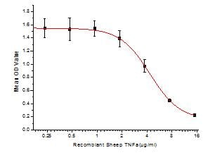
(Figure 2. Inhibition of A549 cells proliferation after stimulated with TNFalpha)
Bioactivity
(Figure 2. Inhibition of A549 cells proliferation after stimulated with TNFalpha)
Tumor Necrosis Factor Alpha (TNFa), Active Protein (Cat# AAA152989)
Full Name
Active Tumor Necrosis Factor Alpha (TNFa)
Gene Names
TNF; TNF-alpha
Applications
Activity Assay, Cell Culture
Purity
Greater than 90% by SDS-PAGE
Pricing
 WB (Western Blot)
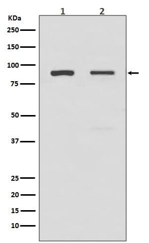
(Western blot analysis of TLR2 expression in (1) A549 cell lysate; (2) HeLa cell lysate (AAA46882).Electrophoresis was performed on a 5-20% SDS-PAGE gel at 70V (Stacking gel) / 90V (Resolving gel) for 2-3 hours. The sample well of each lane was loaded with 50ug of sample under reducing conditions.After Electrophoresis, proteins were transferred to a Nitrocellulose membrane at 150mA for 50-90 minutes. Blocked the membrane with 5% Non-fat Milk/ TBS for 1.5 hour at RT. The membrane was incubated with rabbit anti-TLR2 monoclonal antibody overnight at 4 degree C, then washed with TBS-0.1%Tween 3 times with 5 minutes each and probed with a goat anti-rabbit IgG-HRP secondary antibody at a dilution of 1:10000 for 1.5 hour at RT. The signal is developed using an Enhanced Chemiluminescent detection (ECL) kit with Tanon 5200 system. A specific band was detected for TLR2)
WB (Western Blot)
(Western blot analysis of TLR2 expression in (1) A549 cell lysate; (2) HeLa cell lysate (AAA46882).Electrophoresis was performed on a 5-20% SDS-PAGE gel at 70V (Stacking gel) / 90V (Resolving gel) for 2-3 hours. The sample well of each lane was loaded with 50ug of sample under reducing conditions.After Electrophoresis, proteins were transferred to a Nitrocellulose membrane at 150mA for 50-90 minutes. Blocked the membrane with 5% Non-fat Milk/ TBS for 1.5 hour at RT. The membrane was incubated with rabbit anti-TLR2 monoclonal antibody overnight at 4 degree C, then washed with TBS-0.1%Tween 3 times with 5 minutes each and probed with a goat anti-rabbit IgG-HRP secondary antibody at a dilution of 1:10000 for 1.5 hour at RT. The signal is developed using an Enhanced Chemiluminescent detection (ECL) kit with Tanon 5200 system. A specific band was detected for TLR2)
TLR2, Monoclonal Antibody (Cat# AAA46882)
Full Name
Anti-TLR2 Rabbit Monoclonal Antibody
Gene Names
TLR2; TIL4; CD282
Reactivity
Human, Mouse, Rat
Applications
Western Blot, Immunohistochemistry
Purity
Affinity-chromatography
Pricing
 Bioactivity
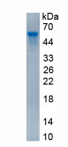
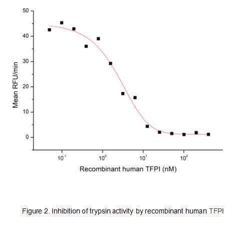
(Human TFPI, also known as lipoprotein-associated coagulation inhibitor (LACI) and extrinsic pathway inhibitor (EPI), is a physiological inhibitor of extrinsic pathway of coagulation and has biological functions of anticoagulation and anti-inflammation. It is a secreted protein with a N-terminal acidic region, three Kunitz (K) domains separated with by two linker regions, and a C-terminal basic region. The activity of recombinant human TFPI was measured by its ability to inhibit trypsin cleavage of a fluorogenic peptide substrate Mca-RPKPVE-Nval-WRK(Dnp)-NH2 in the assay buffer 50 mM Tris, 10 mM CaCl2, 150 mM NaCl, 0.05% (w/v) Brij-35, pH 7.5. Trypsin was diluted to 50 ug/ml in the assay buffer and 20 ul different concentrations of recombinant human TFPI (MW: 61.9 KD) was incubated with 20 ul diluted trypsin at 37 degree C for 15 minutes. Loading 50 uL of the incubated mixtures which were diluted five-fold in assay buffer into empty wells of a plate, and start the reaction by adding 50 uL of 20 uM substrate. Include a substrate blank containing 50 uL of assay buffer and 50 uL of 20 uM substrate. Then read at excitiation and emission wavelengths of 320 nm and 405 nm, respectively, in kinetic mode for 5 minutes. The result was shown in Figure 1 and it was obvious that recombinant human TFPI significantly decreased trypsin activity. The inhibition IC50 was )
Bioactivity
(Human TFPI, also known as lipoprotein-associated coagulation inhibitor (LACI) and extrinsic pathway inhibitor (EPI), is a physiological inhibitor of extrinsic pathway of coagulation and has biological functions of anticoagulation and anti-inflammation. It is a secreted protein with a N-terminal acidic region, three Kunitz (K) domains separated with by two linker regions, and a C-terminal basic region. The activity of recombinant human TFPI was measured by its ability to inhibit trypsin cleavage of a fluorogenic peptide substrate Mca-RPKPVE-Nval-WRK(Dnp)-NH2 in the assay buffer 50 mM Tris, 10 mM CaCl2, 150 mM NaCl, 0.05% (w/v) Brij-35, pH 7.5. Trypsin was diluted to 50 ug/ml in the assay buffer and 20 ul different concentrations of recombinant human TFPI (MW: 61.9 KD) was incubated with 20 ul diluted trypsin at 37 degree C for 15 minutes. Loading 50 uL of the incubated mixtures which were diluted five-fold in assay buffer into empty wells of a plate, and start the reaction by adding 50 uL of 20 uM substrate. Include a substrate blank containing 50 uL of assay buffer and 50 uL of 20 uM substrate. Then read at excitiation and emission wavelengths of 320 nm and 405 nm, respectively, in kinetic mode for 5 minutes. The result was shown in Figure 1 and it was obvious that recombinant human TFPI significantly decreased trypsin activity. The inhibition IC50 was )
Tissue Factor Pathway Inhibitor (TFPI), Active Protein (Cat# AAA161730)
Full Name
Active Tissue Factor Pathway Inhibitor (TFPI)
Gene Names
TFPI; EPI; TFI; LACI; TFPI1
Applications
Activity Assay, Cell Culture
Purity
Greater than 80% by SDS-PAGE
Pricing