

Rabbit PDIA5 Polyclonal Antibody | anti-PDIA5 antibody
Anti-PDIA5 Antibody
IHC-P: 2-5ug/ml|Human, Mouse, Rat|
ICC/IF: 5ug/ml|Human|
FC/FACS/FCM: 1-3ug/1x106 cells|Human|
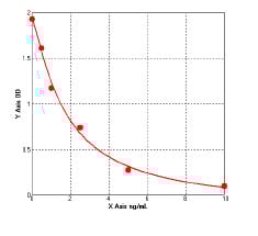
Direct ELISA: 0.1-0.5ug/ml|Human|
FCM (Flow Cytometry)
(Figure 10. Flow Cytometry analysis of U20S cells using anti-PDIA5 antibody (AAA19331).Overlay histogram showing U20S cells stained with AAA19331 (Blue line). The cells were blocked with 10% normal goat serum. And then incubated with rabbit anti-PDIA5 Antibody (AAA19331, 1μg/1x106 cells) for 30 min at 20 degree C. DyLight®488 conjugated goat anti-rabbit IgG (5-10μg/1x106 cells) was used as secondary antibody for 30 minutes at 20 degree C. Isotype control antibody (Green line) was rabbit IgG (1μg/1x106) used under the same conditions. Unlabelled sample (Red line) was also used as a control.)
IF (Immunofluorescence)
(Figure 9. IF analysis of PDIA5 using anti- PDIA5 antibody (AAA19331).PDIA5 was detected in immunocytochemical section of CACO-2 cells. Enzyme antigen retrieval was performed using IHC enzyme antigen retrieval reagent for 15 mins. The cells were blocked with 10% goat serum. And then incubated with 5μg/mL rabbit anti- PDIA5 Antibody (AAA19331) overnight at 4 degree C. DyLight®488 Conjugated Goat Anti-Rabbit IgG was used as secondary antibody at 1:100 dilution and incubated for 30 minutes at 37 degree C. The section was counterstained with DAPI. Visualize using a fluorescence microscope and filter sets appropriate for the label used.)
IHC (Immunohistochemistry)
(Figure 8. IHC analysis of PDIA5 using anti-PDIA5 antibody (AAA19331).PDIA5 was detected in paraffin-embedded section of human adrenocortical adenoma tissue. Heat mediated antigen retrieval was performed in EDTA buffer (pH8. 0, epitope retrieval solution). The tissue section was blocked with 10% goat serum. The tissue section was then incubated with 2μg/ml rabbit anti-PDIA5 Antibody (AAA19331) overnight at 4 degree C. Biotinylated goat anti-rabbit IgG was used as secondary antibody and incubated for 30 minutes at 37 degree C. The tissue section was developed using Strepavidin-Biotin-Complex (SABC) (Catalog # with DAB as the chromogen.)
IHC (Immunohistochemistry)
(Figure 7. IHC analysis of PDIA5 using anti-PDIA5 antibody (AAA19331).PDIA5 was detected in paraffin-embedded section of human ovarian adenocarcinoma tissue. Heat mediated antigen retrieval was performed in EDTA buffer (pH8. 0, epitope retrieval solution). The tissue section was blocked with 10% goat serum. The tissue section was then incubated with 2μg/ml rabbit anti-PDIA5 Antibody (AAA19331) overnight at 4 degree C. Biotinylated goat anti-rabbit IgG was used as secondary antibody and incubated for 30 minutes at 37 degree C. The tissue section was developed using Strepavidin-Biotin-Complex (SABC) (Catalog # with DAB as the chromogen.)
IHC (Immunohistchemistry)
(Figure 6. IHC analysis of PDIA5 using anti-PDIA5 antibody (AAA19331).PDIA5 was detected in paraffin-embedded section of human lung cancer tissue. Heat mediated antigen retrieval was performed in EDTA buffer (pH8. 0, epitope retrieval solution). The tissue section was blocked with 10% goat serum. The tissue section was then incubated with 2μg/ml rabbit anti-PDIA5 Antibody (AAA19331) overnight at 4 degree C. Biotinylated goat anti-rabbit IgG was used as secondary antibody and incubated for 30 minutes at 37 degree C. The tissue section was developed using Strepavidin-Biotin-Complex (SABC) (Catalog # with DAB as the chromogen.)
IHC (Immunohistochemistry)
(Figure 5. IHC analysis of PDIA5 using anti-PDIA5 antibody (AAA19331).PDIA5 was detected in paraffin-embedded section of human rectal cancer tissue. Heat mediated antigen retrieval was performed in EDTA buffer (pH8. 0, epitope retrieval solution). The tissue section was blocked with 10% goat serum. The tissue section was then incubated with 2μg/ml rabbit anti-PDIA5 Antibody (AAA19331) overnight at 4 degree C. Biotinylated goat anti-rabbit IgG was used as secondary antibody and incubated for 30 minutes at 37 degree C. The tissue section was developed using Strepavidin-Biotin-Complex (SABC) (Catalog # with DAB as the chromogen.)
IHC (Immunohistochemistry)
(Figure 4. IHC analysis of PDIA5 using anti-PDIA5 antibody (AAA19331).PDIA5 was detected in paraffin-embedded section of human gallbladder adenocarcinoma tissue. Heat mediated antigen retrieval was performed in EDTA buffer (pH8. 0, epitope retrieval solution). The tissue section was blocked with 10% goat serum. The tissue section was then incubated with 2μg/ml rabbit anti-PDIA5 Antibody (AAA19331) overnight at 4 degree C. Biotinylated goat anti-rabbit IgG was used as secondary antibody and incubated for 30 minutes at 37 degree C. The tissue section was developed using Strepavidin-Biotin-Complex (SABC) (Catalog # with DAB as the chromogen.)
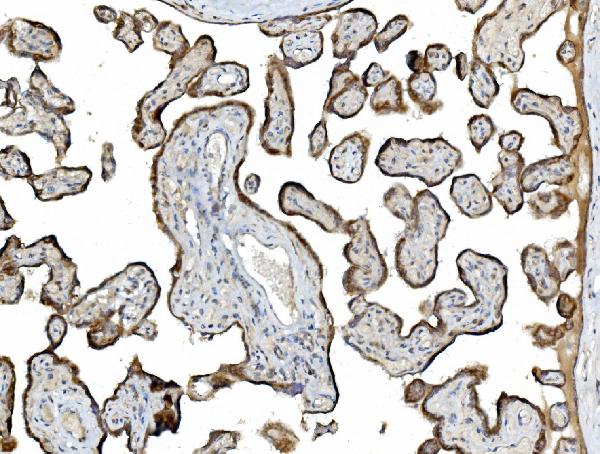
IHC (Immunohistochemistry)
(Figure 3. IHC analysis of PDIA5 using anti-PDIA5 antibody (AAA19331).PDIA5 was detected in paraffin-embedded section of human placenta tissue. Heat mediated antigen retrieval was performed in EDTA buffer (pH8. 0, epitope retrieval solution). The tissue section was blocked with 10% goat serum. The tissue section was then incubated with 2μg/ml rabbit anti-PDIA5 Antibody (AAA19331) overnight at 4 degree C. Biotinylated goat anti-rabbit IgG was used as secondary antibody and incubated for 30 minutes at 37 degree C. The tissue section was developed using Strepavidin-Biotin-Complex (SABC) (Catalog # with DAB as the chromogen.)
IHC (Immunohistochemistry)
(Figure 2. IHC analysis of PDIA5 using anti-PDIA5 antibody (AAA19331).PDIA5 was detected in paraffin-embedded section of human breast cancer tissue. Heat mediated antigen retrieval was performed in EDTA buffer (pH8. 0, epitope retrieval solution). The tissue section was blocked with 10% goat serum. The tissue section was then incubated with 2μg/ml rabbit anti-PDIA5 Antibody (AAA19331) overnight at 4 degree C. Biotinylated goat anti-rabbit IgG was used as secondary antibody and incubated for 30 minutes at 37 degree C. The tissue section was developed using Strepavidin-Biotin-Complex (SABC) (Catalog # with DAB as the chromogen.)
WB (Western Blot)
(Figure 1. Western blot analysis of PDIA5 using anti-PDIA5 antibody (AAA19331).Electrophoresis was performed on a 5-20% SDS-PAGE gel at 70V (Stacking gel) / 90V (Resolving gel) for 2-3 hours. The sample well of each lane was loaded with 30ug of sample under reducing conditions.Lane 1: human Hela whole cell lysatesLane 2: human HepG2 whole cell lysatesLane 3: human Caco-2 whole cell lysatesLane 4: human THP-1 whole cell lysatesLane 5: human MCF-7 whole cell lysatesLane 6: human HL-60 whole cell lysatesLane 7: human A431 whole cell lysatesLane 8: human T47D whole cell lysatesLane 9: rat liver tissue lysatesLane 10: rat lung tissue lysatesLane 11: rat stomach tissue lysatesLane 12: rat pancreas tissue lysatesLane 13: mouse liver tissue lysatesLane 14: mouse lung tissue lysatesLane 15: mouse stomach tissue lysatesLane 16: mouse pancreas tissue lysates.After Electrophoresis, proteins were transferred to a Nitrocellulose membrane at 150mA for 50-90 minutes. Blocked the membrane with 5% Non-fat Milk/ TBS for 1. 5 hour at RT. The membrane was incubated with rabbit anti-PDIA5 antigen affinity purified polyclonal antibody (Catalog # AAA19331) at 0. 25 μg/mL overnight at 4 degree C, then washed with TBS-0. 1%Tween 3 times with 5 minutes each and probed with a goat anti-rabbit IgG-HRP secondary antibody at a dilution of 1:5000 for 1. 5 hour at RT. The signal is developed using an Enhanced Chemiluminescent detection (ECL) kit (Catalog # with Tanon 5200 system. A specific band was detected for PDIA5 at approximately 60KD. The expected band size for PDIA5 is at 60KD.)
2. Hayano, T., Kikuchi, M. Molecular cloning of the cDNA encoding a novel protein disulfide isomerase-related protein (PDIR). FEBS Lett. 372: 210-214, 1995.
3. Higa, A., Taouji, S., Lhomond, S., Jensen, D., Fernandez-Zapico, M. E., Simpson, J. C., Pasquet, J. -M., Schekman, R., Chevet, E. Endoplasmic reticulum stress-activated transcription factor ATF6-alpha requires the disulfide isomerase PDIA5 to modulate chemoresistance. Molec. Cell. Biol. 34: 1839-1849, 2014.