 Application Data
Radius 24-Well Cell Migration Assay Assay Kit
Radius 24-Well Cell Migration Assay
Synonyms
Radius 24-Well Cell Migration Assay; N/A; Radius 24-Well Cell Migration Assay assay kit
Preparation and Storage
Upon receipt, aliquot and store the Radius Gel Removal Solution and DAPI Fluorescence Stain at -20 degree C (avoid multiple freeze/thaw cycles), and transfer the Fixation Solution to 4 degree C. All other kit components should be stored at room temperature until the kit's expiration date.
Application Data
Application Data
Application Data
Application Data
Application Data
Application Data
Related Product Information for Radius 24-Well Cell Migration Assay assay kit
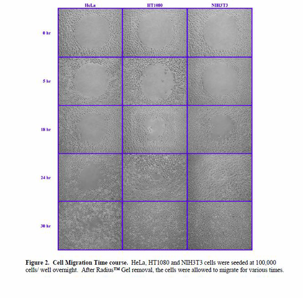
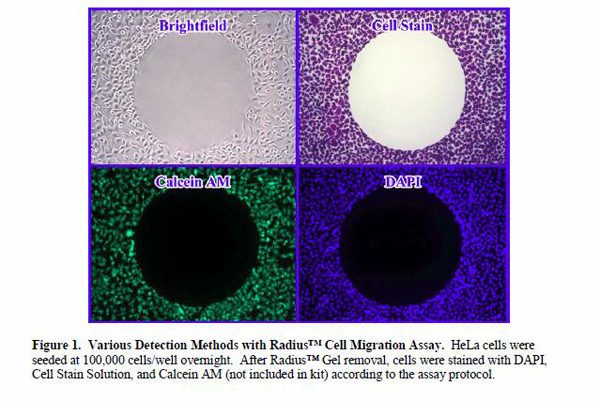
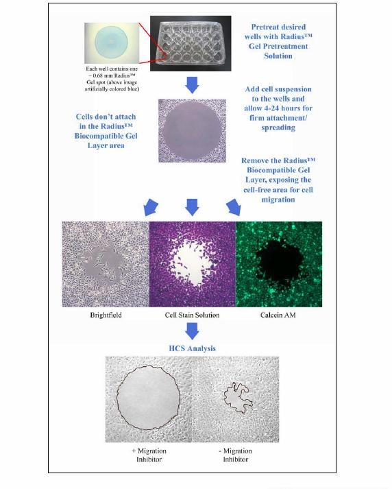
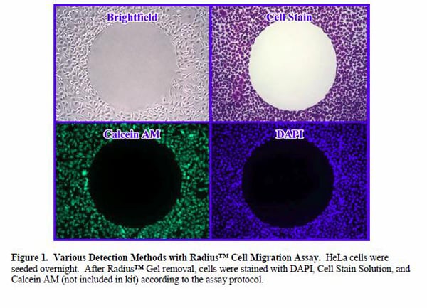
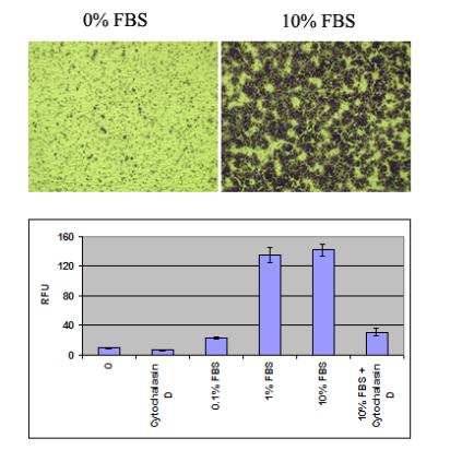
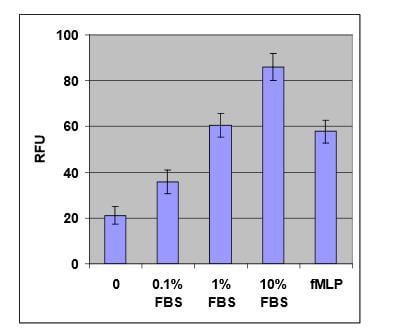
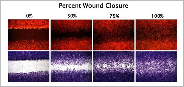
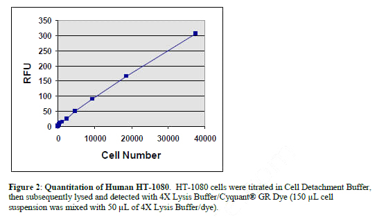
Background: Cell migration is a highly integrated, multistep process that orchestrates embryonic morphogenesis, tissue repair and regeneration. It plays a pivotal role in the disease progression of cancer, mental retardation, atherosclerosis, and arthritis. The initial response of a cell to a migration-promoting agent is to polarize and extend protrusions in the direction of the attractant; these protrusions can consist of large, broad lamellipodia or spike-like filopodia. In either case, these protrusions are driven by actin polymerization and can be stabilized by extracellular matrix (ECM) adhesion or cell-cell interactions (via transmembrane receptors). The Radius™ Cell Migration Assay Kit utilizes a proprietary 24-well plate to monitor the migratory properties of cells. Each plate well contains a 0.68 mm non-toxic, biocompatible hydrogel spot (Radius™ Gel) where cells cannot attach. When adherent cells are seeded in the Radius™ Cell Migration well, they attach outside of the Radius™ Gel coated area. Once firm cell attachment is achieved, the hydrogel is quickly removed to expose a cell-free region to study cell migration/closure. This format provides a robust in vitro system to measure 2-D cell migration, screen potential inhibitors and study cytoskeleton reorganization events.
Product Categories/Family for Radius 24-Well Cell Migration Assay assay kit
References
1. Ridley AJ, Schwartz MA, Burridge K, Firtel RA, Ginsberg MH, Borisy G, Parsons JT, Horwitz AR. (2003) Science 302, 1704-9.
2. Horwitz R, Webb D. (2003) Curr Biol. 13, R756-9.
3. Lauffenburger DA, Horwitz AF. (1996) Cell 84, 359-369.
1. Langfelder, A. et al. (2015). Extracellular acidosis impairs P2Y receptor-mediated Ca 2+ signalling and migration of microglia. Cell Calcium. doi: 10.1016/j.ceca.2015.01.004.
2. Cersosimo, E. et al. (2014). Acute insulin resistance stimulates and insulin sensitization attenuates vascular smooth muscle cell migration and proliferation. Physiol Rep. 2:e12123.
3. Qiang, L. et al. (2014). Regulation of cell proliferation and migration by p62 through stabilization of Twist1. Proc Natl Acad Sci U S A. 111:9241-9246.
4. Sriram, S. et al.(2013). Triple combination of siRNAs targeting TGFbeta1, TGFbetaR2, and CTGF enhances reduction of collagen I and smooth muscle actin in corneal fibroblasts. Invest. Opthalmol. Vis. Sci. 54:8214-8223
5. Robinson, P. et al. (2013). MicroRNA signature in wound healing following excimer laser ablation: role of miR-133b on TGFbeta1, CTGF, SMA, and COL1A1 expression levels in rabbit corneal fibroblasts. J. Cell Sci. 126:4769-4781.
6. Sun, J. et al. (2013). Targeting the metastasis suppressor, NDRG1, using novel iron chelators: regulation of stress fiber-mediated tumor cell migration via modulation of the ROCK1/pMLC2 signaling pathway. Mol. Pharmacol. 83:454-469.
7. Apostolos, K. et al. (2013). Increased susceptibility of melanin-concentrating hormone-deficient mice to infection with Salmonella enterica Serovar typhimurium. Infect. Immun. 81: 166-172.
8. Smith, K. et al. (2012). Human Family with Sequence Similarity 60 Member A (FAM60A) protein: a new subunit of the Sin3 deacetylase complex. Mol. Cell. Proteomics. 11:1815-1828.
9. Chuang, T.D. et al. (2012). miR-93/106b and their host gene, MCM7, are differentially expressed in leiomyomas and functionally target F3 and IL-8. Mol. Endocrin. 26: 1028-1042.
10. Young, S. et al. (2012). Rapid Protein Kinase D1 signaling promotes migration of intestinal epithelial cells. Am J Physiol Gastrointest Liver Physiol. 303: G356-G366.
11. Larive, R.M. et al. (2012). The Ras-like protein R-Ras2/TC21 is important for proper mammary gland development. Mol. Biol. Cell. 23:2373-2387
Customer Reviews
Loading reviews...
Share Your Experience
Similar Products


Product Notes
The Radius 24-Well Cell Migration Assay (Catalog #AAA44581) is an Assay Kit and is intended for research purposes only. The product is available for immediate purchase. It is sometimes possible for the material contained within the vial of "Radius 24-Well Cell Migration Assay, Assay Kit" to become dispersed throughout the inside of the vial, particularly around the seal of said vial, during shipment and storage. We always suggest centrifuging these vials to consolidate all of the liquid away from the lid and to the bottom of the vial prior to opening. Please be advised that certain products may require dry ice for shipping and that, if this is the case, an additional dry ice fee may also be required.Precautions
All products in the AAA Biotech catalog are strictly for research-use only, and are absolutely not suitable for use in any sort of medical, therapeutic, prophylactic, in-vivo, or diagnostic capacity. By purchasing a product from AAA Biotech, you are explicitly certifying that said products will be properly tested and used in line with industry standard. AAA Biotech and its authorized distribution partners reserve the right to refuse to fulfill any order if we have any indication that a purchaser may be intending to use a product outside of our accepted criteria.Disclaimer
Though we do strive to guarantee the information represented in this datasheet, AAA Biotech cannot be held responsible for any oversights or imprecisions. AAA Biotech reserves the right to adjust any aspect of this datasheet at any time and without notice. It is the responsibility of the customer to inform AAA Biotech of any product performance issues observed or experienced within 30 days of receipt of said product. To see additional details on this or any of our other policies, please see our Terms & Conditions page.Item has been added to Shopping Cart
If you are ready to order, navigate to Shopping Cart and get ready to checkout.