Western Blot Antibodies: Selection, Dilution & Troubleshooting
 Cynthia
9 min read
Cynthia
9 min read
In this Article
- Why Is Antibody Selection So Important In Western Blotting?
- How Do You Choose The Right Antibody Dilution?
- How Does Antibody Concentration Affect Western Blot Results?
- What Is The Best Workflow For Western Blot Antibody Optimization?
- Common Western Blot Problems And Their Solutions
- What Controls Should Every Western Blot Include?


All of the products listed in AAA Biotech’s catalog are strictly for research-use only (RUO).

Selecting and diluting the right Western blot antibody is one of the biggest factors in getting accurate protein detection. The right antibody gives you clear, reproducible bands, while the wrong one can leave you with weak signals, high background, or no result at all. That's more common than many researchers realize.
Independent studies have found that more than 50% of commercial antibodies fail basic characterization and specificity tests, contributing to an estimated $1 billion in wasted research funding every year. No one wants to spend days repeating an experiment because of an unreliable antibody.
That's why antibody selection matters from the very beginning. It not only improves your experimental results but also helps you meet the growing expectations for antibody validation from journals, reviewers, and funding agencies.
In this Western blot guide, you'll learn how to choose the right Western blot antibody, determine the best dilution, troubleshoot common problems, and follow best practices for consistent, reliable results.
Key Takeaways
- Learn why selecting a validated Western blot antibody is essential for accurate and reproducible protein detection.
- Understand how specificity, affinity, host species, and antibody type influence experimental success.
- Follow a step-by-step process to determine the optimal antibody dilution using serial titration.
- Discover how antibody concentration affects signal strength, background noise, and overall blot quality.
- Apply an effective workflow for antibody optimization, from sample preparation to protein detection.
- Troubleshoot common Western blot issues, including weak bands, high background, and uneven transfer.
- Use appropriate controls to validate results and improve the reliability and reproducibility of your experiments.
Why Is Antibody Selection So Important In Western Blotting?
The success of a Western blot often comes down to one decision: “choosing the right antibody”. A well-validated antibody produces clear, accurate bands, while the wrong one can lead to weak signals, unexpected bands, or results you can't trust. In many cases, a poor antibody choice means repeating the entire experiment. Understanding the western blotting purpose behind each selection criterion below makes it easier to avoid that outcome.
What Should You Look For In A Western Blot Antibody?
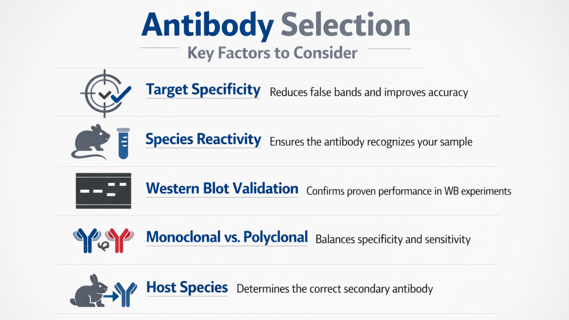
- Specificity: Choose an antibody that binds only to your target protein. High specificity reduces false-positive bands and improves confidence in your results.
- Affinity: High-affinity antibodies bind more strongly to the target protein, making it easier to detect proteins present at low concentrations.
- Western Blot Validation: Always check that the antibody has been validated for Western blotting. An antibody that works well in ELISA or immunohistochemistry may not perform reliably in a Western blot.
- Host Species: The host species of your primary antibody determines which secondary antibody you need. For example, a rabbit primary antibody requires an anti-rabbit secondary antibody.
- Antibody Type: Select the antibody format that best fits your experiment. Each is a distinct type of Western blotting antibody, suited to different targets.
- Monoclonal antibodies: Bind a single epitope, providing high specificity and consistent results.
- Polyclonal antibodies: Recognize multiple epitopes, offering higher sensitivity for detecting low-abundance proteins.
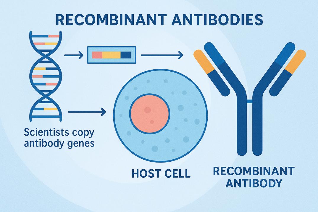
- Recombinant antibodies: Produced from defined genetic sequences, ensuring excellent batch-to-batch consistency and reproducibility.
- Application Fit: There isn't a single "best" antibody for every experiment. Your choice should match your target protein, sample type, and research objectives.
- Validation and Reproducibility: Many journals, funding agencies, and organizations such as the National Library of Medicine (NIH) now expect researchers to use well-characterized, validated antibodies. Using validated reagents strengthens the reliability and reproducibility of your data.
Note: At AAA Biotech, our validated monoclonal and purified polyclonal antibodies are tested for Western blot applications. Designed for reliable performance, they deliver consistent, reproducible results and support a wide range of immunoassays for diverse research needs.
How Do You Choose The Right Antibody Dilution?
Choosing the right antibody dilution is all about finding the balance between a strong target signal and low background noise. Follow these steps to identify the optimal dilution for your experiment.
Step 1: Start With the Manufacturer's Recommendation
Use the recommended dilution provided in the antibody datasheet as your starting point. This gives you a reliable baseline before making any adjustments.
Step 2: Run a Dilution Series
Prepare a series of antibody dilutions (serial titration) instead of testing only one concentration. This helps you identify the dilution that produces the strongest specific signal with the cleanest background.
Step 3: Evaluate Your Results
Review the blot carefully.
- Too concentrated? You'll often see high background, non-specific bands, or smearing.
- Too dilute? Your target band may become faint or disappear completely.
The best dilution is the one that provides a clear, specific band with minimal background.
Step 4: Consider Your Experimental Conditions
The ideal dilution varies from one experiment to another. Factors that influence antibody concentration include:
- Target protein abundance
- Antibody affinity
- Blocking buffer
- Incubation time
- Membrane type (PVDF or nitrocellulose)
Never assume the same dilution will work for every sample.
Typical Starting Dilutions
| Antibody Type | Common Starting Dilution |
|---|---|
| Primary antibody | 1:500–1:2,000 |
| Secondary antibody | 1:2,000–1:10,000 |
Step 5: Re-optimize When Using a New Antibody Lot
Even antibodies from the same supplier can show slight lot-to-lot variation. Running a quick dilution test with each new lot helps maintain consistent and reproducible results.
Note: If you're using unpurified antibodies, such as serum or tissue culture supernatant, you'll typically need lower starting dilutions (around 1:100–1:1,000). Ascites fluid is often used at much higher dilutions. Regardless of the source, always confirm the optimal dilution with a titration experiment rather than relying on general guidelines.
How Does Antibody Concentration Affect Western Blot Results?
Antibody concentration directly shapes signal strength and background noise. Too much antibody floods the membrane with excess signal. This produces high background and non-specific binding patterns. Too little antibody produces faint, hard-to-read bands. Getting concentration right is often the biggest lever for blot quality.
Use this decision tree when your blot doesn't look right:
- Signal too strong or high background: dilute the antibody further
- Signal too weak or bands are faint: increase concentration, extend incubation
- No signal at all: check antibody validation, confirm positive control
This flow works for both primary and secondary antibodies. Walking through it systematically beats guessing at random fixes.
What Is The Best Workflow For Western Blot Antibody Optimization?
A structured workflow helps reduce experimental errors and improves the chances of obtaining clear, reproducible Western blot results. This overall Western blotting technique relies on following these eight steps for effective antibody optimization.
Step1: Sample Preparation
Begin by lysing your cells or tissues to extract proteins. Measure the protein concentration accurately so each lane receives an equal amount, ensuring consistent and reliable comparisons across all samples.
Step2: Protein Transfer
After electrophoresis, transfer the separated proteins from the gel onto a PVDF or nitrocellulose membrane. An efficient transfer preserves protein integrity and ensures your target protein is available for antibody binding.
Step3: Blocking
Incubate the membrane with a blocking solution, such as BSA or non-fat milk, to cover unoccupied binding sites. This minimizes non-specific antibody binding and reduces unwanted background signals.
Step4: Primary Antibody Incubation
Incubate the membrane with your primary antibody at the optimized dilution. Proper concentration and incubation time allow the antibody to bind specifically to the target protein without increasing background.
Step5: Washing
Wash the membrane thoroughly between incubation steps using an appropriate wash buffer. Effective washing removes unbound antibodies while preserving specific interactions, resulting in cleaner blots and more accurate detection.
Step6: Secondary Antibody Incubation
Apply a secondary antibody that matches the host species of your primary antibody. The secondary antibody carries the detection label needed to visualize the target protein during the final detection step.
Step7: Detection
Visualize the protein bands using a compatible detection method, such as chemiluminescence or fluorescence. Choose the detection system that best supports clear western blot visualization and matches your experiment's sensitivity and imaging requirements.
Step8: Optimization
Review your results and refine the protocol if needed. Adjust antibody dilution, incubation time, blocking conditions, or wash steps until you achieve strong target signals with minimal background noise.
Quick Troubleshooting Guide for Western Blot Optimization
☐ Protein concentration measured accurately
☐ Protein transfer confirmed
☐ Appropriate blocking buffer selected
☐ Primary antibody dilution optimized
☐ Wash steps completed thoroughly
☐ Correct secondary antibody used
☐ Target bands clearly detected
☐ Background minimized before analysis
Common Western Blot Problems And Their Solutions
Even with a well-planned protocol, Western blot experiments don't always go as expected. Most issues can be traced back to antibody selection, dilution, blocking, or protein transfer. Instead of repeating the entire experiment, identify the root cause first and make targeted adjustments.
Troubleshooting Common Western Blot Problems
| Problem | Possible Cause | Recommended Solution |
|---|---|---|
| Weak or faint bands | Antibody concentration is too low | Optimize the antibody dilution using a serial titration. |
| High background | Antibody concentration is too high, or blocking is inadequate | Increase the dilution ratio and review your blocking conditions. |
| No bands detected | Incorrect or unvalidated antibody, or poor protein transfer | Verify antibody specificity, confirm Western blot validation, and check transfer efficiency. |
| Multiple or unexpected bands | Non-specific antibody binding | Improve blocking conditions and use a more specific, validated antibody if needed. |
| Uneven or patchy bands | Incomplete protein transfer or uneven sample loading | Check transfer conditions, loading consistency, and membrane handling. |
Not every failed Western blot is caused by the antibody. Before replacing it:
- Check protein loading
- Transfer efficiency
- Blocking antibody dilution
- Incubation conditions
- Detection settings
A systematic review often identifies the real issue. Using well-validated antibodies also improves reproducibility.
What Controls Should Every Western Blot Include?
Controls help confirm that your antibody is detecting the correct protein and that your experiment worked as expected.
| Control | Why It's Important |
|---|---|
| Positive control | Confirms the antibody can detect the target protein. |
| Negative control | Identifies non-specific antibody binding. |
| Loading control (β-actin, GAPDH, Tubulin) | Verifies equal protein loading and transfer between lanes. |
| Secondary-only control | Checks for non-specific binding from the secondary antibody. |
Note: Always compare the detected band with the expected molecular weight of your target protein. Running biological and technical replicates further strengthens the reliability and reproducibility of your results.
Final Thought
Reliable Western blot results come down to three things. These are validated antibodies, optimized dilution, and systematic troubleshooting. Skipping any one makes the other two less effective. Select a well-characterized antibody, titrate carefully, and follow a consistent workflow.
AAA Biotech offers validated monoclonal, polyclonal, and recombinant antibodies. Each is built for dependable Western blot performance. Reach out to our team for antibody selection guidance.
Faq's
What antibody dilution should I use for Western blot?
Start with the manufacturer's recommended range, typically 1:500–1:2000. Always confirm the optimal dilution through titration for your sample.
Can antibody concentration affect background?
Yes, excess antibody is a common cause of high background. Diluting further usually resolves this without losing signal strength.
Should I use monoclonal or polyclonal antibodies?
Monoclonal antibodies offer high specificity for a single epitope. Polyclonal antibodies offer broader sensitivity across multiple target epitopes.
Why is my Western blot showing multiple bands?
Multiple bands often indicate non-specific binding or an unvalidated antibody. Improving your blocking buffer usually resolves this issue.
How should antibodies be stored?
Store antibodies at the temperature listed on the datasheet. This is usually 4°C or -20°C, avoiding repeated freeze-thaw cycles.
What antibody dilution should I use for Western blot?
Start with the manufacturer's recommended range, typically 1:500–1:2000. Always confirm the optimal dilution through titration for your sample.
Can antibody concentration affect background?
Yes, excess antibody is a common cause of high background. Diluting further usually resolves this without losing signal strength.
Should I use monoclonal or polyclonal antibodies?
Monoclonal antibodies offer high specificity for a single epitope. Polyclonal antibodies offer broader sensitivity across multiple target epitopes.
Why is my Western blot showing multiple bands?
Multiple bands often indicate non-specific binding or an unvalidated antibody. Improving your blocking buffer usually resolves this issue.
How should antibodies be stored?
Store antibodies at the temperature listed on the datasheet. This is usually 4°C or -20°C, avoiding repeated freeze-thaw cycles.
What causes faint Western blot bands?
Faint bands usually result from low antibody concentration or poor transfer. Increasing concentration or verifying transfer efficiency typically fixes this.
Lead Clinical Research Coordinator (LCRC)
Cynthia Lee is the President of AAA Biotech and specializes in understanding highly validated and characterized monoclonal/polyclonal antibodies, recombinant proteins, and ELISA kits.
Related Posts