What Are Phospho Antibodies?
 Cynthia
9 min read
Cynthia
9 min read
In this Article
All of the products listed in AAA Biotech’s catalog are strictly for research-use only (RUO).
Phosphorylation is when a phosphate group is added to a protein. It usually happens on specific amino acids comprising the protein – specifically, serine, threonine, or tyrosine – and is performed by specialized enzymes called kinases.
Phosphorylation helps to “turn on” or control important processes in cells.
Phospho-specific antibodies are special tools, primarily made using rabbits, that can recognize and bind excousively to proteins that have been phosphorylated. These antibodies are very useful for studying how cells send and receive signals.
The History and Importance of Phospho Antibodies

The discovery of phospho-specific antibodies completely changed how scientists would go about studying what different proteins actually do, and each of their purposes, in the body.
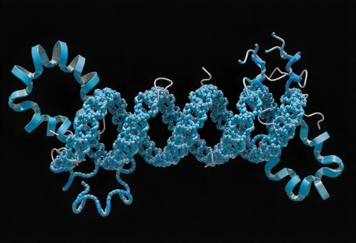
Why Shape Matters in Proteins?
Most proteins’ functions are entirely dependent on their shape. Some proteins give structure to cells, while others control important activities like cell division. These kinds of "active" proteins need to be turned “on” or “off” depending on the cell's needs.
Cells control protein activity through a process called phosphorylation. This process occurs when a special enzyme, known as a kinase, attaches a small chemical group called a phosphate (derived from an ATP molecule) to specific amino acid residues, typically serine, threonine, or tyrosine, within the protein. Adding this phosphate causes a change in the protein’s shape, which then changes how it works.
Why Phosphoproteins Matter?
Proteins that have been changed by phosphorylation are called phosphoproteins. These proteins play a role in almost every important process in the body—from how cancer cells grow to how brain cells communicate during learning and memory.
Because they’re involved in so many actions, scientists often say, “Phosphoproteins are the verbs of the protein world.”
By checking whether a protein is phosphorylated (turned on or off), researchers can better understand what that specific protein does and how it's affected by signals inside the cell. This also helps scientists develop new drugs that can control how certain proteins behave.
Why Just Detecting a Protein Isn’t Enough
Simply knowing a protein is present in the cell doesn’t tell you if it’s actually currently doing anything. Imagine an FBI agent watching a house from the outside—they know their suspect is inside, but not what they’re doing.
Now, imagine that the agent has a secret camera inside the house—they can actually see the suspect in action.
In the same way, regular antibodies only show that a protein is present. But phospho-specific antibodies go further—they show whether the protein is actually active and currently “working”. This gives scientists much more useful information about what's happening inside the cell.
It All Started with Radioactive Labels
In the early days, scientists used radioactive phosphorus (called ³²P) to detect phosphorylated proteins. This method was very time-consuming and difficult because:
- Thousands of proteins take up the radioactive phosphate, not just the one you're trying to study.
- To find the protein of interest, scientists had to use a method called immunoprecipitation to separate it from all the others.
- Even then, they couldn't tell which exact spot on the protein was phosphorylated—which matters because one site might turn the protein on, while another site on the same protein might turn it off.
- Measuring exactly how much phosphorylation happened was difficult, since cell activity could directly affect the ³²P signal.
- Most importantly, this technique couldn’t really be used in living
organisms, and was only possible in strictly-controlled lab environments.
The First Phospho-Specific Antibodies: A Step Forward
In 1981, scientists developed the first antibodies that could recognize phosphotyrosine (a phosphorylated form of the amino acid tyrosine). These were made by injecting rabbits with a special chemical (benzyl phosphonate). These antibodies were helpful, particularly in cancer research, but they had some downsides:
- They could detect phosphorylation on many different proteins, not just the one scientists were interested in.
- They couldn’t tell which specific site on a protein was phosphorylated, just that phosphorylation had happened.
Later attempts to make antibodies for phosphoserine or phosphothreonine (other
types of phosphorylated amino acids) didn’t work very well either.
Trying Whole Proteins Didn’t Solve the Problem
Another approach was to inject animals with entire phosphorylated proteins to make antibodies. Though, one success story involved a protein known as G-substrate, found in the brain.
But this method had its own problems too:
The proteins would often lose their phosphate group (dephosphorylate) during the process, so the immune system didn’t recognize the phosphorylated version.
Whole proteins have many different parts that the immune system can respond to, so the antibodies might miss the specific phosphorylated site that scientists are primarily concerned about.
How Phospho-Specific Antibodies Are Made?
Making phospho-specific antibodies is more complex than making regular
antibodies.
These antibodies are specially designed to recognize proteins only when a phosphate group is attached to certain amino acids—specifically serine (S), threonine (T), or tyrosine (Y).
Making Phospho Polyclonal Antibodies (from Rabbits)
To make polyclonal phospho-antibodies, scientists follow these steps:
- Create the phospho-peptides: These are short pieces of proteins with a phosphate group attached at the right spot. (“Peptide” refer to short chains of linked amino acids, whereas “protein” refers to amino acid chains of significant length.)
- Inject them into rabbits to trigger an immune response.
- Collect the antibodies from the rabbit’s blood (antiserum).
- Use a special process known as “affinity purification” to isolate only the antibodies that bind to the phospho-peptide.
- Occasionally, antibodies might also bind to the non-phosphorylated version of the protein. To remove these unwanted antibodies, a second step called cross-absorption is used. This requires making a version of the same peptide without the phosphate, so it can be used to help act like a filter.
- After this process, you end up with highly specific polyclonal antibodiesthat only recognize the phosphorylated site—perfect for research.
Making Phospho Monoclonal Antibodies (from Mice)
The process for making monoclonal phospho-antibodies is a bit simpler:
- Scientists inject phospho-peptides into mice to force their immune systems respond.
- They will then look for antibody-producing cells (which will later be converted into hybridomas) that create antibodies that bind to the phosphorylated peptide.
- To make sure the antibodies don’t bind to the non-phosphorylated version, a step called negative selection is done using the plain peptide.
This negative selection step is very important, even though it’s sometimes skipped.
That’s because in real samples, not all proteins are fully phosphorylated, so it’s critical to make sure the antibody only detects the phosphorylated form.
Tips for Using Phosphoprotein Antibodies in Western Blotting
(WB)
When working with phosphoproteins in a Western blot, it’s important to protect them from damage or changes that could affect your results. Here’s how to get the best outcome:
1. Use Inhibitors to Protect Your Proteins
Add protease and phosphatase inhibitors to your lysis buffer. These protect your proteins from being broken down or having their phosphate groups removed.
You might also want to add kinase inhibitors to stop any extra phosphorylation from happening during sample prep.
2. Work Quickly and Keep Everything Cold
- Process your samples as quickly as possible.
- Keep everything cold to slow down any unwanted enzyme activity.
- Sonicate (use sound waves to break up cells) to make sure you get total lysis—meaning all cells are fully broken open.
- The faster and cooler you work, the more likely it is that the
phosphorylation state of the protein stays the same.
3. Monitor the pH
Some phosphorylation sites are stable even in unusual pH, but changes in pH can still affect how well an antibody binds to its target.
Also, if the pH is wrong in your gel or transfer buffer, your proteins might not separate or transfer correctly during the blot.
4. Blocking and Antibody Incubation
Before adding your antibodies, block the membrane with 5% nonfat dry milk in TBS with 0.1% Tween 20 (TBS-T).
This helps prevent background noise and works well—even with phospho-specific antibodies, because milk doesn’t contain active phosphatase enzymes.
However, when using the phospho-specific primary antibody, it's often best to dilute it in 5% BSA instead of milk.
5. Choose TBS-T, Not PBS-T
Use TBS-T for washing and incubating, not PBS-T, because phosphate in PBS can interfere with phospho-antibody binding.
6. Probe for Phosphoprotein First
If you're planning to strip and re-probe your blot, start with the phosphoprotein antibody. Stripping can damage or remove phosphoprotein signals.
Phosphoproteins are usually present in relatively small quantities, so we recommend incubating the membrane with the primary antibody overnight at 4°C.
After that, you can probe for the total protein to check loading and transfer.
7. Use Fluorescent Multiplexing (If You Can)
If you have the equipment, you can use fluorescent detection to look at both the total protein and phosphorylated protein at the same time.
8. Always Include Controls
- Use a positive control to make sure you’re detecting actual phosphorylation.
- You can also treat your sample with a phosphatase enzyme as a negative control—if the signal disappears, that shows it was due to phosphorylation.
- Specific activators or inhibitors of protein signaling can also help verify
your results.
9. What If You See Nothing?
Phosphoproteins are often found in very low amounts, so if you get no signal, try using immunoprecipitation to concentrate the protein before running the blot.
To Wrap Up
Once you’ve carefully followed all the steps and optimized your experiment, you should be able to detect your phosphoprotein, even if it's hard to locate/isolate. Most of the time, you’ll be looking at only a single phosphorylation site at a time.
But keep in mind—many proteins have more than one phosphorylation site, and each site can affect the protein differently.
It can make things much more complicated, but it also gives you more detailed information about how the protein works.
Lead Clinical Research Coordinator (LCRC)
Cynthia Lee is the President of AAA Biotech and specializes in understanding highly validated and characterized monoclonal/polyclonal antibodies, recombinant proteins, and ELISA kits.