Filters
▼Clonality
▼Type
▼Reactivity
▼Gene Name
▼Isotype
▼Host
▼Application
▼Clone
▼Monoclonal Antibodies
Get accurate results in your research with our Monoclonal Antibodies, which are specially made to target exactly what you require for your research, and will produce consistent, reliable performance in lab tests.
Viewing 5000-5050 of 27645 product results


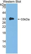
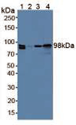
 WB (Western Blot)
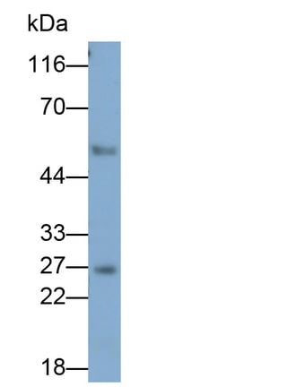
(Western Blot; Sample: Porcine Serum Primary Ab: 2ug/ml Mouse AntiPorcine CLU Antibody Second Ab: 0.2ug/mL HRPLinked Caprine AntiMouse IgG Polyclonal Antibody (Catalog: SAA544Mu19))
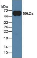
WB (Western Blot)
(Western Blot; Sample: Porcine Serum Primary Ab: 2ug/ml Mouse AntiPorcine CLU Antibody Second Ab: 0.2ug/mL HRPLinked Caprine AntiMouse IgG Polyclonal Antibody (Catalog: SAA544Mu19))
Clusterin (CLU), Monoclonal Antibody (Cat# AAA151689)
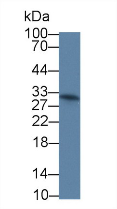
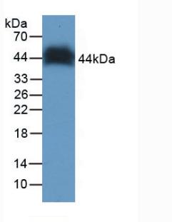
 WB (Western Blot)
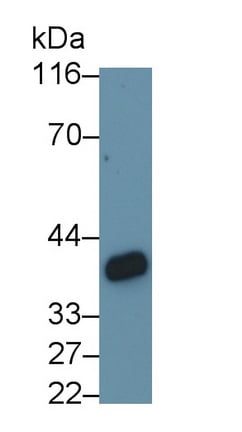
(Western Blot; Sample: Rat Lung lysate Primary Ab: 0.1ug/ml Mouse AntiMouse GZMK Antibody Second Ab: 0.2ug/mL HRPLinked Caprine AntiMouse IgG Polyclonal Antibody (Catalog: SAA544Mu19))
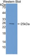
WB (Western Blot)
(Western Blot; Sample: Rat Lung lysate Primary Ab: 0.1ug/ml Mouse AntiMouse GZMK Antibody Second Ab: 0.2ug/mL HRPLinked Caprine AntiMouse IgG Polyclonal Antibody (Catalog: SAA544Mu19))
Granzyme K (GZMK), Monoclonal Antibody (Cat# AAA151692)
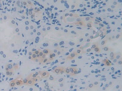
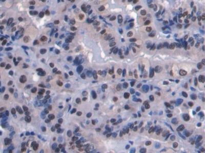
 IHC (Immunohiostchemistry)
(DAB staining on IHCP;Sample: Human Cerebrum Tissue; Primary Ab: 40ug/ml Mouse AntiHuman FSTL1 AntibodySecond Ab: 2ug/mL HRPLinked Caprine AntiMouse IgG Polyclonal Antibody(Catalog: SAA544Mu19))
IHC (Immunohiostchemistry)
(DAB staining on IHCP;Sample: Human Cerebrum Tissue; Primary Ab: 40ug/ml Mouse AntiHuman FSTL1 AntibodySecond Ab: 2ug/mL HRPLinked Caprine AntiMouse IgG Polyclonal Antibody(Catalog: SAA544Mu19))
Follistatin Like Protein 1 (FSTL1), Monoclonal Antibody (Cat# AAA151888)
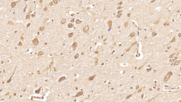
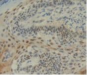
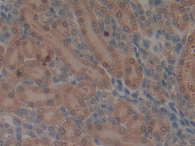
 IHC (Immunohistochemisry)
(DAB staining on IHCP;Sample: Human Cardiac Muscle Tissue; Primary Ab: 40ug/ml Mouse AntiHuman FSTL1 AntibodySecond Ab: 2ug/mL HRPLinked Caprine AntiMouse IgG Polyclonal Antibody(Catalog: SAA544Mu19))
IHC (Immunohistochemisry)
(DAB staining on IHCP;Sample: Human Cardiac Muscle Tissue; Primary Ab: 40ug/ml Mouse AntiHuman FSTL1 AntibodySecond Ab: 2ug/mL HRPLinked Caprine AntiMouse IgG Polyclonal Antibody(Catalog: SAA544Mu19))
Follistatin Like Protein 1 (FSTL1), Monoclonal Antibody (Cat# AAA151889)
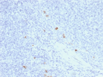
 IHC (Immunohistochemistry)
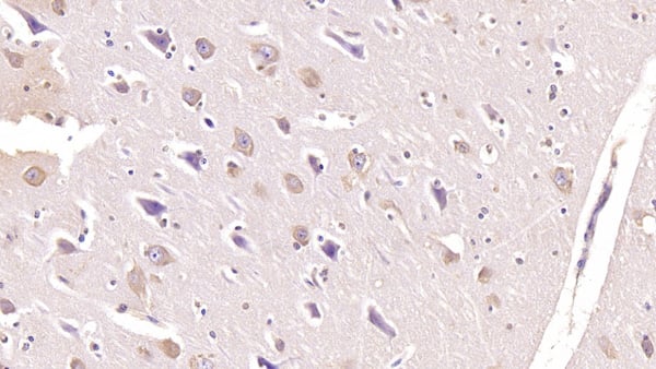
(DAB staining on IHCP;Sample: Human Cerebrum Tissue; Primary Ab: 10ug/ml Mouse AntiHuman SEMA3A AntibodySecond Ab: 2ug/mL HRPLinked Caprine AntiMouse IgG Polyclonal Antibody(Catalog: SAA544Mu19))
IHC (Immunohistochemistry)
(DAB staining on IHCP;Sample: Human Cerebrum Tissue; Primary Ab: 10ug/ml Mouse AntiHuman SEMA3A AntibodySecond Ab: 2ug/mL HRPLinked Caprine AntiMouse IgG Polyclonal Antibody(Catalog: SAA544Mu19))
Semaphorin 3A (SEMA3A), Monoclonal Antibody (Cat# AAA151903)
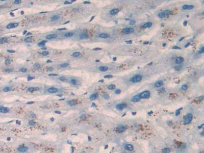
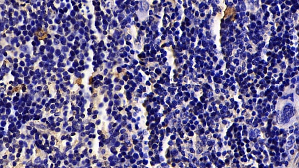
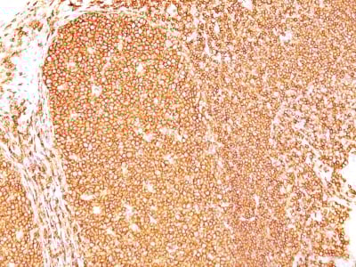
 IHC (Immunohiostchemistry)
(DAB staining on IHCP;Samples: Human Liver cancer Tissue; Primary Ab: 40ug/ml Mouse AntiMultispecies Ang17 AntibodySecond Ab: 2ug/mL HRPLinked Caprine AntiMouse IgG Polyclonal Antibody(Catalog: SAA544Mu19))
IHC (Immunohiostchemistry)
(DAB staining on IHCP;Samples: Human Liver cancer Tissue; Primary Ab: 40ug/ml Mouse AntiMultispecies Ang17 AntibodySecond Ab: 2ug/mL HRPLinked Caprine AntiMouse IgG Polyclonal Antibody(Catalog: SAA544Mu19))
Angiotensin 17 (Ang17), Monoclonal Antibody (Cat# AAA151912)
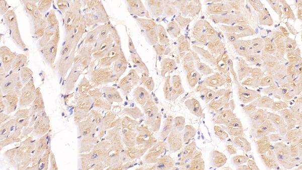
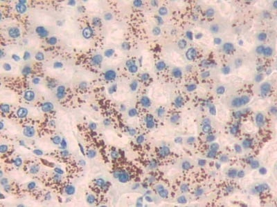
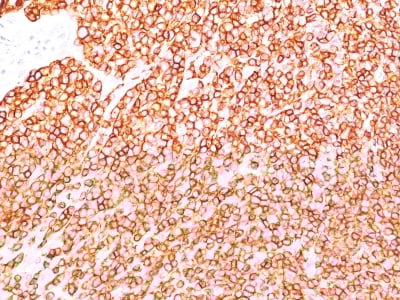
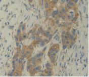
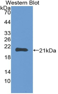
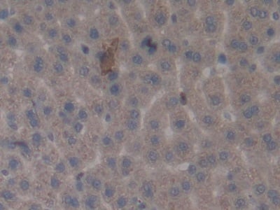
 IHC (Immunohiostchemistry)
(DAB staining on IHC-P;Sample: Human Liver Tissue;Primary Ab: 30ug/ml Mouse Anti-Human TSLP AntibodySecond Ab: 2ug/mL HRP-Linked Caprine Anti-Mouse IgG Polyclonal Antibody)
IHC (Immunohiostchemistry)
(DAB staining on IHC-P;Sample: Human Liver Tissue;Primary Ab: 30ug/ml Mouse Anti-Human TSLP AntibodySecond Ab: 2ug/mL HRP-Linked Caprine Anti-Mouse IgG Polyclonal Antibody)
Thymic Stromal Lymphopoietin (TSLP), Monoclonal Antibody (Cat# AAA151699)
CD64, Monoclonal Antibody (Cat# AAA128244)
CD64, Monoclonal Antibody (Cat# AAA128251)
CD268, Monoclonal Antibody (Cat# AAA128259)
CD206, Monoclonal Antibody (Cat# AAA128268)
CD85g, Monoclonal Antibody (Cat# AAA128277)
CD154, Monoclonal Antibody (Cat# AAA128291)
CD274, Monoclonal Antibody (Cat# AAA128300)
CD274, Monoclonal Antibody (Cat# AAA128303)
CD45, Monoclonal Antibody (Cat# AAA128312)
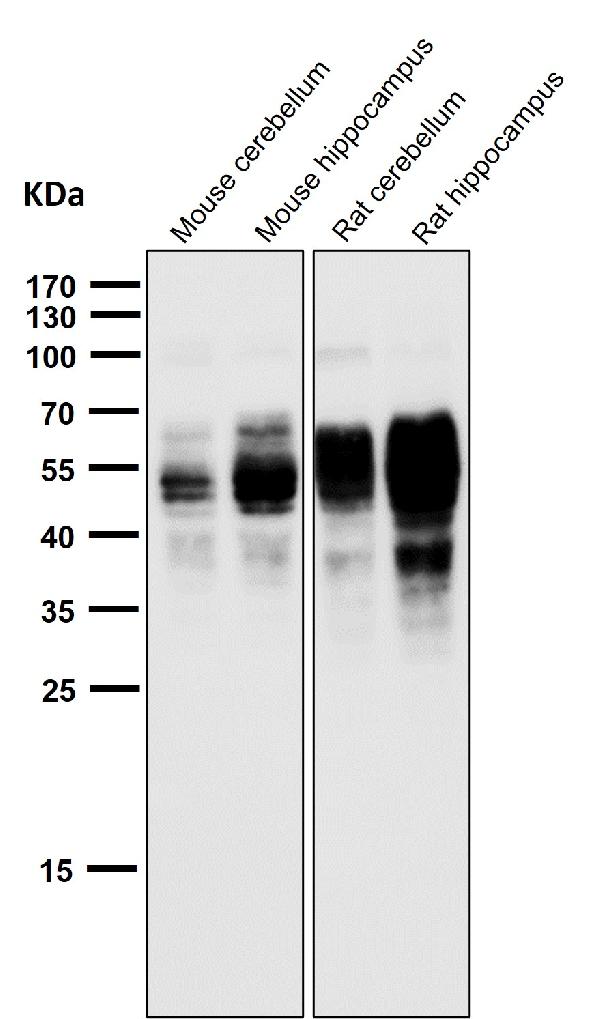
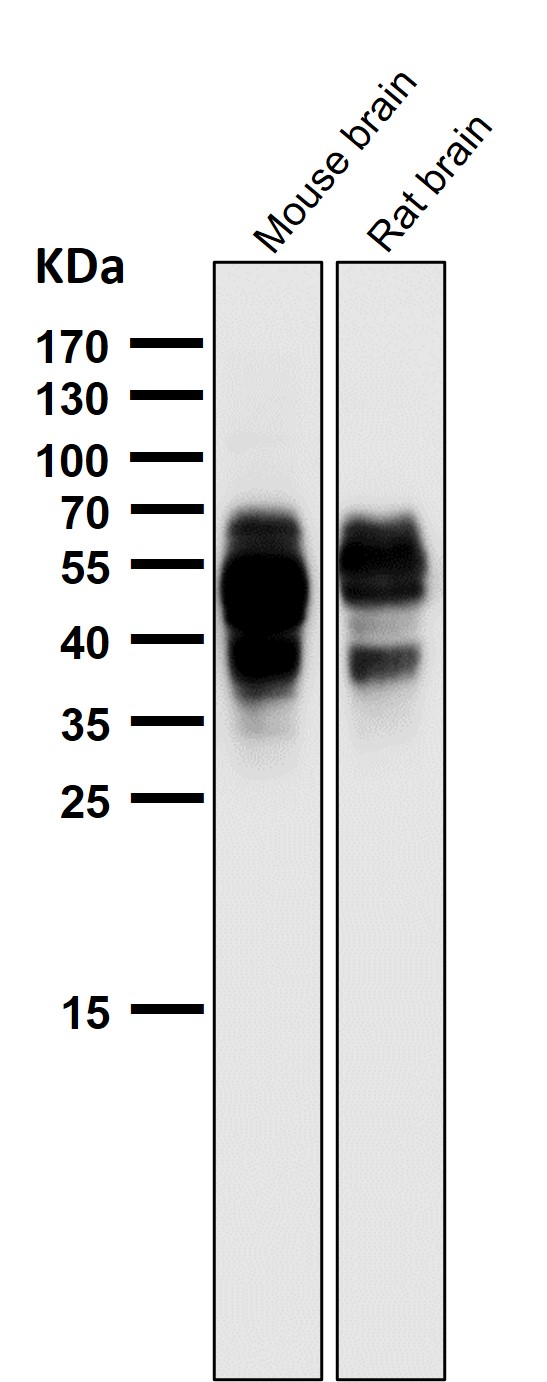
 WB (Western Blot)
(Western blot analysis of Phospho-Tau (S202) expression in mouse hippocampus cell lysate.)
WB (Western Blot)
(Western blot analysis of Phospho-Tau (S202) expression in mouse hippocampus cell lysate.)
Tau, Monoclonal Antibody (Cat# AAA128105)
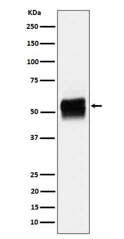
 WB (Western Blot)
(All lanes use the Antibody at 1:5K dilution for 1 hour at room temperature.)
WB (Western Blot)
(All lanes use the Antibody at 1:5K dilution for 1 hour at room temperature.)
RalA, Monoclonal Antibody (Cat# AAA128168)
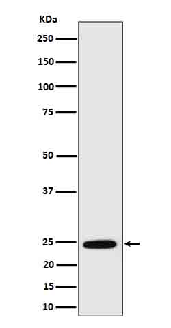
 WB (Western Blot)
(Western blot analysis of HSPC150 expression in HeLa cell lysate.)
WB (Western Blot)
(Western blot analysis of HSPC150 expression in HeLa cell lysate.)
HSPC150, Monoclonal Antibody (Cat# AAA128177)
 WB (Western Blot)
(Western blot analysis of SEC14L2/TAP expression in U87-MG cell lysate.)
WB (Western Blot)
(Western blot analysis of SEC14L2/TAP expression in U87-MG cell lysate.)
SEC14L2/TAP, Monoclonal Antibody (Cat# AAA128180)
 WB (Western Blot)
(Western blot analysis of GAS2 expression in Jurkat cell lysate.)
WB (Western Blot)
(Western blot analysis of GAS2 expression in Jurkat cell lysate.)
GAS2, Monoclonal Antibody (Cat# AAA128184)
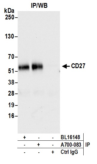

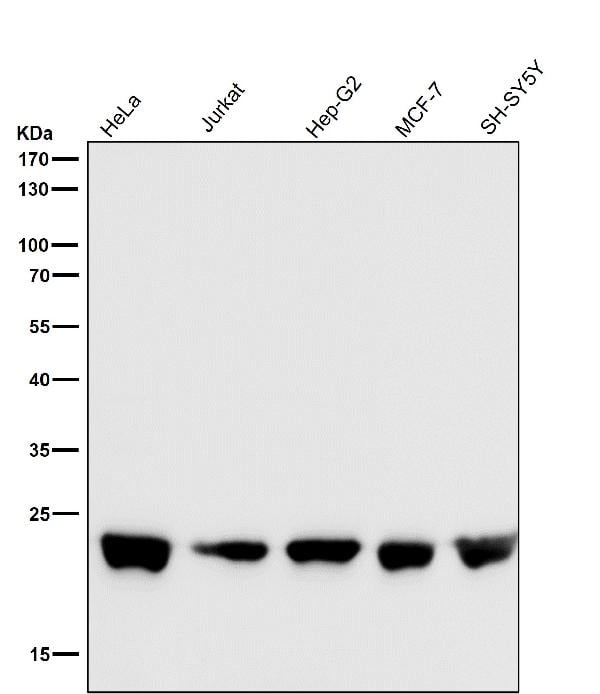
 WB (Western Blot)
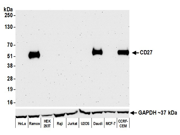
(Detection of human CD27 by western blot. Samples: Whole cell lysate (50 ug) from HeLa, Ramos, HEK293T, Raji, Jurkat, K-562, U2OS, Daudi, MCF-7, and CCRF-CEM cells prepared using NETN lysis buffer. Antibody: Rabbit anti-CD27 recombinant monoclonal antibody (AAA213567 lot 1) used at 1:1000. Secondary: HRP-conjugated goat anti-rabbit IgG . Detection: Chemiluminescence with an exposure time of 3 minutes. Lower Panel: Rabbit anti-GAPDH .)
WB (Western Blot)
(Detection of human CD27 by western blot. Samples: Whole cell lysate (50 ug) from HeLa, Ramos, HEK293T, Raji, Jurkat, K-562, U2OS, Daudi, MCF-7, and CCRF-CEM cells prepared using NETN lysis buffer. Antibody: Rabbit anti-CD27 recombinant monoclonal antibody (AAA213567 lot 1) used at 1:1000. Secondary: HRP-conjugated goat anti-rabbit IgG . Detection: Chemiluminescence with an exposure time of 3 minutes. Lower Panel: Rabbit anti-GAPDH .)
CD27, Monoclonal Recombinant Antibody (Cat# AAA213567)
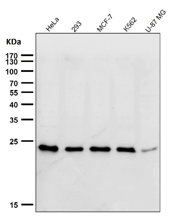
 WB (Western Blot)
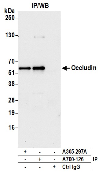
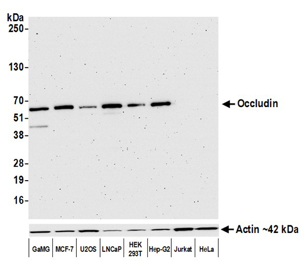
(Detection of human Occludin by western blot. Samples: Whole cell lysate (10 ug) from GaMG, MCF-7, U2OS, LNCaP, HEK293T, Hep-G2, Jurkat, and HeLa cells prepared using NETN lysis buffer. Antibody: Rabbit anti-Occludin recombinant monoclonal antibody (AAA213592 lot 1) used at 1:1000. Secondary: HRP-conjugated goat anti-rabbit IgG . Detection: Chemiluminescence with an exposure time of 3 minutes.)
WB (Western Blot)
(Detection of human Occludin by western blot. Samples: Whole cell lysate (10 ug) from GaMG, MCF-7, U2OS, LNCaP, HEK293T, Hep-G2, Jurkat, and HeLa cells prepared using NETN lysis buffer. Antibody: Rabbit anti-Occludin recombinant monoclonal antibody (AAA213592 lot 1) used at 1:1000. Secondary: HRP-conjugated goat anti-rabbit IgG . Detection: Chemiluminescence with an exposure time of 3 minutes.)
Occludin, Monoclonal Recombinant Antibody (Cat# AAA213592)
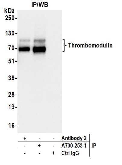
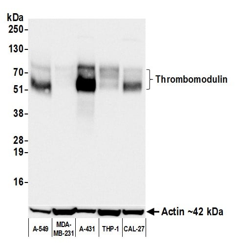 WB (Western Blot)
(Detection of human Thrombomodulin by western blot. Samples: Whole cell lysate (50 ug) from A-549, MDA-MB-231, A-431 (25 ug), THP-1, and CAL-27 cells prepared using NETN lysis buffer. Antibody: Rabbit anti-Thrombomodulin recombinant monoclonal antibody (AAA213666 lot 1) used at 1:1000. Secondary: HRP-conjugated goat anti-rabbit IgG . Detection: Chemiluminescence with an exposure time of 30 seconds. Lower Panel: Rabbit anti-Actin recombinant monoclonal antibody .)
WB (Western Blot)
(Detection of human Thrombomodulin by western blot. Samples: Whole cell lysate (50 ug) from A-549, MDA-MB-231, A-431 (25 ug), THP-1, and CAL-27 cells prepared using NETN lysis buffer. Antibody: Rabbit anti-Thrombomodulin recombinant monoclonal antibody (AAA213666 lot 1) used at 1:1000. Secondary: HRP-conjugated goat anti-rabbit IgG . Detection: Chemiluminescence with an exposure time of 30 seconds. Lower Panel: Rabbit anti-Actin recombinant monoclonal antibody .)
Thrombomodulin, Monoclonal Recombinant Antibody (Cat# AAA213666)
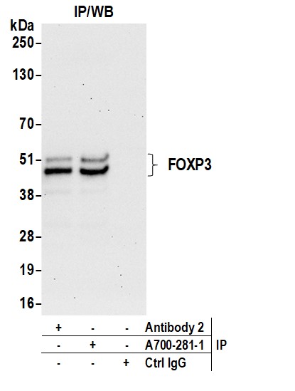
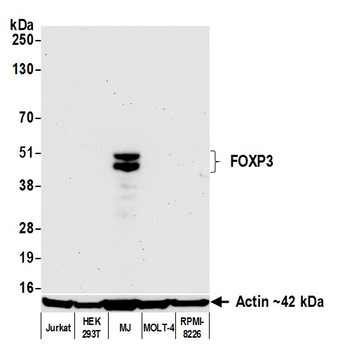 WB (Western Blot)
(Detection of human FOXP3 by western blot. Samples: Whole cell lysate (10 ug) from Jurkat, HEK293T, MJ, MOLT-4, and RPMI-8226 cells prepared using NETN lysis buffer. Antibody: Rabbit anti-FOXP3 recombinant monoclonal antibody (AAA213683 lot 1) used at 1:1000. Secondary: HRP-conjugated goat anti-rabbit IgG . Detection: Chemiluminescence with an exposure time of 30 seconds. Lower Panel: Rabbit anti-Actin recombinant monoclonal antibody .)
WB (Western Blot)
(Detection of human FOXP3 by western blot. Samples: Whole cell lysate (10 ug) from Jurkat, HEK293T, MJ, MOLT-4, and RPMI-8226 cells prepared using NETN lysis buffer. Antibody: Rabbit anti-FOXP3 recombinant monoclonal antibody (AAA213683 lot 1) used at 1:1000. Secondary: HRP-conjugated goat anti-rabbit IgG . Detection: Chemiluminescence with an exposure time of 30 seconds. Lower Panel: Rabbit anti-Actin recombinant monoclonal antibody .)
FOXP3, Monoclonal Recombinant Antibody (Cat# AAA213683)
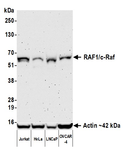
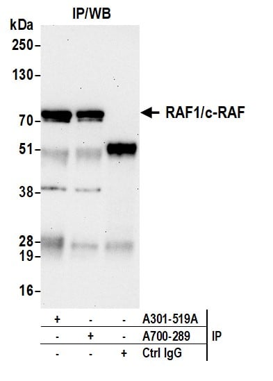
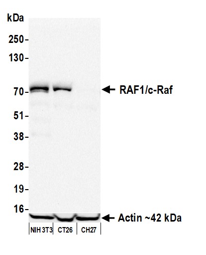 WB (Western Blot)
(Detection of mouse RAF1/c-Raf by western blot. Samples: Whole cell lysate (50 ug) from NIH 3T3, CT26, and CH27 cells prepared using NETN lysis buffer. Antibody: Rabbit anti-RAF1/c-Raf recombinant monoclonal antibody (AAA213689 lot 1) used at 1:1000. Secondary: HRP-conjugated goat anti-rabbit IgG . Detection: Chemiluminescence with an exposure time of 30 seconds. Lower Panel: Rabbit anti-Actin recombinant monoclonal antibody .)
WB (Western Blot)
(Detection of mouse RAF1/c-Raf by western blot. Samples: Whole cell lysate (50 ug) from NIH 3T3, CT26, and CH27 cells prepared using NETN lysis buffer. Antibody: Rabbit anti-RAF1/c-Raf recombinant monoclonal antibody (AAA213689 lot 1) used at 1:1000. Secondary: HRP-conjugated goat anti-rabbit IgG . Detection: Chemiluminescence with an exposure time of 30 seconds. Lower Panel: Rabbit anti-Actin recombinant monoclonal antibody .)
RAF1/c-Raf, Monoclonal Recombinant Antibody (Cat# AAA213689)
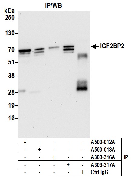
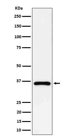
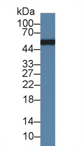
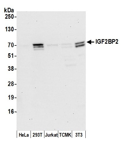 WB (Western Blot)
(Detection of human and mouse IGF2BP2 by western blot. Samples: Whole cell lysate (15 ug) from HeLa, HEK293T, Jurkat, mouse TCMK-1, and mouse NIH 3T3 cells prepared using NETN lysis buffer. Antibody: Mouse monoclonal anti-IGF2BP2 antibody [3A3.5F1] (AAA213499 lot 2) used for WB at 1:1000. Secondary: HRP-conjugated goat anti-mouse IgG . Detection: Chemiluminescence with an exposure time of 30 seconds.)
WB (Western Blot)
(Detection of human and mouse IGF2BP2 by western blot. Samples: Whole cell lysate (15 ug) from HeLa, HEK293T, Jurkat, mouse TCMK-1, and mouse NIH 3T3 cells prepared using NETN lysis buffer. Antibody: Mouse monoclonal anti-IGF2BP2 antibody [3A3.5F1] (AAA213499 lot 2) used for WB at 1:1000. Secondary: HRP-conjugated goat anti-mouse IgG . Detection: Chemiluminescence with an exposure time of 30 seconds.)
IGF2BP2, Monoclonal Antibody (Cat# AAA213499)
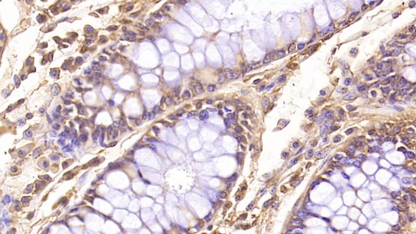
 IHC (Immunohiostchemistry)
(Formalin-fixed, paraffin-embedded Rat Colon stained with FOXA1 Monoclonal Antibody (FOXA1/1519).)
IHC (Immunohiostchemistry)
(Formalin-fixed, paraffin-embedded Rat Colon stained with FOXA1 Monoclonal Antibody (FOXA1/1519).)
FOXA1/HNF3A, Monoclonal Antibody (Cat# AAA214387)
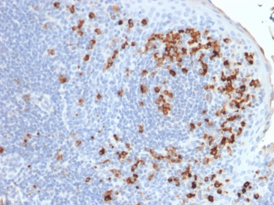
 IHC (Immunohiostchemistry)
(Formalin-fixed, paraffin-embedded human Tonsil stained with CD45RB Ab (PD7/26).)
IHC (Immunohiostchemistry)
(Formalin-fixed, paraffin-embedded human Tonsil stained with CD45RB Ab (PD7/26).)
CD45RB, Monoclonal Antibody (Cat# AAA214391)
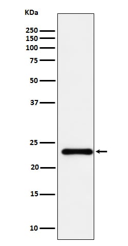
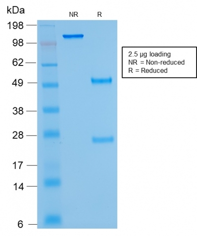
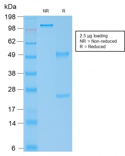 SDS-PAGE
(SDS-PAGE Analysis Purified IgG4 Mouse Recombinant Monoclonal Antibody (rIGHG4/1345).)
SDS-PAGE
(SDS-PAGE Analysis Purified IgG4 Mouse Recombinant Monoclonal Antibody (rIGHG4/1345).)
IgG4 (Ig Heavy Constant Gamma 4), Monoclonal Antibody (Cat# AAA214395)
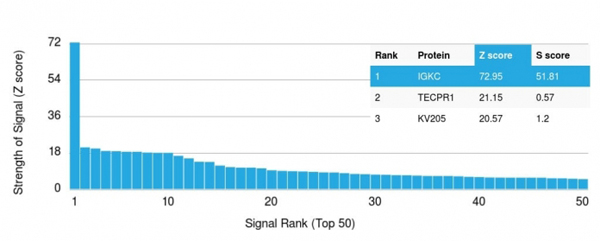 Application Data
(Analysis of Protein Array containing >19, 000 full-length human proteins using Kappa Light Chain Mouse Monoclonal Antibody (rKLC/264) Z- and S- Score: The Z-score represents the strength of a signal that a monoclonal antibody (MAb) (in combination with a fluorescently-tagged anti-IgG secondary antibody) produces when binding to a particular protein on the HuProtTM array. Z-scores are described in units of standard deviations (SD's) above the mean value of all signals generated on that array. If targets on HuProtTM are arranged in descending order of the Z-score, the S-score is the difference (also in units of SD's) between the Z-score. S-score therefore represents the relative target specificity of a MAb to its intended target. A MAb is considered to specific to its intended target, if the MAb has an S-score of at least 2.5. For example, if a MAb binds to protein X with a Z-score of 43 and to protein Y with a Z-score of 14, then the S-score for the binding of that MAb to protein X is equal to 29.)
Application Data
(Analysis of Protein Array containing >19, 000 full-length human proteins using Kappa Light Chain Mouse Monoclonal Antibody (rKLC/264) Z- and S- Score: The Z-score represents the strength of a signal that a monoclonal antibody (MAb) (in combination with a fluorescently-tagged anti-IgG secondary antibody) produces when binding to a particular protein on the HuProtTM array. Z-scores are described in units of standard deviations (SD's) above the mean value of all signals generated on that array. If targets on HuProtTM are arranged in descending order of the Z-score, the S-score is the difference (also in units of SD's) between the Z-score. S-score therefore represents the relative target specificity of a MAb to its intended target. A MAb is considered to specific to its intended target, if the MAb has an S-score of at least 2.5. For example, if a MAb binds to protein X with a Z-score of 43 and to protein Y with a Z-score of 14, then the S-score for the binding of that MAb to protein X is equal to 29.)
Kappa Light Chain, Monoclonal Antibody (Cat# AAA214398)
 WB (Western Blot)
(Western Blot: Sample: Rabbit Serum.)
WB (Western Blot)
(Western Blot: Sample: Rabbit Serum.)
Immunoglobulin G, Monoclonal Antibody (Cat# AAA141331)
 WB (Western Blot)
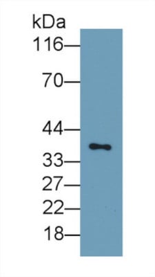
(Western Blot; Sample: SKOV3 cell lysatePrimary Ab: 0.2ug/ml Mouse Anti-Rat uPAR AntibodySecond Ab: 0.2ug/mL HRP-Linked Caprine Anti-Mouse IgG Polyclonal Antibody)
WB (Western Blot)
(Western Blot; Sample: SKOV3 cell lysatePrimary Ab: 0.2ug/ml Mouse Anti-Rat uPAR AntibodySecond Ab: 0.2ug/mL HRP-Linked Caprine Anti-Mouse IgG Polyclonal Antibody)
Plasminogen Activator, Urokinase Receptor (uPAR), Monoclonal Antibody (Cat# AAA141334)
 IHC (Immunohistochemistry)
(DAB staining on IHC-P;Sample: Rat Pancreas Tissue;Primary Ab: 20ug/ml Mouse Anti-Rat PTHrP AntibodySecond Ab: 2ug/mL HRP-Linked Caprine Anti-Mouse IgG Polyclonal Antibody)
IHC (Immunohistochemistry)
(DAB staining on IHC-P;Sample: Rat Pancreas Tissue;Primary Ab: 20ug/ml Mouse Anti-Rat PTHrP AntibodySecond Ab: 2ug/mL HRP-Linked Caprine Anti-Mouse IgG Polyclonal Antibody)
Parathyroid Hormone, Monoclonal Antibody (Cat# AAA141343)
 WB (Western Blot)
(Western Blot: Sample: Recombinant protein.)
WB (Western Blot)
(Western Blot: Sample: Recombinant protein.)
Vascular Endothelial Growth Factor A, Monoclonal Antibody (Cat# AAA141344)
 IHC (Immunohistochemistry)
(DAB staining on IHC-P; Samples: Human Breast Cancer Tissue)
IHC (Immunohistochemistry)
(DAB staining on IHC-P; Samples: Human Breast Cancer Tissue)
Interleukin 12A, Monoclonal Antibody (Cat# AAA141353)
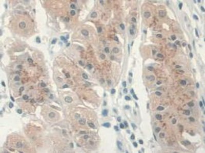
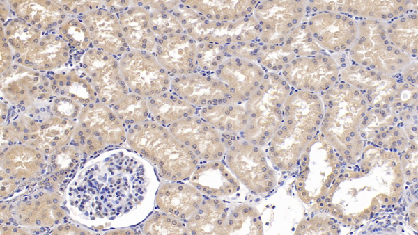
 IHC (Immunohistochemisry)
(DAB staining on IHC-P;Samples: Human Kidney Tissue;Primary Ab: 30ug/ml Mouse Anti-Human GREM1 AntibodySecond Ab: 2ug/mL HRP-Linked Caprine Anti-Mouse IgG Polyclonal Antibody)
IHC (Immunohistochemisry)
(DAB staining on IHC-P;Samples: Human Kidney Tissue;Primary Ab: 30ug/ml Mouse Anti-Human GREM1 AntibodySecond Ab: 2ug/mL HRP-Linked Caprine Anti-Mouse IgG Polyclonal Antibody)
Gremlin 1, Monoclonal Antibody (Cat# AAA141355)
 WB (Western Blot)
(Western Blot: Sample: Recombinant NRP1, Human.)
WB (Western Blot)
(Western Blot: Sample: Recombinant NRP1, Human.)
Neuropilin 1, Monoclonal Antibody (Cat# AAA141358)
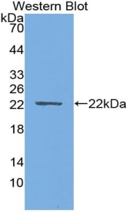
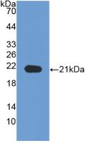
Matrix Metalloproteinase 7 (MMP7), Monoclonal Antibody (Cat# AAA149324)
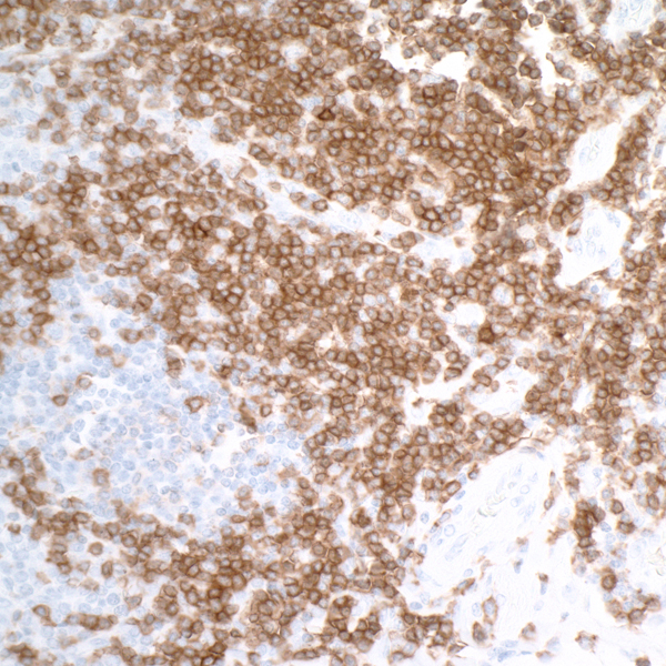
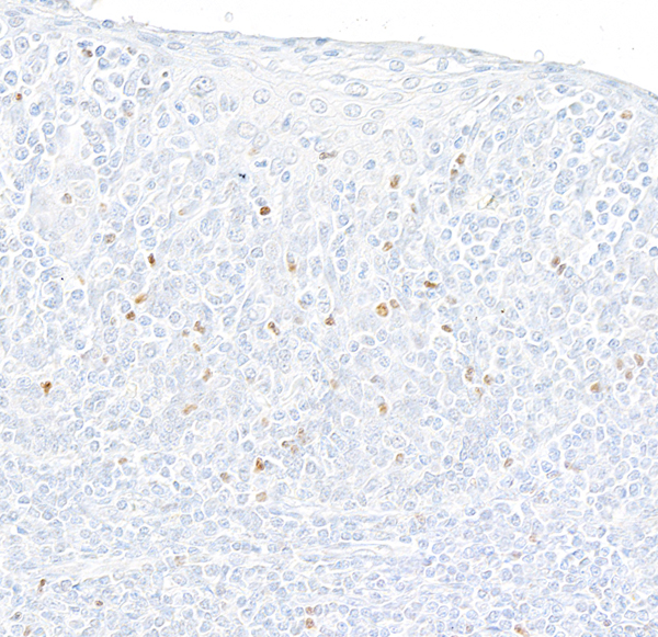
 IHC (Immunohiostchemistry)
(DAB staining on IHC-P; Samples: Human Tonsil Tissue))
IHC (Immunohiostchemistry)
(DAB staining on IHC-P; Samples: Human Tonsil Tissue))
Programmed Cell Death Protein 1 Ligand 1 (PDL1), Monoclonal Antibody (Cat# AAA149438)
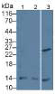
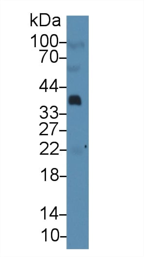
 WB (Western Blot)
(Lane 1: Human Liver TissueLane 2: Mouse Brain TissueLane 3: Human MCF7 CellsLane 4: Human A-375 CellsPrimary Ab: 1: 1500 Dilution of Mouse Anti-Human CNX AbSecond Ab: 1: 5000 Dilution of HRP-Linked Rabbit Anti-Mouse IgG Ab (Catalog: ))
WB (Western Blot)
(Lane 1: Human Liver TissueLane 2: Mouse Brain TissueLane 3: Human MCF7 CellsLane 4: Human A-375 CellsPrimary Ab: 1: 1500 Dilution of Mouse Anti-Human CNX AbSecond Ab: 1: 5000 Dilution of HRP-Linked Rabbit Anti-Mouse IgG Ab (Catalog: ))
Calnexin (CNX), Monoclonal Antibody (Cat# AAA145959)
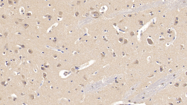
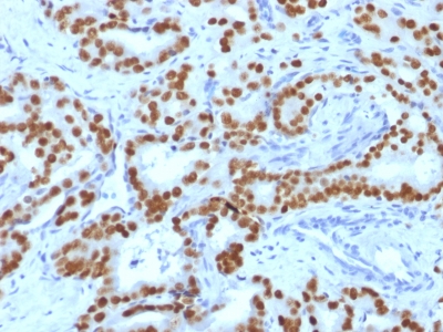
 IHC (Immunohiostchemistry)
(DAB staining on IHC-P; Samples: Human Thyroid cancer Tissue))
IHC (Immunohiostchemistry)
(DAB staining on IHC-P; Samples: Human Thyroid cancer Tissue))
Proliferating Cell Nuclear Antigen (PCNA), Monoclonal Antibody (Cat# AAA145961)
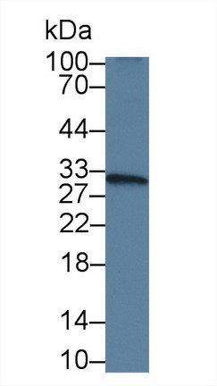
Interleukin 10 (IL10), Monoclonal Antibody (Cat# AAA145996)
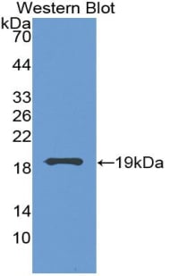
Interleukin 18 (IL18), Monoclonal Antibody (Cat# AAA146000)
Interleukin 18 (IL18), Monoclonal Antibody (Cat# AAA146002)
Interleukin 6 (IL6), Monoclonal Antibody (Cat# AAA146012)
Interleukin 8 (IL8), Monoclonal Antibody (Cat# AAA146013)
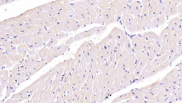
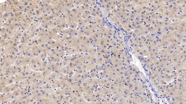
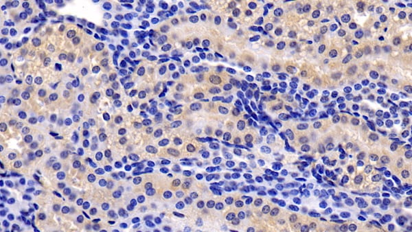
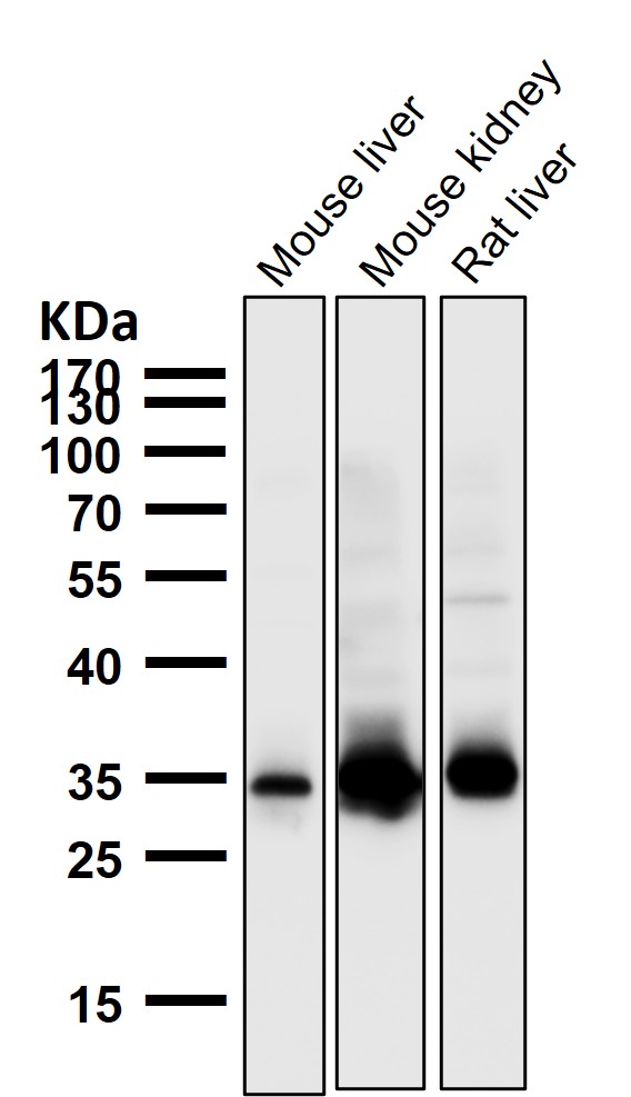
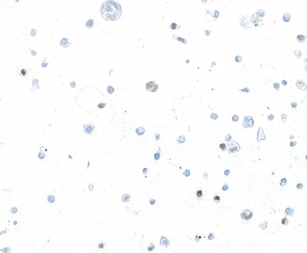
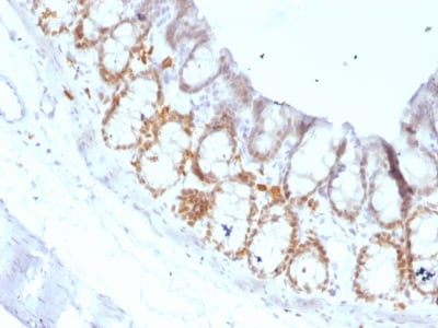
 IHC (Immunohistochemisry)
(DAB staining on IHC-P; Samples: Rat Liver Tissue)
IHC (Immunohistochemisry)
(DAB staining on IHC-P; Samples: Rat Liver Tissue)
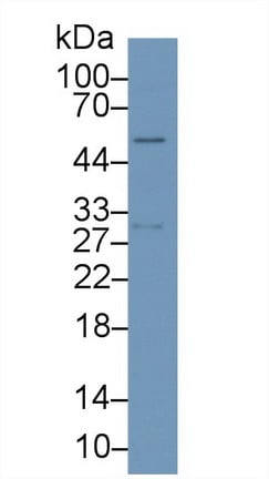
Matrix Metalloproteinase 3, Monoclonal Antibody (Cat# AAA141268)

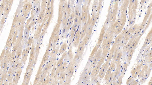
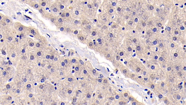
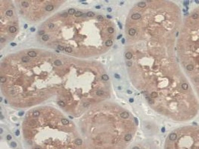
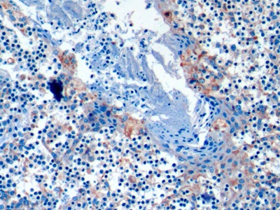
 IHC (Immunohiostchemistry)
(DAB staining on IHC-P; Samples: Porcine Pancreas Tissue)
IHC (Immunohiostchemistry)
(DAB staining on IHC-P; Samples: Porcine Pancreas Tissue)
Clusterin, Monoclonal Antibody (Cat# AAA141269)
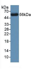
Advanced Glycation End Product, Monoclonal Antibody (Cat# AAA141278)
What are Monoclonal Antibodies?
Monoclonal antibodies are specialized laboratory-produced proteins developed for binding to specific biological antigens or other molecular targets. Since they come from a single cell (or clone), they are especially consistent and accurate in the data they are involved in producing.
This type of antibody material has been shown to be a powerful tool in finding and subsequently destroying harmful cells in an organism, such as those found in cancers or various autoimmune diseases. This makes them excellent aids in medical testing and research, which is why they are so widely used.
AAA Biotech offers a comprehensive range of high-quality monoclonal antibodies that perform effectively in various laboratory tests, including (amongst others) ELISA, western blotting, immunohistochemistry, and flow cytometry. All of the products in our catalog are thoroughly quality tested to make sure that they are reliable and will consistently perform well in your research.
What Are The Uses of Monoclonal Antibodies
Monoclonal antibodies are used in many lab tests, including (amongst others) ELISA, western blotting, immunohistochemistry, and flow cytometry.
ELISA is a test that helps detect a specific substance/analyte in a sample. It uses antibodies (often monoclonal) bound to a solid surface (such as the well of a microplate) to “capture” the substance/analyte in the sample and immobilize it so that the detection antibody component can then bind to it and produce a signal, which can then be measured.
Western blotting identifies specific proteins in a sample. The sample is first separated on a gel, and then antibodies are applied that will typically bind to the target, which will all be localized to a single band in a lane.
Immunohistochemistry helps locate specific proteins in cells or tissue samples using antibodies.
Flow cytometry looks at and sorts cells. It uses antibodies that are conjugated to reporter molecules called “fluorophores”, which, under special lights, emit light themselves, which can then be measured by a detector instrument. For a deeper understanding of these techniques, explore our complete guide to monoclonal antibodies and their benefits.
How Monoclonal Antibodies Are Used as Medicine?
Please note that all of the products listed in AAA Biotech’s also known as AAA Bio or AAABio catalog are strictly for research-use only (RUO).
Monoclonal antibodies can also be used as therapeutic/medical treatments, particularly in the context of cancers. They are designed to find and bind to specific cells or proteins, helping the immune system recognize and attack the cancer. These treatments work in different ways, such as:
- Radioimmunotherapy attaches a small amount of radioactive molecule to the antibody, so it delivers the radiation directly to the cancer cells that the antibody is specifically binding to.
- Antibody-directed enzyme prodrug therapy uses antibodies that are specifically bound to special enzymes. These enzymes activate a harmless drug in the body and turn it into a cancer-killing drug only near the cancer cells—this helps avoid harming healthy cells.
- Immunoliposomes are tiny “bubbles” filled with medicine/drug and coated with antibodies. They carry the drug straight to the cancer cells.
Why Buy Monoclonal Antibodies From Us?
At AAA Biotech, we provide high-performance monoclonal antibodies designed to support a wide range of research needs.
1. Validated for Versatile Applications
The antibodies in our catalog are extensively validated and compatible with multiple techniques, including (but not limited to) ELISA, flow cytometry (FC), immunocytochemistry (ICC), immunofluorescence (IF), immunohistochemistry (IHC), immunoprecipitation (IP), and western blotting (WB).
2. Wide Selection & Specialized Options
We offer antibodies for common and rare species, that are available in various conjugated forms, and also in recombinant formats. Essentially, there is almost anything one might need to meet their experimental model’s requirements.
3. High-Quality Proteins
Our proteins meet high purity standards—90% or more as confirmed by SDS-PAGE. Many are available with tags like His, Flag, GST, or MBP, and we also supply native and biologically active proteins for functional studies.
Frequently Asked Questions
1. Are your monoclonal antibodies validated for specific applications?
Yes, our antibodies are tested and validated for use in methods such as ELISA, western blot, IHC, flow cytometry, and more. Refer to specific product pages or datasheets for individual product information.
2. How do I choose the right monoclonal antibody for my application?
Review the product details directly for application validation, species reactivity, and target information. You may also contact our support team at any time for help.
3. How quickly can I receive my order?
Most orders are processed and shipped within 1–3 business days, depending on product availability and your shipping location.