Filters
▼Clonality
▼Type
▼Reactivity
▼Gene Name
▼Isotype
▼Host
▼Application
▼Clone
▼Monoclonal Antibodies
Get accurate results in your research with our Monoclonal Antibodies, which are specially made to target exactly what you require for your research, and will produce consistent, reliable performance in lab tests.
Viewing 7050-7100 of 27645 product results

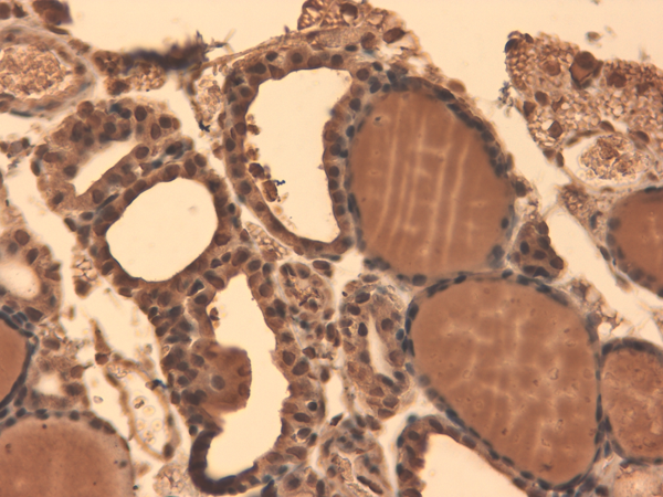
 IHC (Immunohistochemistry)
(Immunohistochemistry analysis using Mouse Anti-Sodium Iodide Symporter Monoclonal Antibody, Clone 14F. Tissue: Thyroid. Species: Mouse. Fixation: 10% Formalin Solution for 12-24 hours at RT. Primary Antibody: Mouse Anti-Sodium Iodide Symporter Monoclonal Antibody at 1:1000 for 1 hour at RT. Secondary Antibody: HRP/DAB Detection System: Biotinylated Goat Anti-Mouse, Streptavidin Peroxidase, DAB Chromogen (brown) for 30 minutes at RT. Counterstain: Mayer Hematoxylin (purple/blue) nuclear stain at 250-500 ul for 5 minutes at RT.)
IHC (Immunohistochemistry)
(Immunohistochemistry analysis using Mouse Anti-Sodium Iodide Symporter Monoclonal Antibody, Clone 14F. Tissue: Thyroid. Species: Mouse. Fixation: 10% Formalin Solution for 12-24 hours at RT. Primary Antibody: Mouse Anti-Sodium Iodide Symporter Monoclonal Antibody at 1:1000 for 1 hour at RT. Secondary Antibody: HRP/DAB Detection System: Biotinylated Goat Anti-Mouse, Streptavidin Peroxidase, DAB Chromogen (brown) for 30 minutes at RT. Counterstain: Mayer Hematoxylin (purple/blue) nuclear stain at 250-500 ul for 5 minutes at RT.)
Sodium-Iodide Symporter, Monoclonal Antibody (Cat# AAA103175)
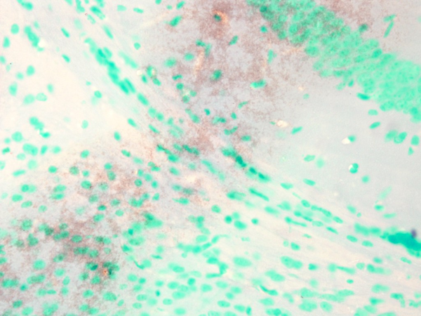
 IHC (Immunohistochemistry)
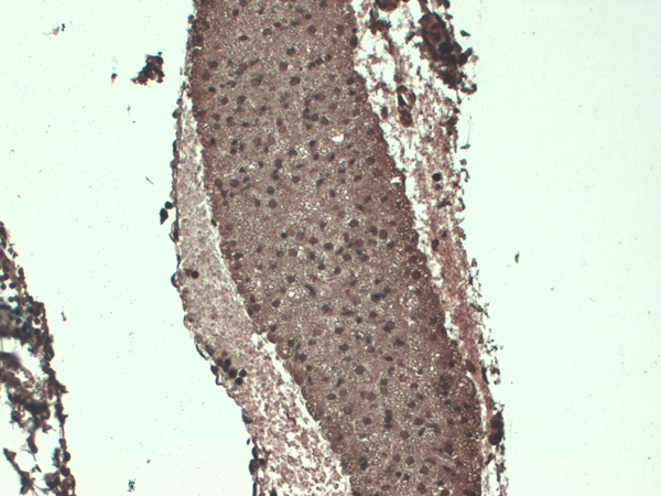
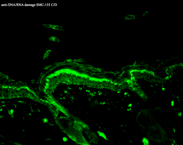
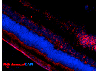
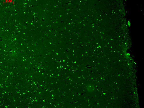
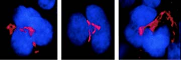
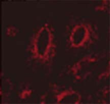
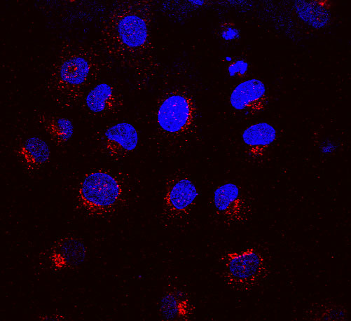
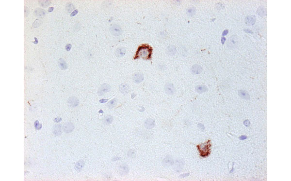
(Immunohistochemistry analysis using Mouse Anti-DNA Damage Monoclonal Antibody, Clone 15A3. Tissue: Ischemic fresh brain tissue. Species: Rat. Primary Antibody: Mouse Anti-DNA Damage Monoclonal Antibody at 1:1000 for 16 hours at RT. Secondary Antibody: Alexa Fluor 546 Goat Anti-mouse (Red) at 1:500 for 1 hour at RT. Localization: Cerebral Cortex. Courtesy of: Dr. Yi Yang, U. New Mexico.)
IHC (Immunohistochemistry)
(Immunohistochemistry analysis using Mouse Anti-DNA Damage Monoclonal Antibody, Clone 15A3. Tissue: Ischemic fresh brain tissue. Species: Rat. Primary Antibody: Mouse Anti-DNA Damage Monoclonal Antibody at 1:1000 for 16 hours at RT. Secondary Antibody: Alexa Fluor 546 Goat Anti-mouse (Red) at 1:500 for 1 hour at RT. Localization: Cerebral Cortex. Courtesy of: Dr. Yi Yang, U. New Mexico.)
DNA/RNA Damage, Monoclonal Antibody (Cat# AAA103182)

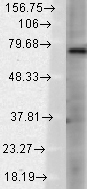
 WB (Western Blot)
(Western Blot analysis of Rat Brain showing detection of ~200 kDa NALCN protein using Mouse Anti-NALCN Monoclonal Antibody, Clone S187-7 . Lane 1: Molecular Weight (MW) Ladder. Lane 3: Rat Brain Membrane. Load: 15 ug. Block: 2% BSA and 2% Skim Milk in 1X TBST. Primary Antibody: Mouse Anti-NALCN Monoclonal Antibody at 1:1000 for 16 hours at 4 degree C. Secondary Antibody: Goat Anti-Mouse IgG: HRP at 1:2000 for 60 min at RT. Color Development: ECL solution for 6 min at RT. Predicted/Observed Size: ~200 kDa.)
WB (Western Blot)
(Western Blot analysis of Rat Brain showing detection of ~200 kDa NALCN protein using Mouse Anti-NALCN Monoclonal Antibody, Clone S187-7 . Lane 1: Molecular Weight (MW) Ladder. Lane 3: Rat Brain Membrane. Load: 15 ug. Block: 2% BSA and 2% Skim Milk in 1X TBST. Primary Antibody: Mouse Anti-NALCN Monoclonal Antibody at 1:1000 for 16 hours at 4 degree C. Secondary Antibody: Goat Anti-Mouse IgG: HRP at 1:2000 for 60 min at RT. Color Development: ECL solution for 6 min at RT. Predicted/Observed Size: ~200 kDa.)
NALCN, Monoclonal Antibody (Cat# AAA103186)
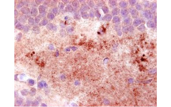
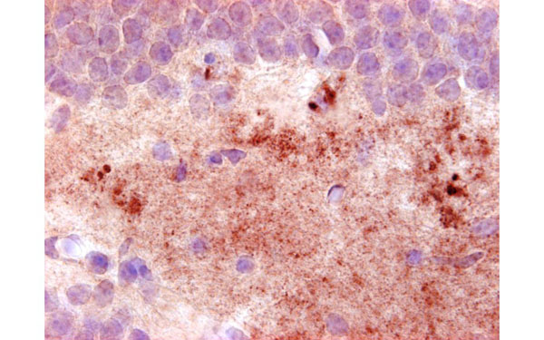
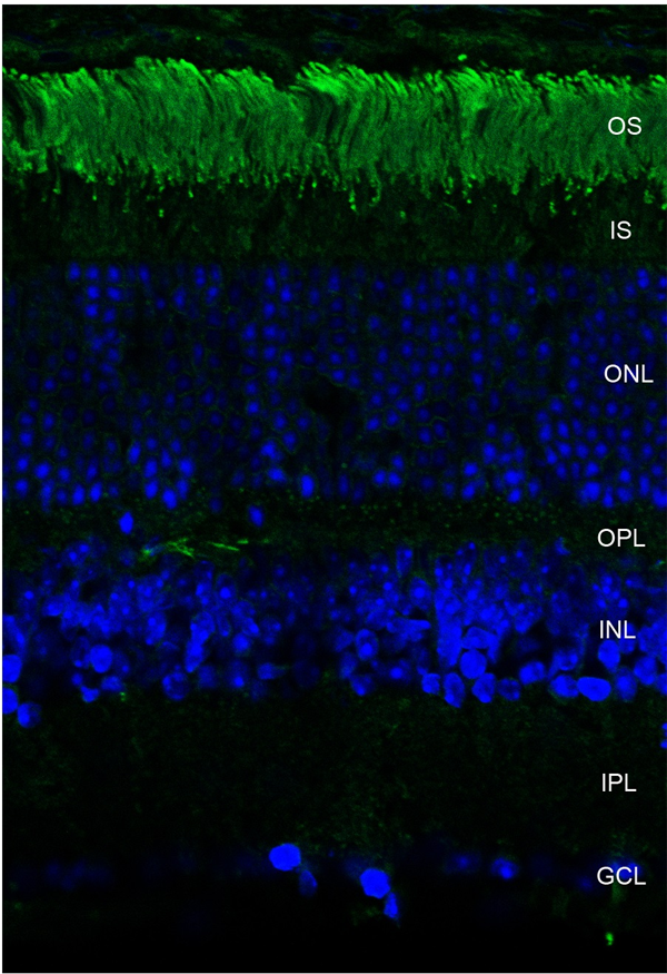 IHC (Immunohistochemistry)
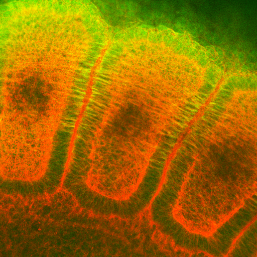
(Immunohistochemistry analysis using Mouse Anti-Rhodopsin Monoclonal Antibody, Clone 4D2. Tissue: retina. Species: Mouse. Primary Antibody: Mouse Anti-Rhodopsin Monoclonal Antibody at 1:1000. Secondary Antibody: FITC Goat Anti-Mouse (green). Counterstain: DAPI (blue) nuclear stain. Localization: Staining of photoreceptor outer segment (OS). Other layers of the retina: IS - inner segment; ONL - outer nuclear layer; OPL - outer plexiform layer; INL - inner nuclear layer; IPL - inner plexiform layer; GCL - ganglion cell layer.)
IHC (Immunohistochemistry)
(Immunohistochemistry analysis using Mouse Anti-Rhodopsin Monoclonal Antibody, Clone 4D2. Tissue: retina. Species: Mouse. Primary Antibody: Mouse Anti-Rhodopsin Monoclonal Antibody at 1:1000. Secondary Antibody: FITC Goat Anti-Mouse (green). Counterstain: DAPI (blue) nuclear stain. Localization: Staining of photoreceptor outer segment (OS). Other layers of the retina: IS - inner segment; ONL - outer nuclear layer; OPL - outer plexiform layer; INL - inner nuclear layer; IPL - inner plexiform layer; GCL - ganglion cell layer.)
Rhodopsin, Monoclonal Antibody (Cat# AAA103189)
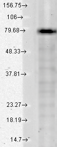
 WB (Western Blot)
(Western Blot analysis of hamster T-CHO cell lysate showing detection of KCNQ1 protein using Mouse Anti-KCNQ1 Monoclonal Antibody, Clone S37A-10. Load: 15 ug. Block: 1.5% BSA for 30 minutes at RT. Primary Antibody: Mouse Anti-KCNQ1 Monoclonal Antibody at 1:1000 for 2 hours at RT. Secondary Antibody: Sheep Anti-Mouse IgG: HRP for 1 hour at RT.)
WB (Western Blot)
(Western Blot analysis of hamster T-CHO cell lysate showing detection of KCNQ1 protein using Mouse Anti-KCNQ1 Monoclonal Antibody, Clone S37A-10. Load: 15 ug. Block: 1.5% BSA for 30 minutes at RT. Primary Antibody: Mouse Anti-KCNQ1 Monoclonal Antibody at 1:1000 for 2 hours at RT. Secondary Antibody: Sheep Anti-Mouse IgG: HRP for 1 hour at RT.)
KCNQ1, Monoclonal Antibody (Cat# AAA103207)
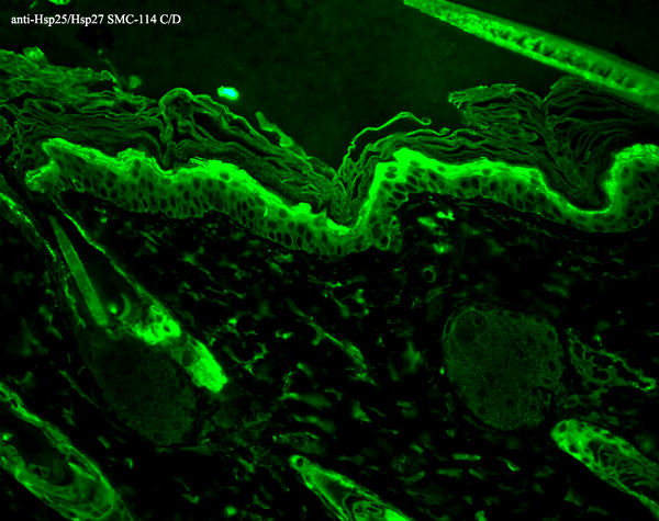
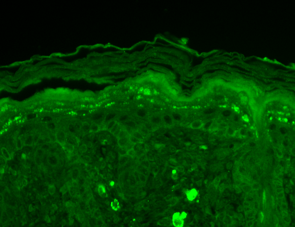
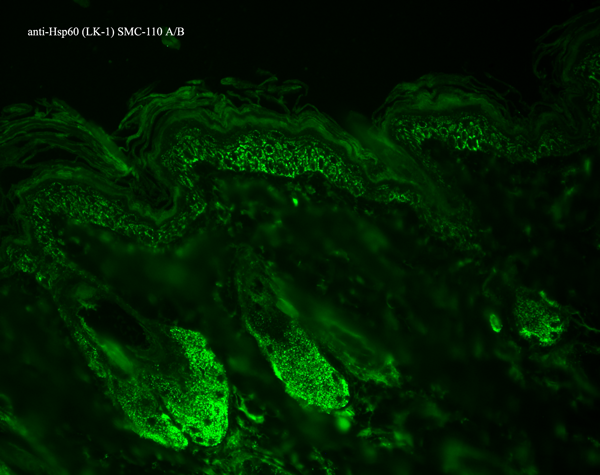
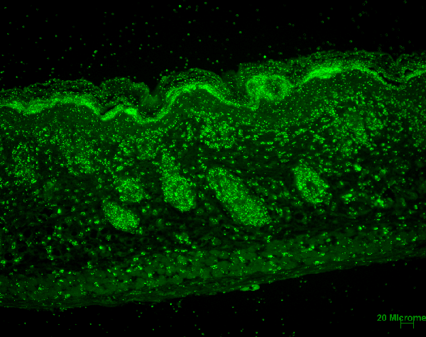
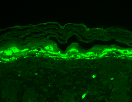
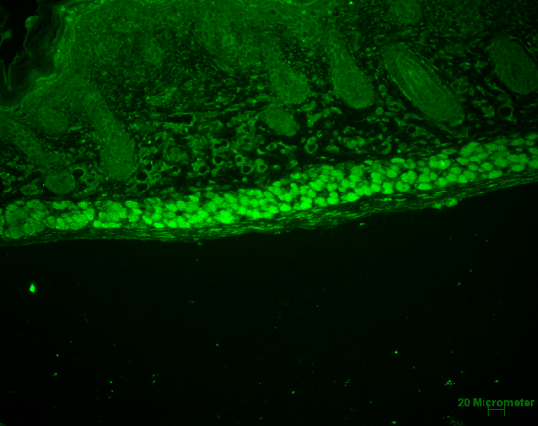
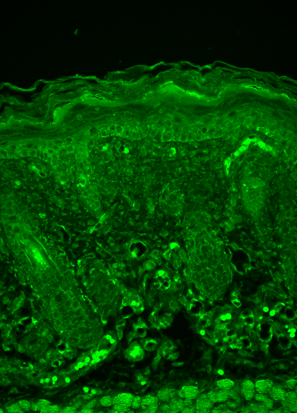
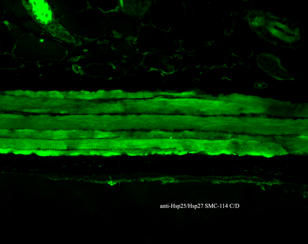 IHC (Immunohistochemistry)
(Immunohistochemistry analysis using Mouse Anti-Hsp27 Monoclonal Antibody, Clone 8A7. Tissue: backskin. Species: Mouse. Fixation: Bouin's Fixative and paraffin-embedded. Primary Antibody: Mouse Anti-Hsp27 Monoclonal Antibody at 1:100 for 1 hour at RT. Secondary Antibody: FITC Goat Anti-Mouse (green) at 1:50 for 1 hour at RT. Localization: Epidermis.)
IHC (Immunohistochemistry)
(Immunohistochemistry analysis using Mouse Anti-Hsp27 Monoclonal Antibody, Clone 8A7. Tissue: backskin. Species: Mouse. Fixation: Bouin's Fixative and paraffin-embedded. Primary Antibody: Mouse Anti-Hsp27 Monoclonal Antibody at 1:100 for 1 hour at RT. Secondary Antibody: FITC Goat Anti-Mouse (green) at 1:50 for 1 hour at RT. Localization: Epidermis.)
Hsp25/Hsp27, Monoclonal Antibody (Cat# AAA103208)
 WB (Western Blot)
(Western Blot analysis of Rat brain membrane lysate showing detection of SHANK3 protein using Mouse Anti-SHANK3 Monoclonal Antibody, Clone S69-46. Load: 15 ug. Block: 1.5% BSA for 30 minutes at RT. Primary Antibody: Mouse Anti-SHANK3 Monoclonal Antibody at 1:1000 for 2 hours at RT. Secondary Antibody: Sheep Anti-Mouse IgG: HRP for 1 hour at RT.)
WB (Western Blot)
(Western Blot analysis of Rat brain membrane lysate showing detection of SHANK3 protein using Mouse Anti-SHANK3 Monoclonal Antibody, Clone S69-46. Load: 15 ug. Block: 1.5% BSA for 30 minutes at RT. Primary Antibody: Mouse Anti-SHANK3 Monoclonal Antibody at 1:1000 for 2 hours at RT. Secondary Antibody: Sheep Anti-Mouse IgG: HRP for 1 hour at RT.)
SHANK3, Monoclonal Antibody (Cat# AAA103234)
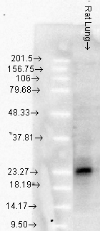
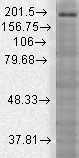
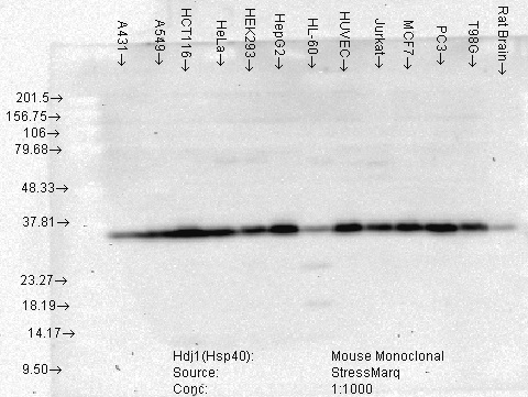 WB (Western Blot)
(Western Blot analysis of Human Cell lysates showing detection of Hsp40 protein using Mouse Anti-Hsp40 Monoclonal Antibody, Clone 3B9.E6. Load: 15 ug. Block: 1.5% BSA for 30 minutes at RT. Primary Antibody: Mouse Anti-Hsp40 Monoclonal Antibody at 1:1000 for 2 hours at RT. Secondary Antibody: Sheep Anti-Mouse IgG: HRP for 1 hour at RT.)
WB (Western Blot)
(Western Blot analysis of Human Cell lysates showing detection of Hsp40 protein using Mouse Anti-Hsp40 Monoclonal Antibody, Clone 3B9.E6. Load: 15 ug. Block: 1.5% BSA for 30 minutes at RT. Primary Antibody: Mouse Anti-Hsp40 Monoclonal Antibody at 1:1000 for 2 hours at RT. Secondary Antibody: Sheep Anti-Mouse IgG: HRP for 1 hour at RT.)
Hsp40 (Hdj1), Monoclonal Antibody (Cat# AAA103239)
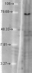
 WB (Western Blot)
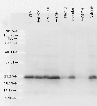
(Western Blot analysis of Human Cell lysates showing detection of Hsp27 protein using Mouse Anti-Hsp27 Monoclonal Antibody, Clone 5D12-A3. Load: 15 ug. Block: 1.5% BSA for 30 minutes at RT. Primary Antibody: Mouse Anti-Hsp27 Monoclonal Antibody at 1:1000 for 2 hours at RT. Secondary Antibody: Sheep Anti-Mouse IgG: HRP for 1 hour at RT.)
WB (Western Blot)
(Western Blot analysis of Human Cell lysates showing detection of Hsp27 protein using Mouse Anti-Hsp27 Monoclonal Antibody, Clone 5D12-A3. Load: 15 ug. Block: 1.5% BSA for 30 minutes at RT. Primary Antibody: Mouse Anti-Hsp27 Monoclonal Antibody at 1:1000 for 2 hours at RT. Secondary Antibody: Sheep Anti-Mouse IgG: HRP for 1 hour at RT.)
Hsp27, Monoclonal Antibody (Cat# AAA103257)
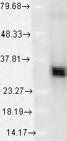
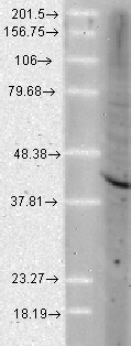
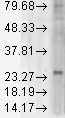
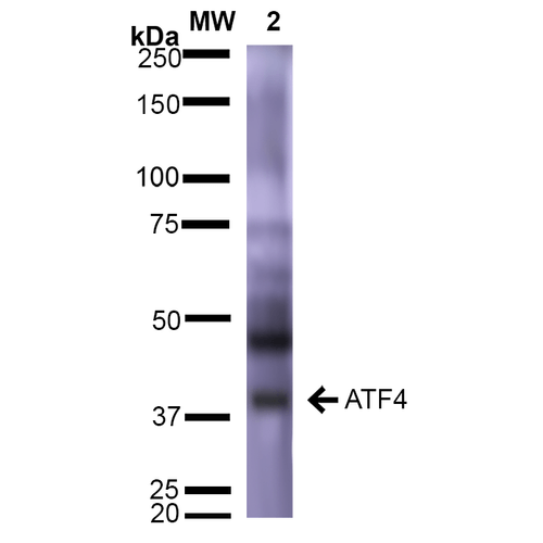 WB (Western Blot)
(Western Blot analysis of Rat Brain showing detection of ~39 kDa (isoform 2) ATF4 protein using Mouse Anti-ATF4 Monoclonal Antibody, Clone S360A-24 . Lane 1: Molecular Weight Ladder (MW). Lane 2: Rat Brain. Load: 15 ug. Block: 5% Skim Milk in 1X TBST. Primary Antibody: Mouse Anti-ATF4 Monoclonal Antibody at 1:1000 for 2 hours at RT. Secondary Antibody: Goat Anti-Mouse IgG: HRP at 1:2000 for 60 min at RT. Color Development: ECL solution for 5 min at RT. Predicted/Observed Size: ~39 kDa (isoform 2).)
WB (Western Blot)
(Western Blot analysis of Rat Brain showing detection of ~39 kDa (isoform 2) ATF4 protein using Mouse Anti-ATF4 Monoclonal Antibody, Clone S360A-24 . Lane 1: Molecular Weight Ladder (MW). Lane 2: Rat Brain. Load: 15 ug. Block: 5% Skim Milk in 1X TBST. Primary Antibody: Mouse Anti-ATF4 Monoclonal Antibody at 1:1000 for 2 hours at RT. Secondary Antibody: Goat Anti-Mouse IgG: HRP at 1:2000 for 60 min at RT. Color Development: ECL solution for 5 min at RT. Predicted/Observed Size: ~39 kDa (isoform 2).)
ATF4, Monoclonal Antibody (Cat# AAA103265)
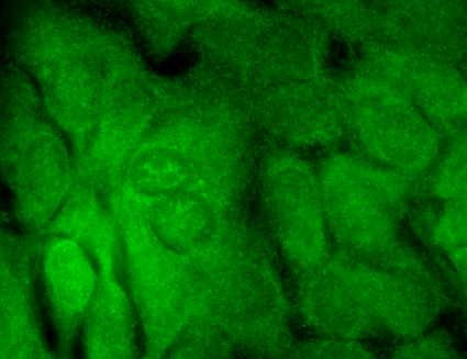
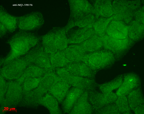
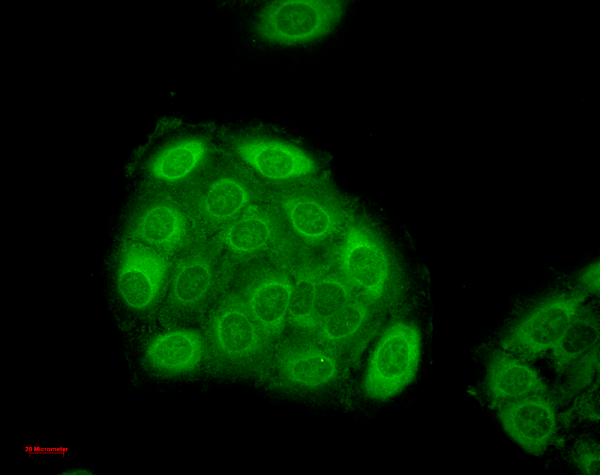
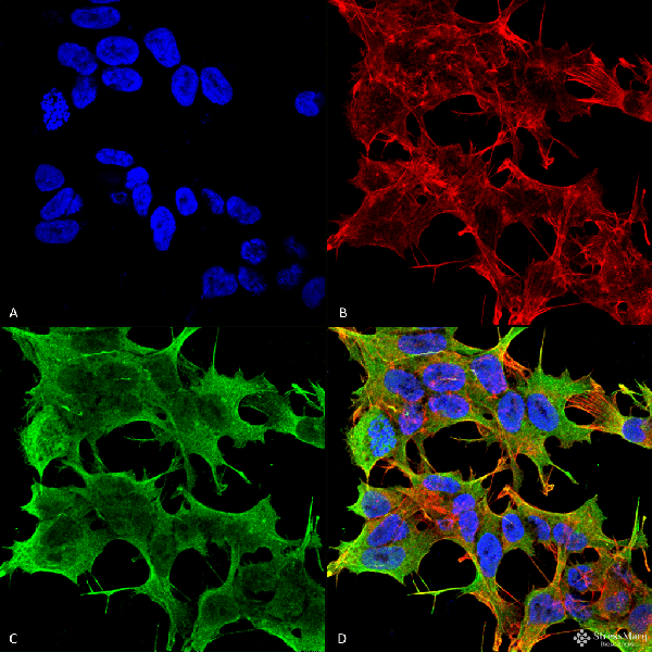
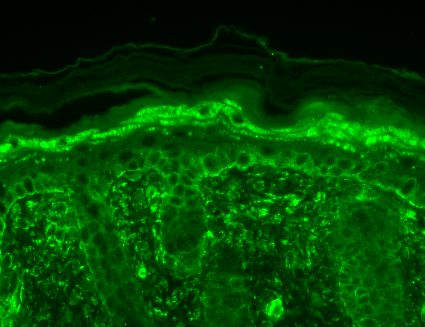
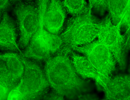
 ICC (Immunocytochemistry)
(Immunocytochemistry/Immunofluorescence analysis using Mouse Anti-HO-1 Monoclonal Antibody, Clone 1F12-A6. Tissue: HaCaT cells. Species: Human. Fixation: Cold 100% methanol for 10 minutes at -20 degree C. Primary Antibody: Mouse Anti-HO-1 Monoclonal Antibody at 1:100 for 1 hour at RT. Secondary Antibody: FITC Goat Anti-Mouse (green) at 1:50 for 1 hour at RT. Localization: Cell-cell border staining in epidermis, punctuate nuclear staining.)
ICC (Immunocytochemistry)
(Immunocytochemistry/Immunofluorescence analysis using Mouse Anti-HO-1 Monoclonal Antibody, Clone 1F12-A6. Tissue: HaCaT cells. Species: Human. Fixation: Cold 100% methanol for 10 minutes at -20 degree C. Primary Antibody: Mouse Anti-HO-1 Monoclonal Antibody at 1:100 for 1 hour at RT. Secondary Antibody: FITC Goat Anti-Mouse (green) at 1:50 for 1 hour at RT. Localization: Cell-cell border staining in epidermis, punctuate nuclear staining.)
HO-1, Monoclonal Antibody (Cat# AAA103268)
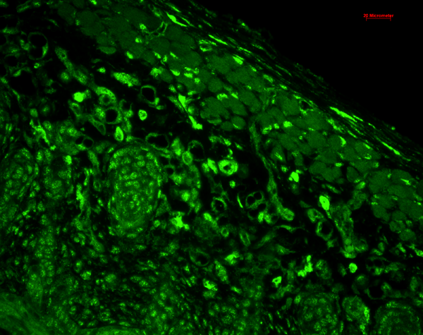
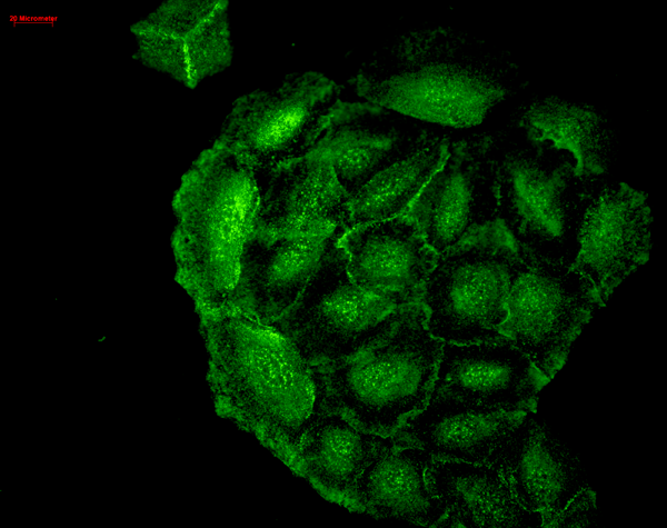
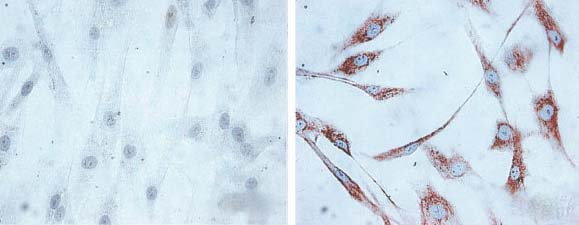 ICC (Immunocytochemistry)
(Immunocytochemistry/Immunofluorescence analysis using Mouse Anti-Hsp60 Monoclonal Antibody, Clone LK1,. Tissue: skin Fibroblasts. Species: Human. Fixation: Cold 100% methanol for 30 minutes at -20 degree C . Primary Antibody: Mouse Anti-Hsp60 Monoclonal Antibody at 1:1000 for 1 hour at RT. Secondary Antibody: DAKO LSAB2 streptavidin-peroxidase system. Counterstain: Mayer Hematoxylin (purple/blue) nuclear stain. Left: control; Right: 24 hours after 7th passage of senescence. Courtesy of: Valentina di Felice, University of Palermo, Italy.)
ICC (Immunocytochemistry)
(Immunocytochemistry/Immunofluorescence analysis using Mouse Anti-Hsp60 Monoclonal Antibody, Clone LK1,. Tissue: skin Fibroblasts. Species: Human. Fixation: Cold 100% methanol for 30 minutes at -20 degree C . Primary Antibody: Mouse Anti-Hsp60 Monoclonal Antibody at 1:1000 for 1 hour at RT. Secondary Antibody: DAKO LSAB2 streptavidin-peroxidase system. Counterstain: Mayer Hematoxylin (purple/blue) nuclear stain. Left: control; Right: 24 hours after 7th passage of senescence. Courtesy of: Valentina di Felice, University of Palermo, Italy.)
HSP60, Monoclonal Antibody (Cat# AAA103277)
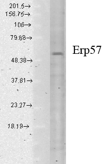
 ICC (Immunocytochemistry)
(Immunocytochemistry/Immunofluorescence analysis using Mouse Anti-Erp57 Monoclonal Antibody, Clone Map.ERP57. Tissue: HaCaT cells. Species: Human. Fixation: Cold 100% methanol for 10 minutes at -20 degree C. Primary Antibody: Mouse Anti-Erp57 Monoclonal Antibody at 1:100 for 1 hour at RT. Secondary Antibody: FITC Goat Anti-Mouse (green) at 1:50 for 1 hour at RT. Localization: Cytoplasmic and perinuclear staining.)
ICC (Immunocytochemistry)
(Immunocytochemistry/Immunofluorescence analysis using Mouse Anti-Erp57 Monoclonal Antibody, Clone Map.ERP57. Tissue: HaCaT cells. Species: Human. Fixation: Cold 100% methanol for 10 minutes at -20 degree C. Primary Antibody: Mouse Anti-Erp57 Monoclonal Antibody at 1:100 for 1 hour at RT. Secondary Antibody: FITC Goat Anti-Mouse (green) at 1:50 for 1 hour at RT. Localization: Cytoplasmic and perinuclear staining.)
Erp57 (Grp58), Monoclonal Antibody (Cat# AAA103283)

 WB (Western Blot)
(Western Blot analysis of Human Cell lysates showing detection of Hsp27 protein using Mouse Anti-Hsp27 Monoclonal Antibody, Clone 5D12-A3. Load: 15 ug. Block: 1.5% BSA for 30 minutes at RT. Primary Antibody: Mouse Anti-Hsp27 Monoclonal Antibody at 1:1000 for 2 hours at RT. Secondary Antibody: Sheep Anti-Mouse IgG: HRP for 1 hour at RT.)
WB (Western Blot)
(Western Blot analysis of Human Cell lysates showing detection of Hsp27 protein using Mouse Anti-Hsp27 Monoclonal Antibody, Clone 5D12-A3. Load: 15 ug. Block: 1.5% BSA for 30 minutes at RT. Primary Antibody: Mouse Anti-Hsp27 Monoclonal Antibody at 1:1000 for 2 hours at RT. Secondary Antibody: Sheep Anti-Mouse IgG: HRP for 1 hour at RT.)
Hsp27, Monoclonal Antibody (Cat# AAA103284)
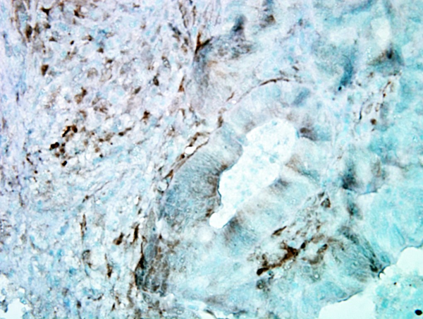
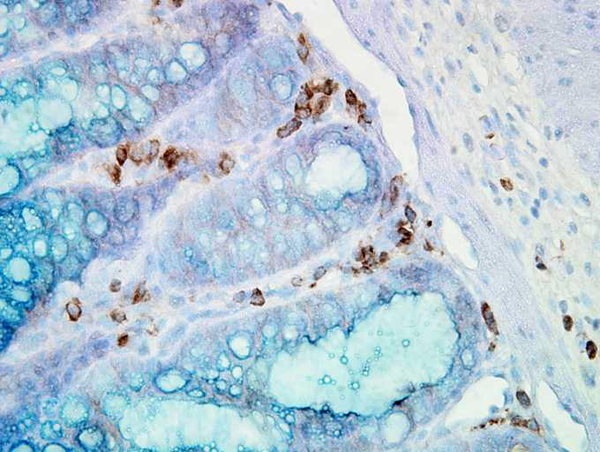
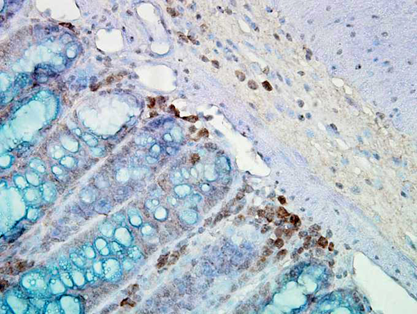 IHC (Immunohiostchemistry)
(Immunohistochemistry analysis using Mouse Anti-Hsp90 Monoclonal Antibody, Clone AC-16. Tissue: inflamed colon. Species: Mouse. Fixation: Formalin. Primary Antibody: Mouse Anti-Hsp90 Monoclonal Antibody at 1:2000 for 12 hours at 4 degree C. Secondary Antibody: Biotin Goat Anti-Mouse at 1:2000 for 1 hour at RT. Counterstain: Mayer Hematoxylin (purple/blue) nuclear stain at 200 ul for 2 minutes at RT. Localization: Inflammatory cells. Magnification: 40x. Mostly inflammatory cells, some mucosa.)
IHC (Immunohiostchemistry)
(Immunohistochemistry analysis using Mouse Anti-Hsp90 Monoclonal Antibody, Clone AC-16. Tissue: inflamed colon. Species: Mouse. Fixation: Formalin. Primary Antibody: Mouse Anti-Hsp90 Monoclonal Antibody at 1:2000 for 12 hours at 4 degree C. Secondary Antibody: Biotin Goat Anti-Mouse at 1:2000 for 1 hour at RT. Counterstain: Mayer Hematoxylin (purple/blue) nuclear stain at 200 ul for 2 minutes at RT. Localization: Inflammatory cells. Magnification: 40x. Mostly inflammatory cells, some mucosa.)
Hsp90, Monoclonal Antibody (Cat# AAA103311)
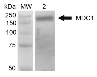
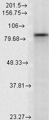
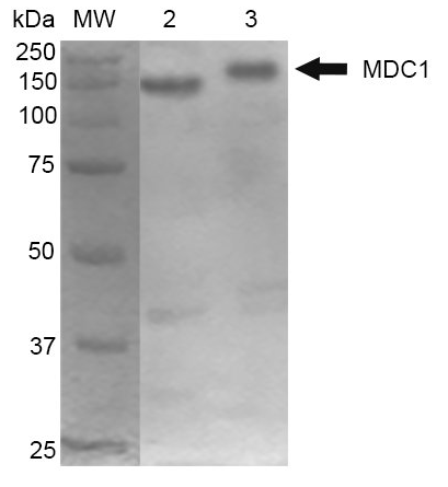 WB (Western Blot)
(Western Blot analysis of Mouse Cortex and Cerebellum showing detection of 184 kDa MDC1 protein using Mouse Anti-MDC1 Monoclonal Antibody, Clone P2B11. Lane 1: MW ladder. Lane 2: Mouse Cortex. Lane 3: Mouse Cerebellum. Load: 10 ug. Block: 5% Skim Milk in 1X TBST. Primary Antibody: Mouse Anti-MDC1 Monoclonal Antibody at 1:1000 for 2 hours RT. Secondary Antibody: Goat Anti-Mouse at 1:2000 for 60 min at RT. Color Development: ECL solution for 5 min in RT. Predicted/Observed Size: 184 kDa.)
WB (Western Blot)
(Western Blot analysis of Mouse Cortex and Cerebellum showing detection of 184 kDa MDC1 protein using Mouse Anti-MDC1 Monoclonal Antibody, Clone P2B11. Lane 1: MW ladder. Lane 2: Mouse Cortex. Lane 3: Mouse Cerebellum. Load: 10 ug. Block: 5% Skim Milk in 1X TBST. Primary Antibody: Mouse Anti-MDC1 Monoclonal Antibody at 1:1000 for 2 hours RT. Secondary Antibody: Goat Anti-Mouse at 1:2000 for 60 min at RT. Color Development: ECL solution for 5 min in RT. Predicted/Observed Size: 184 kDa.)
MDC1, Monoclonal Antibody (Cat# AAA103312)
 WB (Western Blot)
(Western Blot analysis of Rat brain membrane lysate showing detection of SHANK1 protein using Mouse Anti-SHANK1 Monoclonal Antibody, Clone S22-21. Load: 15 ug. Block: 1.5% BSA for 30 minutes at RT. Primary Antibody: Mouse Anti-SHANK1 Monoclonal Antibody at 1:1000 for 2 hours at RT. Secondary Antibody: Sheep Anti-Mouse IgG: HRP for 1 hour at RT.)
WB (Western Blot)
(Western Blot analysis of Rat brain membrane lysate showing detection of SHANK1 protein using Mouse Anti-SHANK1 Monoclonal Antibody, Clone S22-21. Load: 15 ug. Block: 1.5% BSA for 30 minutes at RT. Primary Antibody: Mouse Anti-SHANK1 Monoclonal Antibody at 1:1000 for 2 hours at RT. Secondary Antibody: Sheep Anti-Mouse IgG: HRP for 1 hour at RT.)
Shank1, Monoclonal Antibody (Cat# AAA103106)
 WB (Western Blot)
(Western Blot analysis of Rat Brain showing detection of ~92 kDa and ~82 kDa (pro and active) MMP9 protein using Mouse Anti-MMP9 Monoclonal Antibody, Clone S51-82 . Lane 1: Molecular Weight Ladder (MW). Lane 2: Rat Brain. Load: 15 ug. Block: 5% Skim Milk in 1X TBST. Primary Antibody: Mouse Anti-MMP9 Monoclonal Antibody at 1:1000 for 2 hours at RT. Secondary Antibody: Goat Anti-Mouse IgG: HRP at 1:2000 for 60 min at RT. Color Development: ECL solution for 5 min at RT. Predicted/Observed Size: ~92 kDa and ~82 kDa (pro and active).)
WB (Western Blot)
(Western Blot analysis of Rat Brain showing detection of ~92 kDa and ~82 kDa (pro and active) MMP9 protein using Mouse Anti-MMP9 Monoclonal Antibody, Clone S51-82 . Lane 1: Molecular Weight Ladder (MW). Lane 2: Rat Brain. Load: 15 ug. Block: 5% Skim Milk in 1X TBST. Primary Antibody: Mouse Anti-MMP9 Monoclonal Antibody at 1:1000 for 2 hours at RT. Secondary Antibody: Goat Anti-Mouse IgG: HRP at 1:2000 for 60 min at RT. Color Development: ECL solution for 5 min at RT. Predicted/Observed Size: ~92 kDa and ~82 kDa (pro and active).)
MMP9, Monoclonal Antibody (Cat# AAA103107)
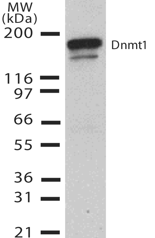
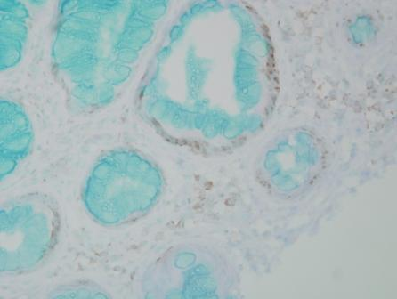
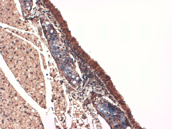
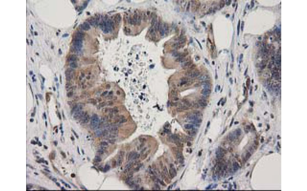
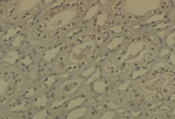 IHC (Immunohistochemisry)
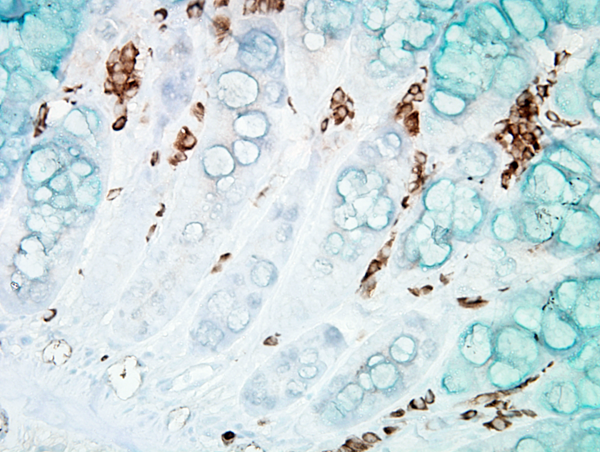
(Immunohistochemistry analysis using Mouse Anti-DNMT1 Monoclonal Antibody, Clone 60B1220.1. Tissue: medullar kidney tissue. Species: Mouse. Primary Antibody: Mouse Anti-DNMT1 Monoclonal Antibody at 1:1000. Secondary Antibody: HRP/DAB Detection System: Biotinylated Goat Anti-Mouse, Streptavidin Peroxidase, DAB Chromogen (brown). Counterstain: Mayer Hematoxylin (purple/blue) nuclear stain.)
IHC (Immunohistochemisry)
(Immunohistochemistry analysis using Mouse Anti-DNMT1 Monoclonal Antibody, Clone 60B1220.1. Tissue: medullar kidney tissue. Species: Mouse. Primary Antibody: Mouse Anti-DNMT1 Monoclonal Antibody at 1:1000. Secondary Antibody: HRP/DAB Detection System: Biotinylated Goat Anti-Mouse, Streptavidin Peroxidase, DAB Chromogen (brown). Counterstain: Mayer Hematoxylin (purple/blue) nuclear stain.)
DNMT1, Monoclonal Antibody (Cat# AAA103111)
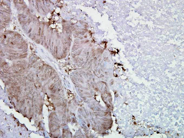
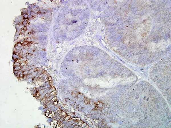
 IHC (Immunohistochemisry)
(Immunohistochemistry analysis using Mouse Anti-Hsp90 alpha Monoclonal Antibody, Clone K41009. Tissue: inflamed colon. Species: Mouse. Fixation: Formalin. Primary Antibody: Mouse Anti-Hsp90 alpha Monoclonal Antibody at 1:5000 for 12 hours at 4 degree C. Secondary Antibody: Biotin Goat Anti-Mouse at 1:2000 for 1 hour at RT. Counterstain: Mayer Hematoxylin (purple/blue) nuclear stain at 200 ul for 2 minutes at RT. Localization: Inflammatory cells. Magnification: 40x. Inflammatory cells.)
IHC (Immunohistochemisry)
(Immunohistochemistry analysis using Mouse Anti-Hsp90 alpha Monoclonal Antibody, Clone K41009. Tissue: inflamed colon. Species: Mouse. Fixation: Formalin. Primary Antibody: Mouse Anti-Hsp90 alpha Monoclonal Antibody at 1:5000 for 12 hours at 4 degree C. Secondary Antibody: Biotin Goat Anti-Mouse at 1:2000 for 1 hour at RT. Counterstain: Mayer Hematoxylin (purple/blue) nuclear stain at 200 ul for 2 minutes at RT. Localization: Inflammatory cells. Magnification: 40x. Inflammatory cells.)
Hsp90 alpha, Monoclonal Antibody (Cat# AAA103135)
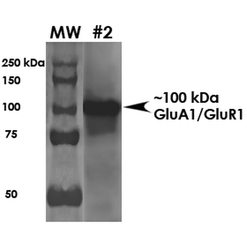 WB (Western Blot)
(Western Blot analysis of Rat Brain Membrane showing detection of ~100 kDa GluA1-GluR1 protein using Mouse Anti-GluA1-GluR1 Monoclonal Antibody, Clone S355-1 . Load: 10 ug. Block: 5% milk + TBST. Primary Antibody: Mouse Anti-GluA1-GluR1 Monoclonal Antibody at 1:2000 for 1 hour at RT. Secondary Antibody: Goat Anti-Mouse HRP at 1:200 for 1 hour at RT. Predicted/Observed Size: ~100 kDa.)
WB (Western Blot)
(Western Blot analysis of Rat Brain Membrane showing detection of ~100 kDa GluA1-GluR1 protein using Mouse Anti-GluA1-GluR1 Monoclonal Antibody, Clone S355-1 . Load: 10 ug. Block: 5% milk + TBST. Primary Antibody: Mouse Anti-GluA1-GluR1 Monoclonal Antibody at 1:2000 for 1 hour at RT. Secondary Antibody: Goat Anti-Mouse HRP at 1:200 for 1 hour at RT. Predicted/Observed Size: ~100 kDa.)
GluA1/GluR1 Glutamate Receptor, Monoclonal Antibody (Cat# AAA103152)
 WB (Western Blot)
(Western Blot analysis of Human Cervical cancer cell line (HeLa) lysate showing detection of FKBP51 protein using Mouse Anti-FKBP51 Monoclonal Antibody, Clone Hi51B. Load: 15 ug. Block: 1.5% BSA for 30 minutes at RT. Primary Antibody: Mouse Anti-FKBP51 Monoclonal Antibody at 1:1000 for 2 hours at RT. Secondary Antibody: Sheep Anti-Mouse IgG: HRP for 1 hour at RT.)
WB (Western Blot)
(Western Blot analysis of Human Cervical cancer cell line (HeLa) lysate showing detection of FKBP51 protein using Mouse Anti-FKBP51 Monoclonal Antibody, Clone Hi51B. Load: 15 ug. Block: 1.5% BSA for 30 minutes at RT. Primary Antibody: Mouse Anti-FKBP51 Monoclonal Antibody at 1:1000 for 2 hours at RT. Secondary Antibody: Sheep Anti-Mouse IgG: HRP for 1 hour at RT.)
FKBP51, Monoclonal Antibody (Cat# AAA103154)

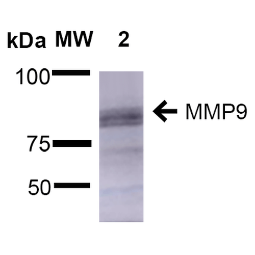
 WB (Western Blot)
(Western Blot analysis of Rat Brain showing detection of ~92 kDa and ~82 kDa (pro and active) MMP9 protein using Mouse Anti-MMP9 Monoclonal Antibody, Clone S51-82 . Lane 1: Molecular Weight Ladder (MW). Lane 2: Rat Brain. Load: 15 ug. Block: 5% Skim Milk in 1X TBST. Primary Antibody: Mouse Anti-MMP9 Monoclonal Antibody at 1:1000 for 2 hours at RT. Secondary Antibody: Goat Anti-Mouse IgG: HRP at 1:2000 for 60 min at RT. Color Development: ECL solution for 5 min at RT. Predicted/Observed Size: ~92 kDa and ~82 kDa (pro and active).)
WB (Western Blot)
(Western Blot analysis of Rat Brain showing detection of ~92 kDa and ~82 kDa (pro and active) MMP9 protein using Mouse Anti-MMP9 Monoclonal Antibody, Clone S51-82 . Lane 1: Molecular Weight Ladder (MW). Lane 2: Rat Brain. Load: 15 ug. Block: 5% Skim Milk in 1X TBST. Primary Antibody: Mouse Anti-MMP9 Monoclonal Antibody at 1:1000 for 2 hours at RT. Secondary Antibody: Goat Anti-Mouse IgG: HRP at 1:2000 for 60 min at RT. Color Development: ECL solution for 5 min at RT. Predicted/Observed Size: ~92 kDa and ~82 kDa (pro and active).)
MMP9, Monoclonal Antibody (Cat# AAA103480)
 WB (Western Blot)
(Western Blot analysis of Rat liver microsome lysate showing detection of LAMP1 protein using Mouse Anti-LAMP1 Monoclonal Antibody, Clone Ly1C6. Load: 15 ug. Block: 1.5% BSA for 30 minutes at RT. Primary Antibody: Mouse Anti-LAMP1 Monoclonal Antibody at 1:1000 for 2 hours at RT. Secondary Antibody: Sheep Anti-Mouse IgG: HRP for 1 hour at RT.)
WB (Western Blot)
(Western Blot analysis of Rat liver microsome lysate showing detection of LAMP1 protein using Mouse Anti-LAMP1 Monoclonal Antibody, Clone Ly1C6. Load: 15 ug. Block: 1.5% BSA for 30 minutes at RT. Primary Antibody: Mouse Anti-LAMP1 Monoclonal Antibody at 1:1000 for 2 hours at RT. Secondary Antibody: Sheep Anti-Mouse IgG: HRP for 1 hour at RT.)
LAMP1, Monoclonal Antibody (Cat# AAA103332)
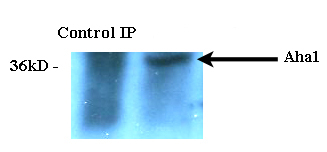
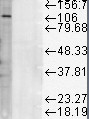
 WB (Western Blot)
(Western Blot analysis of Human Cell lysates showing detection of Aha1 protein using Rat Anti-Aha1 Monoclonal Antibody, Clone 25F2.D9. Load: 15 ug. Block: 1.5% BSA for 30 minutes at RT. Primary Antibody: Rat Anti-Aha1 Monoclonal Antibody at 1:1000 for 2 hours at RT. Secondary Antibody: Sheep Anti-Mouse IgG: HRP for 1 hour at RT.)
WB (Western Blot)
(Western Blot analysis of Human Cell lysates showing detection of Aha1 protein using Rat Anti-Aha1 Monoclonal Antibody, Clone 25F2.D9. Load: 15 ug. Block: 1.5% BSA for 30 minutes at RT. Primary Antibody: Rat Anti-Aha1 Monoclonal Antibody at 1:1000 for 2 hours at RT. Secondary Antibody: Sheep Anti-Mouse IgG: HRP for 1 hour at RT.)
Aha1, Monoclonal Antibody (Cat# AAA103358)
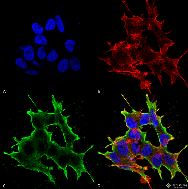
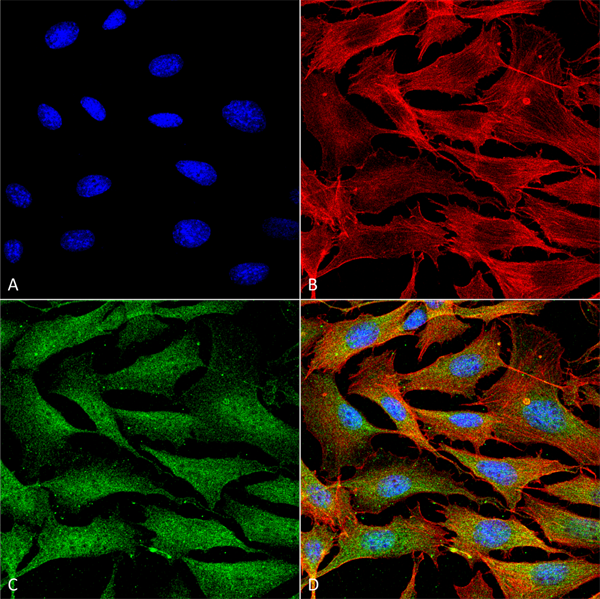
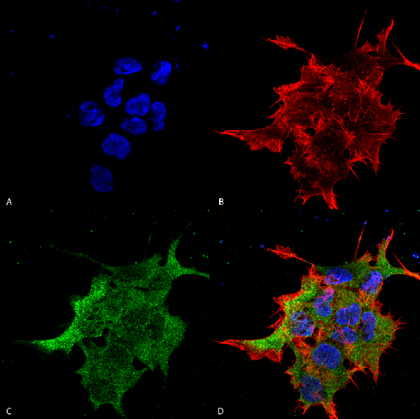
 ICC (Immunocytochemistry)
(Immunocytochemistry/Immunofluorescence analysis using Mouse Anti-TrpV3 Monoclonal Antibody, Clone S15-39. Tissue: HaCaT cells. Species: Human. Fixation: Cold 100% methanol for 10 minutes at -20 degree C. Primary Antibody: Mouse Anti-TrpV3 Monoclonal Antibody at 1:100 for 1 hour at RT. Secondary Antibody: FITC Goat Anti-Mouse (green) at 1:50 for 1 hour at RT. Localization: Dotty staining in all cells. Some intermediate filament-like staining in some cells.)
ICC (Immunocytochemistry)
(Immunocytochemistry/Immunofluorescence analysis using Mouse Anti-TrpV3 Monoclonal Antibody, Clone S15-39. Tissue: HaCaT cells. Species: Human. Fixation: Cold 100% methanol for 10 minutes at -20 degree C. Primary Antibody: Mouse Anti-TrpV3 Monoclonal Antibody at 1:100 for 1 hour at RT. Secondary Antibody: FITC Goat Anti-Mouse (green) at 1:50 for 1 hour at RT. Localization: Dotty staining in all cells. Some intermediate filament-like staining in some cells.)
TrpV3, Monoclonal Antibody (Cat# AAA103359)
 WB (Western Blot)
(Western Blot analysis of Human Cell lysates showing detection of Rhodopsin protein using Mouse Anti-Rhodopsin Monoclonal Antibody, Clone 1D4. Load: 15 ug. Block: 1.5% BSA for 30 minutes at RT. Primary Antibody: Mouse Anti-Rhodopsin Monoclonal Antibody at 1:1000 for 2 hours at RT. Secondary Antibody: Sheep Anti-Mouse IgG: HRP for 1 hour at RT.)
WB (Western Blot)
(Western Blot analysis of Human Cell lysates showing detection of Rhodopsin protein using Mouse Anti-Rhodopsin Monoclonal Antibody, Clone 1D4. Load: 15 ug. Block: 1.5% BSA for 30 minutes at RT. Primary Antibody: Mouse Anti-Rhodopsin Monoclonal Antibody at 1:1000 for 2 hours at RT. Secondary Antibody: Sheep Anti-Mouse IgG: HRP for 1 hour at RT.)
Rhodopsin, Monoclonal Antibody (Cat# AAA103368)
 WB (Western Blot)
(Western Blot analysis of Rat tissue lysate showing detection of KDEL Receptor protein using Mouse Anti-KDEL Receptor Monoclonal Antibody, Clone KR-10. Load: 15 ug. Block: 1.5% BSA for 30 minutes at RT. Primary Antibody: Mouse Anti-KDEL Receptor Monoclonal Antibody at 1:1000 for 2 hours at RT. Secondary Antibody: Sheep Anti-Mouse IgG: HRP for 1 hour at RT.)
WB (Western Blot)
(Western Blot analysis of Rat tissue lysate showing detection of KDEL Receptor protein using Mouse Anti-KDEL Receptor Monoclonal Antibody, Clone KR-10. Load: 15 ug. Block: 1.5% BSA for 30 minutes at RT. Primary Antibody: Mouse Anti-KDEL Receptor Monoclonal Antibody at 1:1000 for 2 hours at RT. Secondary Antibody: Sheep Anti-Mouse IgG: HRP for 1 hour at RT.)
KDEL, Monoclonal Antibody (Cat# AAA103421)
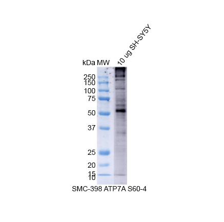 WB (Western Blot)
(Western Blot analysis of Human SH-SY5Y showing detection of Copper Transporting ATPase 1 protein using Mouse Anti-Copper Transporting ATPase 1 Monoclonal Antibody, Clone S60-4 . Lane 1: MW Ladder. Lane 2: 10 ug SH-SY5Y. Load: 10 ug. Block: 5% Skim Milk powder in TBST. Primary Antibody: Mouse Anti-Copper Transporting ATPase 1 Monoclonal Antibody at 1:500 for 2 hours at RT with shaking. Secondary Antibody: Goat anti-mouse IgG:HRP at 1:4000 for 1 hour at RT with shaking. Color Development: Chemiluminescent for HRP (Moss) for 5 min in RT.)
WB (Western Blot)
(Western Blot analysis of Human SH-SY5Y showing detection of Copper Transporting ATPase 1 protein using Mouse Anti-Copper Transporting ATPase 1 Monoclonal Antibody, Clone S60-4 . Lane 1: MW Ladder. Lane 2: 10 ug SH-SY5Y. Load: 10 ug. Block: 5% Skim Milk powder in TBST. Primary Antibody: Mouse Anti-Copper Transporting ATPase 1 Monoclonal Antibody at 1:500 for 2 hours at RT with shaking. Secondary Antibody: Goat anti-mouse IgG:HRP at 1:4000 for 1 hour at RT with shaking. Color Development: Chemiluminescent for HRP (Moss) for 5 min in RT.)
Copper-Transporting ATPase1, Monoclonal Antibody (Cat# AAA103423)
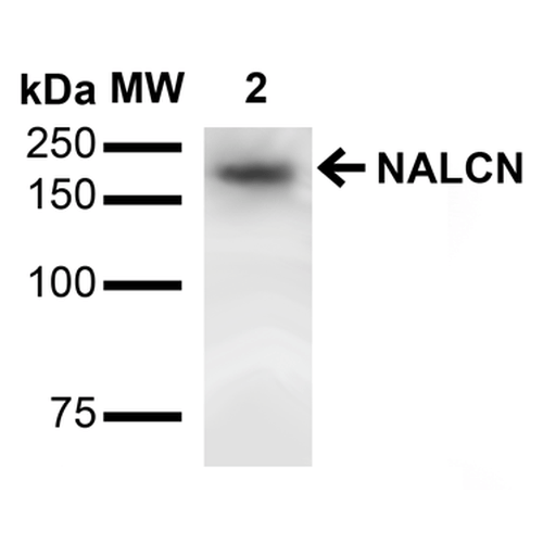
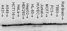
 WB (Western Blot)
(Western Blot analysis of Rat Brain showing detection of ~200 kDa NALCN protein using Mouse Anti-NALCN Monoclonal Antibody, Clone S187-7 . Lane 1: Molecular Weight (MW) Ladder. Lane 3: Rat Brain Membrane. Load: 15 ug. Block: 2% BSA and 2% Skim Milk in 1X TBST. Primary Antibody: Mouse Anti-NALCN Monoclonal Antibody at 1:1000 for 16 hours at 4 degree C. Secondary Antibody: Goat Anti-Mouse IgG: HRP at 1:2000 for 60 min at RT. Color Development: ECL solution for 6 min at RT. Predicted/Observed Size: ~200 kDa.)
WB (Western Blot)
(Western Blot analysis of Rat Brain showing detection of ~200 kDa NALCN protein using Mouse Anti-NALCN Monoclonal Antibody, Clone S187-7 . Lane 1: Molecular Weight (MW) Ladder. Lane 3: Rat Brain Membrane. Load: 15 ug. Block: 2% BSA and 2% Skim Milk in 1X TBST. Primary Antibody: Mouse Anti-NALCN Monoclonal Antibody at 1:1000 for 16 hours at 4 degree C. Secondary Antibody: Goat Anti-Mouse IgG: HRP at 1:2000 for 60 min at RT. Color Development: ECL solution for 6 min at RT. Predicted/Observed Size: ~200 kDa.)
NALCN, Monoclonal Antibody (Cat# AAA103460)
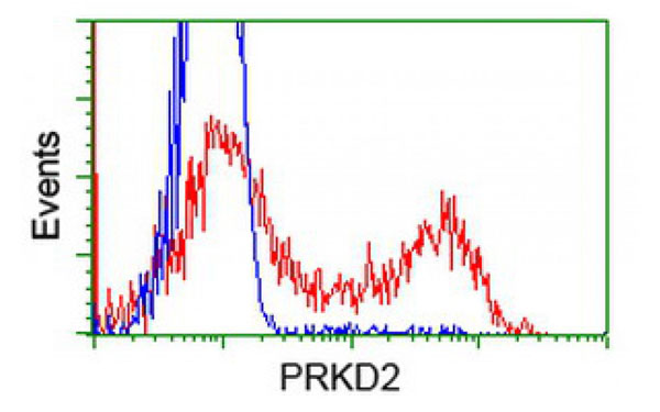
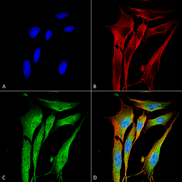
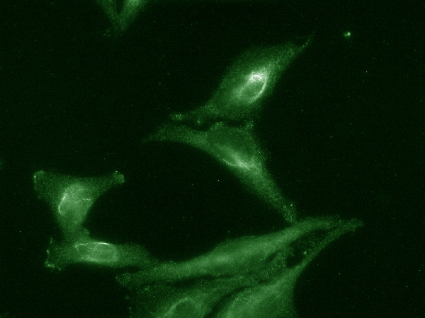
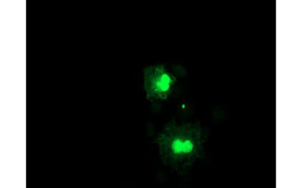
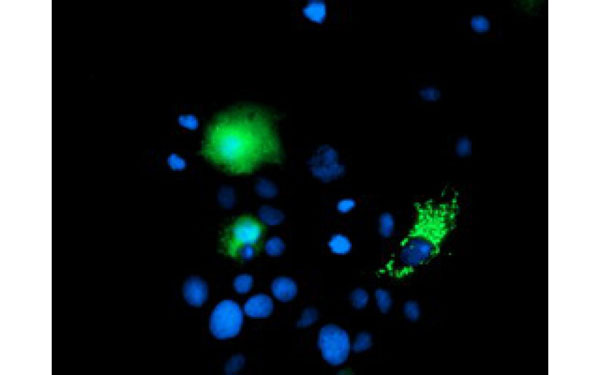
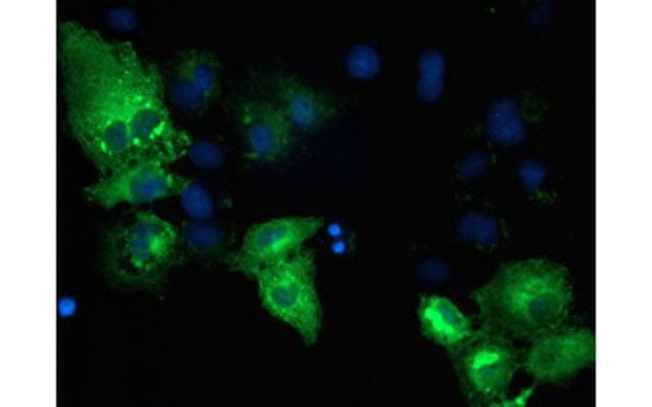
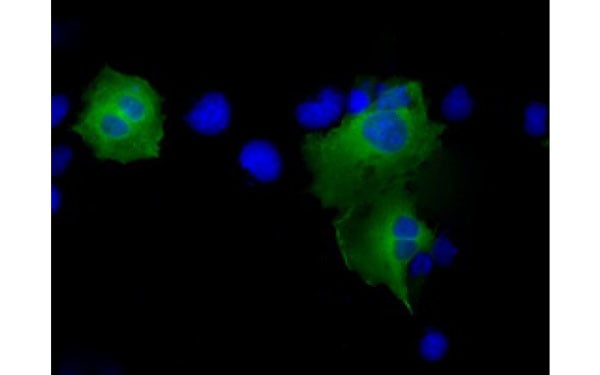
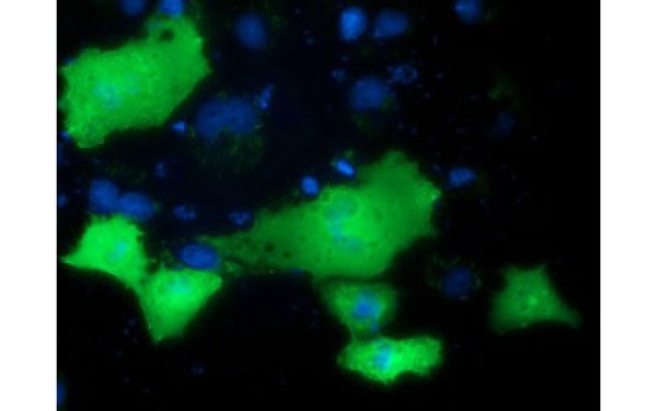 IF (Immunofluorescence)
(Immunofluorescent staining of COS7 cells transiently transfected with recombinant PRKD2 protein using PRKD2 antibody)
IF (Immunofluorescence)
(Immunofluorescent staining of COS7 cells transiently transfected with recombinant PRKD2 protein using PRKD2 antibody)
PRKD2, Monoclonal Antibody (Cat# AAA108174)
MDMA, Monoclonal Antibody (Cat# AAA108179)
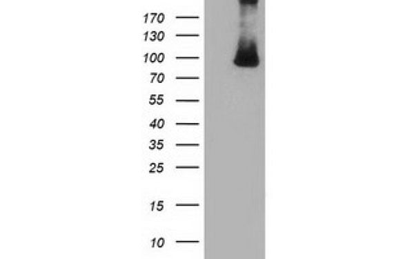
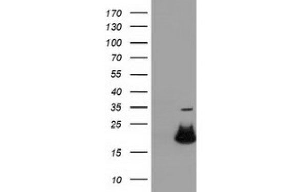
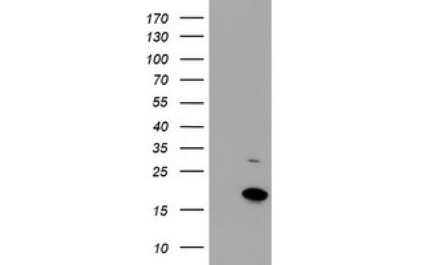
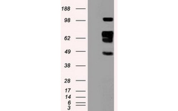
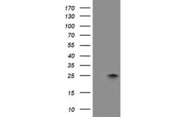 WB (Western Blot)
(Western Blot analysis of HEK293T cell lysates (5 ug) transfected with either recombinant FXN protein (Right) or empty vector (Left) detected with FXN antibody)
WB (Western Blot)
(Western Blot analysis of HEK293T cell lysates (5 ug) transfected with either recombinant FXN protein (Right) or empty vector (Left) detected with FXN antibody)
FXN, Monoclonal Antibody (Cat# AAA108187)
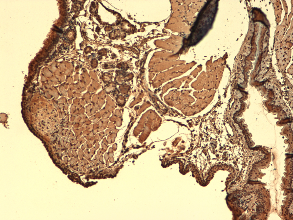
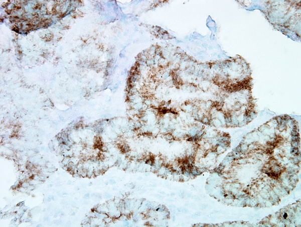
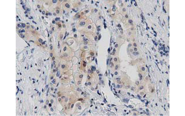
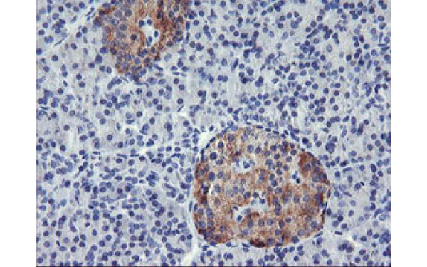
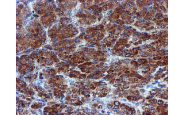
 IHC (Immunohiostchemistry)
(Immunohistochemical analysis of AGPAT5 protein in paraffin embedded Human pancreas tissue using AGPAT5 antibody)
IHC (Immunohiostchemistry)
(Immunohistochemical analysis of AGPAT5 protein in paraffin embedded Human pancreas tissue using AGPAT5 antibody)
AGPAT5, Monoclonal Antibody (Cat# AAA108188)
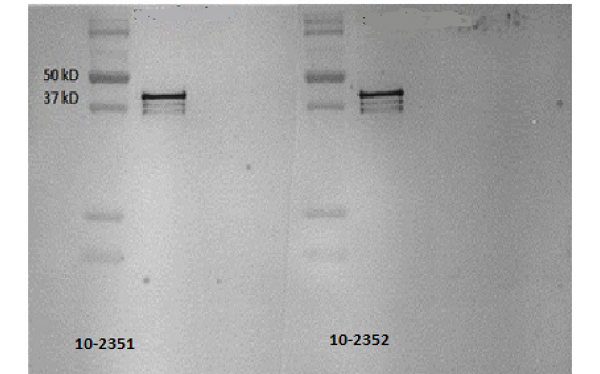
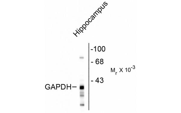 WB (Western Blot)
(Western blot of rat hippocampal lysate showing the immunolabeling of ~38k GAPDH protein)
WB (Western Blot)
(Western blot of rat hippocampal lysate showing the immunolabeling of ~38k GAPDH protein)
GAPDHS, Monoclonal Antibody (Cat# AAA108189)

 WB (Western Blot)
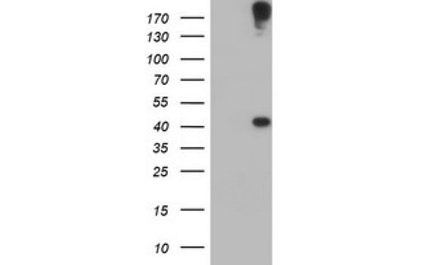
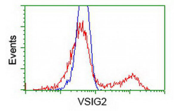
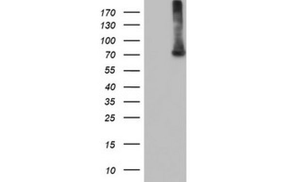
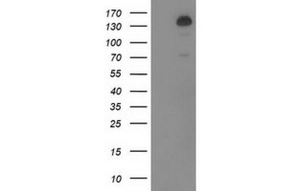
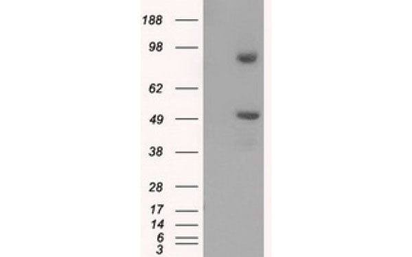
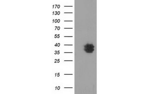
(Western Blot analysis of HEK293T cell lysates (5 ug) transfected with either recombinant VSIG2 protein (Right) or empty vector (Left) detected with VSIG2 antibody)
WB (Western Blot)
(Western Blot analysis of HEK293T cell lysates (5 ug) transfected with either recombinant VSIG2 protein (Right) or empty vector (Left) detected with VSIG2 antibody)
VSIG2, Monoclonal Antibody (Cat# AAA108190)
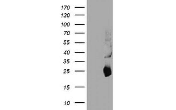
 WB (Western Blot)
(Western Blot analysis of HEK293T cell lysates (5 ug) transfected with either recombinant VSIG2 protein (Right) or empty vector (Left) detected with VSIG2 antibody)
WB (Western Blot)
(Western Blot analysis of HEK293T cell lysates (5 ug) transfected with either recombinant VSIG2 protein (Right) or empty vector (Left) detected with VSIG2 antibody)
VSIG2, Monoclonal Antibody (Cat# AAA108191)
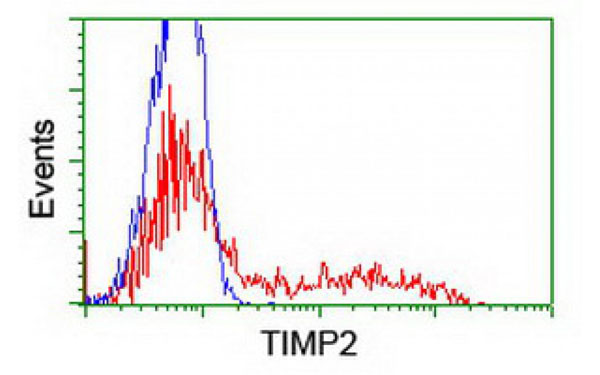
 IHC (Immunohistochemisry)
(Immunohistochemical analysis of TIMP2 protein in paraffin embedded Human pancreas tissue using TIMP2 antibody)
IHC (Immunohistochemisry)
(Immunohistochemical analysis of TIMP2 protein in paraffin embedded Human pancreas tissue using TIMP2 antibody)
TIMP2, Monoclonal Antibody (Cat# AAA108199)
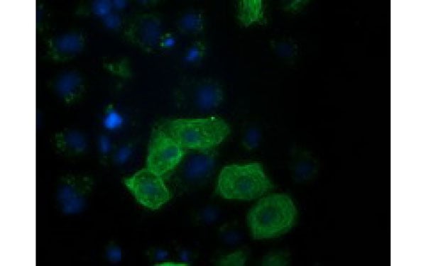
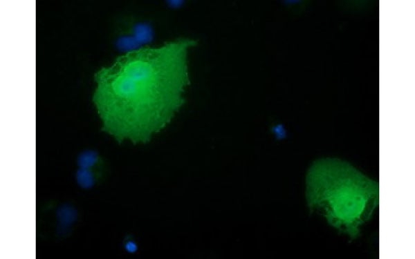 IF (Immunofluorescence)
(Immunofluorescent staining of COS7 cells transiently transfected with recombinant RBBP9 protein using RBBP9 antibody)
IF (Immunofluorescence)
(Immunofluorescent staining of COS7 cells transiently transfected with recombinant RBBP9 protein using RBBP9 antibody)
RBBP9, Monoclonal Antibody (Cat# AAA108202)
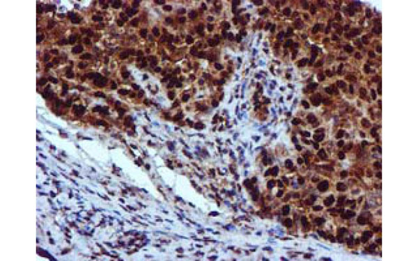 IHC (Immunohistochemisry)
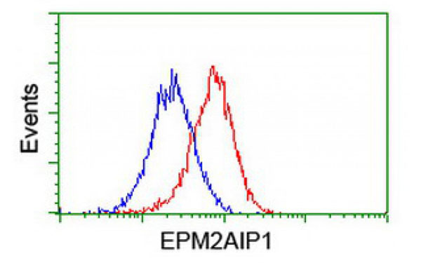
(Immunohistochemical analysis of EPM2AIP1 protein in paraffin embedded Adenocarcinoma of Human ovary tissue using EPM2AIP1 antibody)
IHC (Immunohistochemisry)
(Immunohistochemical analysis of EPM2AIP1 protein in paraffin embedded Adenocarcinoma of Human ovary tissue using EPM2AIP1 antibody)
EPM2AIP1, Monoclonal Antibody (Cat# AAA108206)
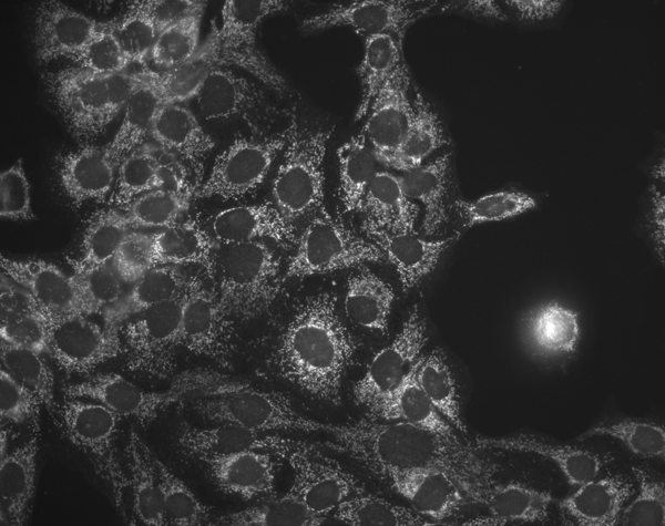
 IF (Immunofluorescence)
(Immunofluorescent staining of COS7 cells transiently transfected with recombinant CHCHD5 protein using CHCHD5 antibody)
IF (Immunofluorescence)
(Immunofluorescent staining of COS7 cells transiently transfected with recombinant CHCHD5 protein using CHCHD5 antibody)
CHCHD5, Monoclonal Antibody (Cat# AAA108208)
 WB (Western Blot)
(Western Blot analysis of HEK293T cell lysates (5 ug) transfected with either recombinant RASSF1 protein (Right) or empty vector (Left) detected with RASSF1 antibody)
WB (Western Blot)
(Western Blot analysis of HEK293T cell lysates (5 ug) transfected with either recombinant RASSF1 protein (Right) or empty vector (Left) detected with RASSF1 antibody)
RASSF1, Monoclonal Antibody (Cat# AAA108209)
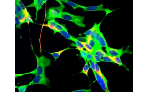
 IHC (Immunohistochemisry)
(Neuropeptide Y antibody (1:1000) staining of an enlarged section of a paraffin embedded mouse brain tissue (fibres in the hilus of the hippocampus) from a mouse suffering an epileptic attack just before perfusion.)
IHC (Immunohistochemisry)
(Neuropeptide Y antibody (1:1000) staining of an enlarged section of a paraffin embedded mouse brain tissue (fibres in the hilus of the hippocampus) from a mouse suffering an epileptic attack just before perfusion.)
Neuropeptide Y, Monoclonal Antibody (Cat# AAA108217)
 IF (Immunofluorescence)
(Immunofluorescent staining of COS7 cells transiently transfected with recombinant MAPRE2 protein using MAPRE2 antibody)
IF (Immunofluorescence)
(Immunofluorescent staining of COS7 cells transiently transfected with recombinant MAPRE2 protein using MAPRE2 antibody)
MAPRE2, Monoclonal Antibody (Cat# AAA108225)
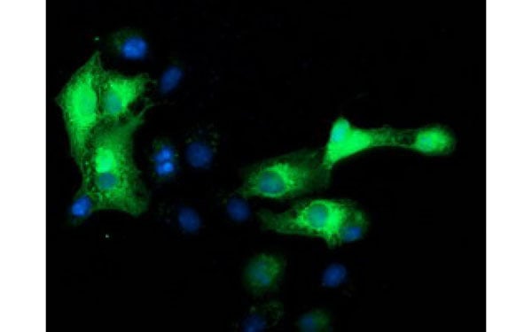
 IF (Immunofluorescence)
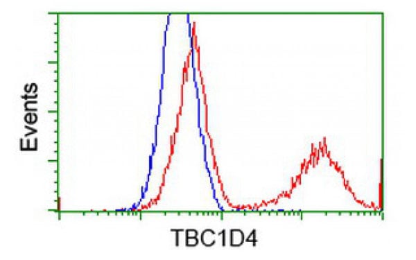
(Immunofluorescent staining of COS7 cells transiently transfected with recombinant TBC1D4 protein using TBC1D4 antibody)
IF (Immunofluorescence)
(Immunofluorescent staining of COS7 cells transiently transfected with recombinant TBC1D4 protein using TBC1D4 antibody)
TBC1D4, Monoclonal Antibody (Cat# AAA108226)
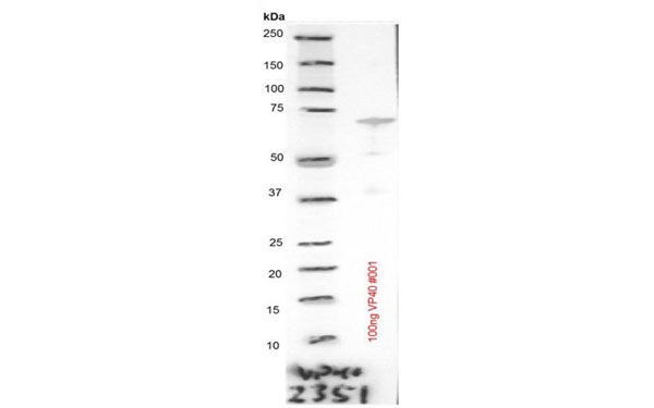
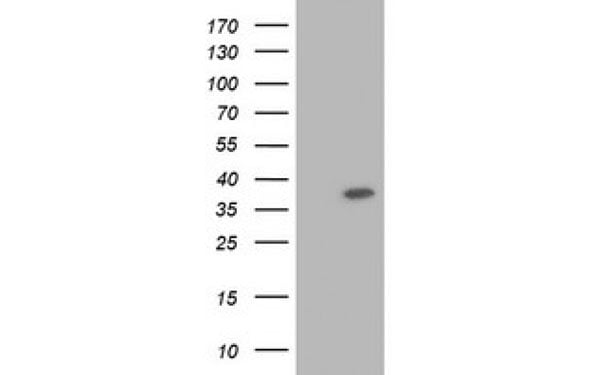
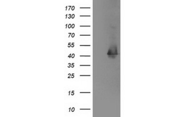
 WB (Western Blot)
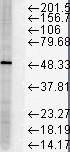
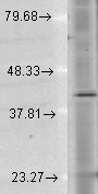
(Antibodies diluted 1:100 detect distinct band at 40 kDa, and 2 weak bands around the 37 kDa marker)
WB (Western Blot)
(Antibodies diluted 1:100 detect distinct band at 40 kDa, and 2 weak bands around the 37 kDa marker)
Ebola Virus VP40, Monoclonal Antibody (Cat# AAA108229)
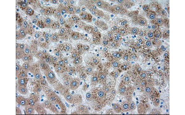
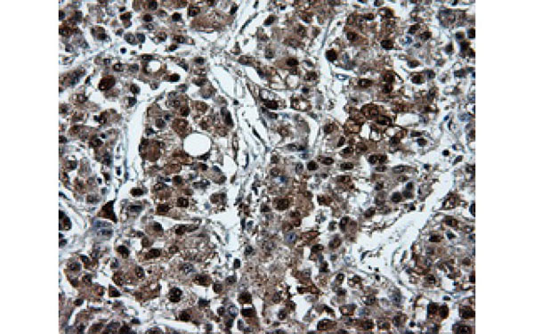 IHC (Immunohistochemisry)
(Immunohistochemical analysis of PSMC3 protein in paraffin embedded Carcinoma of Human liver tissue using PSMC3 antibody)
IHC (Immunohistochemisry)
(Immunohistochemical analysis of PSMC3 protein in paraffin embedded Carcinoma of Human liver tissue using PSMC3 antibody)
PSMC3, Monoclonal Antibody (Cat# AAA108230)
 WB (Western Blot)
(Western Blot analysis of HEK293T cell lysates (5 ug) transfected with either recombinant SIGLEC9 protein (Right) or empty vector (Left) detected with SIGLEC9 antibody)
WB (Western Blot)
(Western Blot analysis of HEK293T cell lysates (5 ug) transfected with either recombinant SIGLEC9 protein (Right) or empty vector (Left) detected with SIGLEC9 antibody)
SIGLEC9, Monoclonal Antibody (Cat# AAA108240)
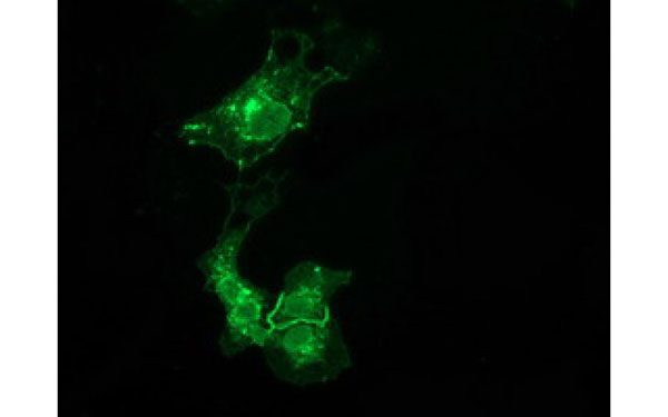 IF (Immunofluorescence)
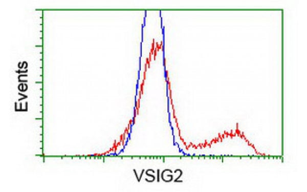
(Immunofluorescent staining of COS7 cells transiently transfected with recombinant VSIG2 protein using VSIG2 antibody)
IF (Immunofluorescence)
(Immunofluorescent staining of COS7 cells transiently transfected with recombinant VSIG2 protein using VSIG2 antibody)
VSIG2, Monoclonal Antibody (Cat# AAA108242)
What are Monoclonal Antibodies?
Monoclonal antibodies are specialized laboratory-produced proteins developed for binding to specific biological antigens or other molecular targets. Since they come from a single cell (or clone), they are especially consistent and accurate in the data they are involved in producing.
This type of antibody material has been shown to be a powerful tool in finding and subsequently destroying harmful cells in an organism, such as those found in cancers or various autoimmune diseases. This makes them excellent aids in medical testing and research, which is why they are so widely used.
AAA Biotech offers a comprehensive range of high-quality monoclonal antibodies that perform effectively in various laboratory tests, including (amongst others) ELISA, western blotting, immunohistochemistry, and flow cytometry. All of the products in our catalog are thoroughly quality tested to make sure that they are reliable and will consistently perform well in your research.
What Are The Uses of Monoclonal Antibodies
Monoclonal antibodies are used in many lab tests, including (amongst others) ELISA, western blotting, immunohistochemistry, and flow cytometry.
ELISA is a test that helps detect a specific substance/analyte in a sample. It uses antibodies (often monoclonal) bound to a solid surface (such as the well of a microplate) to “capture” the substance/analyte in the sample and immobilize it so that the detection antibody component can then bind to it and produce a signal, which can then be measured.
Western blotting identifies specific proteins in a sample. The sample is first separated on a gel, and then antibodies are applied that will typically bind to the target, which will all be localized to a single band in a lane.
Immunohistochemistry helps locate specific proteins in cells or tissue samples using antibodies.
Flow cytometry looks at and sorts cells. It uses antibodies that are conjugated to reporter molecules called “fluorophores”, which, under special lights, emit light themselves, which can then be measured by a detector instrument. For a deeper understanding of these techniques, explore our complete guide to monoclonal antibodies and their benefits.
How Monoclonal Antibodies Are Used as Medicine?
Please note that all of the products listed in AAA Biotech’s also known as AAA Bio or AAABio catalog are strictly for research-use only (RUO).
Monoclonal antibodies can also be used as therapeutic/medical treatments, particularly in the context of cancers. They are designed to find and bind to specific cells or proteins, helping the immune system recognize and attack the cancer. These treatments work in different ways, such as:
- Radioimmunotherapy attaches a small amount of radioactive molecule to the antibody, so it delivers the radiation directly to the cancer cells that the antibody is specifically binding to.
- Antibody-directed enzyme prodrug therapy uses antibodies that are specifically bound to special enzymes. These enzymes activate a harmless drug in the body and turn it into a cancer-killing drug only near the cancer cells—this helps avoid harming healthy cells.
- Immunoliposomes are tiny “bubbles” filled with medicine/drug and coated with antibodies. They carry the drug straight to the cancer cells.
Why Buy Monoclonal Antibodies From Us?
At AAA Biotech, we provide high-performance monoclonal antibodies designed to support a wide range of research needs.
1. Validated for Versatile Applications
The antibodies in our catalog are extensively validated and compatible with multiple techniques, including (but not limited to) ELISA, flow cytometry (FC), immunocytochemistry (ICC), immunofluorescence (IF), immunohistochemistry (IHC), immunoprecipitation (IP), and western blotting (WB).
2. Wide Selection & Specialized Options
We offer antibodies for common and rare species, that are available in various conjugated forms, and also in recombinant formats. Essentially, there is almost anything one might need to meet their experimental model’s requirements.
3. High-Quality Proteins
Our proteins meet high purity standards—90% or more as confirmed by SDS-PAGE. Many are available with tags like His, Flag, GST, or MBP, and we also supply native and biologically active proteins for functional studies.
Frequently Asked Questions
1. Are your monoclonal antibodies validated for specific applications?
Yes, our antibodies are tested and validated for use in methods such as ELISA, western blot, IHC, flow cytometry, and more. Refer to specific product pages or datasheets for individual product information.
2. How do I choose the right monoclonal antibody for my application?
Review the product details directly for application validation, species reactivity, and target information. You may also contact our support team at any time for help.
3. How quickly can I receive my order?
Most orders are processed and shipped within 1–3 business days, depending on product availability and your shipping location.