Filters
▼Clonality
▼Type
▼Reactivity
▼Gene Name
▼Isotype
▼Host
▼Application
▼Clone
▼Monoclonal Antibodies
Get accurate results in your research with our Monoclonal Antibodies, which are specially made to target exactly what you require for your research, and will produce consistent, reliable performance in lab tests.
Viewing 3850-3900 of 27645 product results
 IHC (Immunohiostchemistry)
(Immunohistochemical analysis of COASY protein in paraffin embedded Carcinoma of Human bladder tissue using COASY antibody)
IHC (Immunohiostchemistry)
(Immunohistochemical analysis of COASY protein in paraffin embedded Carcinoma of Human bladder tissue using COASY antibody)
COASY, Monoclonal Antibody (Cat# AAA107988)
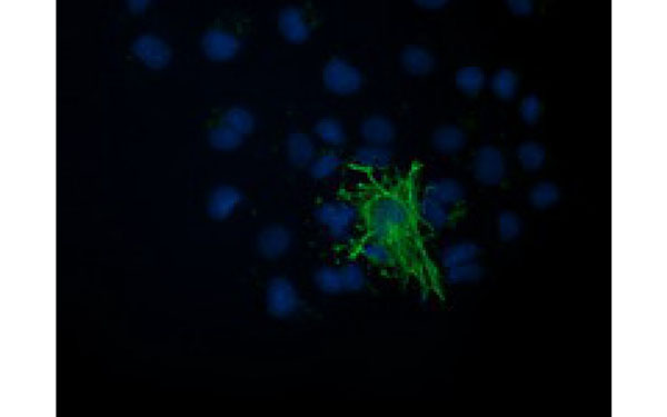
 IF (Immunofluorescence)
(Immunofluorescent staining of COS7 cells transiently transfected with recombinant MAPRE2 protein using MAPRE2 antibody)
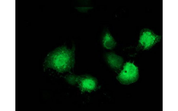
IF (Immunofluorescence)
(Immunofluorescent staining of COS7 cells transiently transfected with recombinant MAPRE2 protein using MAPRE2 antibody)
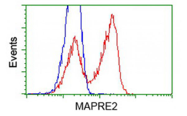
MAPRE2, Monoclonal Antibody (Cat# AAA108013)
 IF (Immunofluorescence)
(Immunofluorescent staining of COS7 cells transiently transfected with recombinant VASP protein using VASP antibody)
IF (Immunofluorescence)
(Immunofluorescent staining of COS7 cells transiently transfected with recombinant VASP protein using VASP antibody)
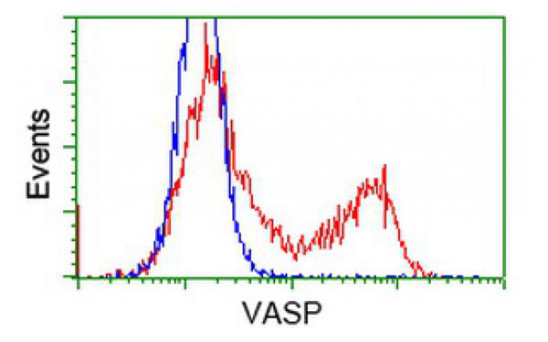
VASP, Monoclonal Antibody (Cat# AAA108015)
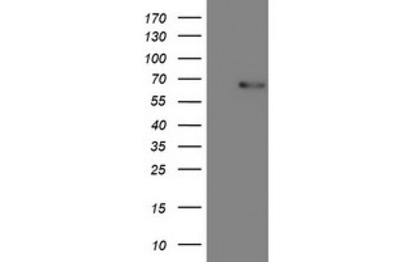
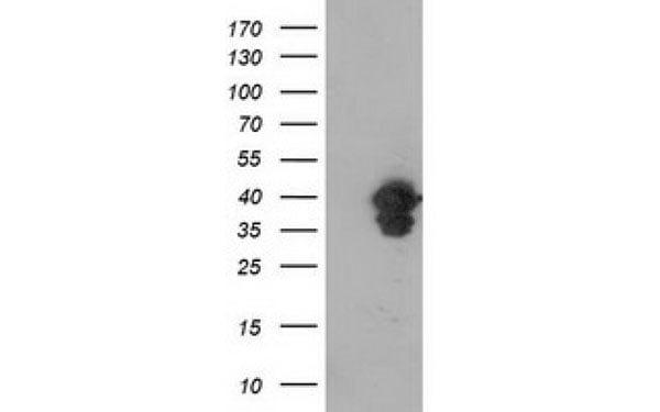
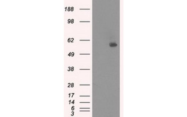
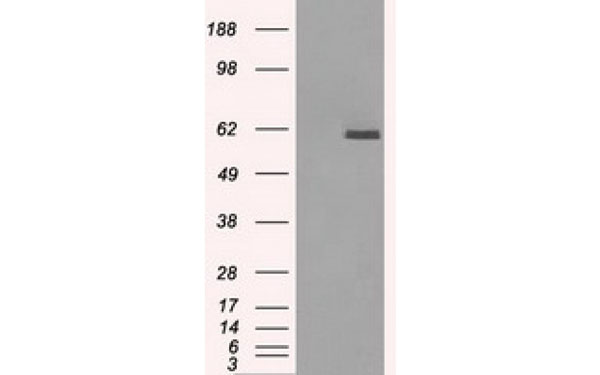
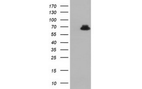
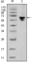
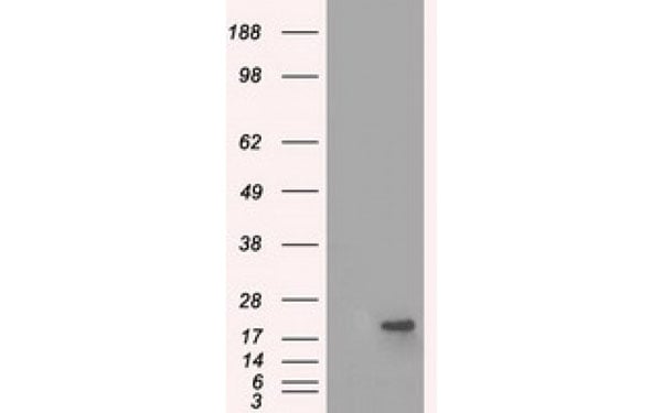
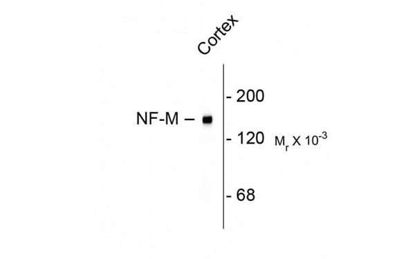 WB (Western Blot)
(Western blot of rat of the ~ 145k NEFM protein cortex lysate showing specific immunolabeling of ~ 68k NEFM protein)
WB (Western Blot)
(Western blot of rat of the ~ 145k NEFM protein cortex lysate showing specific immunolabeling of ~ 68k NEFM protein)
NEFM, Monoclonal Antibody (Cat# AAA108016)
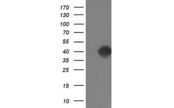 WB (Western Blot)
(Western Blot analysis of HEK293T cell lysates (5 ug) transfected with either recombinant TOMM34 protein (Right) or empty vector (Left) detected with TOMM34 antibody)
WB (Western Blot)
(Western Blot analysis of HEK293T cell lysates (5 ug) transfected with either recombinant TOMM34 protein (Right) or empty vector (Left) detected with TOMM34 antibody)
TOMM34, Monoclonal Antibody (Cat# AAA108022)
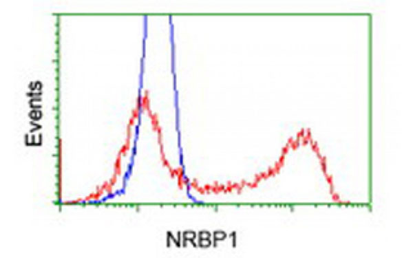
 IHC (Immunohistochemisry)
(Immunohistochemical analysis of NRBP1 protein in paraffin embedded Human ovary tissue using NRBP1 antibody)
IHC (Immunohistochemisry)
(Immunohistochemical analysis of NRBP1 protein in paraffin embedded Human ovary tissue using NRBP1 antibody)
NRBP1, Monoclonal Antibody (Cat# AAA108029)
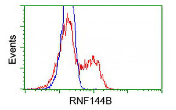
 IF (Immunofluorescence)
(Immunofluorescent staining of COS7 cells transiently transfected with recombinant RNF144B protein using RNF144B antibody)
IF (Immunofluorescence)
(Immunofluorescent staining of COS7 cells transiently transfected with recombinant RNF144B protein using RNF144B antibody)
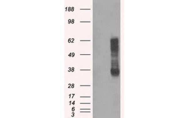
RNF144B, Monoclonal Antibody (Cat# AAA108030)
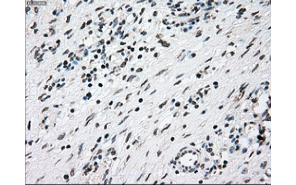
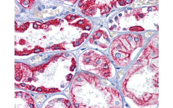
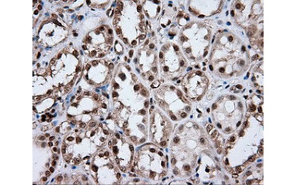
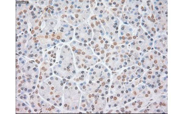
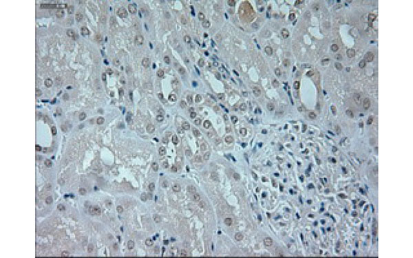 IHC (Immunohistochemisry)
(Immunohistochemical analysis of NEUROG1 protein in paraffin embedded Human Kidney tissue using NEUROG1 antibody)
IHC (Immunohistochemisry)
(Immunohistochemical analysis of NEUROG1 protein in paraffin embedded Human Kidney tissue using NEUROG1 antibody)
NEUROG1, Monoclonal Antibody (Cat# AAA108036)
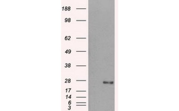
PICP, Monoclonal Antibody (Cat# AAA108039)
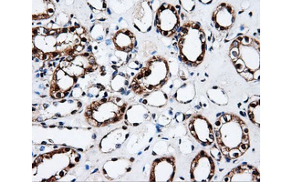
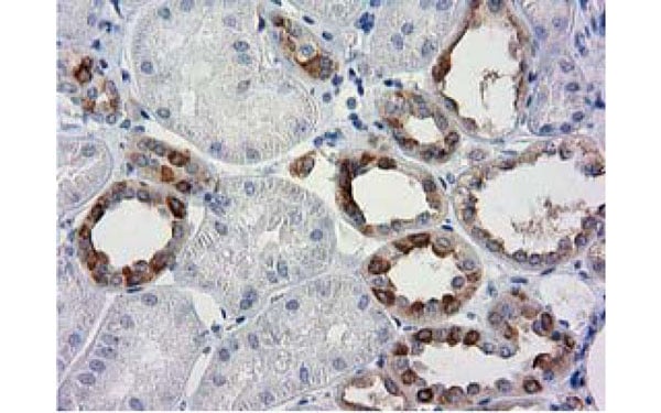
 IHC (Immunohistochemisry)
(Immunohistochemical analysis of paraffin-embedded human Kidney tissues using CK8 antibody)
IHC (Immunohistochemisry)
(Immunohistochemical analysis of paraffin-embedded human Kidney tissues using CK8 antibody)
Keratin K8, Monoclonal Antibody (Cat# AAA108041)
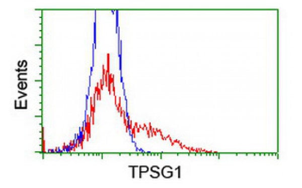
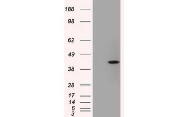
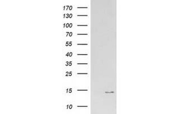
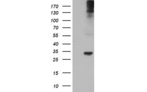 WB (Western Blot)
(Western Blot analysis of HEK293T cell lysates (5 ug) transfected with either recombinant TPSG1 protein (Right) or empty vector (Left) detected with TPSG1 antibody)
WB (Western Blot)
(Western Blot analysis of HEK293T cell lysates (5 ug) transfected with either recombinant TPSG1 protein (Right) or empty vector (Left) detected with TPSG1 antibody)
TPSG1, Monoclonal Antibody (Cat# AAA107469)
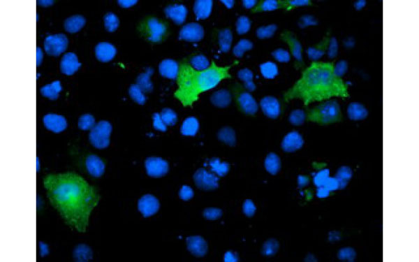
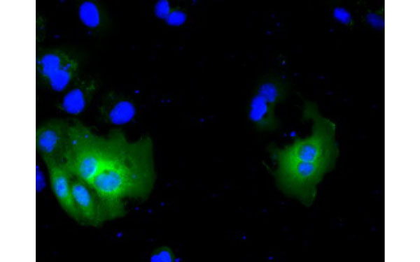
 IF (Immunofluorescence)
(Immunofluorescent staining of COS7 cells transiently transfected with recombinant ARCN1 protein using ARCN1 antibody)
IF (Immunofluorescence)
(Immunofluorescent staining of COS7 cells transiently transfected with recombinant ARCN1 protein using ARCN1 antibody)
ARCN1, Monoclonal Antibody (Cat# AAA107472)
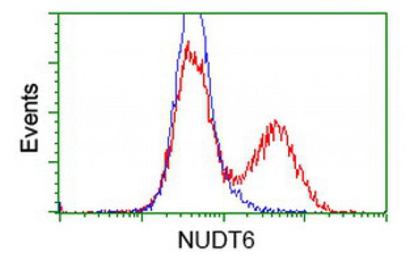
 IF (Immunofluorescence)
(Immunofluorescent staining of COS7 cells transiently transfected with recombinant NUDT6 protein using NUDT6 antibody)
IF (Immunofluorescence)
(Immunofluorescent staining of COS7 cells transiently transfected with recombinant NUDT6 protein using NUDT6 antibody)
NUDT6, Monoclonal Antibody (Cat# AAA107475)
 IHC (Immunohistochemisry)
(Immunohistochemical analysis of PPP5C protein in paraffin embedded Human endometrium tissue using PPP5C antibody)
IHC (Immunohistochemisry)
(Immunohistochemical analysis of PPP5C protein in paraffin embedded Human endometrium tissue using PPP5C antibody)
PPP5C, Monoclonal Antibody (Cat# AAA107478)
 IHC (Immunohiostchemistry)
(Immunohistochemical analysis of PDSS2 protein in paraffin embedded Human endometrium tissue using PDSS2 antibody)
IHC (Immunohiostchemistry)
(Immunohistochemical analysis of PDSS2 protein in paraffin embedded Human endometrium tissue using PDSS2 antibody)
PDSS2, Monoclonal Antibody (Cat# AAA107479)
 IF (Immunofluorescence)
(Immunofluorescent staining of COS7 cells transiently transfected with recombinant SMS protein using SMS antibody)
IF (Immunofluorescence)
(Immunofluorescent staining of COS7 cells transiently transfected with recombinant SMS protein using SMS antibody)
SMS, Monoclonal Antibody (Cat# AAA107486)

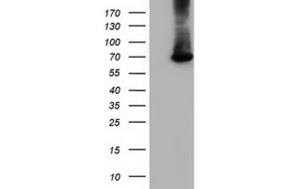
 WB (Western Blot)
(Western Blot analysis of HEK293T cell lysates (5 ug) transfected with either recombinant RTN4IP1 protein (Right) or empty vector (Left) detected with RTN4IP1 antibody)
WB (Western Blot)
(Western Blot analysis of HEK293T cell lysates (5 ug) transfected with either recombinant RTN4IP1 protein (Right) or empty vector (Left) detected with RTN4IP1 antibody)
RTN4IP1, Monoclonal Antibody (Cat# AAA107490)
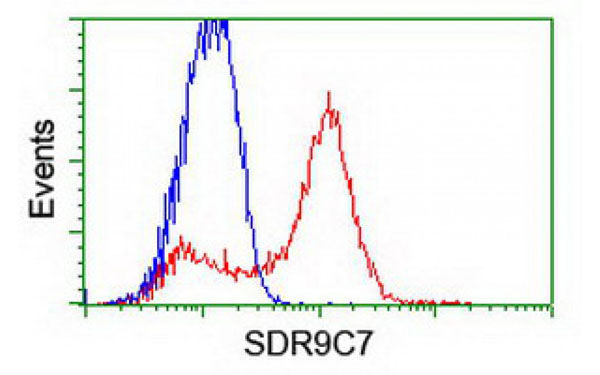
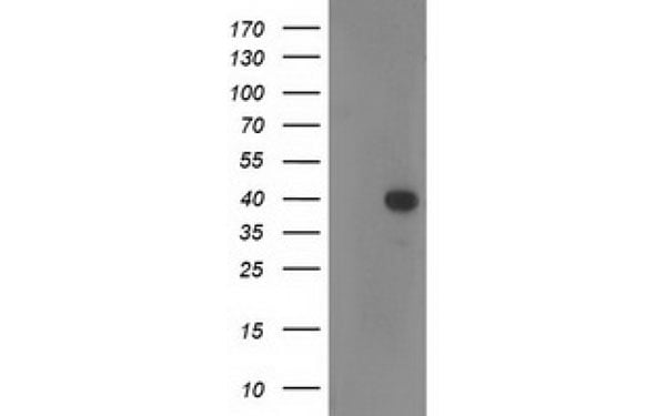
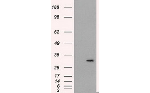
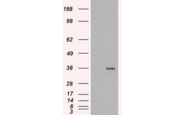
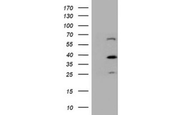
 WB (Western Blot)
(Western Blot analysis of HEK293T cell lysates (5 ug) transfected with either recombinant SDR9C7 protein (Right) or empty vector (Left) detected with SDR9C7 antibody)
WB (Western Blot)
(Western Blot analysis of HEK293T cell lysates (5 ug) transfected with either recombinant SDR9C7 protein (Right) or empty vector (Left) detected with SDR9C7 antibody)
SDR9C7, Monoclonal Antibody (Cat# AAA107494)
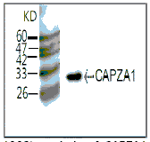
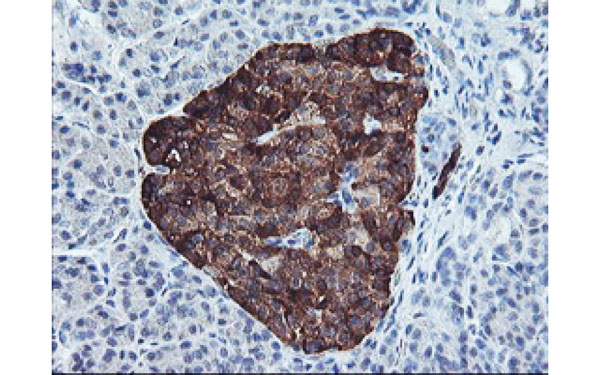
 IHC (Immunohiostchemistry)
(IHC (1:100 400x) analysis of CAPZA1 expression in Liver Cancer with Anti-CAPZA1.)
IHC (Immunohiostchemistry)
(IHC (1:100 400x) analysis of CAPZA1 expression in Liver Cancer with Anti-CAPZA1.)
CAPZA1, Monoclonal Antibody (Cat# AAA108861)
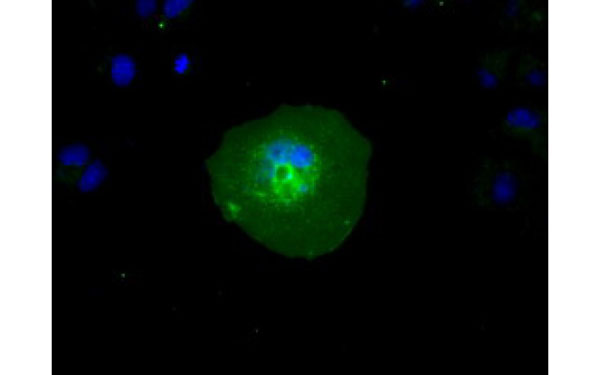
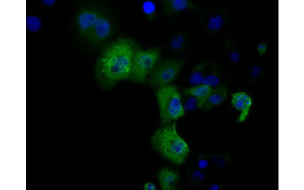
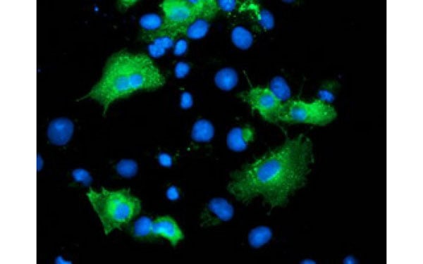
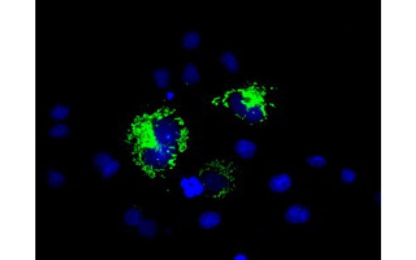
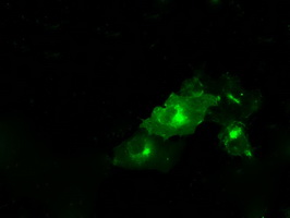
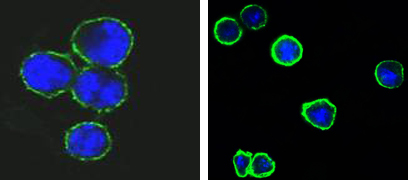 IF (Immunofluorescence)
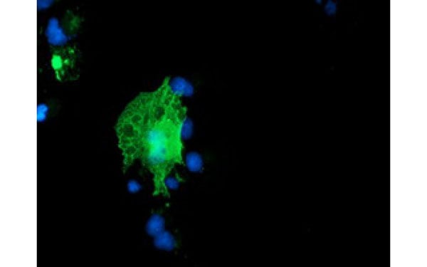
(Figure 1. Confocal immunofluorescence analysis of methanol-fixed BCBL-1 (left) and L1210 (right) cells using CD37 mouse mAb(green), showing membrane localization. Blue. DRAQ5 fluorescent DNA dye.)
IF (Immunofluorescence)
(Figure 1. Confocal immunofluorescence analysis of methanol-fixed BCBL-1 (left) and L1210 (right) cells using CD37 mouse mAb(green), showing membrane localization. Blue. DRAQ5 fluorescent DNA dye.)
CD37, Monoclonal Antibody (Cat# AAA108724)
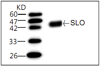 Application Data
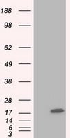
(WB (1:1000) analysis of recombinant protein HS SLO with Anti-HS SLO (AAA109011))
Application Data
(WB (1:1000) analysis of recombinant protein HS SLO with Anti-HS SLO (AAA109011))
HS SLO, Monoclonal Antibody (Cat# AAA109011)
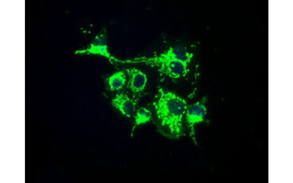
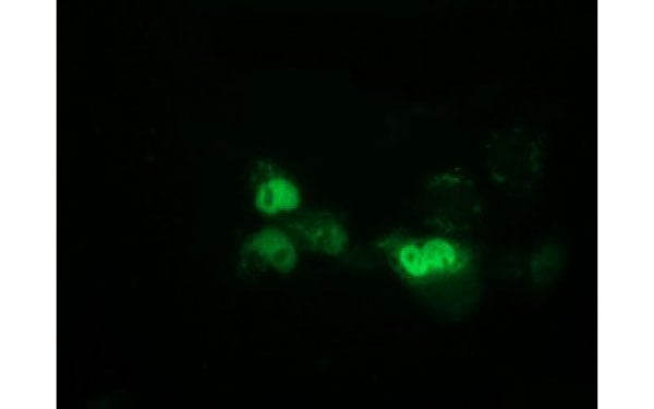
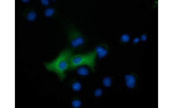
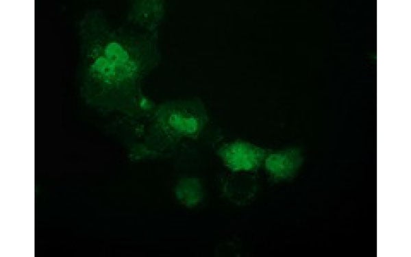
 IF (Immunofluorescence)
(Immunofluorescent staining of COS7 cells transiently transfected with recombinant DCXR protein using DCXR antibody)
IF (Immunofluorescence)
(Immunofluorescent staining of COS7 cells transiently transfected with recombinant DCXR protein using DCXR antibody)
DCXR, Monoclonal Antibody (Cat# AAA106384)
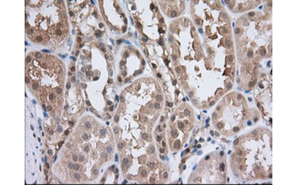
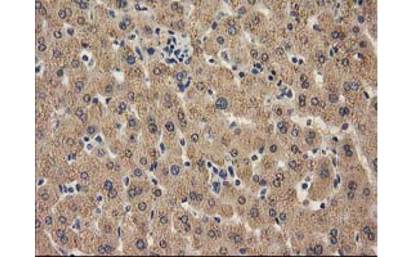 IHC (Immunohiostchemistry)
(Immunohistochemical analysis of CAT protein in paraffin embedded Human liver tissue using CAT antibody)
IHC (Immunohiostchemistry)
(Immunohistochemical analysis of CAT protein in paraffin embedded Human liver tissue using CAT antibody)
CAT, Monoclonal Antibody (Cat# AAA106387)
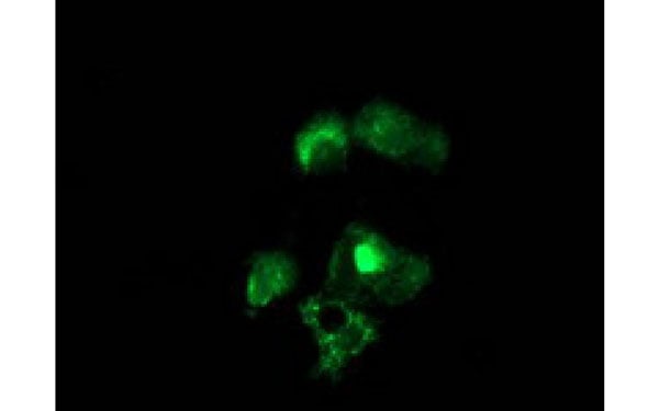
 IF (Immunofluorescence)
(Immunofluorescent staining of COS7 cells transiently transfected with recombinant FAHD2A protein using FAHD2A antibody)
IF (Immunofluorescence)
(Immunofluorescent staining of COS7 cells transiently transfected with recombinant FAHD2A protein using FAHD2A antibody)
FAHD2A, Monoclonal Antibody (Cat# AAA106391)
 IF (Immunofluorescence)
(Immunofluorescent staining of COS7 cells transiently transfected with recombinant ACAT2 protein using ACAT2 antibody)
IF (Immunofluorescence)
(Immunofluorescent staining of COS7 cells transiently transfected with recombinant ACAT2 protein using ACAT2 antibody)
ACAT2, Monoclonal Antibody (Cat# AAA106401)
LDL, Monoclonal Antibody (Cat# AAA106418)
 IF (Immunofluorescence)
(Immunofluorescent staining of COS7 cells transiently transfected with recombinant EPM2AIP1 protein using EPM2AIP1 antibody)
IF (Immunofluorescence)
(Immunofluorescent staining of COS7 cells transiently transfected with recombinant EPM2AIP1 protein using EPM2AIP1 antibody)
EPM2AIP1, Monoclonal Antibody (Cat# AAA106420)
 IF (Immunofluorescence)
(Immunofluorescent staining of COS7 cells transiently transfected with recombinant EPHX2 protein using EPHX2 antibody)
IF (Immunofluorescence)
(Immunofluorescent staining of COS7 cells transiently transfected with recombinant EPHX2 protein using EPHX2 antibody)
EPHX2, Monoclonal Antibody (Cat# AAA106421)
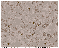
 IHC (Immunohiostchemistry)
(Immunohistochemical analysis of FAHD2A protein in paraffin embedded Human kidney tissue using FAHD2A antibody)
IHC (Immunohiostchemistry)
(Immunohistochemical analysis of FAHD2A protein in paraffin embedded Human kidney tissue using FAHD2A antibody)
FAHD2A, Monoclonal Antibody (Cat# AAA106432)
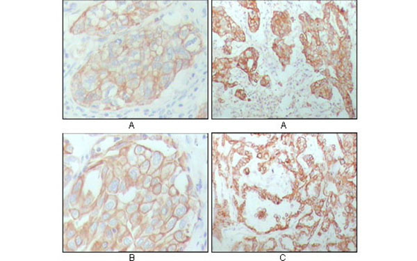
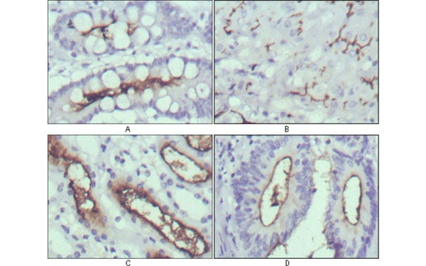 IHC (Immunohiostchemistry)
(Immunohistochemical analysis of paraffin-embedded human normal stomach (A), normal liver (B), normal kidney (C) and rectum cancer tissues (D) using WNT10B antibody with DAB staining.)
IHC (Immunohiostchemistry)
(Immunohistochemical analysis of paraffin-embedded human normal stomach (A), normal liver (B), normal kidney (C) and rectum cancer tissues (D) using WNT10B antibody with DAB staining.)
WNT10B, Monoclonal Antibody (Cat# AAA106447)
 IHC (Immunohiostchemistry)
(Immunohistochemical analysis of CAMK1D protein in paraffin embedded Human thyroid tissue using CAMK1D antibody)
IHC (Immunohiostchemistry)
(Immunohistochemical analysis of CAMK1D protein in paraffin embedded Human thyroid tissue using CAMK1D antibody)
CAMK1D, Monoclonal Antibody (Cat# AAA106461)
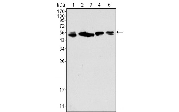
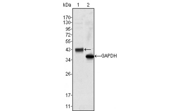
 WB (Western Blot)
(Western Blot analysis using Mouse anti Human IgGWestern Blot showing human IgG (Fc specific) antibody used against human serum (1).)
WB (Western Blot)
(Western Blot analysis using Mouse anti Human IgGWestern Blot showing human IgG (Fc specific) antibody used against human serum (1).)
Mouse anti Human IgG (Fc Specific) antibody, Monoclonal Antibody (Cat# AAA106306)
 IF (Immunofluorescence)
(Immunofluorescent staining of COS7 cells transiently transfected with recombinant ATP6V1F protein using ATP6V1F antibody)
IF (Immunofluorescence)
(Immunofluorescent staining of COS7 cells transiently transfected with recombinant ATP6V1F protein using ATP6V1F antibody)
ATP6V1F, Monoclonal Antibody (Cat# AAA106315)
 IF (Immunofluorescence)
(Immunofluorescent staining of COS7 cells transiently transfected with recombinant ARFGAP1 protein using ARFGAP1 antibody)
IF (Immunofluorescence)
(Immunofluorescent staining of COS7 cells transiently transfected with recombinant ARFGAP1 protein using ARFGAP1 antibody)
ARFGAP1, Monoclonal Antibody (Cat# AAA106319)
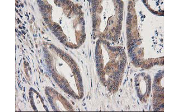
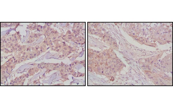
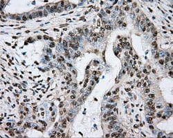
 IHC (Immunohiostchemistry)
(Immunohistochemical analysis of CRYAB protein in paraffin embedded Adenocarcinoma of Human colon tissue using CRYAB antibody)
IHC (Immunohiostchemistry)
(Immunohistochemical analysis of CRYAB protein in paraffin embedded Adenocarcinoma of Human colon tissue using CRYAB antibody)
CRYAB, Monoclonal Antibody (Cat# AAA106324)
 IF (Immunofluorescence)
(Immunofluorescent staining of COS7 cells transiently transfected with recombinant HOXC11 protein using HOXC11 antibody)
IF (Immunofluorescence)
(Immunofluorescent staining of COS7 cells transiently transfected with recombinant HOXC11 protein using HOXC11 antibody)
HOXC11, Monoclonal Antibody (Cat# AAA106336)
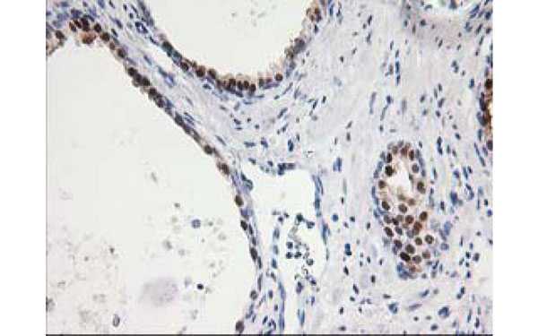
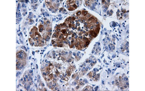
 IHC (Immunohiostchemistry)
(Immunohistochemical analysis of FGF2 protein in paraffin embedded Human pancreas tissue using FGF2 antibody)
IHC (Immunohiostchemistry)
(Immunohistochemical analysis of FGF2 protein in paraffin embedded Human pancreas tissue using FGF2 antibody)
FGF2, Monoclonal Antibody (Cat# AAA106359)

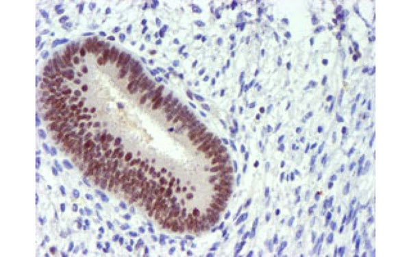
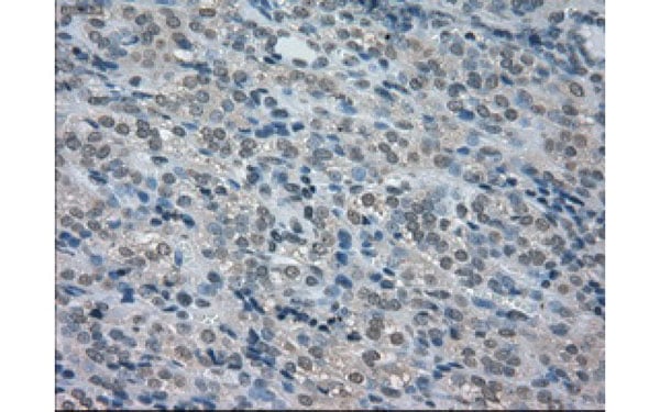
 IHC (Immunohiostchemistry)
(Immunohistochemical analysis of BECN1 protein in paraffin embedded Human Kidney tissue using BECN1 antibody)
IHC (Immunohiostchemistry)
(Immunohistochemical analysis of BECN1 protein in paraffin embedded Human Kidney tissue using BECN1 antibody)
BECN1, Monoclonal Antibody (Cat# AAA106369)
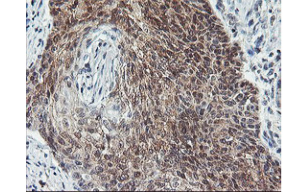
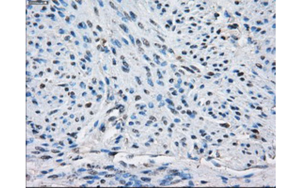
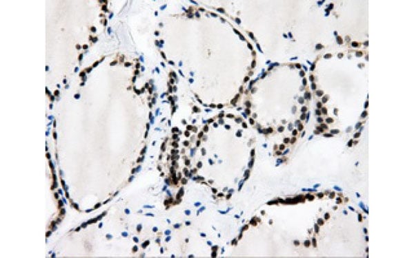
 IHC (Immunohiostchemistry)
(Immunohistochemical analysis of BUB1B protein in paraffin embedded Carcinoma of Human thyroid tissue using BUB1B antibody)
IHC (Immunohiostchemistry)
(Immunohistochemical analysis of BUB1B protein in paraffin embedded Carcinoma of Human thyroid tissue using BUB1B antibody)
BUB1B, Monoclonal Antibody (Cat# AAA106371)
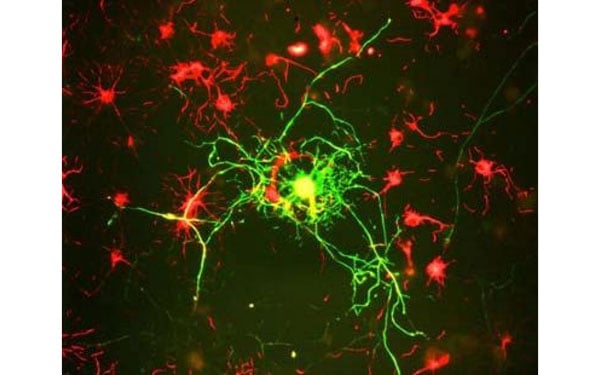
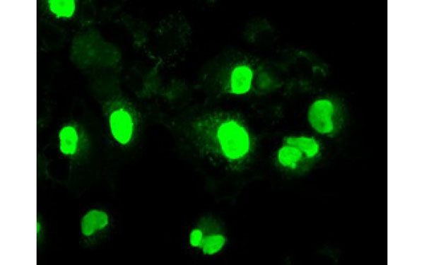
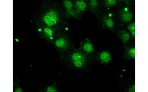
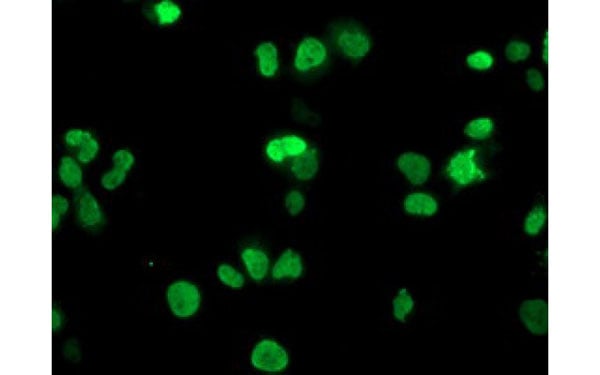
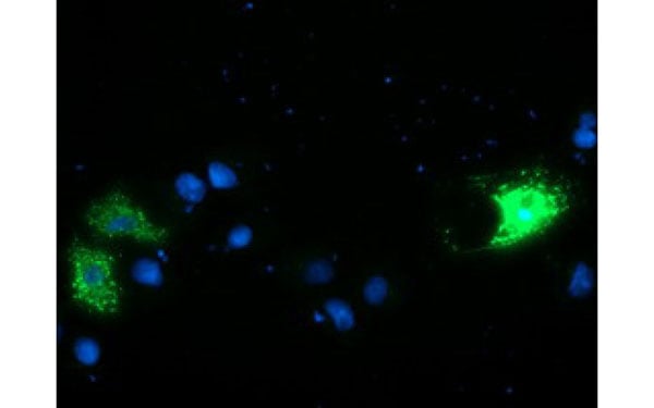
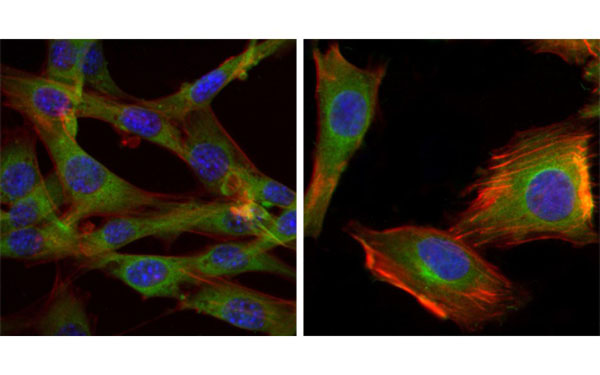 IF (Immunofluorescence)
(Immunofluorescence analysis of NIH/3T3 (left) and U251 (right) cells using GSK3B antibody (green). Blue: DRAQ5 fluorescent DNA dye. Red: Actin filaments have been labeled with Alexa Fluor-555 phalloidin.)
IF (Immunofluorescence)
(Immunofluorescence analysis of NIH/3T3 (left) and U251 (right) cells using GSK3B antibody (green). Blue: DRAQ5 fluorescent DNA dye. Red: Actin filaments have been labeled with Alexa Fluor-555 phalloidin.)
GSK3B, Monoclonal Antibody (Cat# AAA106377)
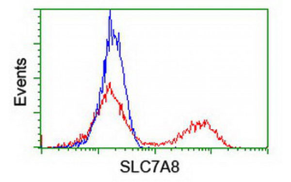
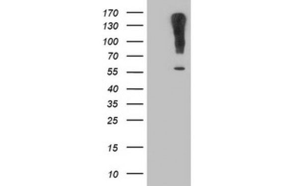 WB (Western Blot)
(Western Blot analysis of HEK293T cell lysates (5 ug) transfected with either recombinant SLC7A8 protein (Right) or empty vector (Left) detected with SLC7A8 antibody)
WB (Western Blot)
(Western Blot analysis of HEK293T cell lysates (5 ug) transfected with either recombinant SLC7A8 protein (Right) or empty vector (Left) detected with SLC7A8 antibody)
SLC7A8, Monoclonal Antibody (Cat# AAA106604)
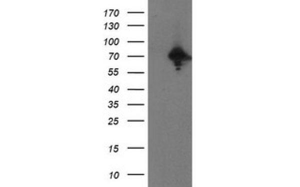
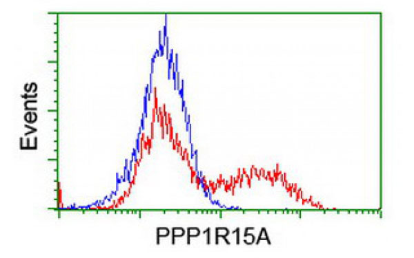
 IHC (Immunohistochemisry)
(Immunohistochemical analysis of PPP1R15A protein in paraffin embedded Human pancreas tissue using PPP1R15A antibody)
IHC (Immunohistochemisry)
(Immunohistochemical analysis of PPP1R15A protein in paraffin embedded Human pancreas tissue using PPP1R15A antibody)
PPP1R15A, Monoclonal Antibody (Cat# AAA106615)
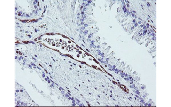 IHC (Immunohiostchemistry)
(Immunohistochemical analysis of DTNB protein in paraffin embedded Human prostate tissue using DTNB antibody)
IHC (Immunohiostchemistry)
(Immunohistochemical analysis of DTNB protein in paraffin embedded Human prostate tissue using DTNB antibody)
DTNB, Monoclonal Antibody (Cat# AAA106633)
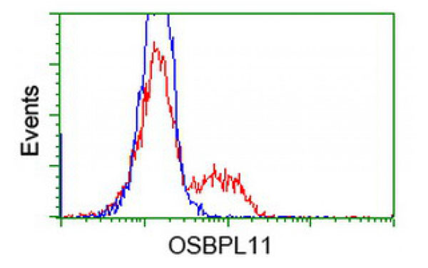
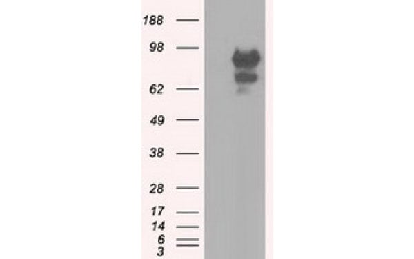
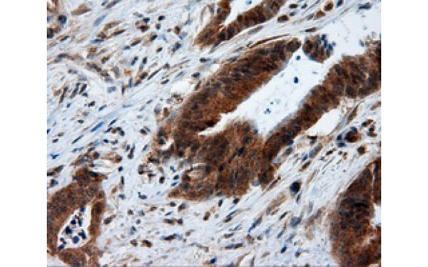
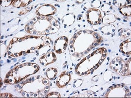
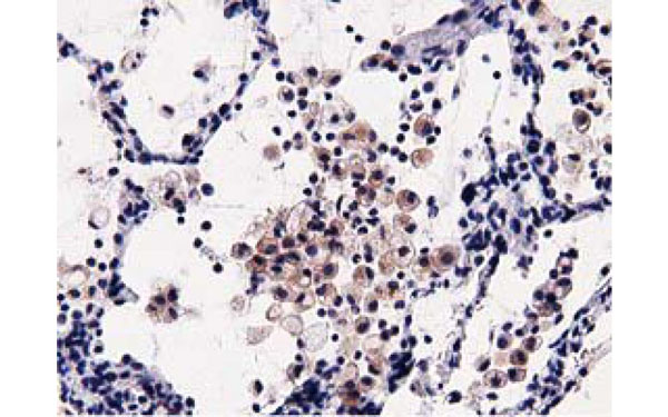 IHC (Immunohistochemisry)
(Immunohistochemical analysis of OSBPL11 protein in paraffin embedded Carcinoma of Human lung tissue using OSBPL11 antibody)
IHC (Immunohistochemisry)
(Immunohistochemical analysis of OSBPL11 protein in paraffin embedded Carcinoma of Human lung tissue using OSBPL11 antibody)
OSBPL11, Monoclonal Antibody (Cat# AAA106635)
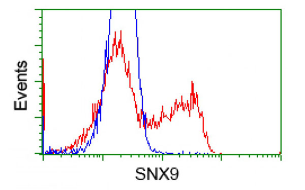
 IF (Immunofluorescence)
(Immunofluorescent staining of COS7 cells transiently transfected with recombinant SNX9 protein using SNX9 antibody)
IF (Immunofluorescence)
(Immunofluorescent staining of COS7 cells transiently transfected with recombinant SNX9 protein using SNX9 antibody)
SNX9, Monoclonal Antibody (Cat# AAA106636)
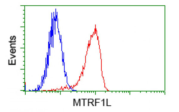
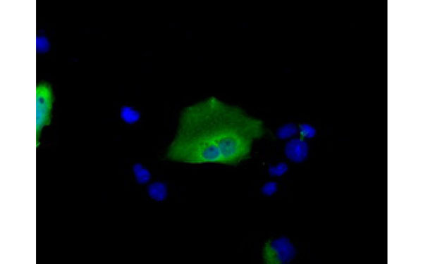
 IF (Immunofluorescence)
(Immunofluorescent staining of COS7 cells transiently transfected with recombinant MTRF1L protein using MTRF1L antibody)
IF (Immunofluorescence)
(Immunofluorescent staining of COS7 cells transiently transfected with recombinant MTRF1L protein using MTRF1L antibody)
MTRF1L, Monoclonal Antibody (Cat# AAA106644)
HRP2, Monoclonal Antibody (Cat# AAA106647)
HRP2 antibody was purified by Protein G chromatography
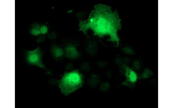
 IF (Immunofluorescence)
(Immunofluorescent staining of COS7 cells transiently transfected with recombinant CAMLG protein using CAMLG antibody)
IF (Immunofluorescence)
(Immunofluorescent staining of COS7 cells transiently transfected with recombinant CAMLG protein using CAMLG antibody)
CAMLG, Monoclonal Antibody (Cat# AAA106660)
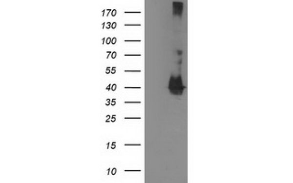
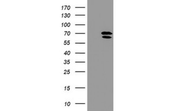
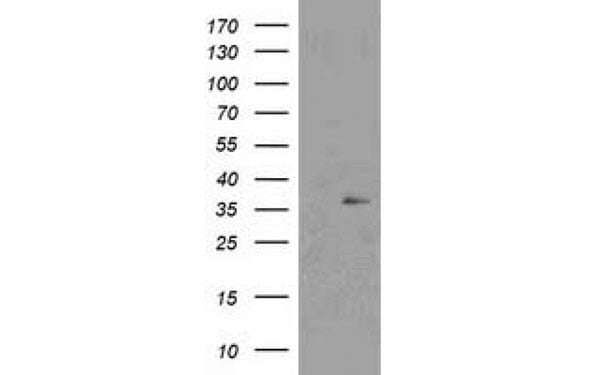
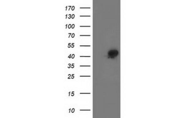
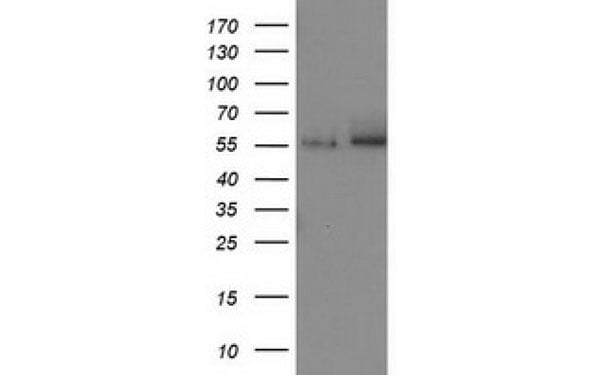
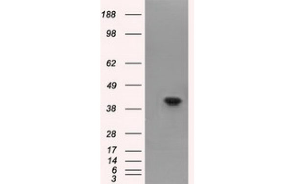
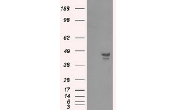
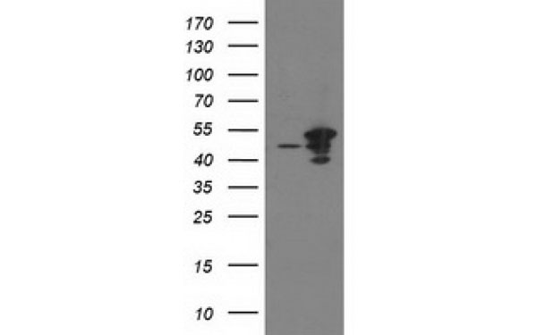
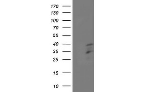
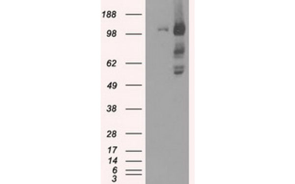
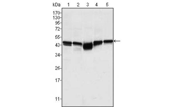
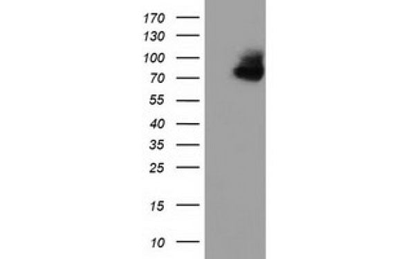
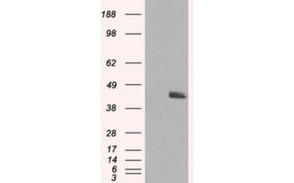
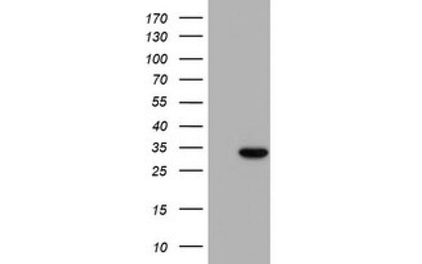
 WB (Western Blot)
(Western Blot analysis using PTPRE antibodyWestern Blot analysis of HEK293T cell lysates (5 ug) transfected with either recombinant PTPRE protein (Right) or empty vector (Left) detected with PTPRE antibody)
WB (Western Blot)
(Western Blot analysis using PTPRE antibodyWestern Blot analysis of HEK293T cell lysates (5 ug) transfected with either recombinant PTPRE protein (Right) or empty vector (Left) detected with PTPRE antibody)
PTPRE, Monoclonal Antibody (Cat# AAA106661)
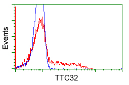
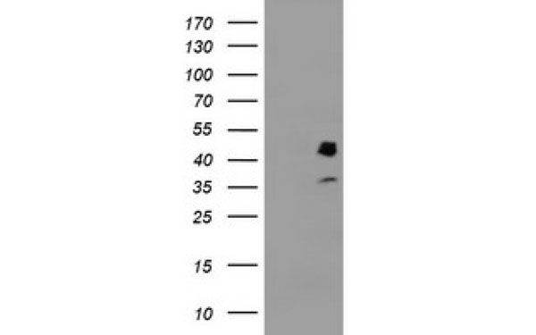
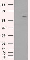
 WB (Western Blot)
(Western Blot analysis using TTC32 antibodyWestern Blot analysis of HEK293T cell lysates (5 ug) transfected with either recombinant TTC32 protein (Right) or empty vector (Left) detected with TTC32 antibody)
WB (Western Blot)
(Western Blot analysis using TTC32 antibodyWestern Blot analysis of HEK293T cell lysates (5 ug) transfected with either recombinant TTC32 protein (Right) or empty vector (Left) detected with TTC32 antibody)
TTC32, Monoclonal Antibody (Cat# AAA106666)
What are Monoclonal Antibodies?
Monoclonal antibodies are specialized laboratory-produced proteins developed for binding to specific biological antigens or other molecular targets. Since they come from a single cell (or clone), they are especially consistent and accurate in the data they are involved in producing.
This type of antibody material has been shown to be a powerful tool in finding and subsequently destroying harmful cells in an organism, such as those found in cancers or various autoimmune diseases. This makes them excellent aids in medical testing and research, which is why they are so widely used.
AAA Biotech offers a comprehensive range of high-quality monoclonal antibodies that perform effectively in various laboratory tests, including (amongst others) ELISA, western blotting, immunohistochemistry, and flow cytometry. All of the products in our catalog are thoroughly quality tested to make sure that they are reliable and will consistently perform well in your research.
What Are The Uses of Monoclonal Antibodies
Monoclonal antibodies are used in many lab tests, including (amongst others) ELISA, western blotting, immunohistochemistry, and flow cytometry.
ELISA is a test that helps detect a specific substance/analyte in a sample. It uses antibodies (often monoclonal) bound to a solid surface (such as the well of a microplate) to “capture” the substance/analyte in the sample and immobilize it so that the detection antibody component can then bind to it and produce a signal, which can then be measured.
Western blotting identifies specific proteins in a sample. The sample is first separated on a gel, and then antibodies are applied that will typically bind to the target, which will all be localized to a single band in a lane.
Immunohistochemistry helps locate specific proteins in cells or tissue samples using antibodies.
Flow cytometry looks at and sorts cells. It uses antibodies that are conjugated to reporter molecules called “fluorophores”, which, under special lights, emit light themselves, which can then be measured by a detector instrument. For a deeper understanding of these techniques, explore our complete guide to monoclonal antibodies and their benefits.
How Monoclonal Antibodies Are Used as Medicine?
Please note that all of the products listed in AAA Biotech’s also known as AAA Bio or AAABio catalog are strictly for research-use only (RUO).
Monoclonal antibodies can also be used as therapeutic/medical treatments, particularly in the context of cancers. They are designed to find and bind to specific cells or proteins, helping the immune system recognize and attack the cancer. These treatments work in different ways, such as:
- Radioimmunotherapy attaches a small amount of radioactive molecule to the antibody, so it delivers the radiation directly to the cancer cells that the antibody is specifically binding to.
- Antibody-directed enzyme prodrug therapy uses antibodies that are specifically bound to special enzymes. These enzymes activate a harmless drug in the body and turn it into a cancer-killing drug only near the cancer cells—this helps avoid harming healthy cells.
- Immunoliposomes are tiny “bubbles” filled with medicine/drug and coated with antibodies. They carry the drug straight to the cancer cells.
Why Buy Monoclonal Antibodies From Us?
At AAA Biotech, we provide high-performance monoclonal antibodies designed to support a wide range of research needs.
1. Validated for Versatile Applications
The antibodies in our catalog are extensively validated and compatible with multiple techniques, including (but not limited to) ELISA, flow cytometry (FC), immunocytochemistry (ICC), immunofluorescence (IF), immunohistochemistry (IHC), immunoprecipitation (IP), and western blotting (WB).
2. Wide Selection & Specialized Options
We offer antibodies for common and rare species, that are available in various conjugated forms, and also in recombinant formats. Essentially, there is almost anything one might need to meet their experimental model’s requirements.
3. High-Quality Proteins
Our proteins meet high purity standards—90% or more as confirmed by SDS-PAGE. Many are available with tags like His, Flag, GST, or MBP, and we also supply native and biologically active proteins for functional studies.
Frequently Asked Questions
1. Are your monoclonal antibodies validated for specific applications?
Yes, our antibodies are tested and validated for use in methods such as ELISA, western blot, IHC, flow cytometry, and more. Refer to specific product pages or datasheets for individual product information.
2. How do I choose the right monoclonal antibody for my application?
Review the product details directly for application validation, species reactivity, and target information. You may also contact our support team at any time for help.
3. How quickly can I receive my order?
Most orders are processed and shipped within 1–3 business days, depending on product availability and your shipping location.