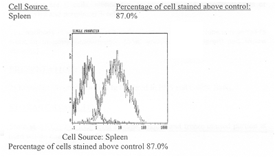
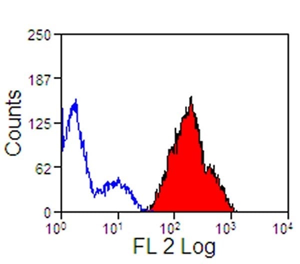
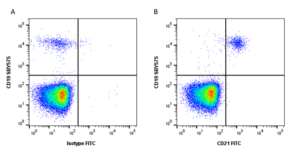
 FCM/FACS (Flow Cytometry)
Rat anti-Mouse CD44 Monoclonal Antibody | anti-CD44 antibody
Anti-Mouse CD44, Purified (Clone IM7.8.1) (rat IgG2b)
Gene Names
CD44; IN; LHR; MC56; MDU2; MDU3; MIC4; Pgp1; CDW44; CSPG8; HCELL; HUTCH-I; ECMR-III
Reactivity
Mouse
Applications
Immunoprecipitation, Western Blot, ELISA, Flow Cytometry, Functional Assay, Immunohistochemistry
Synonyms
CD44, Antibody; Anti-Mouse CD44, Purified (Clone IM7.8.1) (rat IgG2b); CD44, Purified (Clone IM7.8.1) (rat IgG2b); Purified Anti-Mouse CD44 Monoclonal Antibody; anti-CD44 antibody
Host
Rat
Reactivity
Mouse
Clonality
Monoclonal
Isotype
Rat IgG2b
Clone Number
IM7.8.1
Specificity
CD44 (Ly 24, Pgp-1) (Mouse CD44)
Form/Format
Purified
Concentration
Antibody Concentration: 0.2 mg/ml (varies by lot)
Sequence Length
43
Applicable Applications for anti-CD44 antibody
IP (Immunoprecipitation), WB (Western Blot), ELISA, FCM/FACS (Flow Cytometry), IHC (Immunohistochemistry)
Presentation
200 ug purified Ig buffered in PBS and 0.05% sodium azide 0.1% (NaN3).
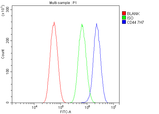
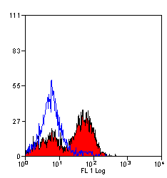
FLOW CYTOMETRY ANALYSIS
Method:
1. Prepare a cell suspension in media A. For cell preparations, deplete the red blood cell population with Lympholyte®-M cell separation medium.
2. Wash 2 times.
3. Resuspend the cells to a concentration of 2x 10^7 cells/ml in media A. Add 50ul of this suspension to each tube (each tube will then contain 1 x 10^6 cells, representing 1 test).
4. To each tube, add ~1.0ug*.
5. Vortex the tubes to ensure thorough mixing of antibody and cells.
6. Incubate the tubes for 30 minutes at 4 degree C.
7. Wash 2 times at 4 degree C.
8. Add 100 J11 of secondary antibody (FITC Goat anti-rat IgG (H+L)) at 1:500 dilution.
9. Incubate the tubes at 4 degree C for 30-60 minutes.
(It is recommended that the tubes are protected from light since most fluorochromes are light sensitive).
10. Wash 2 times at 4 degree C in media B.
11. Resuspend the cell pellet in 50 ul ice cold media B.
12. Transfer to suitable tubes for flow cytometric analysis containing 15 ul of propidium iodide at 0.5 mg/ml in PBS. This stains dead cells by intercalating in DNA.
Media
A. Phosphate buffered saline (pH 72) + 5% normal serum of host species + sodium azide ( 100 ul of 2M sodium azide in 100 mls).
B. Phosphate buffered saline (pH 7.2) + 0.5% Bovine serum albumin + sodium azide (100 ul of 2M sodium azide in 100 mls).
N.B. Appropriate contml samples should always be included in any labelling studies.
* For optimal results in various applications, it is recommended that each investigator determine dilutions appropriate for individual use.
Preparation and Storage
Store at 4 degree C. For long term storage, aliquot and freeze unused portion at -20 degree C in volumes appropriate for single usage. Avoid freeze/thaw cycles. If the reagent is being diluted, it is recommended that only the quantity to be used within one week be diluted.
FCM/FACS (Flow Cytometry)
FCM/FACS (Flow Cytometry)
Related Product Information for anti-CD44 antibody
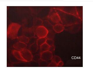
anti-mouse CD44 monoclonal antibody reacts with all isoforms of CD44 (also known as Pgp-1, Ly-24) glycoprotein. By flow cytometry, the main cellular reactivities are B cellls, monocytes, macrophages and variable subsets of thymocyctes and peripheral T-cells.
NCBI and Uniprot Product Information
NCBI GI #
NCBI GeneID
UniProt Accession #
Molecular Weight
81,538 Da
NCBI Official Full Name
CD44
NCBI Official Synonym Full Names
CD44 molecule (Indian blood group)
NCBI Official Symbol
CD44
NCBI Official Synonym Symbols
IN; LHR; MC56; MDU2; MDU3; MIC4; Pgp1; CDW44; CSPG8; HCELL; HUTCH-I; ECMR-III
NCBI Protein Information
CD44 antigen; epican; Hermes antigen; hyaluronate receptor; phagocytic glycoprotein 1; heparan sulfate proteoglycan; cell surface glycoprotein CD44; extracellular matrix receptor III; chondroitin sulfate proteoglycan 8; GP90 lymphocyte homing/adhesion receptor; hematopoietic cell E- and L-selectin ligand; homing function and Indian blood group system
UniProt Protein Name
CD44 antigen
UniProt Gene Name
CD44
UniProt Synonym Gene Names
LHR; MDU2; MDU3; MIC4; ECMR-III; PGP-1; PGP-I
UniProt Entry Name
CD44_HUMAN
Customer Reviews
Loading reviews...
Share Your Experience
Similar Products


Product Notes
The CD44 cd44 (Catalog #AAA74207) is an Antibody produced from Rat and is intended for research purposes only. The product is available for immediate purchase. The Anti-Mouse CD44, Purified (Clone IM7.8.1) (rat IgG2b) reacts with Mouse and may cross-react with other species as described in the data sheet. AAA Biotech's CD44 can be used in a range of immunoassay formats including, but not limited to, IP (Immunoprecipitation), WB (Western Blot), ELISA, FCM/FACS (Flow Cytometry), IHC (Immunohistochemistry). Researchers should empirically determine the suitability of the CD44 cd44 for an application not listed in the data sheet. Researchers commonly develop new applications and it is an integral, important part of the investigative research process. It is sometimes possible for the material contained within the vial of "CD44, Monoclonal Antibody" to become dispersed throughout the inside of the vial, particularly around the seal of said vial, during shipment and storage. We always suggest centrifuging these vials to consolidate all of the liquid away from the lid and to the bottom of the vial prior to opening. Please be advised that certain products may require dry ice for shipping and that, if this is the case, an additional dry ice fee may also be required.Precautions
All products in the AAA Biotech catalog are strictly for research-use only, and are absolutely not suitable for use in any sort of medical, therapeutic, prophylactic, in-vivo, or diagnostic capacity. By purchasing a product from AAA Biotech, you are explicitly certifying that said products will be properly tested and used in line with industry standard. AAA Biotech and its authorized distribution partners reserve the right to refuse to fulfill any order if we have any indication that a purchaser may be intending to use a product outside of our accepted criteria.Disclaimer
Though we do strive to guarantee the information represented in this datasheet, AAA Biotech cannot be held responsible for any oversights or imprecisions. AAA Biotech reserves the right to adjust any aspect of this datasheet at any time and without notice. It is the responsibility of the customer to inform AAA Biotech of any product performance issues observed or experienced within 30 days of receipt of said product. To see additional details on this or any of our other policies, please see our Terms & Conditions page.Item has been added to Shopping Cart
If you are ready to order, navigate to Shopping Cart and get ready to checkout.