 Application Data
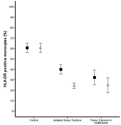
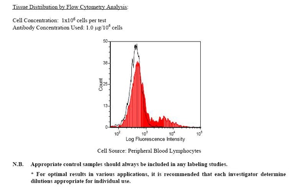
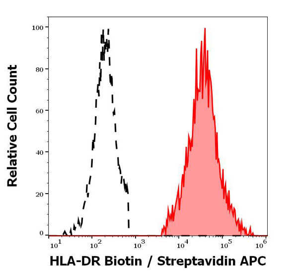
(Published customer image: Rat anti Human HLA-DR antibody, clone YE2/36-HLK used for flow cytometryImage caption:HLA-DR positive monocytes. The percentage HLA-DR positive monocytes was decreased in all patients compared to controls (P = 0.002). The pre-operative ("black square") lowest percentage was seen in patients who developed respiratory failure (P = 0.002). Eighteen hours after intramedullary nailing ("open triangle"), a further decrease in HLA-DR positive monocytes was seen in patients with isolated femur fracture (P < 0.001) and multitrauma patients (P = 0.047).From: Hietbrink, Falco, Leo Koenderman, and Luke PH Leenen. œIntramedullary Nailing of the Femur and the Systemic Activation of Monocytes and Neutrophils. World Journal of Emergency Surgery?: WJES 6 (2011): 34. PMC. Web. 22 Jan. 2015.)
Application Data
(Published customer image: Rat anti Human HLA-DR antibody, clone YE2/36-HLK used for flow cytometryImage caption:HLA-DR positive monocytes. The percentage HLA-DR positive monocytes was decreased in all patients compared to controls (P = 0.002). The pre-operative ("black square") lowest percentage was seen in patients who developed respiratory failure (P = 0.002). Eighteen hours after intramedullary nailing ("open triangle"), a further decrease in HLA-DR positive monocytes was seen in patients with isolated femur fracture (P < 0.001) and multitrauma patients (P = 0.047).From: Hietbrink, Falco, Leo Koenderman, and Luke PH Leenen. œIntramedullary Nailing of the Femur and the Systemic Activation of Monocytes and Neutrophils. World Journal of Emergency Surgery?: WJES 6 (2011): 34. PMC. Web. 22 Jan. 2015.)
Rat HLA DR Monoclonal Antibody | anti-HLA DR antibody
RAT ANTI HUMAN HLA DR:FITC
DISCONTINUED
This product has been discontinued and is no longer available for purchase. Please contact us for alternative products or more information.
|
This product is discontinued. Contact us for alternatives. |
1% Bovine Serum Albumin
Excitation (nm): 490
Emission (nm): 520
Shelf Life: 18 months from date of despatch.
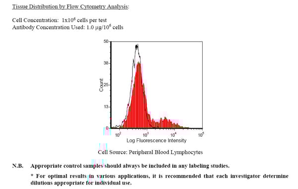
Application Data
(Published customer image: Rat anti Human HLA-DR antibody, clone YE2/36-HLK used for flow cytometryImage caption:HLA-DR positive monocytes. The percentage HLA-DR positive monocytes was decreased in all patients compared to controls (P = 0.002). The pre-operative ("black square") lowest percentage was seen in patients who developed respiratory failure (P = 0.002). Eighteen hours after intramedullary nailing ("open triangle"), a further decrease in HLA-DR positive monocytes was seen in patients with isolated femur fracture (P < 0.001) and multitrauma patients (P = 0.047).From: Hietbrink, Falco, Leo Koenderman, and Luke PH Leenen. œIntramedullary Nailing of the Femur and the Systemic Activation of Monocytes and Neutrophils. World Journal of Emergency Surgery?: WJES 6 (2011): 34. PMC. Web. 22 Jan. 2015.)
Application Data
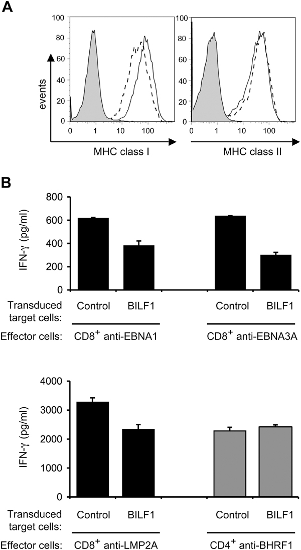
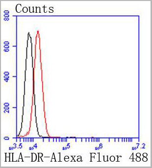
(Published customer image: Rat anti Human HLA-DR antibody, clone YE2/36-HLK used for flow cytometryImage caption:BILF1 downregulates surface MHC class I expression and inhibits the T cell recognition of endogenous EBV antigen in LCLs. (A) LCLs were transduced with PLZRS-HABILF1-IRES-GFP retrovirus. After 6 days, surface MHC class I was stained with PE-conjugated W6/32 mAb and MHC class II was stained with PE-conjugated anti-DR mAb, YE2/36-HLK. Two-colour flow cytometry was used to analyze staining in the untransduced, GFP-, population, shown as the solid line histogram, and in the transduced GFP+ (BILF1+) population, shown as the dashed line histogram. The grey histogram denotes background staining obtained with an isotype control PE-conjugated antibody. (B) LCL cultures transduced with control retrovirus or with the BILF1 retrovirus were sorted by flow cytometry to generate GFP+/BILF1- and GFP+/BILF1+ lines to use as targets in assays with EBV-specific T cells. The control and BILF1+ LCL targets were incubated with HLA-matched CD8+ effector T cells clones specific for EBNA1 (HPV), EBNA3A (YPL), or LMP2A (CLG) peptides, or a CD4+ effector T cell clone specific for a BHRF1 (PYY) peptide. After 18 hrs the supernatants were tested for the release of IFN-? as a measure of T cell recognition. All results are expressed as IFN-? release in pg/ml and error bars indicate standard deviation of triplicate cultures.From: Zuo J, Currin A, Griffin BD, Shannon-Lowe C, Thomas WA, et al. (2009) The Epstein-Barr Virus G-Protein-Coupled Receptor Contributes to Immune Evasion by Targeting MHC Class I Molecules for Degradation. PLoS Pathog 5(1): e1000255.)
Application Data
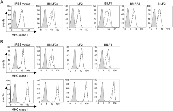
(Published customer image: Rat anti Human HLA-DR antibody, clone YE2/36-HLK used for flow cytometryImage caption:BILF1 identified as a lytic gene that downregulates surface MHC class I. 293 (A) or MJS (B) cells were transfected with different EBV genes in the bi-cistronic vector, pCDNA3-IRES-nlsGFP. At 48 hr post-transfection, surface MHC class I was stained with PE-conjugated W6/32 mAb and (in MJS only) MHC class II was stained with PE-conjugated anti-DR mAb, YE2/36-HLK. Two-colour flow cytometry was used to analyse staining in the untransfected GFP- population, shown as the solid line histogram, and in the transfected GFP+ population, shown as the dashed line histogram. The grey histogram denotes background staining obtained with an isotype control PE-conjugated antibody.From: Zuo J, Currin A, Griffin BD, Shannon-Lowe C, Thomas WA, et al. (2009) The Epstein-Barr Virus G-Protein-Coupled Receptor Contributes to Immune Evasion by Targeting MHC Class I Molecules for Degradation. PLoS Pathog 5(1): e1000255.)
Application Data
(Published customer image: Rat anti Human HLA-DR antibody, clone YE2/36-HLK used for flow cytometryImage caption:BILF1 identified as a lytic gene that downregulates surface MHC class I. 293 (A) or MJS (B) cells were transfected with different EBV genes in the bi-cistronic vector, pCDNA3-IRES-nlsGFP. At 48 hr post-transfection, surface MHC class I was stained with PE-conjugated W6/32 mAb and (in MJS only) MHC class II was stained with PE-conjugated anti-DR mAb, YE2/36-HLK. Two-colour flow cytometry was used to analyse staining in the untransfected GFP- population, shown as the solid line histogram, and in the transfected GFP+ population, shown as the dashed line histogram. The grey histogram denotes background staining obtained with an isotype control PE-conjugated antibody.From: Zuo J, Currin A, Griffin BD, Shannon-Lowe C, Thomas WA, et al. (2009) The Epstein-Barr Virus G-Protein-Coupled Receptor Contributes to Immune Evasion by Targeting MHC Class I Molecules for Degradation. PLoS Pathog 5(1): e1000255.)
NCBI and Uniprot Product Information
Customer Reviews
Loading reviews...
Share Your Experience
Similar Products


Product Notes
The HLA DR (Catalog #AAA49117) is an Antibody produced from Rat and is intended for research purposes only. The product is available for immediate purchase. AAA Biotech's HLA DR can be used in a range of immunoassay formats including, but not limited to, FCM/FACS (Flow Cytometry). Researchers should empirically determine the suitability of the HLA DR for an application not listed in the data sheet. Researchers commonly develop new applications and it is an integral, important part of the investigative research process. It is sometimes possible for the material contained within the vial of "HLA DR, Monoclonal Antibody" to become dispersed throughout the inside of the vial, particularly around the seal of said vial, during shipment and storage. We always suggest centrifuging these vials to consolidate all of the liquid away from the lid and to the bottom of the vial prior to opening. Please be advised that certain products may require dry ice for shipping and that, if this is the case, an additional dry ice fee may also be required.Precautions
All products in the AAA Biotech catalog are strictly for research-use only, and are absolutely not suitable for use in any sort of medical, therapeutic, prophylactic, in-vivo, or diagnostic capacity. By purchasing a product from AAA Biotech, you are explicitly certifying that said products will be properly tested and used in line with industry standard. AAA Biotech and its authorized distribution partners reserve the right to refuse to fulfill any order if we have any indication that a purchaser may be intending to use a product outside of our accepted criteria.Disclaimer
Though we do strive to guarantee the information represented in this datasheet, AAA Biotech cannot be held responsible for any oversights or imprecisions. AAA Biotech reserves the right to adjust any aspect of this datasheet at any time and without notice. It is the responsibility of the customer to inform AAA Biotech of any product performance issues observed or experienced within 30 days of receipt of said product. To see additional details on this or any of our other policies, please see our Terms & Conditions page.Item has been added to Shopping Cart
If you are ready to order, navigate to Shopping Cart and get ready to checkout.