


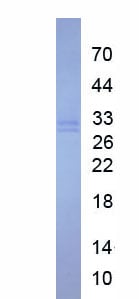
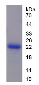
 WB (Western Blot)
(Western Blot analysis of HMGB1 expression in transfected 293T cell line by HMGB1 monoclonal antibody (M03), clone 1B11.Lane 1: HMGB1 transfected lysate (25 KDa).Lane 2: Non-transfected lysate.)
WB (Western Blot)
(Western Blot analysis of HMGB1 expression in transfected 293T cell line by HMGB1 monoclonal antibody (M03), clone 1B11.Lane 1: HMGB1 transfected lysate (25 KDa).Lane 2: Non-transfected lysate.)
Mouse HMGB1 Monoclonal Antibody | anti-HMGB1 antibody
HMGB1 (High-Mobility Group Box 1, DKFZp686A04236, HMG1, HMG3, SBP-1) (Biotin)
WB (Western Blot)
(Western Blot analysis of HMGB1 expression in transfected 293T cell line by HMGB1 monoclonal antibody (M03), clone 1B11.Lane 1: HMGB1 transfected lysate (25 KDa).Lane 2: Non-transfected lysate.)
WB (Western Blot)
(Western Blot analysis of HMGB1 expression in transfected 293T cell line by HMGB1 monoclonal antibody (M03), clone 1B11.Lane 1: HMGB1 transfected lysate (25 KDa).Lane 2: Non-transfected lysate.)
IF (Immunofluorescence)
(Immunofluorescence of monoclonal antibody to HMGB1 on HeLa cell. [antibody concentration 10 ug/ml])
IF (Immunofluorescence)
(Immunofluorescence of monoclonal antibody to HMGB1 on HeLa cell. [antibody concentration 10 ug/ml])
IF (Immunofluorescence)
(Immunofluorescence of monoclonal antibody to HMGB1 on HeLa cell. [antibody concentration 10 ug/ml])
IF (Immunofluorescence)
(Immunofluorescence of monoclonal antibody to HMGB1 on HeLa cell. [antibody concentration 10 ug/ml])
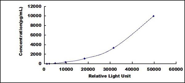
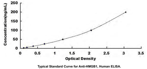
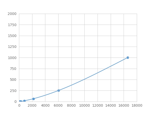
Application Data
(Detection limit for recombinant GST tagged HMGB1 is approximately 3ng/ml as a capture antibody.)
Application Data
(Detection limit for recombinant GST tagged HMGB1 is approximately 3ng/ml as a capture antibody.)
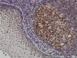
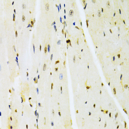
IHC (Immunohistochemistry)
(Immunoperoxidase of monoclonal antibody to HMGB1 on formalin-fixed paraffin-embedded human tonsil. [antibody concentration 3 ug/ml])
IHC (Immunohistochemistry)
(Immunoperoxidase of monoclonal antibody to HMGB1 on formalin-fixed paraffin-embedded human tonsil. [antibody concentration 3 ug/ml])
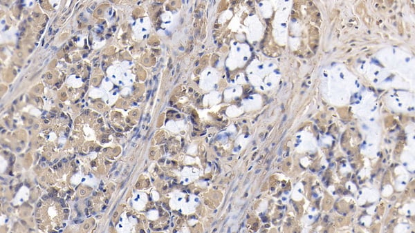
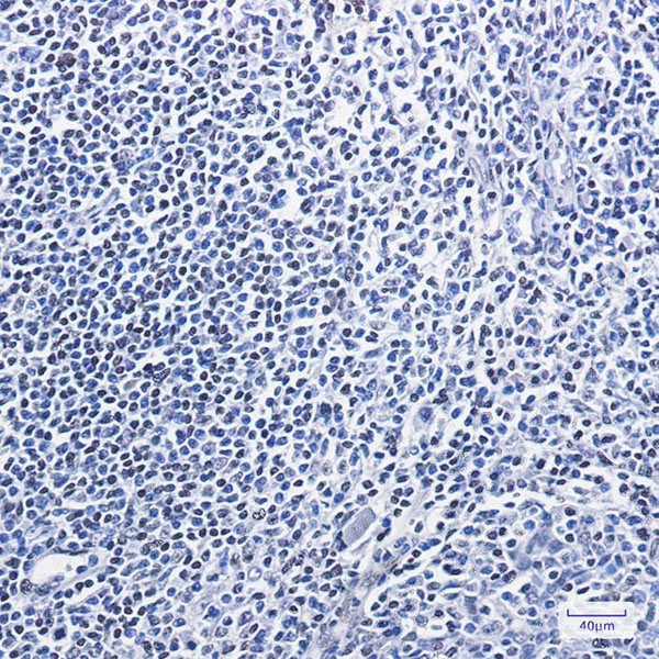
Application Data
(Immunoperoxidase of monoclonal antibody to HMGB1 on formalin-fixed paraffin-embedded human tonsil. [antibody concentration 3 ug/ml])
Application Data
(Immunoperoxidase of monoclonal antibody to HMGB1 on formalin-fixed paraffin-embedded human tonsil. [antibody concentration 3 ug/ml])
NCBI and Uniprot Product Information
Customer Reviews
Loading reviews...
Share Your Experience
Similar Products


Product Notes
The HMGB1 (Catalog #AAA26248) is an Antibody produced from Mouse and is intended for research purposes only. The product is available for immediate purchase. AAA Biotech's HMGB1 can be used in a range of immunoassay formats including, but not limited to, IF (Immunofluorescence), IHC (Immunohistochemistry), WB (Western Blot). Applications are based on unconjugated antibody. Researchers should empirically determine the suitability of the HMGB1 for an application not listed in the data sheet. Researchers commonly develop new applications and it is an integral, important part of the investigative research process. It is sometimes possible for the material contained within the vial of "HMGB1, Monoclonal Antibody" to become dispersed throughout the inside of the vial, particularly around the seal of said vial, during shipment and storage. We always suggest centrifuging these vials to consolidate all of the liquid away from the lid and to the bottom of the vial prior to opening. Please be advised that certain products may require dry ice for shipping and that, if this is the case, an additional dry ice fee may also be required.Precautions
All products in the AAA Biotech catalog are strictly for research-use only, and are absolutely not suitable for use in any sort of medical, therapeutic, prophylactic, in-vivo, or diagnostic capacity. By purchasing a product from AAA Biotech, you are explicitly certifying that said products will be properly tested and used in line with industry standard. AAA Biotech and its authorized distribution partners reserve the right to refuse to fulfill any order if we have any indication that a purchaser may be intending to use a product outside of our accepted criteria.Disclaimer
Though we do strive to guarantee the information represented in this datasheet, AAA Biotech cannot be held responsible for any oversights or imprecisions. AAA Biotech reserves the right to adjust any aspect of this datasheet at any time and without notice. It is the responsibility of the customer to inform AAA Biotech of any product performance issues observed or experienced within 30 days of receipt of said product. To see additional details on this or any of our other policies, please see our Terms & Conditions page.Frequently Asked Questions
What cell stress or damage conditions increase HMGB1 expression?
HMGB1 (High Mobility Group Box 1) expression increases following cell death via apoptosis, autophagy, or necrosis; ischemia/reperfusion injury; bacterial/viral infection; and inflammatory stimulation via TLRs. These conditions trigger both passive leakage and active secretion of HMGB1, making it a universal marker of cellular stress and damage.
Can this HMGB1 antibody be used to detect extracellular HMGB1 release?
Yes, HMGB1 monoclonal antibodies specifically detect extracellular HMGB1 released from dying or stimulated cells. This enables researchers to measure circulating HMGB1 levels in serum/plasma, quantify HMGB1 in tissue culture media, and assess HMGB1-mediated inflammatory signaling in disease models of ischemia, sepsis, and inflammation.
Is HMGB1 monoclonal antibody suitable for Western blot or ELISA?
Yes, HMGB1 monoclonal antibodies are optimized for both Western blotting (detecting HMGB1 protein ~25 kDa in cell lysates) and ELISA (quantifying extracellular HMGB1 in biofluids). ELISA formats enable clinical assessment of circulating HMGB1 as a biomarker for inflammation, sepsis severity, and tissue damage extent.
Can this antibody detect HMGB1 in inflammatory or cancer models?
Yes, this antibody effectively detects HMGB1 in both acute inflammatory models (endotoxemia, wound healing) and chronic inflammatory/cancer contexts. Elevated extracellular HMGB1 in inflammatory and cancer environments signals pro-tumoral inflammation, immune evasion, and tissue remodeling, making HMGB1 measurement clinically relevant.
How does HMGB1 localization change between the nucleus and cytoplasm?
Under basal conditions, HMGB1 localizes in the nucleus where it regulates DNA structure. Upon cellular stress, HMGB1 rapidly translocates to the cytoplasm within 1-2 hours (detectable by immunofluorescence) and is subsequently exported from cells as an extracellular alarmin. This antibody detects both intracellular and extracellular HMGB1 pools.
Does fixation method affect HMGB1 antibody staining quality?
Yes, fixation method significantly impacts HMGB1 staining quality. Standard paraformaldehyde (4%) fixation generally preserves HMGB1 antigenicity well, but prolonged fixation or harsh cross-linking (methanol/acetone) may reduce epitope accessibility. Mild permeabilization and optimized fixation times enhance staining specificity and signal intensity.
Item has been added to Shopping Cart
If you are ready to order, navigate to Shopping Cart and get ready to checkout.