


 WB (Western Blot)
(Western blot analysis of HNF4A over-expressed 293 cell line, cotransfected with HNF4A Validated Chimera RNAi (Lane 2) or non-transfected control (Lane 1). Blot probed with HNF4A monoclonal antibody GAPDH (36.1kD) used as specificity and loading control.)
WB (Western Blot)
(Western blot analysis of HNF4A over-expressed 293 cell line, cotransfected with HNF4A Validated Chimera RNAi (Lane 2) or non-transfected control (Lane 1). Blot probed with HNF4A monoclonal antibody GAPDH (36.1kD) used as specificity and loading control.)
Mouse anti-Human HNF4A Monoclonal Antibody | anti-HNF4A antibody
HNF4A (Hepatocyte Nuclear Factor 4-alpha, HNF-4-alpha, Transcription Factor HNF-4, Nuclear Receptor Subfamily 2 Group A Member 1, Transcription Factor 14, HNF4, NR2A1, TCF14) (FITC)
Applications are based on unconjugated antibody.
WB (Western Blot)
(Western blot analysis of HNF4A over-expressed 293 cell line, cotransfected with HNF4A Validated Chimera RNAi (Lane 2) or non-transfected control (Lane 1). Blot probed with HNF4A monoclonal antibody GAPDH (36.1kD) used as specificity and loading control.)
WB (Western Blot)
(Western blot analysis of HNF4A over-expressed 293 cell line, cotransfected with HNF4A Validated Chimera RNAi (Lane 2) or non-transfected control (Lane 1). Blot probed with HNF4A monoclonal antibody GAPDH (36.1kD) used as specificity and loading control.)
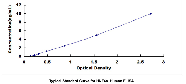
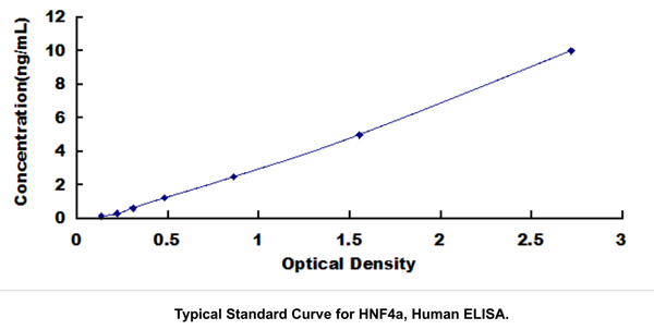
Application Data
(Detection limit for recombinant GST tagged HNF4A is ~1ng/ml as a capture antibody.)
Application Data
(Detection limit for recombinant GST tagged HNF4A is ~1ng/ml as a capture antibody.)
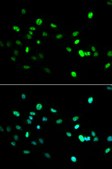
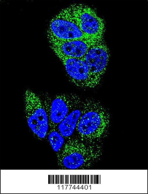
IF (Immunofluorescence)
(Immunofluorescence of monoclonal antibody to HNF4A on HeLa cell. [antibody concentration 10ug/ml])
IF (Immunofluorescence)
(Immunofluorescence of monoclonal antibody to HNF4A on HeLa cell. [antibody concentration 10ug/ml])
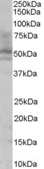
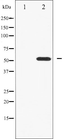
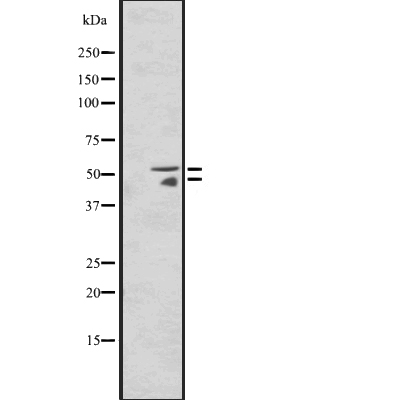
WB (Western Blot)
(Western Blot analysis of HNF4A expression in transfected 293T cell line by HNF4A monoclonal antibody. Lane 1: HNF4A transfected lysate (51.6kD). Lane 2: Non-transfected lysate.)
WB (Western Blot)
(Western Blot analysis of HNF4A expression in transfected 293T cell line by HNF4A monoclonal antibody. Lane 1: HNF4A transfected lysate (51.6kD). Lane 2: Non-transfected lysate.)
WB (Western Blot)
(HNF4A monoclonal antibody Western Blot analysis of HNF4A expression in Jurkat.)
WB (Western Blot)
(HNF4A monoclonal antibody Western Blot analysis of HNF4A expression in Jurkat.)
WB (Western Blot)
(HNF4A monoclonal antibody Western Blot analysis of HNF4A expression in HepG2)
WB (Western Blot)
(HNF4A monoclonal antibody Western Blot analysis of HNF4A expression in HepG2)
WB (Western Blot)
(Western Blot detection against Immunogen (36.74kD).)
WB (Western Blot)
(Western Blot detection against Immunogen (36.74kD).)
NCBI and Uniprot Product Information
Customer Reviews
Loading reviews...
Share Your Experience
Similar Products


Product Notes
The HNF4A hnf4a (Catalog #AAA25126) is an Antibody produced from Mouse and is intended for research purposes only. The product is available for immediate purchase. The HNF4A (Hepatocyte Nuclear Factor 4-alpha, HNF-4-alpha, Transcription Factor HNF-4, Nuclear Receptor Subfamily 2 Group A Member 1, Transcription Factor 14, HNF4, NR2A1, TCF14) (FITC) reacts with Human and may cross-react with other species as described in the data sheet. AAA Biotech's HNF4A can be used in a range of immunoassay formats including, but not limited to, ELISA, IF (Immunofluorescence), WB (Western Blot). IF: 10ug/ml Applications are based on unconjugated antibody. Researchers should empirically determine the suitability of the HNF4A hnf4a for an application not listed in the data sheet. Researchers commonly develop new applications and it is an integral, important part of the investigative research process. It is sometimes possible for the material contained within the vial of "HNF4A, Monoclonal Antibody" to become dispersed throughout the inside of the vial, particularly around the seal of said vial, during shipment and storage. We always suggest centrifuging these vials to consolidate all of the liquid away from the lid and to the bottom of the vial prior to opening. Please be advised that certain products may require dry ice for shipping and that, if this is the case, an additional dry ice fee may also be required.Precautions
All products in the AAA Biotech catalog are strictly for research-use only, and are absolutely not suitable for use in any sort of medical, therapeutic, prophylactic, in-vivo, or diagnostic capacity. By purchasing a product from AAA Biotech, you are explicitly certifying that said products will be properly tested and used in line with industry standard. AAA Biotech and its authorized distribution partners reserve the right to refuse to fulfill any order if we have any indication that a purchaser may be intending to use a product outside of our accepted criteria.Disclaimer
Though we do strive to guarantee the information represented in this datasheet, AAA Biotech cannot be held responsible for any oversights or imprecisions. AAA Biotech reserves the right to adjust any aspect of this datasheet at any time and without notice. It is the responsibility of the customer to inform AAA Biotech of any product performance issues observed or experienced within 30 days of receipt of said product. To see additional details on this or any of our other policies, please see our Terms & Conditions page.Frequently Asked Questions
What tissues or cell types express human HNF4A that this antibody can detect?
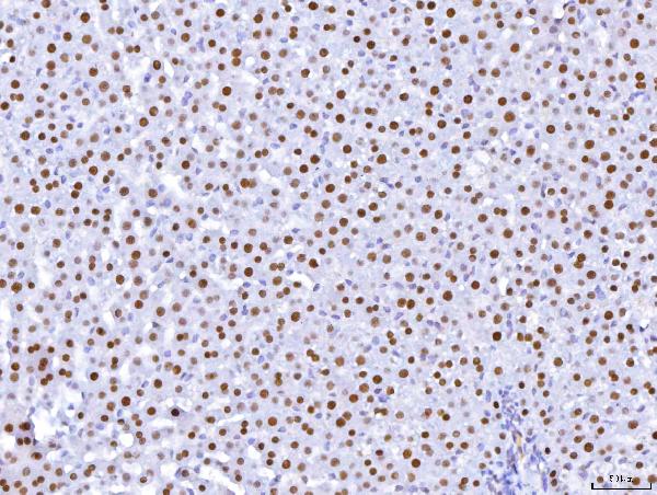
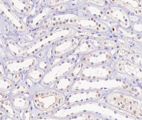
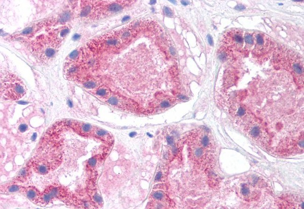
HNF4A (hepatocyte nuclear factor 4-alpha) is predominantly expressed in the liver as a master transcription factor. Significant expression occurs in the kidney (proximal tubules), intestine (enterocytes), pancreas (hepatocytes, β-cells), and stomach epithelium. HNF4A marks differentiated hepatocytes and regulates organ-specific gene expression. AAA Biotech's HNF4A antibody detects nuclear HNF4A across these tissues in normal and diseased states, enabling assessment of liver function, organ development, and cancer-associated expression changes.
Can this HNF4A antibody be used for immunohistochemistry and Western blot?
Yes, HNF4A antibodies work effectively for both applications. In IHC, HNF4A marks hepatocytes and gastrointestinal epithelium; standard antigen retrieval protocols work well. Western blots detect HNF4A at approximately 50-57 kDa (multiple isoforms exist due to alternative splicing). HNF4A levels decrease in disease states (alcoholic liver disease, hepatitis C, cirrhosis, hepatocellular carcinoma), making it valuable for diagnostic assessment and monitoring liver-specific function loss during pathological progression.
How does HNF4A expression change during liver development or disease?
HNF4A expression is essential during fetal liver development. In the adult liver, HNF4A maintains high expression and controls hepatocyte-specific genes (CYP enzymes, albumin, lipoproteins). During disease, HNF4A expression typically decreases: inflammation (IL-1β, TNFα) suppresses HNF4A; alcoholic liver disease reduces expression; hepatocellular carcinoma shows altered P1:P2 isoform ratios. Recovering HNF4A expression correlates with restoration of liver function, making it a prognostic biomarker.
Is this anti-HNF4A antibody suitable for detecting nuclear localization?
Yes, HNF4A is a nuclear receptor exclusively localized to the nucleus, where it functions as a transcription factor. Immunofluorescence and immunohistochemistry reveal concentrated nuclear signal, confirming HNF4A's functional state. Some disease conditions (non-alcoholic fatty liver disease) show cytoplasmic retention, a pathological sign indicating loss of transcriptional activity. Nuclear localization assays using HNF4A antibodies assess whether HNF4A maintains normal nuclear import and transcriptional function in hepatocytes.
Can HNF4A antibody distinguish between normal and diseased liver samples?
Yes, HNF4A serves as a diagnostic marker. Normal liver shows strong, consistent nuclear HNF4A expression. Diseased liver (cirrhosis, hepatitis, hepatocellular carcinoma) displays reduced HNF4A levels or abnormal localization. Quantifying nuclear HNF4A intensity and distribution differentiates healthy from pathological samples. HNF4A loss correlates with hepatocyte dedifferentiation and loss of specialized function. Combined with other markers, HNF4A assessment aids HCC diagnosis, disease staging, and prognosis evaluation.
What dilution or concentration is recommended for best results in IF/WB?
For immunofluorescence: typically 1:100-1:500 primary antibody dilution; optimize based on tissue type and detection method. For Western blot: 1:1000-1:5000 dilution; HNF4A is strongly expressed in hepatocytes, yielding a robust signal. Titrate antibody across lysates to establish optimal dilution within the linear detection range. Multiple isoforms (~50, 57 kDa) appear due to P1/P2 promoter-driven alternative translation; verify the antibody recognizes both forms for comprehensive assessment.
Item has been added to Shopping Cart
If you are ready to order, navigate to Shopping Cart and get ready to checkout.