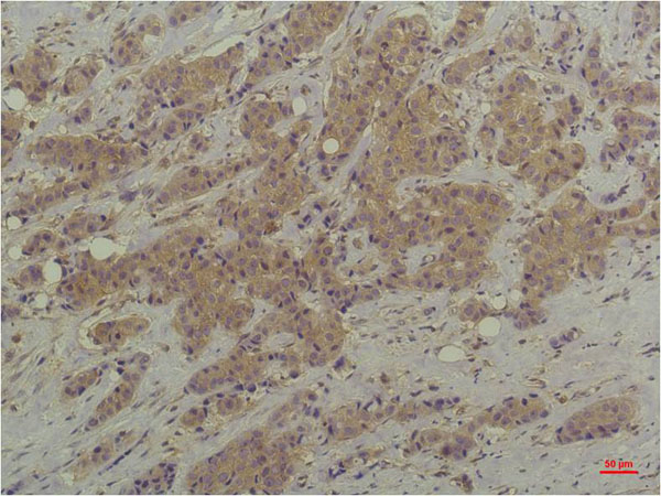
 IHC (Immunohiostchemistry)
(AAA328875 at 1/100 staining Mouse liver tissue sections by IHC-P. The tissue was formaldehyde fixed and a heat mediated antigen retrieval step in citrate buffer was performed. The tissue was then blocked and incubated with the antibody for 1.5 hours at 22°C. An HRP conjugated goat anti-Mouse antibody was used as the secondary antibody.)
IHC (Immunohiostchemistry)
(AAA328875 at 1/100 staining Mouse liver tissue sections by IHC-P. The tissue was formaldehyde fixed and a heat mediated antigen retrieval step in citrate buffer was performed. The tissue was then blocked and incubated with the antibody for 1.5 hours at 22°C. An HRP conjugated goat anti-Mouse antibody was used as the secondary antibody.)
Mouse p44/42 MAPK (Erk1/2) Monoclonal Antibody | anti-MAPK3 antibody
p44/42 MAPK (Erk1/2) Mouse monoclonal Antibody
Gene Names
MAPK3; ERK1; ERT2; ERK-1; PRKM3; P44ERK1; P44MAPK; HS44KDAP; HUMKER1A; p44-ERK1; p44-MAPK
Reactivity
Human, Mouse, Rat
Applications
ELISA, Immunohistochemistry, Western Blot
Purity
Affinity-chromatography
Synonyms
p44/42 MAPK (Erk1/2), Antibody; p44/42 MAPK (Erk1/2) Mouse monoclonal Antibody; ERK 1; ERK; ERK-1; ERK1; ERT 2; ERT2; Extracellular Signal Regulated Kinase 1; Extracellular signal related kinase 1; Extracellular signal-regulated kinase 1; HGNC6877; HS44KDAP; HUMKER1A; Insulin Stimulated MAP2 Kinase; Insulin-stimulated MAP2 kinase; MAP kinase 1; MAP kinase 3; MAP Kinase; MAP kinase isoform p44; MAPK 1; MAPK 3; MAPK; MAPK1; Mapk3; MGC20180; Microtubule Associated Protein 2 Kinase; Microtubule-associated protein 2 kinase; Mitogen Activated Protein Kinase 3; Mitogen-activated protein kinase 1; Mitogen-activated protein kinase 3; MK03_HUMAN; OTTHUMP00000174538; OTTHUMP00000174541; p44 ERK1; p44 MAPK; p44-ERK1; p44-MAPK; P44ERK1; P44MAPK; PRKM 3; PRKM3; Protein Kinase Mitogen Activated 3; ERK 2; ERK-2; ERT1; Extracellular Signal Regulated Kinase 2; Extracellular signal-regulated kinase 2; MAP kinase 2; MAP kinase isoform p42; MAPK 2; Mapk1; MAPK2; Mitogen-activated protein kinase 2; MK01_HUMAN; P38; P40; P41; p42-MAPK; P42MAPK; PRKM1; PRKM2; protein kinase; mitogen-activated; 1; 2; protein tyrosine kinase ERK2; anti-MAPK3 antibody
Host
Mouse
Reactivity
Human, Mouse, Rat
Clonality
Monoclonal
Isotype
IgG1
Specificity
p44/42 MAPK (Erk1/2) antibody detects endogenous levels of total p44/42 MAPK (Erk1/2).
Purity/Purification
Affinity-chromatography
Form/Format
Liquid. Mouse IgG1 in phosphate buffered saline (without Mg2+ and Ca2+), pH7.4, 150mM NaCl, 0.02% sodium azide and 50% glycerol
Concentration
1mg/ml (varies by lot)
Sequence Length
357
Applicable Applications for anti-MAPK3 antibody
ELISA, IHC (Immunohistochemistry), WB (Western Blot)
Immunogen
A synthesized peptide derived from human ERK1/2
Fragment
Fab fragment
Conjugation
Unconjugated
Preparation and Storage
Store at -20 degree C. Stable for 12 months from date of receipt.
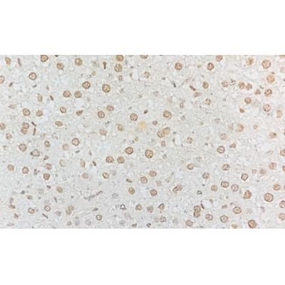
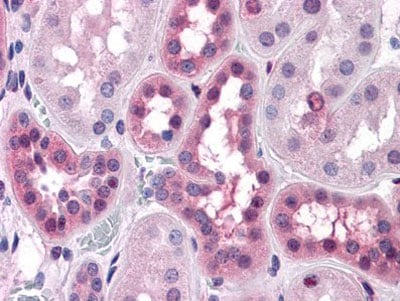
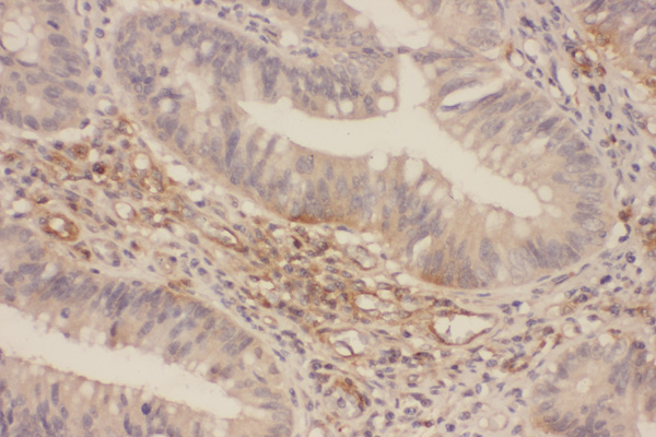
IHC (Immunohiostchemistry)
(AAA328875 at 1/100 staining Mouse liver tissue sections by IHC-P. The tissue was formaldehyde fixed and a heat mediated antigen retrieval step in citrate buffer was performed. The tissue was then blocked and incubated with the antibody for 1.5 hours at 22°C. An HRP conjugated goat anti-Mouse antibody was used as the secondary antibody.)
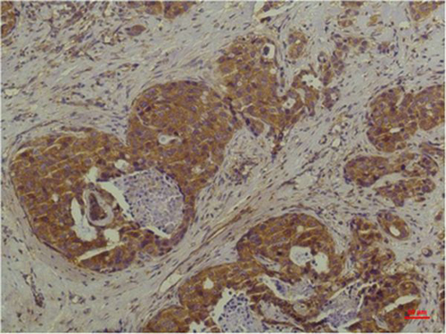
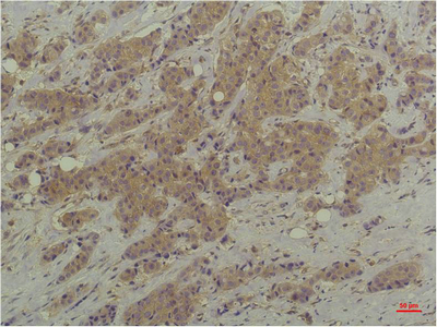
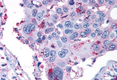
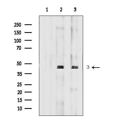
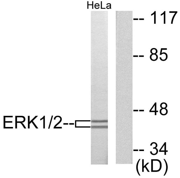
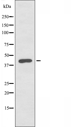
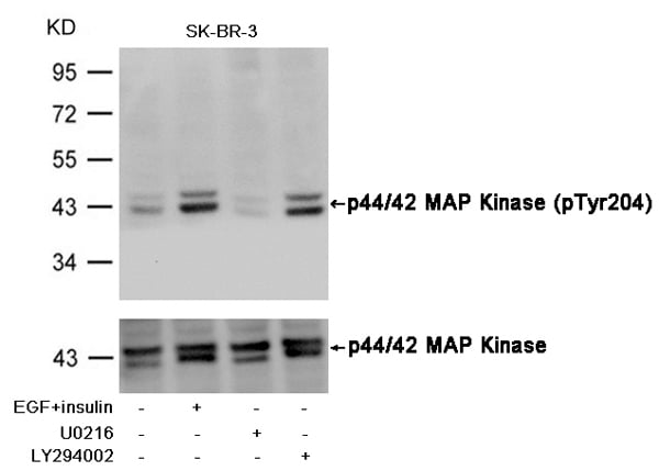
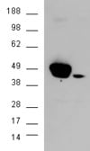
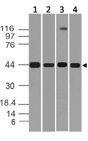
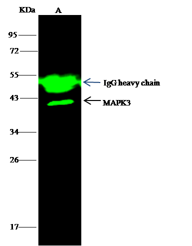
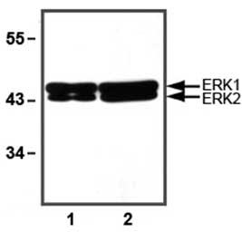
WB (Western Blot)
(Western blot analysis of extracts from various samples, using p44/42 MAPK (Erk1/2) Mouse monoclonal Antibody.Lane 1: 3T3 treated with blocking peptide;Lane 2: 3T3;Lane 3: Rat brain.)
WB (Western Blot)
(Western blot analysis of extracts from various samples, using p44/42 MAPK (Erk1/2) Mouse monoclonal Antibody.Lane 1: 3T3 treated with blocking peptide;Lane 2: 3T3;Lane 3: Rat brain.)
Related Product Information for anti-MAPK3 antibody
Mitogen-activated protein kinases (MAPKs) are a widely conserved family of serine/threonine protein kinases involved in many cellular programs such as cell proliferation, differentiation, motility, and death. The p44/42 MAPK (Erk1/2) signaling pathway can be activated in response to a diverse range of extracellular stimuli including mitogens, growth factors, and cytokines and is an important target in the identification and treatment of cancer. Upon stimulation, a sequential three-part protein kinase cascade is initiated, consisting of a MAP kinase kinase kinase (MAPKKK or MAP3K), a MAP kinase kinase (MAPKK or MAP2K), and a MAP kinase (MAPK). Multiple p44/42 MAP3Ks have been identified, including members of the Raf family as well as Mos and Tpl2/Cot. MEK1 and MEK2 are the primary MAPKKs in this pathway. MEK1 and MEK2 activate p44 and p42 through phosphorylation of activation loop residues Thr202/Tyr204 and Thr185/Tyr187, respectively. Several downstream targets of p44/42 have been identified, including p90RSK and the transcription factor Elk-1. p44/42 are negatively regulated by a family of dual-specificity (Thr/Tyr) MAPK phosphatases, known as DUSPs or MKPs, along with MEK inhibitors such as U0126 and PD98059.
Function: Serine/threonine kinase which acts as an essential component of the MAP kinase signal transduction pathway. MAPK1/ERK2 and MAPK3/ERK1 are the 2 MAPKs which play an important role in the MAPK/ERK cascade. They participate also in a signaling cascade initiated by activated KIT and KITLG/SCF. Depending on the cellular context, the MAPK/ERK cascade mediates diverse biological functions such as cell growth, adhesion, survival and differentiation through the regulation of transcription, translation, cytoskeletal rearrangements. The MAPK/ERK cascade plays also a role in initiation and regulation of meiosis, mitosis, and postmitotic functions in differentiated cells by phosphorylating a number of transcription factors. About 160 substrates have already been discovered for ERKs. Many of these substrates are localized in the nucleus, and seem to participate in the regulation of transcription upon stimulation. However, other substrates are found in the cytosol as well as in other cellular organelles, and those are responsible for processes such as translation, mitosis and apoptosis. Moreover, the MAPK/ERK cascade is also involved in the regulation of the endosomal dynamics, including lysosome processing and endosome cycling through the perinuclear recycling compartment (PNRC); as well as in the fragmentation of the Golgi apparatus during mitosis. The substrates include transcription factors (such as ATF2, BCL6, ELK1, ERF, FOS, HSF4 or SPZ1), cytoskeletal elements (such as CANX, CTTN, GJA1, MAP2, MAPT, PXN, SORBS3 or STMN1), regulators of apoptosis (such as BAD, BTG2, CASP9, DAPK1, IER3, MCL1 or PPARG), regulators of translation (such as EIF4EBP1) and a variety of other signaling-related molecules (like ARHGEF2, DCC, FRS2 or GRB10). Protein kinases (such as RAF1, RPS6KA1/RSK1, RPS6KA3/RSK2, RPS6KA2/RSK3, RPS6KA6/RSK4, SYK, MKNK1/MNK1, MKNK2/MNK2, RPS6KA5/MSK1, RPS6KA4/MSK2, MAPKAPK3 or MAPKAPK5) and phosphatases (such as DUSP1, DUSP4, DUSP6 or DUSP16) are other substrates which enable the propagation the MAPK/ERK signal to additional cytosolic and nuclear targets, thereby extending the specificity of the cascade. Mediates phosphorylation of TPR in respons to EGF stimulation. May play a role in the spindle assembly checkpoint. Phosphorylates PML and promotes its interaction with PIN1, leading to PML degradation. Phosphorylates CDK2AP2 (By similarity).Acts as a transcriptional repressor. Binds to a [GC]AAA[GC] consensus sequence. Repress the expression of interferon gamma-induced genes. Seems to bind to the promoter of CCL5, DMP1, IFIH1, IFITM1, IRF7, IRF9, LAMP3, OAS1, OAS2, OAS3 and STAT1. Transcriptional activity is independent of kinase activity.
Post Translational Modifications: Phosphorylated upon KIT and FLT3 signaling (By similarity). Dually phosphorylated on Thr-185 and Tyr-187, which activates the enzyme. Undergoes regulatory phosphorylation on additional residues such as Ser-246 and Ser-248 in the kinase insert domain (KID) These phosphorylations, which are probably mediated by more than one kinase, are important for binding of MAPK1/ERK2 to importin-7 (IPO7) and its nuclear translocation. In addition, autophosphorylation of Thr-190 was shown to affect the subcellular localization of MAPK1/ERK2 as well. Ligand-activated ALK induces tyrosine phosphorylation. Dephosphorylated by PTPRJ at Tyr-187. Phosphorylation on Ser-29 by SGK1 results in its activation by enhancing its interaction with MAP2K1/MEK1 and MAP2K2/MEK2. DUSP3 and DUSP6 dephosphorylate specifically MAPK1/ERK2 and MAPK3/ERK1 whereas DUSP9 dephosphorylates a broader range of MAPKs. Dephosphorylated by DUSP1 at Thr-185 and Tyr-187.ISGylated.
Subcellular Location: Cytoplasm>Cytoskeleton>Spindle. Nucleus. Cytoplasm>Cytoskeleton>Microtubule organizing center>Centrosome. Cytoplasm. Membrane>Caveola. Note: Associated with the spindle during prometaphase and metaphase (By similarity). PEA15-binding and phosphorylated DAPK1 promote its cytoplasmic retention. Phosphorylation at Ser- 246 and Ser-248 as well as autophosphorylation at Thr-190 promote nuclear localization.
Subunit Structure: Binds both upstream activators and downstream substrates in multimolecular complexes. This interaction inhibits its tyrosine-kinase activity. Interacts with ADAM15, ARHGEF2, ARRB2, DAPK1 (via death domain), HSF4, IER3, IPO7, DUSP6, NISCH, SGK1, and isoform 1 of NEK2. Interacts (phosphorylated form) with CAV2 ('Tyr-19'-phosphorylated form); the interaction, promoted by insulin, leads to nuclear location and MAPK1 activation. Interacts with MORG1, PEA15 and MKNK2 (By similarity). MKNK2 isoform 1 binding prevents from dephosphorylation and inactivation (By similarity). Interacts with DCC (By similarity). The phosphorylated form interacts with PML (isoform PML-4). Interacts with STYX. Interacts with CDK2AP2. Interacts with CAVIN4 (By similarity). Interacts with DUSP7; the interaction enhances DUSP7 phosphatase activity (PubMed:9788880).(Microbial infection) Interacts with HIV-1 Nef through its SH3 domain.
Similarity: The TXY motif contains the threonine and tyrosine residues whose phosphorylation activates the MAP kinases.Belongs to the protein kinase superfamily. CMGC Ser/Thr protein kinase family. MAP kinase subfamily.
NCBI and Uniprot Product Information
NCBI GI #
NCBI GeneID
NCBI Accession #
NCBI GenBank Nucleotide #
Molecular Weight
Observed Molecular Weight: (Observed)42kD, 44kD.
Predicted Molecular Weight: (Calculated)43kDa, 41kDa.
Predicted Molecular Weight: (Calculated)43kDa, 41kDa.
NCBI Official Full Name
mitogen-activated protein kinase 3 isoform 2
NCBI Official Synonym Full Names
mitogen-activated protein kinase 3
NCBI Official Symbol
MAPK3
NCBI Official Synonym Symbols
ERK1; ERT2; ERK-1; PRKM3; P44ERK1; P44MAPK; HS44KDAP; HUMKER1A; p44-ERK1; p44-MAPK
NCBI Protein Information
mitogen-activated protein kinase 3
UniProt Protein Name
Mitogen-activated protein kinase 3
UniProt Gene Name
MAPK3
UniProt Synonym Gene Names
ERK1; PRKM3; MAP kinase 3; MAPK 3; ERK-1; p44-MAPK
UniProt Entry Name
MK03_HUMAN
Customer Reviews
Loading reviews...
Share Your Experience
Similar Products


Product Notes
The MAPK3 mapk3 (Catalog #AAA328875) is an Antibody produced from Mouse and is intended for research purposes only. The product is available for immediate purchase. The p44/42 MAPK (Erk1/2) Mouse monoclonal Antibody reacts with Human, Mouse, Rat and may cross-react with other species as described in the data sheet. AAA Biotech's p44/42 MAPK (Erk1/2) can be used in a range of immunoassay formats including, but not limited to, ELISA, IHC (Immunohistochemistry), WB (Western Blot). Researchers should empirically determine the suitability of the MAPK3 mapk3 for an application not listed in the data sheet. Researchers commonly develop new applications and it is an integral, important part of the investigative research process. It is sometimes possible for the material contained within the vial of "p44/42 MAPK (Erk1/2), Monoclonal Antibody" to become dispersed throughout the inside of the vial, particularly around the seal of said vial, during shipment and storage. We always suggest centrifuging these vials to consolidate all of the liquid away from the lid and to the bottom of the vial prior to opening. Please be advised that certain products may require dry ice for shipping and that, if this is the case, an additional dry ice fee may also be required.Precautions
All products in the AAA Biotech catalog are strictly for research-use only, and are absolutely not suitable for use in any sort of medical, therapeutic, prophylactic, in-vivo, or diagnostic capacity. By purchasing a product from AAA Biotech, you are explicitly certifying that said products will be properly tested and used in line with industry standard. AAA Biotech and its authorized distribution partners reserve the right to refuse to fulfill any order if we have any indication that a purchaser may be intending to use a product outside of our accepted criteria.Disclaimer
Though we do strive to guarantee the information represented in this datasheet, AAA Biotech cannot be held responsible for any oversights or imprecisions. AAA Biotech reserves the right to adjust any aspect of this datasheet at any time and without notice. It is the responsibility of the customer to inform AAA Biotech of any product performance issues observed or experienced within 30 days of receipt of said product. To see additional details on this or any of our other policies, please see our Terms & Conditions page.Item has been added to Shopping Cart
If you are ready to order, navigate to Shopping Cart and get ready to checkout.