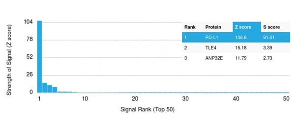
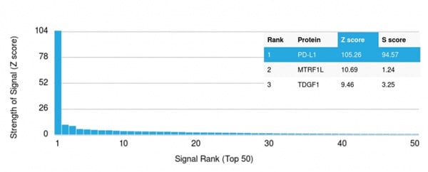
 Application Data
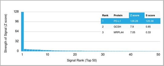
(Analysis of Protein Array containing more than 19,000 full-length human proteins using PD-L1 Mouse Monoclonal Antibody (PDL1/2746). Z- and S- Score: The Z-score represents the strength of a signal that a monoclonal antibody (MAb) (in combination with a fluorescently-tagged anti-IgG secondary antibody) produces when binding to a particular protein on the HuProtTM array. Z-scores are described in units of standard deviations (SD’s) above the mean value of all signals generated on that array. If targets on HuProtTM are arranged in descending order of the Z-score, the S-score is the difference (also in units of SD’s) between the Z-score. S-score therefore represents the relative target specificity of a MAb to its intended target. A MAb is considered to specific to its intended target, if the MAb has an S-score of at least 2.5. For example, if a MAb binds to protein X with a Z-score of 43 and to protein Y with a Z-score of 14, then the S-score for the binding of that MAb to protein X is equal to 29.)
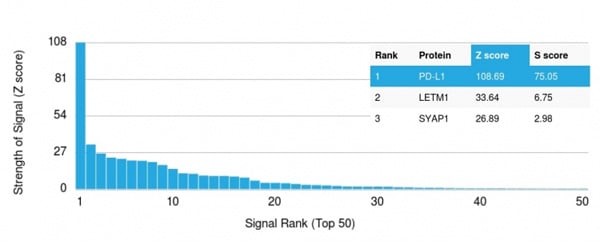
Application Data
(Analysis of Protein Array containing more than 19,000 full-length human proteins using PD-L1 Mouse Monoclonal Antibody (PDL1/2746). Z- and S- Score: The Z-score represents the strength of a signal that a monoclonal antibody (MAb) (in combination with a fluorescently-tagged anti-IgG secondary antibody) produces when binding to a particular protein on the HuProtTM array. Z-scores are described in units of standard deviations (SD’s) above the mean value of all signals generated on that array. If targets on HuProtTM are arranged in descending order of the Z-score, the S-score is the difference (also in units of SD’s) between the Z-score. S-score therefore represents the relative target specificity of a MAb to its intended target. A MAb is considered to specific to its intended target, if the MAb has an S-score of at least 2.5. For example, if a MAb binds to protein X with a Z-score of 43 and to protein Y with a Z-score of 14, then the S-score for the binding of that MAb to protein X is equal to 29.)
Mouse anti-Human, Mouse PD-L1 / PDCD1LG1 / CD274 / B7-H1 Antibody | anti-CD274 antibody
PD-L1 / PDCD1LG1 / CD274 / B7-H1 (Cancer Immunotherapy Target) Mouse Monoclonal Antibody [Clone PDL1/2746]
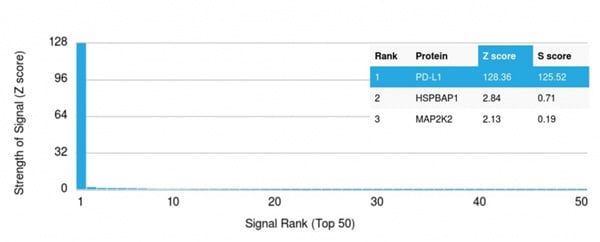
Application Data
(Analysis of Protein Array containing more than 19,000 full-length human proteins using PD-L1 Mouse Monoclonal Antibody (PDL1/2746). Z- and S- Score: The Z-score represents the strength of a signal that a monoclonal antibody (MAb) (in combination with a fluorescently-tagged anti-IgG secondary antibody) produces when binding to a particular protein on the HuProtTM array. Z-scores are described in units of standard deviations (SD’s) above the mean value of all signals generated on that array. If targets on HuProtTM are arranged in descending order of the Z-score, the S-score is the difference (also in units of SD’s) between the Z-score. S-score therefore represents the relative target specificity of a MAb to its intended target. A MAb is considered to specific to its intended target, if the MAb has an S-score of at least 2.5. For example, if a MAb binds to protein X with a Z-score of 43 and to protein Y with a Z-score of 14, then the S-score for the binding of that MAb to protein X is equal to 29.)
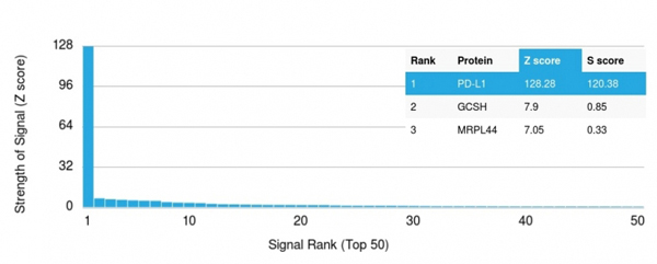
IF (Immunofluorescence)
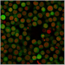
(Immunofluorescence Analysis of Human Jurkat Cells labeling PD-L1 with PD-L1 Mouse Monoclonal Antibody (PDL1/2746) followed by Goat anti-mouse IgG-CF488 (Green). The nuclear counterstain is Reddot (Red))
IF (Immunofluorescence)
(Immunofluorescence Analysis of Human Jurkat Cells labeling PD-L1 with PD-L1 Mouse Monoclonal Antibody (PDL1/2746) followed by Goat anti-mouse IgG-CF488 (Green). The nuclear counterstain is Reddot (Red))
FCM (Flow Cytometry)
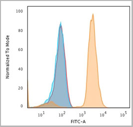
(Flow Cytometric Analysis of Human Jurkat Cells. using PD-L1 Mouse Monoclonal Antibody (PDL1/2746) followed by Goat anti-mouse IgG-CF488 (Orange); Cells alone (Blue); Isotype Control (Red).)
FCM (Flow Cytometry)
(Flow Cytometric Analysis of Human Jurkat Cells. using PD-L1 Mouse Monoclonal Antibody (PDL1/2746) followed by Goat anti-mouse IgG-CF488 (Orange); Cells alone (Blue); Isotype Control (Red).)
WB (Western Blot)
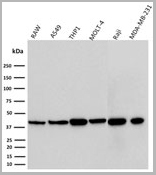
(Western Blot Analysis of human RAW, A549, THP1, MOLT-4, Raji, and MDA-MB-231 Cell Lysate using PD-L1 Mouse Monoclonal Antibody (PDL1/2746).)
WB (Western Blot)
(Western Blot Analysis of human RAW, A549, THP1, MOLT-4, Raji, and MDA-MB-231 Cell Lysate using PD-L1 Mouse Monoclonal Antibody (PDL1/2746).)
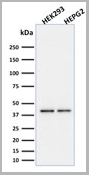
WB (Western Blot)
(Western Blot Analysis of human HEK293 and HepG2 Cell Lysate using PD-L1 Mouse Monoclonal Antibody (PDL1/2746).)
WB (Western Blot)
(Western Blot Analysis of human HEK293 and HepG2 Cell Lysate using PD-L1 Mouse Monoclonal Antibody (PDL1/2746).)
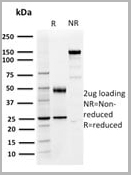
SDS-PAGE
(SDS-PAGE Analysis of Purified PD-L1 Mouse Monoclonal Antibody (PDL1/2746). Confirmation of Purity and Integrity of Antibody.)
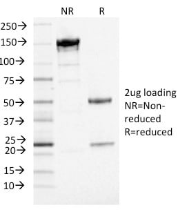
SDS-PAGE
(SDS-PAGE Analysis of Purified PD-L1 Mouse Monoclonal Antibody (PDL1/2746). Confirmation of Purity and Integrity of Antibody.)
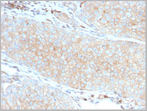
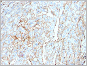
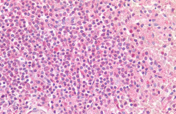
IHC (Immunohistochemistry)
(Formalin-fixed, paraffin-embedded human Basal Cell Carcinoma stained with PD-L1 Mouse Monoclonal Antibody (PDL1/2746).)
IHC (Immunohistochemistry)
(Formalin-fixed, paraffin-embedded human Basal Cell Carcinoma stained with PD-L1 Mouse Monoclonal Antibody (PDL1/2746).)
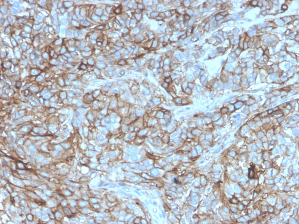
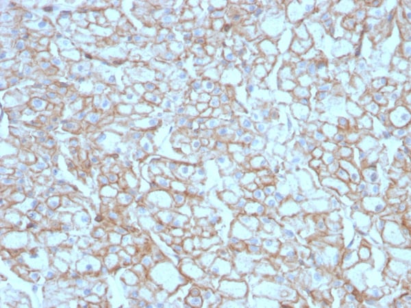
IHC (Immunohistochemistry)
(Formalin-fixed, paraffin-embedded human Breast Carcinoma stained with PD-L1 Mouse Monoclonal Antibody (PDL1/2746).)
IHC (Immunohistochemistry)
(Formalin-fixed, paraffin-embedded human Breast Carcinoma stained with PD-L1 Mouse Monoclonal Antibody (PDL1/2746).)
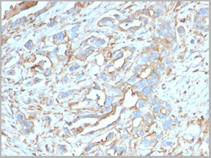
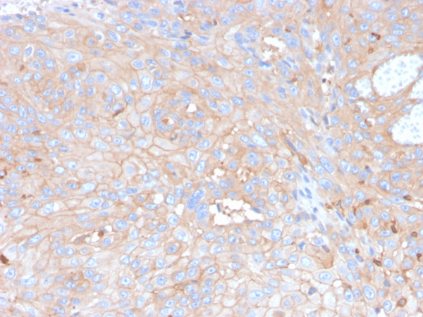
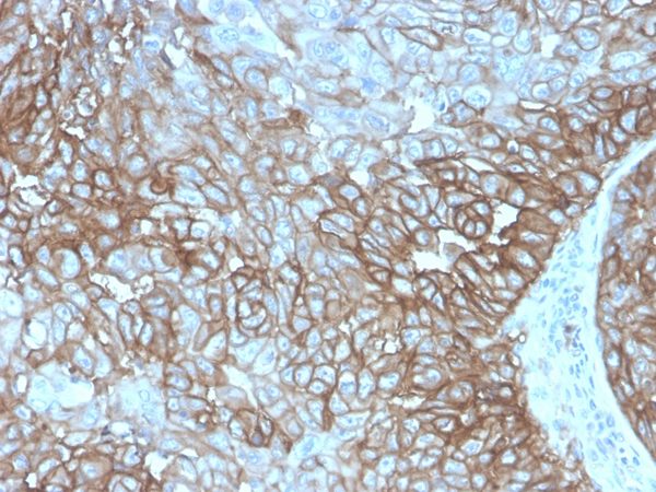
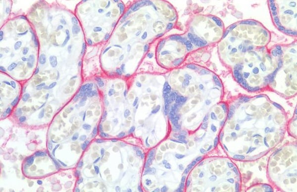
IHC (Immunohistochemistry)
(Formalin-fixed, paraffin-embedded human Lung SCC stained with PD-L1 Mouse Monoclonal Antibody (PDL1/2746).)
IHC (Immunohistochemistry)
(Formalin-fixed, paraffin-embedded human Lung SCC stained with PD-L1 Mouse Monoclonal Antibody (PDL1/2746).)
NCBI and Uniprot Product Information
Customer Reviews
Loading reviews...
Share Your Experience
Similar Products


Product Notes
The CD274 (Catalog #AAA23904) is an Antibody produced from Mouse and is intended for research purposes only. The product is available for immediate purchase. The PD-L1 / PDCD1LG1 / CD274 / B7-H1 (Cancer Immunotherapy Target) Mouse Monoclonal Antibody [Clone PDL1/2746] reacts with Human, Mouse and may cross-react with other species as described in the data sheet. ELISA (For coating, order antibody without BSA); Western Blotting (1-2ug/ml); Flow Cytometry (1-2ug/million cells); Immunofluorescence (1-2ug/ml); Immunohistology (Formalin-fixed) (1-2ug/ml for 30 minutes at RT) (Staining of formalin-fixed tissues requires boiling tissue sections in 10mM Citrate buffer, pH 6.0, for 10-20 min followed by cooling at RT for 20 minutes) Optimal dilution for a specific application should be determined. Researchers should empirically determine the suitability of the CD274 for an application not listed in the data sheet. Researchers commonly develop new applications and it is an integral, important part of the investigative research process. It is sometimes possible for the material contained within the vial of "PD-L1 / PDCD1LG1 / CD274 / B7-H1, Antibody" to become dispersed throughout the inside of the vial, particularly around the seal of said vial, during shipment and storage. We always suggest centrifuging these vials to consolidate all of the liquid away from the lid and to the bottom of the vial prior to opening. Please be advised that certain products may require dry ice for shipping and that, if this is the case, an additional dry ice fee may also be required.Precautions
All products in the AAA Biotech catalog are strictly for research-use only, and are absolutely not suitable for use in any sort of medical, therapeutic, prophylactic, in-vivo, or diagnostic capacity. By purchasing a product from AAA Biotech, you are explicitly certifying that said products will be properly tested and used in line with industry standard. AAA Biotech and its authorized distribution partners reserve the right to refuse to fulfill any order if we have any indication that a purchaser may be intending to use a product outside of our accepted criteria.Disclaimer
Though we do strive to guarantee the information represented in this datasheet, AAA Biotech cannot be held responsible for any oversights or imprecisions. AAA Biotech reserves the right to adjust any aspect of this datasheet at any time and without notice. It is the responsibility of the customer to inform AAA Biotech of any product performance issues observed or experienced within 30 days of receipt of said product. To see additional details on this or any of our other policies, please see our Terms & Conditions page.Item has been added to Shopping Cart
If you are ready to order, navigate to Shopping Cart and get ready to checkout.