Filters
▼Clonality
▼Type
▼Reactivity
▼Gene Name
▼Isotype
▼Host
▼Application
▼Clone
▼Phospho Antibodies
Phospho-specific antibodies’ typical purpose is to enable researchers to detect changes in proteins. They will exclusively bind to the amino acid sequence on a protein that has been phosphorylated (which is both a physical & chemical change) and do not bind to the same amino acid sequence on said protein if it lacks said phosphorylation. This aids in being able to clearly see and understand the data produced from this particular protein modification.
Viewing 100-150 of 5298 product results
 WB (Western Blot)
(All lanes use the Antibody at 1:6K dilution for 1 hour at room temperature.)
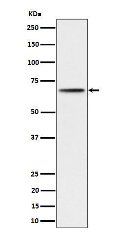
WB (Western Blot)
(All lanes use the Antibody at 1:6K dilution for 1 hour at room temperature.)
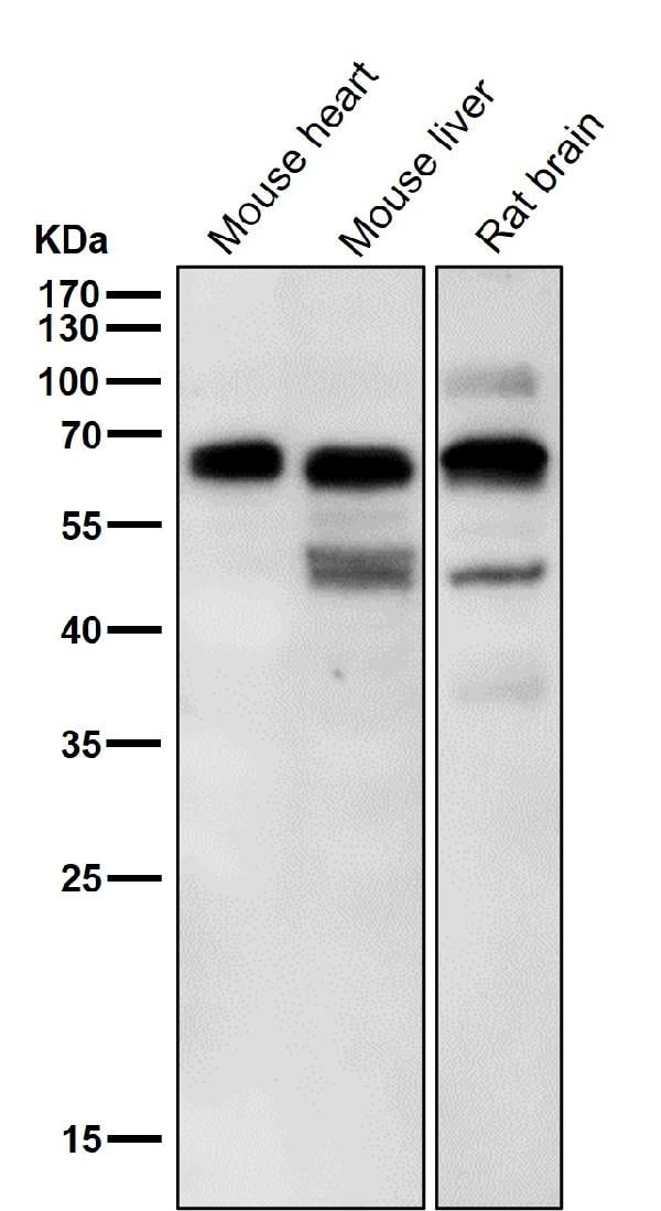
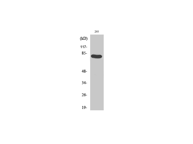
P70 S6 Kinase beta, Monoclonal Antibody (Cat# AAA128176)
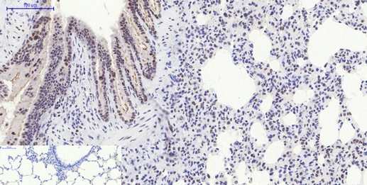
 IHC (Immunohiostchemistry)
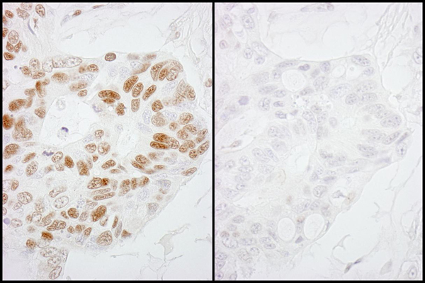
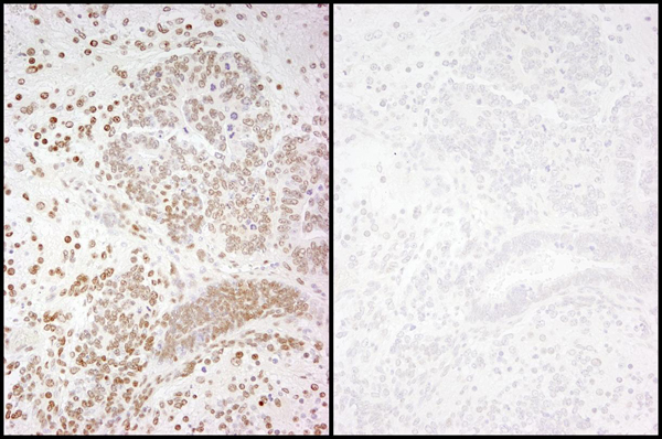
(Detection of mouse Phospho-RNA Polymerase II (S2) by immunohistochemistry. Samples: FFPE serial sections of mouse teratoma. Mock phosphatase treated section (left) or calf intestinal phosphatase-treated section (right) immunostained for Phospho-RNA Polymerase II (S2). Antibody: Affinity purified rabbit anti-Phospho-RNA Polymerase II (S2) (Cat. No. AAA213995) used at a dilution of 1:250. Detection: DAB)
IHC (Immunohiostchemistry)
(Detection of mouse Phospho-RNA Polymerase II (S2) by immunohistochemistry. Samples: FFPE serial sections of mouse teratoma. Mock phosphatase treated section (left) or calf intestinal phosphatase-treated section (right) immunostained for Phospho-RNA Polymerase II (S2). Antibody: Affinity purified rabbit anti-Phospho-RNA Polymerase II (S2) (Cat. No. AAA213995) used at a dilution of 1:250. Detection: DAB)
RNA Polymerase II, Polyclonal Antibody (Cat# AAA213995)
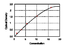
 Standard Curve (Sample)
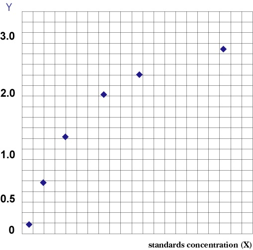 Standard Curve (Sample)
Phospho Calcium/Calmodulin-Dependent Protein Kinase-II, ELISA Kit (Cat# AAA209692)
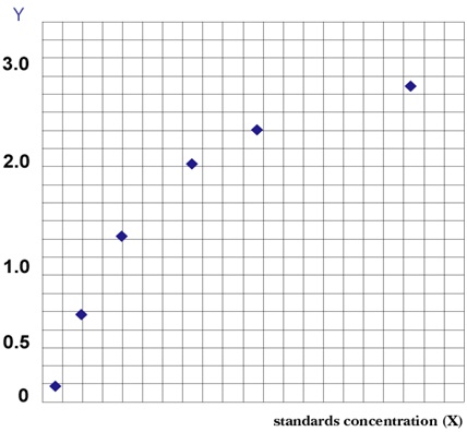 Standard Curve (Sample)
Phospho-Extracellular Signal-Regulated Kinase 2 (PERK1/2), ELISA Kit (Cat# AAA209489)
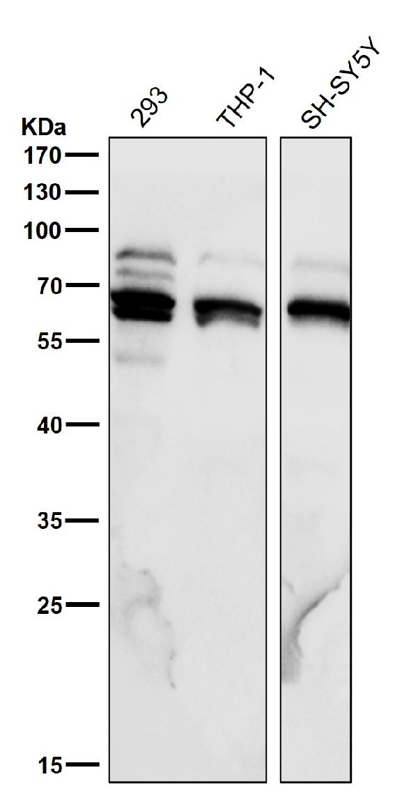
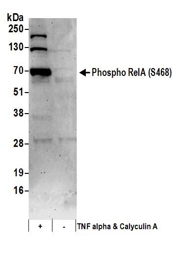 WB (Western Blot)
(Detection of human Phospho RelA (S468) by western blot. Samples: Whole cell lysate (50 ug) from Jurkat cells treated with TNF alpha and Calyculin A (+) or mock treated (-). Antibodies: Affinity purified rabbit anti-Phospho RelA (S468) antibody AAA211747 (lot AAA211747-3) used for WB at 0.1 ug/ml. Detection: Chemiluminescence with an exposure time of 10 seconds.)
WB (Western Blot)
(Detection of human Phospho RelA (S468) by western blot. Samples: Whole cell lysate (50 ug) from Jurkat cells treated with TNF alpha and Calyculin A (+) or mock treated (-). Antibodies: Affinity purified rabbit anti-Phospho RelA (S468) antibody AAA211747 (lot AAA211747-3) used for WB at 0.1 ug/ml. Detection: Chemiluminescence with an exposure time of 10 seconds.)
RelA, Polyclonal Antibody (Cat# AAA211747)
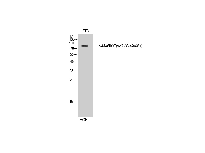 WB (Western Blot)
(Western Blot analysis of 3T3 cells with Phospho-MerTK/Tyro3 (Tyr749/681) Polyclonal Antibody at dilution of 1:500)
WB (Western Blot)
(Western Blot analysis of 3T3 cells with Phospho-MerTK/Tyro3 (Tyr749/681) Polyclonal Antibody at dilution of 1:500)
MerTK/Tyro3, Polyclonal Antibody (Cat# AAA171877)
 WB (Western Blot)
(Western Blot analysis of various cells with Phospho-Stat6 (Tyr641) Polyclonal Antibody)
WB (Western Blot)
(Western Blot analysis of various cells with Phospho-Stat6 (Tyr641) Polyclonal Antibody)
Stat6, Polyclonal Antibody (Cat# AAA171800)
 IF (Immunofluorescence)
(Immunofluorescence analysis of Rat lung tissue with Phospho-p38 (Thr180) Polyclonal Antibody at dilution of 1:200)
IF (Immunofluorescence)
(Immunofluorescence analysis of Rat lung tissue with Phospho-p38 (Thr180) Polyclonal Antibody at dilution of 1:200)
p38, Polyclonal Antibody (Cat# AAA172001)
 IF (Immunofluorescence)
(Immunofluorescence analysis of Rat spleen tissue with Phospho-I?B-? (Ser32/S36) Polyclonal Antibody at dilution of 1:200)
IF (Immunofluorescence)
(Immunofluorescence analysis of Rat spleen tissue with Phospho-I?B-? (Ser32/S36) Polyclonal Antibody at dilution of 1:200)
IkappaB-alpha, Polyclonal Antibody (Cat# AAA171690)
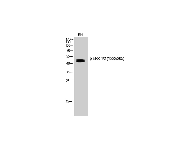
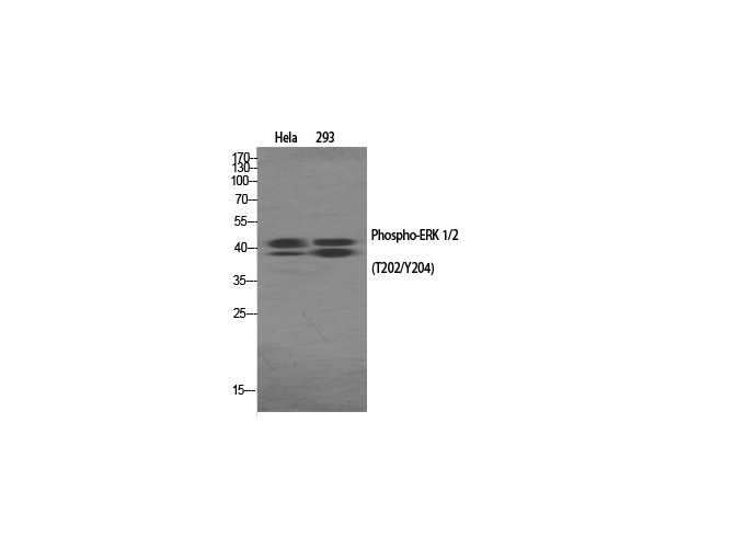
 IF (Immunofluorescence)
(Immunofluorescence analysis of Rat spleen tissue with Phospho-ERK 1/2 (Tyr222/205) Polyclonal Antibody at dilution of 1:200)
IF (Immunofluorescence)
(Immunofluorescence analysis of Rat spleen tissue with Phospho-ERK 1/2 (Tyr222/205) Polyclonal Antibody at dilution of 1:200)
ERK 1/2, Polyclonal Antibody (Cat# AAA171643)
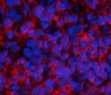
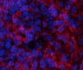 IF (Immunofluorescence)
(Immunofluorescence analysis of Rat spleen tissue with Phospho-ERK 1/2 (Thr202/Tyr204) Polyclonal Antibody at dilution of 1:200)
IF (Immunofluorescence)
(Immunofluorescence analysis of Rat spleen tissue with Phospho-ERK 1/2 (Thr202/Tyr204) Polyclonal Antibody at dilution of 1:200)
ERK 1/2, Polyclonal Antibody (Cat# AAA171662)
 Standard Curve (Sample)
Phospho-Extracellular Signal-Regulated Kinase (PERK1/2), ELISA Kit (Cat# AAA202884)
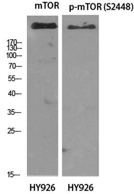
 IF (Immunofluorescence)
(Immunofluorescence analysis of Rat lung tissue with Phospho-mTOR (Ser2448) Polyclonal Antibody at dilution of 1:200)
IF (Immunofluorescence)
(Immunofluorescence analysis of Rat lung tissue with Phospho-mTOR (Ser2448) Polyclonal Antibody at dilution of 1:200)
mTOR, Polyclonal Antibody (Cat# AAA172193)
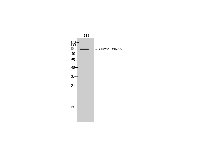 WB (Western Blot)
(Western Blot analysis of 293T cells with Phospho-KIF20A (Ser528) Polyclonal Antibody)
WB (Western Blot)
(Western Blot analysis of 293T cells with Phospho-KIF20A (Ser528) Polyclonal Antibody)
KIF20A, Polyclonal Antibody (Cat# AAA172206)
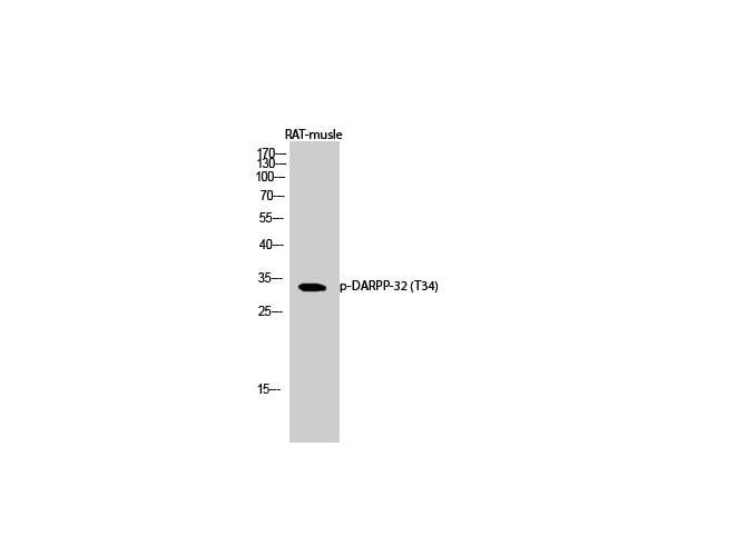 WB (Western Blot)
(Western Blot analysis of Rat musle with Phospho-DARPP-32 (Thr34) Polyclonal Antibody at dilution of 1:1000)
WB (Western Blot)
(Western Blot analysis of Rat musle with Phospho-DARPP-32 (Thr34) Polyclonal Antibody at dilution of 1:1000)
DARPP-32, Polyclonal Antibody (Cat# AAA172345)
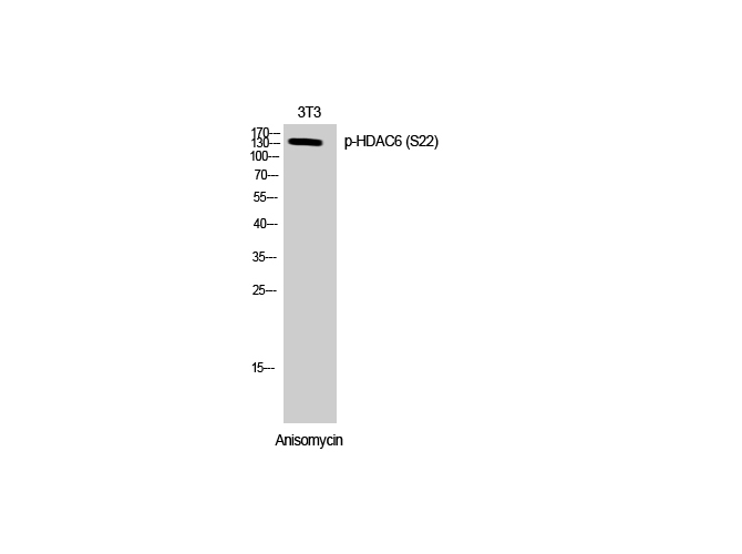 WB (Western Blot)
(Western Blot analysis of 3T3 cells with Phospho-HDAC6 (Ser22) Polyclonal Antibody at dilution of 1:500)
WB (Western Blot)
(Western Blot analysis of 3T3 cells with Phospho-HDAC6 (Ser22) Polyclonal Antibody at dilution of 1:500)
HDAC6, Polyclonal Antibody (Cat# AAA172376)
 IF (Immunofluorescence)
(Immunofluorescence analysis of Rat spleen tissue with Phospho-Stat1 (Tyr701) Polyclonal Antibody at dilution of 1:200)
IF (Immunofluorescence)
(Immunofluorescence analysis of Rat spleen tissue with Phospho-Stat1 (Tyr701) Polyclonal Antibody at dilution of 1:200)
Stat1, Polyclonal Antibody (Cat# AAA172394)
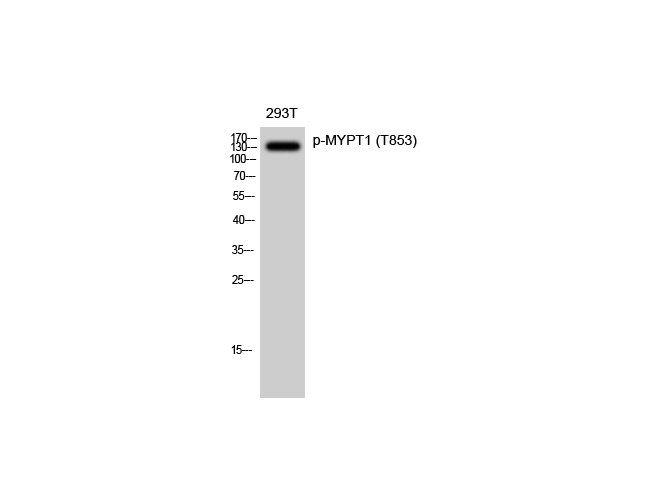
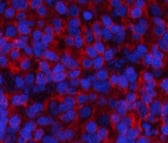
 IF (Immunofluorescence)
(Immunofluorescence analysis of Human lung tissue with Phospho-PPP1R12A (Thr853) Polyclonal Antibody at dilution of 1:200)
IF (Immunofluorescence)
(Immunofluorescence analysis of Human lung tissue with Phospho-PPP1R12A (Thr853) Polyclonal Antibody at dilution of 1:200)
MYPT1, Polyclonal Antibody (Cat# AAA172568)
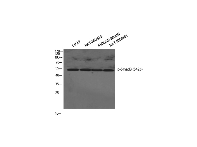
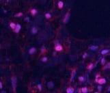
 IF (Immunofluorescence)
(Immunofluorescence analysis of Rat lung tissue with Phospho-Smad3 (Ser425) Polyclonal Antibody at dilution of 1:200)
IF (Immunofluorescence)
(Immunofluorescence analysis of Rat lung tissue with Phospho-Smad3 (Ser425) Polyclonal Antibody at dilution of 1:200)
Smad3, Polyclonal Antibody (Cat# AAA172574)
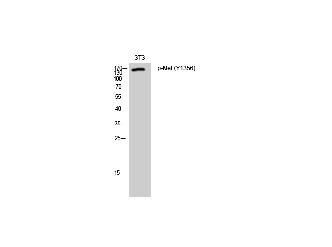 WB (Western Blot)
(Western Blot analysis of 3T3 cells with Phospho-Met (Tyr1356) Polyclonal Antibody)
WB (Western Blot)
(Western Blot analysis of 3T3 cells with Phospho-Met (Tyr1356) Polyclonal Antibody)
Met, Polyclonal Antibody (Cat# AAA172588)
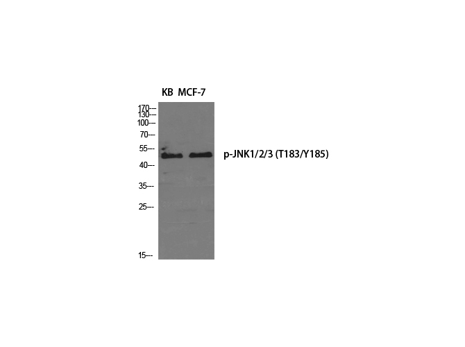
 IF (Immunofluorescence)
(Immunofluorescence analysis of Mouse liver tissue with Phospho-JNK1/2/3 (Thr183/Y185) Polyclonal Antibody at dilution of 1:200)
IF (Immunofluorescence)
(Immunofluorescence analysis of Mouse liver tissue with Phospho-JNK1/2/3 (Thr183/Y185) Polyclonal Antibody at dilution of 1:200)
JNK1/2/3, Polyclonal Antibody (Cat# AAA172608)
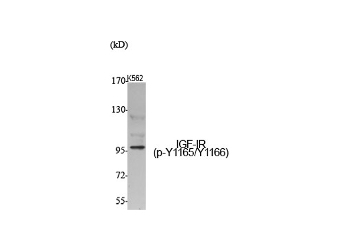
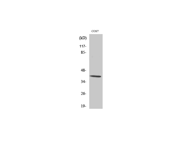
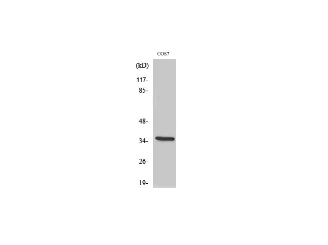
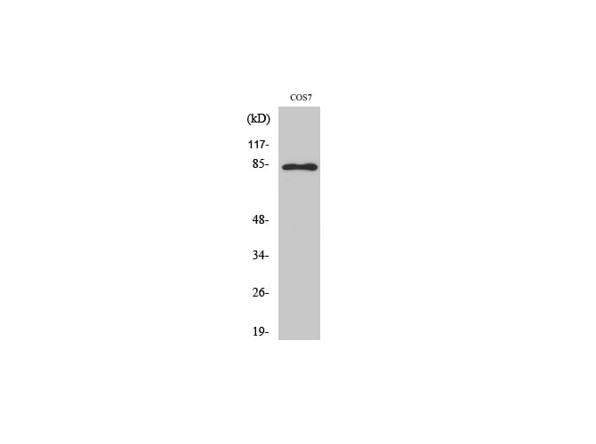
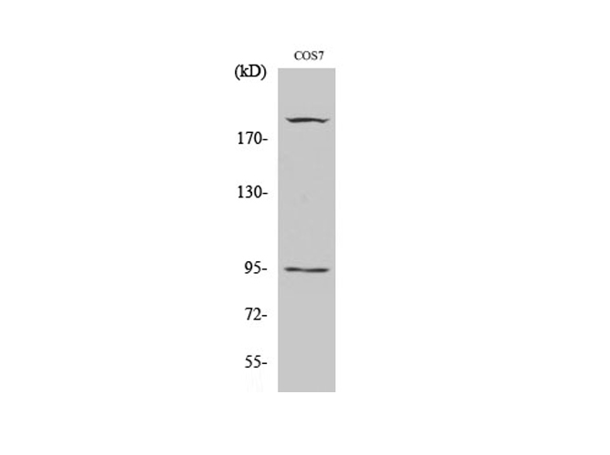 WB (Western Blot)
(Western Blot analysis of COS7 cells using Phospho-IGF-IR (Tyr1165/Y1166) Polyclonal Antibody at dilution of 1:500.)
WB (Western Blot)
(Western Blot analysis of COS7 cells using Phospho-IGF-IR (Tyr1165/Y1166) Polyclonal Antibody at dilution of 1:500.)
IGF-IR, Polyclonal Antibody (Cat# AAA172407)
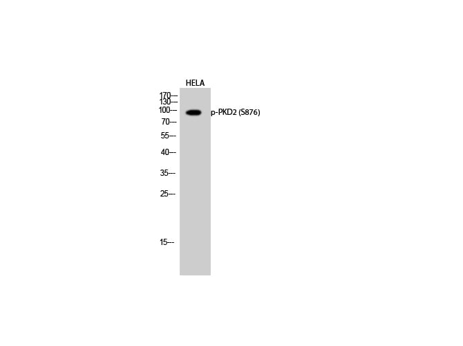 WB (Western Blot)
(Western Blot analysis of Hela cells with Phospho-PKD2 (Ser876) Polyclonal Antibody at dilution of 1:1000)
WB (Western Blot)
(Western Blot analysis of Hela cells with Phospho-PKD2 (Ser876) Polyclonal Antibody at dilution of 1:1000)
PKD2, Polyclonal Antibody (Cat# AAA172432)
 IF (Immunofluorescence)
(Immunofluorescence analysis of Rat lung tissue with Phospho-Chk2 (Thr68) Polyclonal Antibody at dilution of 1:200)
IF (Immunofluorescence)
(Immunofluorescence analysis of Rat lung tissue with Phospho-Chk2 (Thr68) Polyclonal Antibody at dilution of 1:200)
Chk2, Polyclonal Antibody (Cat# AAA172461)
 WB (Western Blot)
(Western Blot analysis of 293T cells with Phospho-Caldesmon (Ser789) Polyclonal Antibody)
WB (Western Blot)
(Western Blot analysis of 293T cells with Phospho-Caldesmon (Ser789) Polyclonal Antibody)
Caldesmon, Polyclonal Antibody (Cat# AAA172094)
 Standard Curve (Sample)
phospho- cAMP response element binding protein (pCREB), ELISA Kit (Cat# AAA121207)
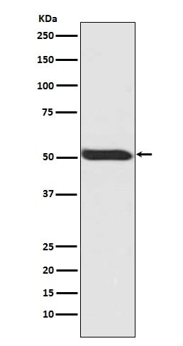 WB (Western Blot)
(Western blot analysis of Phospho-p53 (Ser392) expression in HEK293 whole cell lysate (AAA124519).Electrophoresis was performed on a 5-20% SDS-PAGE gel at 70V (Stacking gel) / 90V (Resolving gel) for 2-3 hours. The sample well of each lane was loaded with 50ug of sample under reducing conditions.After Electrophoresis, proteins were transferred to a Nitrocellulose membrane at 150mA for 50-90 minutes. Blocked the membrane with 5% Non-fat Milk/ TBS for 1.5 hour at RT. The membrane was incubated with rabbit anti-TP53 monoclonal antibody overnight at 4 degree C, then washed with TBS-0.1%Tween 3 times with 5 minutes each and probed with a goat anti-rabbit IgG-HRP secondary antibody at a dilution of 1:10000 for 1.5 hour at RT. The signal is developed using an Enhanced Chemiluminescent detection (ECL) kit with Tanon 5200 system. A specific band was detected for TP53)
WB (Western Blot)
(Western blot analysis of Phospho-p53 (Ser392) expression in HEK293 whole cell lysate (AAA124519).Electrophoresis was performed on a 5-20% SDS-PAGE gel at 70V (Stacking gel) / 90V (Resolving gel) for 2-3 hours. The sample well of each lane was loaded with 50ug of sample under reducing conditions.After Electrophoresis, proteins were transferred to a Nitrocellulose membrane at 150mA for 50-90 minutes. Blocked the membrane with 5% Non-fat Milk/ TBS for 1.5 hour at RT. The membrane was incubated with rabbit anti-TP53 monoclonal antibody overnight at 4 degree C, then washed with TBS-0.1%Tween 3 times with 5 minutes each and probed with a goat anti-rabbit IgG-HRP secondary antibody at a dilution of 1:10000 for 1.5 hour at RT. The signal is developed using an Enhanced Chemiluminescent detection (ECL) kit with Tanon 5200 system. A specific band was detected for TP53)
p53, Monoclonal Antibody (Cat# AAA124519)
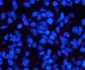
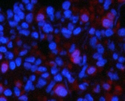
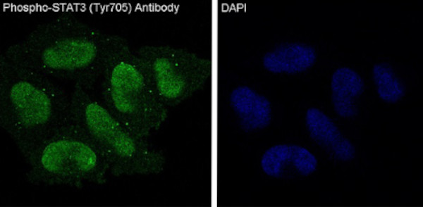 IF (Immunofluorescence)
(Immunofluorescent analysis of HeLa cells treated with IFN-alpha, using Phospho-STAT3 (Y705) Antibody.)
IF (Immunofluorescence)
(Immunofluorescent analysis of HeLa cells treated with IFN-alpha, using Phospho-STAT3 (Y705) Antibody.)
STAT3, Monoclonal Antibody (Cat# AAA124520)
 WB (Western Blot)
(Western blot analysis of Nrf2 phosphorylation expression in HepG2 cell lysate (AAA124527).Electrophoresis was performed on a 5-20% SDS-PAGE gel at 70V (Stacking gel) / 90V (Resolving gel) for 2-3 hours. The sample well of each lane was loaded with 50ug of sample under reducing conditions.After Electrophoresis, proteins were transferred to a Nitrocellulose membrane at 150mA for 50-90 minutes. Blocked the membrane with 5% Non-fat Milk/ TBS for 1.5 hour at RT. The membrane was incubated with rabbit anti-NFE2L2 monoclonal antibody overnight at 4 degree C, then washed with TBS-0.1%Tween 3 times with 5 minutes each and probed with a goat anti-rabbit IgG-HRP secondary antibody at a dilution of 1:10000 for 1.5 hour at RT. The signal is developed using an Enhanced Chemiluminescent detection (ECL) kit with Tanon 5200 system. A specific band was detected for NFE2L2)
WB (Western Blot)
(Western blot analysis of Nrf2 phosphorylation expression in HepG2 cell lysate (AAA124527).Electrophoresis was performed on a 5-20% SDS-PAGE gel at 70V (Stacking gel) / 90V (Resolving gel) for 2-3 hours. The sample well of each lane was loaded with 50ug of sample under reducing conditions.After Electrophoresis, proteins were transferred to a Nitrocellulose membrane at 150mA for 50-90 minutes. Blocked the membrane with 5% Non-fat Milk/ TBS for 1.5 hour at RT. The membrane was incubated with rabbit anti-NFE2L2 monoclonal antibody overnight at 4 degree C, then washed with TBS-0.1%Tween 3 times with 5 minutes each and probed with a goat anti-rabbit IgG-HRP secondary antibody at a dilution of 1:10000 for 1.5 hour at RT. The signal is developed using an Enhanced Chemiluminescent detection (ECL) kit with Tanon 5200 system. A specific band was detected for NFE2L2)
Nrf2, Monoclonal Antibody (Cat# AAA124527)
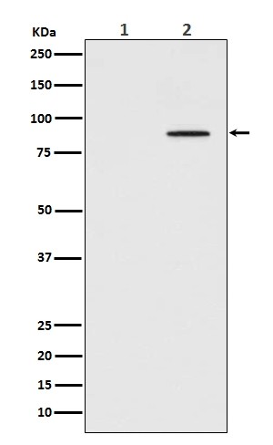
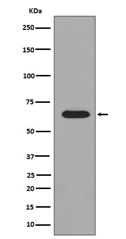
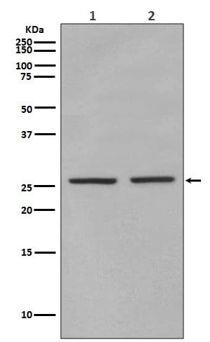 WB (Western Blot)
(Western blot analysis of eIF4E (phosphoS209) expression in (1) HEK293 cell lysate; (2) Mouse spleen lysate (AAA124528).Electrophoresis was performed on a 5-20% SDS-PAGE gel at 70V (Stacking gel) / 90V (Resolving gel) for 2-3 hours. The sample well of each lane was loaded with 50ug of sample under reducing conditions.After Electrophoresis, proteins were transferred to a Nitrocellulose membrane at 150mA for 50-90 minutes. Blocked the membrane with 5% Non-fat Milk/ TBS for 1.5 hour at RT. The membrane was incubated with rabbit anti-EIF4E monoclonal antibody overnight at 4 degree C, then washed with TBS-0.1%Tween 3 times with 5 minutes each and probed with a goat anti-rabbit IgG-HRP secondary antibody at a dilution of 1:10000 for 1.5 hour at RT. The signal is developed using an Enhanced Chemiluminescent detection (ECL) kit with Tanon 5200 system. A specific band was detected for EIF4E)
WB (Western Blot)
(Western blot analysis of eIF4E (phosphoS209) expression in (1) HEK293 cell lysate; (2) Mouse spleen lysate (AAA124528).Electrophoresis was performed on a 5-20% SDS-PAGE gel at 70V (Stacking gel) / 90V (Resolving gel) for 2-3 hours. The sample well of each lane was loaded with 50ug of sample under reducing conditions.After Electrophoresis, proteins were transferred to a Nitrocellulose membrane at 150mA for 50-90 minutes. Blocked the membrane with 5% Non-fat Milk/ TBS for 1.5 hour at RT. The membrane was incubated with rabbit anti-EIF4E monoclonal antibody overnight at 4 degree C, then washed with TBS-0.1%Tween 3 times with 5 minutes each and probed with a goat anti-rabbit IgG-HRP secondary antibody at a dilution of 1:10000 for 1.5 hour at RT. The signal is developed using an Enhanced Chemiluminescent detection (ECL) kit with Tanon 5200 system. A specific band was detected for EIF4E)
eIF4E, Monoclonal Antibody (Cat# AAA124528)
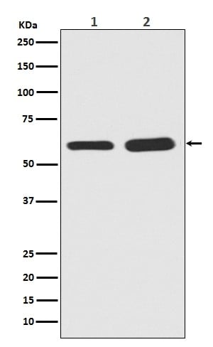 WB (Western Blot)
(Western blot analysis of Phospho-Smad5 in (1)Mouse brain tissue lysate; (2)Rat brain tissue lysate (AAA124535).Electrophoresis was performed on a 5-20% SDS-PAGE gel at 70V (Stacking gel) / 90V (Resolving gel) for 2-3 hours. The sample well of each lane was loaded with 50ug of sample under reducing conditions.After Electrophoresis, proteins were transferred to a Nitrocellulose membrane at 150mA for 50-90 minutes. Blocked the membrane with 5% Non-fat Milk/ TBS for 1.5 hour at RT. The membrane was incubated with rabbit anti-SMAD5 monoclonal antibody overnight at 4 degree C, then washed with TBS-0.1%Tween 3 times with 5 minutes each and probed with a goat anti-rabbit IgG-HRP secondary antibody at a dilution of 1:10000 for 1.5 hour at RT. The signal is developed using an Enhanced Chemiluminescent detection (ECL) kit with Tanon 5200 system. A specific band was detected for SMAD5)
WB (Western Blot)
(Western blot analysis of Phospho-Smad5 in (1)Mouse brain tissue lysate; (2)Rat brain tissue lysate (AAA124535).Electrophoresis was performed on a 5-20% SDS-PAGE gel at 70V (Stacking gel) / 90V (Resolving gel) for 2-3 hours. The sample well of each lane was loaded with 50ug of sample under reducing conditions.After Electrophoresis, proteins were transferred to a Nitrocellulose membrane at 150mA for 50-90 minutes. Blocked the membrane with 5% Non-fat Milk/ TBS for 1.5 hour at RT. The membrane was incubated with rabbit anti-SMAD5 monoclonal antibody overnight at 4 degree C, then washed with TBS-0.1%Tween 3 times with 5 minutes each and probed with a goat anti-rabbit IgG-HRP secondary antibody at a dilution of 1:10000 for 1.5 hour at RT. The signal is developed using an Enhanced Chemiluminescent detection (ECL) kit with Tanon 5200 system. A specific band was detected for SMAD5)
Smad5, Monoclonal Antibody (Cat# AAA124535)
 WB (Western Blot)
(Western blot analysis of Phospho-PAK1/2/3 expression in HeLa Cell lysate treated with lambda phosphatase (AAA124537).Electrophoresis was performed on a 5-20% SDS-PAGE gel at 70V (Stacking gel) / 90V (Resolving gel) for 2-3 hours. The sample well of each lane was loaded with 50ug of sample under reducing conditions.After Electrophoresis, proteins were transferred to a Nitrocellulose membrane at 150mA for 50-90 minutes. Blocked the membrane with 5% Non-fat Milk/ TBS for 1.5 hour at RT. The membrane was incubated with rabbit anti-PAK3 monoclonal antibody overnight at 4 degree C, then washed with TBS-0.1%Tween 3 times with 5 minutes each and probed with a goat anti-rabbit IgG-HRP secondary antibody at a dilution of 1:10000 for 1.5 hour at RT. The signal is developed using an Enhanced Chemiluminescent detection (ECL) kit with Tanon 5200 system. A specific band was detected for PAK3)
WB (Western Blot)
(Western blot analysis of Phospho-PAK1/2/3 expression in HeLa Cell lysate treated with lambda phosphatase (AAA124537).Electrophoresis was performed on a 5-20% SDS-PAGE gel at 70V (Stacking gel) / 90V (Resolving gel) for 2-3 hours. The sample well of each lane was loaded with 50ug of sample under reducing conditions.After Electrophoresis, proteins were transferred to a Nitrocellulose membrane at 150mA for 50-90 minutes. Blocked the membrane with 5% Non-fat Milk/ TBS for 1.5 hour at RT. The membrane was incubated with rabbit anti-PAK3 monoclonal antibody overnight at 4 degree C, then washed with TBS-0.1%Tween 3 times with 5 minutes each and probed with a goat anti-rabbit IgG-HRP secondary antibody at a dilution of 1:10000 for 1.5 hour at RT. The signal is developed using an Enhanced Chemiluminescent detection (ECL) kit with Tanon 5200 system. A specific band was detected for PAK3)
PAK1/2/3, Monoclonal Antibody (Cat# AAA124537)
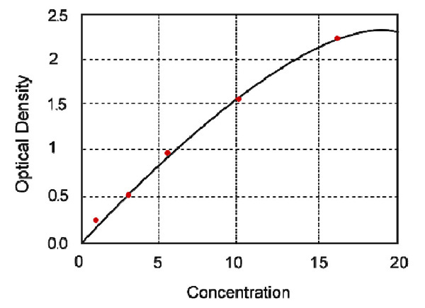 Standard Curve (Sample)
Phospho-Insulin receptor substrate 1 (Ty312), IRS1, ELISA Kit (Cat# AAA123755)
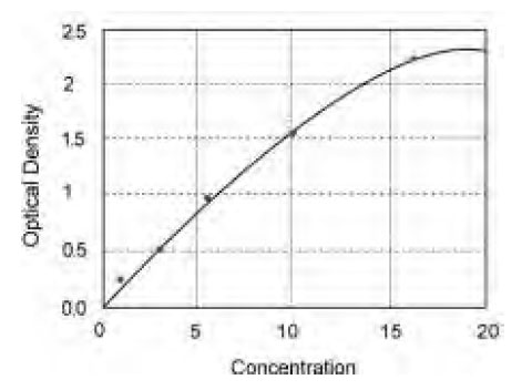
 Standard Curve (Sample)
phospho-Insulin receptor substrate 1(panTyr), p-IRS-1, ELISA Kit (Cat# AAA123764)
 Standard Curve (Sample)
Phospho-Stat3 (Tyr705), p-Stat3, ELISA Kit (Cat# AAA123894)
 Standard Curve (Sample)
Phospho-eNOS, ELISA Kit (Cat# AAA208307)
 Standard Curve (Sample)
Phospho-Cyclic Adenosine Monophosphate, ELISA Kit (Cat# AAA207648)
 WB (Western Blot)
(Western blot analysis of extracts from 293 cells (10% serum-treated, 15min) and HeLa cells (EGF-treated, 200ng/ml, 15min), using FKHR (Ab-319) antibody (Line 1, 2 and 3) and FKHR (phospho-Ser319) antibody (Line 4 and 5).)
WB (Western Blot)
(Western blot analysis of extracts from 293 cells (10% serum-treated, 15min) and HeLa cells (EGF-treated, 200ng/ml, 15min), using FKHR (Ab-319) antibody (Line 1, 2 and 3) and FKHR (phospho-Ser319) antibody (Line 4 and 5).)
FKHR, Antibody (Cat# AAA112224)
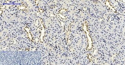
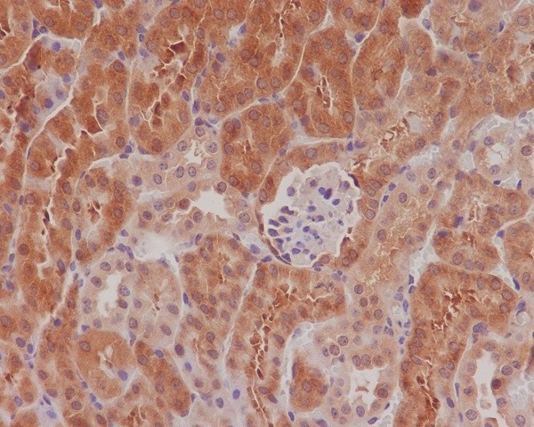
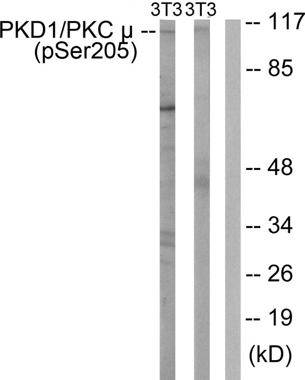 IHC (Immunohiostchemistry)
(Immunohistochemistry analysis of paraffin-embedded human brain tissue using PKD1/PKC mu (Phospho-Ser205) antibody. Western blot analysis of extracts from NIH/3T3 cells treated with Anisomycin (25mug/ml, 30mins) (lane 1) and NIH/3T3 cells treated with H2O2 (100muM, 30mins) (lane 2), using PKD1/PKC mu (Phospho-Ser205) antibody.)
IHC (Immunohiostchemistry)
(Immunohistochemistry analysis of paraffin-embedded human brain tissue using PKD1/PKC mu (Phospho-Ser205) antibody. Western blot analysis of extracts from NIH/3T3 cells treated with Anisomycin (25mug/ml, 30mins) (lane 1) and NIH/3T3 cells treated with H2O2 (100muM, 30mins) (lane 2), using PKD1/PKC mu (Phospho-Ser205) antibody.)
PKD1/PKC, Antibody (Cat# AAA112236)
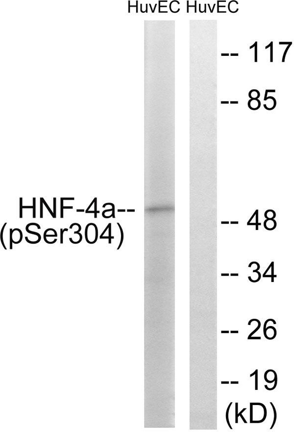
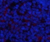
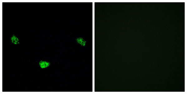 IF (Immunofluorescence)
(P-peptide-+ Immunofluorescence analysis of LOVO cells, using HNF4alpha (Phospho-Ser313) antibody.)
IF (Immunofluorescence)
(P-peptide-+ Immunofluorescence analysis of LOVO cells, using HNF4alpha (Phospho-Ser313) antibody.)
HNF4alpha, Antibody (Cat# AAA111996)
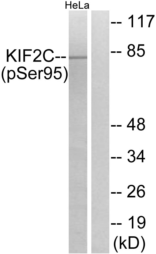
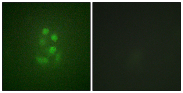 WB (Western Blot)
(P-peptide-+ Western blot analysis of extracts from HeLa cells, treated with TNF (10ng/ml, 30mins), using KIF2C (Phospho-Ser95) antibody. Immunofluorescence analysis of A549 cells, using KIF2C (Phospho-Ser95) antibody.)
WB (Western Blot)
(P-peptide-+ Western blot analysis of extracts from HeLa cells, treated with TNF (10ng/ml, 30mins), using KIF2C (Phospho-Ser95) antibody. Immunofluorescence analysis of A549 cells, using KIF2C (Phospho-Ser95) antibody.)
KIF2C, Antibody (Cat# AAA112000)
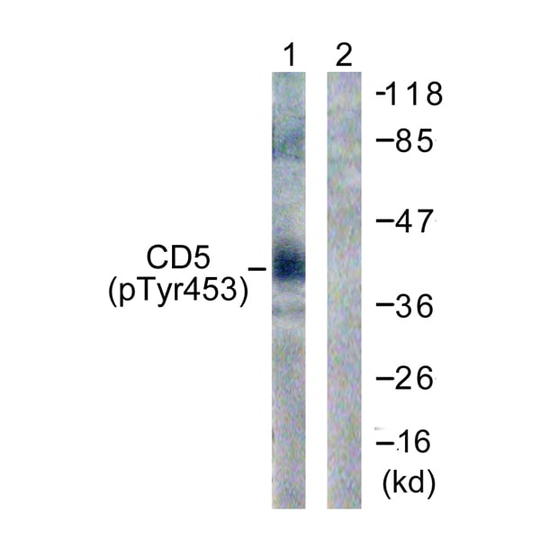
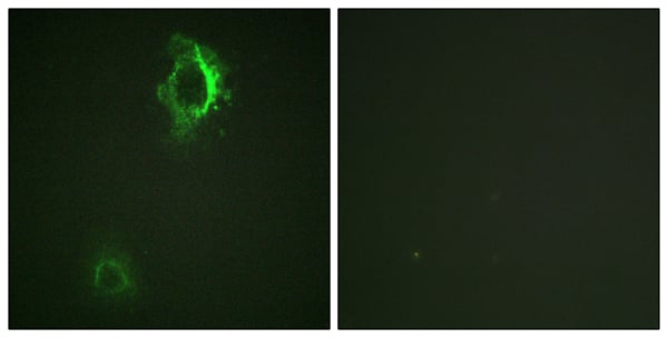 WB (Western Blot)
(P-peptide-+ Western blot analysis of extracts from 293 cells, treated with PMA (125ng/ml, 30mins), using CD5 (Phospho-Tyr453) antibody. Immunofluorescence analysis of HepG2 cells, using CD5 (Phospho-Tyr453) antibody.)
WB (Western Blot)
(P-peptide-+ Western blot analysis of extracts from 293 cells, treated with PMA (125ng/ml, 30mins), using CD5 (Phospho-Tyr453) antibody. Immunofluorescence analysis of HepG2 cells, using CD5 (Phospho-Tyr453) antibody.)
CD5, Antibody (Cat# AAA112002)
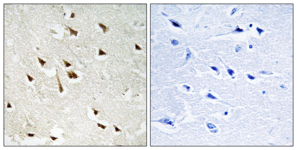
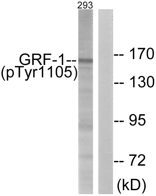 IHC (Immunohiostchemistry)
(Immunohistochemistry analysis of paraffin-embedded human brain tissue using GRF-1 (Phospho-Tyr1105) antibody. Western blot analysis of extracts from 293 cells, treated with EGF (200ng/ml, 30mins), using GRF-1 (Phospho-Tyr1105) antibody.)
IHC (Immunohiostchemistry)
(Immunohistochemistry analysis of paraffin-embedded human brain tissue using GRF-1 (Phospho-Tyr1105) antibody. Western blot analysis of extracts from 293 cells, treated with EGF (200ng/ml, 30mins), using GRF-1 (Phospho-Tyr1105) antibody.)
GRF-1, Antibody (Cat# AAA112009)
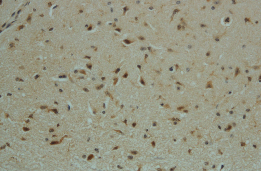
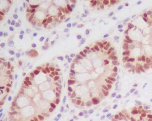
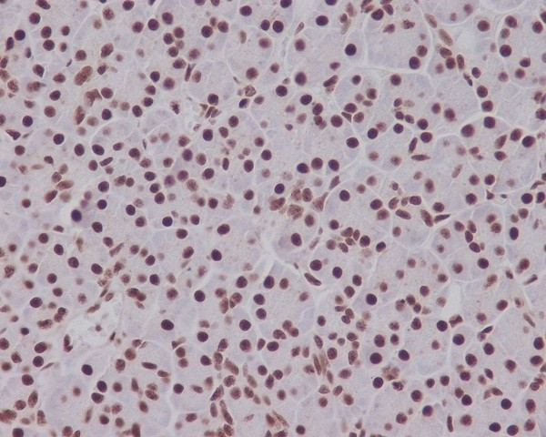
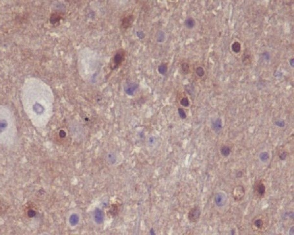
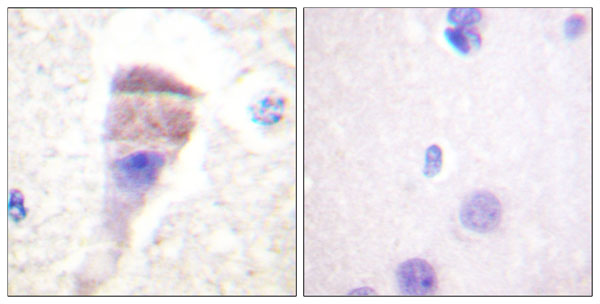
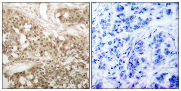
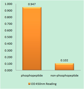 IHC (Immunohiostchemistry)
(Immunohistochemistry analysis of paraffin-embedded human brain tissue using Aquaporin 2 (Phospho-Ser256) antibody. Aquaporin 2 (Phospho-Ser256) antibody reacts with epitope-specific phosphopeptide and corresponding non-phosphopeptide. The absorbance readings at 450 nM are shown in the ELISA figure.)
IHC (Immunohiostchemistry)
(Immunohistochemistry analysis of paraffin-embedded human brain tissue using Aquaporin 2 (Phospho-Ser256) antibody. Aquaporin 2 (Phospho-Ser256) antibody reacts with epitope-specific phosphopeptide and corresponding non-phosphopeptide. The absorbance readings at 450 nM are shown in the ELISA figure.)
Aquaporin 2, Antibody (Cat# AAA112011)
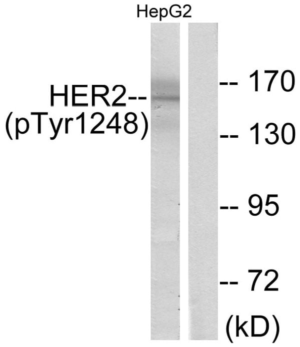
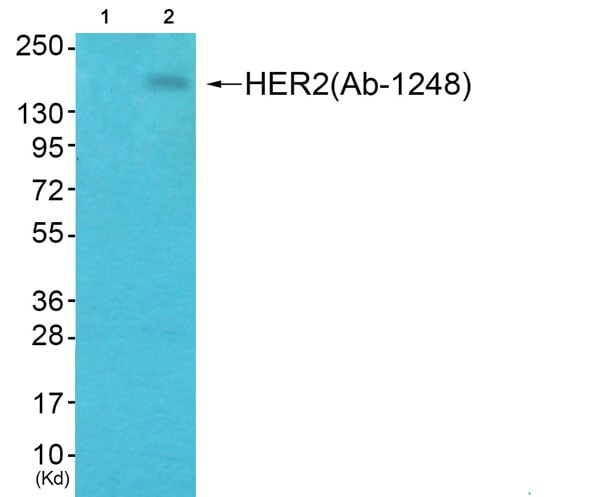
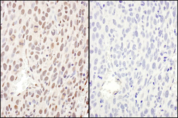
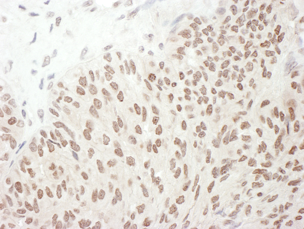
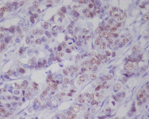
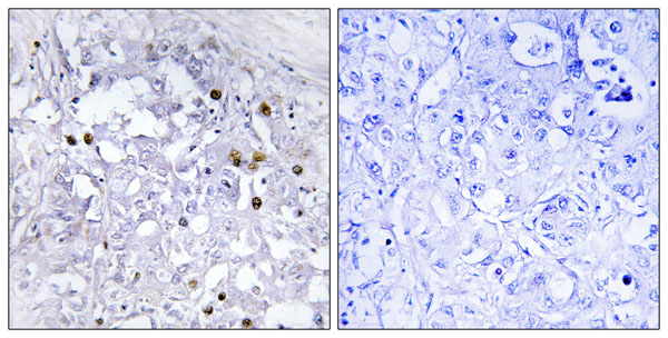
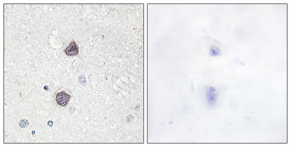
 IHC (Immunohistochemisry)
(Immunohistochemical analysis of paraffin-embedded human breast carcinoma tissue, using HER2 (phospho-Tyr1248) antibody.)
IHC (Immunohistochemisry)
(Immunohistochemical analysis of paraffin-embedded human breast carcinoma tissue, using HER2 (phospho-Tyr1248) antibody.)
HER2, Antibody (Cat# AAA112013)
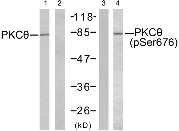 WB (Western Blot)
(Western blot analysis of extracts from JK cells untreated or treated with PMA (200nM, 30mins), using PKC theta (Ab-676) antibody (Line 1 and 2) and PKC theta (phospho-Ser676) antibody (Line 3 and 4).)
WB (Western Blot)
(Western blot analysis of extracts from JK cells untreated or treated with PMA (200nM, 30mins), using PKC theta (Ab-676) antibody (Line 1 and 2) and PKC theta (phospho-Ser676) antibody (Line 3 and 4).)
PKC theta, Antibody (Cat# AAA111782)
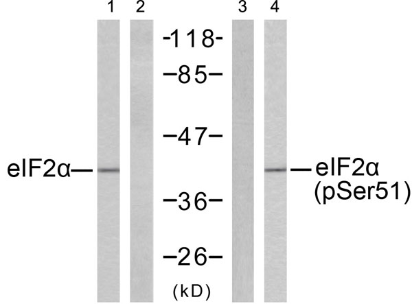 WB (Western Blot)
(Western blot analysis of extracts from K562 cells untreated or treated with IFN-alpha (1000U/ml, 18 hours), using eIF2alpha (Ab-51) antibody (Line 1 and 2) and eIF2alpha (phospho-Ser51) antibody (Line 3 and 4).)
WB (Western Blot)
(Western blot analysis of extracts from K562 cells untreated or treated with IFN-alpha (1000U/ml, 18 hours), using eIF2alpha (Ab-51) antibody (Line 1 and 2) and eIF2alpha (phospho-Ser51) antibody (Line 3 and 4).)
eIF2alpha, Antibody (Cat# AAA111783)
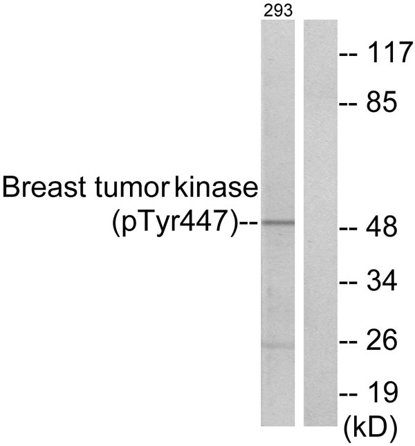
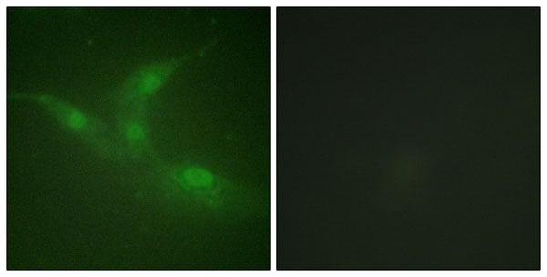 WB (Western Blot)
(P-peptide-+ Western blot analysis of extracts from 293 cells, treated with EGF (200ng/ml, 30mins), using Breast Tumor Kinase (Phospho-Tyr447) antibody. Immunofluorescence analysis of NIH/3T3 cells, using Breast Tumor Kinase (Phospho-Tyr447) antibody.)
WB (Western Blot)
(P-peptide-+ Western blot analysis of extracts from 293 cells, treated with EGF (200ng/ml, 30mins), using Breast Tumor Kinase (Phospho-Tyr447) antibody. Immunofluorescence analysis of NIH/3T3 cells, using Breast Tumor Kinase (Phospho-Tyr447) antibody.)
Breast Tumor Kinase, Antibody (Cat# AAA111798)
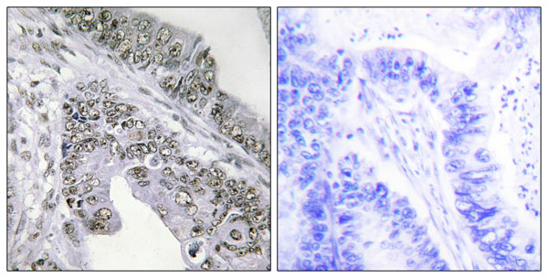
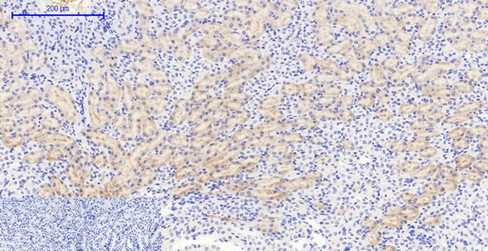
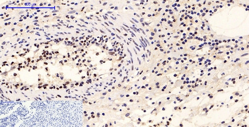
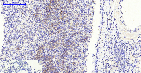
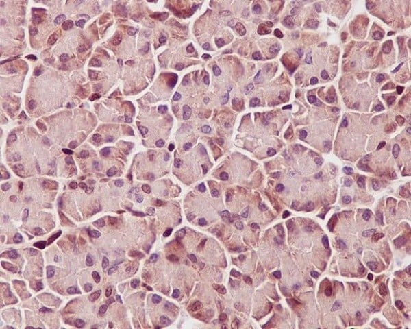
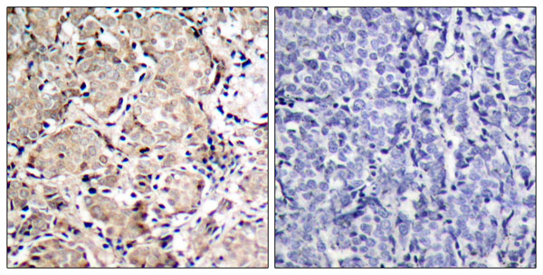
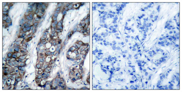
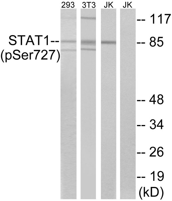 IHC (Immunohiostchemistry)
(Immunohistochemistry analysis of paraffin-embedded human colon carcinoma tissue using STAT1 (Phospho-Ser727) antibody. Western blot analysis of extracts from 293 cells, 3T3 cells treated with UV (15mins) and Jurkat cells treated with eto (25uM, 24hours), using STAT1 (Phospho-Ser727) antibody.)
IHC (Immunohiostchemistry)
(Immunohistochemistry analysis of paraffin-embedded human colon carcinoma tissue using STAT1 (Phospho-Ser727) antibody. Western blot analysis of extracts from 293 cells, 3T3 cells treated with UV (15mins) and Jurkat cells treated with eto (25uM, 24hours), using STAT1 (Phospho-Ser727) antibody.)
STAT1, Antibody (Cat# AAA111855)
What Are Phospho Antibodies?
Protein phosphorylation is a process where a phosphate group is added to certain amino acid residues of a protein – usually serine (S), threonine (T), or tyrosine (Y) - by enzymes called kinases. This process is integral in controlling cellular signaling, cellular growth, and other biological functions, as explained in our detailed guide to phospho antibodies.
Our catalog includes a wide range of phospho-specific antibodies that can accurately detect this important marker, including phospho antibodies as well as other formats such as monoclonal antibodies and polyclonal antibodies for different research needs.
They perform strongly in widely used laboratory applications such as Western blot, flow cytometry, immunohistochemistry, and immunofluorescence microscopy. We value your trust in us and are committed to providing top-quality products and services. All of our antibodies are guaranteed to work for the applications and species indicated on our website & associated product pages.
What Are The Key Applications of Phospho Antibodies?
1. Western Blotting
One of the first steps a researcher can take in utilizing these phospho-specific antibodies is to check if the antibody works using a technique referred to as Western blot, learn more in our guide on Western blot roles and uses. For those unfamiliar, Western Blot aids in showing whether the protein that the antibody recognizes is appearing at the correct/expected size. These phospho-specific antibodies should also be able to detect changes in the target protein’s phosphorylation (on/off state) when cells are stimulated in certain ways.
2. Staining of Fixed Cells (Immunocytochemistry)
Another routine use of these phospho-specific antibodies, is to test if the antibody is able to demonstrate similar performance when used on fixed cells (intact cells that have been preserved) as it did in the Western blot tests. It is an important aspect in many cases to confirm that the antibody works in actual intact cell samples. Ideally, the method used for cellular fixation should be the same as what is used in pathology labs (like using 10% formalin). To check if the antibody works well in tissue sections (FFPE), researchers will often test it on fixed cells that are processed similar to tissue samples.
3. Specificity Tests Using Peptides
In order to make sure that the antibody is only binding to the right target:
- Laboratory technicians will mix the antibody with phospho-peptides (short segments of the protein containing the phosphate group modification).
- If the antibody signal disappears, it is confirmation that it is binding to the correct phosphorylated location. Such validation approaches are commonly used across different antibody types, including monoclonal antibodies.
- A more robust test is to use both the phosphorylated and non-phosphorylated (dephosphorylated) versions of the protein. The antibody should react only with the phosphorylated one.
- Another method sometimes utilized is to treat the sample with an enzyme, such as alkaline phosphatase, that specifically removes phosphate groups. If the antibody signal disappears after this, it also confirms specificity.
4. Genetic Confirmation
As a final step, scientists can genetically manipulate the nucleotide sequence and alter the target protein by removing the exact site where phosphorylation happens. If the antibody no longer appears to detect the modified protein, it is strong evidence supporting the antibody being specific for that phosphorylated site.
Why Buy Phospho Antibodies Through Us?
- The production laboratory adheres to strict and consistent protocols prior to releasing any of these phospho-specific antibodies:
- Standard methods and proper controls in all tests to ensure high quality.
- These antibodies are tested and validated in different cell types and species.
- High quality control criterion to ensure each batch is consistent, so you will obtain reliable results every time.
FAQ
1. What Are Phospho-Specific Antibodies?
Phospho-specific antibodies are made to detect proteins only when they have a phosphate group linked to a specific amino acid residue. This empowers scientists understand if a protein is "turned on" or active, based on its phosphorylation state.
2. How to Detect Phosphorylated Proteins in a Western Blot?
To find out if a protein is phosphorylated using Western blot:
- Use a phospho-specific antibody that binds only to the phosphorylated form of the protein.
- You can also use a “regular” antibody for the same amino acid sequence of the protein that the phospho-specific antibody is binding to (but in this case, this antibody will not bind if there is a phosphate group present) in order to compare how much of it is phosphorylated versus how much is non-phosphorylated (or “total” protein, if the “normal” antibody’s epitopes are non-phospho-site-specific).
3. How to Choose the Best Antibody?
Here are some simple tips to help you pick the right antibody:
- Know your target
- Match your sample characteristics
- Confirm the intended use is appropriate
- Check “host” and “type”
- Check the “quality” of the presented data/images
- Appraise whether the available validation meets your needs