 FCM (Flow Cytometry)
(Flow cytometric analysis of paraformaldehyde fixed A431 cells (blue line), permeabilized with 0.5% Triton. Primary incubation 1hr (10ug/ml) followed by Alexa Fluor 488 secondary antibody (1ug/ml). IgG control: Unimmunized goat IgG (black line) followed by Alexa Fluor 488 secondary antibody.)
FCM (Flow Cytometry)
(Flow cytometric analysis of paraformaldehyde fixed A431 cells (blue line), permeabilized with 0.5% Triton. Primary incubation 1hr (10ug/ml) followed by Alexa Fluor 488 secondary antibody (1ug/ml). IgG control: Unimmunized goat IgG (black line) followed by Alexa Fluor 488 secondary antibody.)
Goat Argininosuccinate synthetase 1 Polyclonal Antibody | anti-ASS1 antibody
Goat anti-Argininosuccinate synthetase 1 Antibody
Expected from sequence similarity: Human, Mouse, Rat, Dog, Cow
Expected from sequence similarity: Human, Mouse, Rat, Dog, Cow
Western blot: Approx 45kDa band observed in lysates of cell lines A431 and NIH3T3 and approx.48kDa in Human and Rat Kidney and in Mouse Liver lysates (calculated MW of 46.5kDa according to Human NP_000041.2 and Rat NP_037289.1, and 46.6kDa according to Mouse NP_031520.1 ). Recommended concentration: 0.03-1µg/ml. Primary incubation 1 hour at room temperature.
IHC: In paraffin embedded Human Kidney shows textured cytoplasm staining in PCT. Recommended concentration: 2-4µg/ml. Paraffin embedded Human Liver. Recommended concentration: 2.5µg/ml.
Immunofluorescence: Strong expression of the protein seen in the cytoplasm of HeLa cells. Recommended concentration: 10µg/ml.
Flow Cytometry: Flow cytometric analysis of A431 cells. Recommended concentration: 10ug/ml.
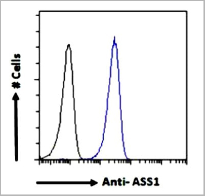
FCM (Flow Cytometry)
(Flow cytometric analysis of paraformaldehyde fixed A431 cells (blue line), permeabilized with 0.5% Triton. Primary incubation 1hr (10ug/ml) followed by Alexa Fluor 488 secondary antibody (1ug/ml). IgG control: Unimmunized goat IgG (black line) followed by Alexa Fluor 488 secondary antibody.)
IF (Immunofluorescence)
(Immunofluorescence analysis of paraformaldehyde fixed HeLa cells, permeabilized with 0.15% Triton. Primary incubation 1hr (10ug/ml) followed by Alexa Fluor 488 secondary antibody (2ug/ml), showing cytoplasmic staining. The nuclear stain is DAPI (blue). Negative control: Unimmunized goat IgG (10ug/ml) followed by Alexa Fluor 488 secondary antibody (2ug/ml).)
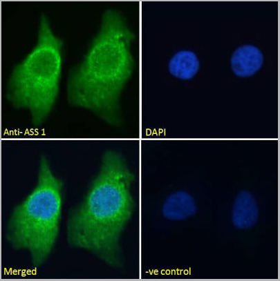
IHC (Immunohistochemistry)
((2.5µg/ml) staining of paraffin embedded Human Liver. Steamed antigen retrieval with citrate buffer pH 6, AP-staining.)
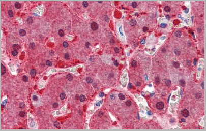
IHC (Immunohistochemistry)
((2.5µg/ml) staining of paraffin embedded Human Liver. Steamed antigen retrieval with citrate buffer pH 6, AP-staining.)
IHC (Immunohistochemistry)
((2.5ug/ml) staining of paraffin embedded Human Kidney. Steamed antigen retrieval with citrate buffer pH 6, AP-staining.)
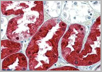
IHC (Immunohistochemistry)
((2.5ug/ml) staining of paraffin embedded Human Kidney. Steamed antigen retrieval with citrate buffer pH 6, AP-staining.)
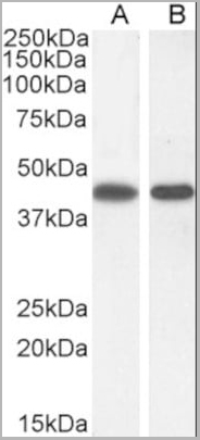
WB (Western Blot)
((0.3µg/ml) staining of A431 (A) and (1ug/ml) NIH3T3(B) cell lysate (35µg protein in RIPA buffer). Detected by chemiluminescence.)
WB (Western Blot)
((0.3µg/ml) staining of A431 (A) and (1ug/ml) NIH3T3(B) cell lysate (35µg protein in RIPA buffer). Detected by chemiluminescence.)
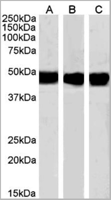
WB (Western Blot)
((0.01µg/ml) staining of Human Kidney (A) Mouse Liver (B) and (0.03ug/ml) Rat Kidney (C) lysate (35µg protein in RIPA buffer). Detected by chemiluminescence.)
WB (Western Blot)
((0.01µg/ml) staining of Human Kidney (A) Mouse Liver (B) and (0.03ug/ml) Rat Kidney (C) lysate (35µg protein in RIPA buffer). Detected by chemiluminescence.)
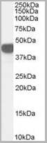
WB (Western Blot)
((0. 03ug/ml) staining of Human Kidney lysate (35ug protein in RIPA buffer). Primary incubation was 1 hour. Detected by chemiluminescence.)
WB (Western Blot)
((0. 03ug/ml) staining of Human Kidney lysate (35ug protein in RIPA buffer). Primary incubation was 1 hour. Detected by chemiluminescence.)
NCBI and Uniprot Product Information
Similar Products


Product Notes
The ASS1 ass1 (Catalog #AAA13660) is an Antibody produced from Goat and is intended for research purposes only. The product is available for immediate purchase. The Goat anti-Argininosuccinate synthetase 1 Antibody reacts with Tested: Human, Mouse, Rat Expected from sequence similarity: Human, Mouse, Rat, Dog, Cow and may cross-react with other species as described in the data sheet. AAA Biotech's Argininosuccinate synthetase 1 can be used in a range of immunoassay formats including, but not limited to, Peptide ELISA (EIA), Western Blot (WB), Immunofluorescence (IF), Immunoprecipitation (IP), Immunohistochemistry (IHC). Peptide ELISA: antibody detection limit dilution 1:64000. Western blot: Approx 45kDa band observed in lysates of cell lines A431 and NIH3T3 and approx.48kDa in Human and Rat Kidney and in Mouse Liver lysates (calculated MW of 46.5kDa according to Human NP_000041.2 and Rat NP_037289.1, and 46.6kDa according to Mouse NP_031520.1 ). Recommended concentration: 0.03-1µg/ml. Primary incubation 1 hour at room temperature. IHC: In paraffin embedded Human Kidney shows textured cytoplasm staining in PCT. Recommended concentration: 2-4µg/ml. Paraffin embedded Human Liver. Recommended concentration: 2.5µg/ml. Immunofluorescence: Strong expression of the protein seen in the cytoplasm of HeLa cells. Recommended concentration: 10µg/ml. Flow Cytometry: Flow cytometric analysis of A431 cells. Recommended concentration: 10ug/ml. Researchers should empirically determine the suitability of the ASS1 ass1 for an application not listed in the data sheet. Researchers commonly develop new applications and it is an integral, important part of the investigative research process. The amino acid sequence is listed below: ENPKNQAPPG LYTKTQD. It is sometimes possible for the material contained within the vial of "Argininosuccinate synthetase 1, Polyclonal Antibody" to become dispersed throughout the inside of the vial, particularly around the seal of said vial, during shipment and storage. We always suggest centrifuging these vials to consolidate all of the liquid away from the lid and to the bottom of the vial prior to opening. Please be advised that certain products may require dry ice for shipping and that, if this is the case, an additional dry ice fee may also be required.Precautions
All products in the AAA Biotech catalog are strictly for research-use only, and are absolutely not suitable for use in any sort of medical, therapeutic, prophylactic, in-vivo, or diagnostic capacity. By purchasing a product from AAA Biotech, you are explicitly certifying that said products will be properly tested and used in line with industry standard. AAA Biotech and its authorized distribution partners reserve the right to refuse to fulfill any order if we have any indication that a purchaser may be intending to use a product outside of our accepted criteria.Disclaimer
Though we do strive to guarantee the information represented in this datasheet, AAA Biotech cannot be held responsible for any oversights or imprecisions. AAA Biotech reserves the right to adjust any aspect of this datasheet at any time and without notice. It is the responsibility of the customer to inform AAA Biotech of any product performance issues observed or experienced within 30 days of receipt of said product. To see additional details on this or any of our other policies, please see our Terms & Conditions page.Item has been added to Shopping Cart
If you are ready to order, navigate to Shopping Cart and get ready to checkout.