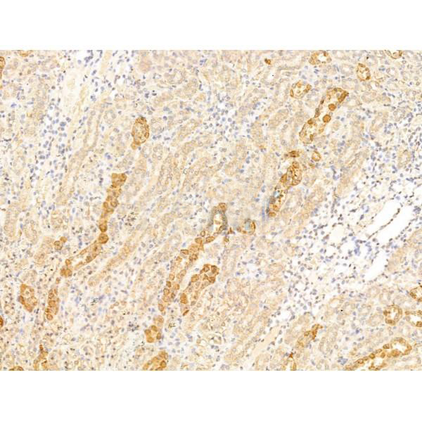
 IHC (Immunohistochemistry)
(AAA329141 at 1/100 staining Rat kidney tissue by IHC-P. The sample was formaldehyde fixed and a heat mediated antigen retrieval step in citrate buffer was performed. The sample was then blocked and incubated with the primary antibody at 4°C overnight. An HRP conjugated anti-Rabbit antibody was used as the secondary antibody.)
IHC (Immunohistochemistry)
(AAA329141 at 1/100 staining Rat kidney tissue by IHC-P. The sample was formaldehyde fixed and a heat mediated antigen retrieval step in citrate buffer was performed. The sample was then blocked and incubated with the primary antibody at 4°C overnight. An HRP conjugated anti-Rabbit antibody was used as the secondary antibody.)
Rabbit Asporin Polyclonal Antibody | anti-ASPN antibody
Asporin Antibody
Predicted Reactivity: Pig(100%), Bovine(100%), Horse(100%), Sheep(100%), Rabbit(88%), Dog(100%), Xenopus(86%)
Predicted Reactivity: Pig(100%), Bovine(100%), Horse(100%), Sheep(100%), Rabbit(88%), Dog(100%), Xenopus(86%)
IHC (Immunohistochemistry)
(AAA329141 at 1/100 staining Rat kidney tissue by IHC-P. The sample was formaldehyde fixed and a heat mediated antigen retrieval step in citrate buffer was performed. The sample was then blocked and incubated with the primary antibody at 4°C overnight. An HRP conjugated anti-Rabbit antibody was used as the secondary antibody.)
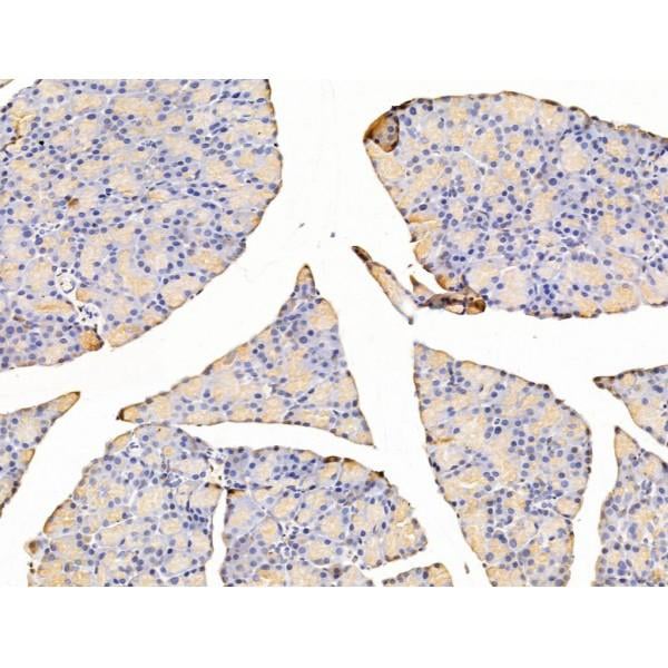
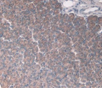
IHC (Immunohistochemisry)
(AAA329141 at 1/100 staining Rat stomach tissue by IHC-P. The sample was formaldehyde fixed and a heat mediated antigen retrieval step in citrate buffer was performed. The sample was then blocked and incubated with the primary antibody at 4°C overnight. An HRP conjugated anti-Rabbit antibody was used as the secondary antibody.)
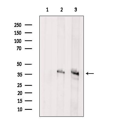
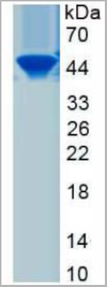
WB (Western Blot)
(Western blot analysis of extracts from various samples, using Asporin Antibody.Lane 1: Hela cells, treated with blocking peptide;Lane 2: Hela cells;Lane 3: B16F10 cells.Observed bands: 37 kDa.)
WB (Western Blot)
(Western blot analysis of extracts from various samples, using Asporin Antibody.Lane 1: Hela cells, treated with blocking peptide;Lane 2: Hela cells;Lane 3: B16F10 cells.Observed bands: 37 kDa.)
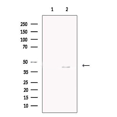
WB (Western Blot)
(Western blot analysis of extracts from HepG2 cells, using Asporin Antibody. The lane on the left was treated with blocking peptide.)
WB (Western Blot)
(Western blot analysis of extracts from HepG2 cells, using Asporin Antibody. The lane on the left was treated with blocking peptide.)
Post Translational Modifications: There is no serine/glycine dipeptide sequence expected for the attachment of O-linked glycosaminoglycans and this is probably not a proteoglycan. The O-linked polysaccharide on 54-Ser is probably the mucin type linked to GalNAc.The N-linked glycan at Asn-282 is composed of variable structures of GlcNAc, mannose, fucose, HexNAc and hexose.
Subcellular Location: Secreted>Extracellular space>Extracellular matrix.
Tissue Specificity: Higher levels in osteoarthritic articular cartilage, aorta, uterus. Moderate expression in small intestine, heart, liver, bladder, ovary, stomach, and in the adrenal, thyroid, and mammary glands. Low expression in trachea, bone marrow, and lung. Colocalizes with TGFB1 in chondrocytes within osteoarthritic (OA) lesions of articular cartilage.
Subunit Structure: Interacts with TGFB1, TGFB2 and TGFB3. DCN, BGN, and FMOD inhibit binding to TGFB1. Interacts with BMP2. Interacts in vitro with type II collagen (By similarity). Interacts with type I collagen. DCN can inhibit collagen binding.
Similarity: The LRR 5 repeat can inhibit BMP2-induced cytodifferentiation and may be involved in the interaction with BMP2 (By similarity). The repeats LRR 10, LRR 11 and LRR 12 are involved in binding type I collagen. The poly-Asp region is involved in binding calcium.Belongs to the small leucine-rich proteoglycan (SLRP) family. SLRP class I subfamily.
NCBI and Uniprot Product Information
Predicted Molecular Weight: (Calculated)43kDa.
Customer Reviews
Loading reviews...
Share Your Experience
Similar Products


Product Notes
The ASPN aspn (Catalog #AAA329141) is an Antibody produced from Rabbit and is intended for research purposes only. The product is available for immediate purchase. The Asporin Antibody reacts with Human, Mouse, Rat Predicted Reactivity: Pig(100%), Bovine(100%), Horse(100%), Sheep(100%), Rabbit(88%), Dog(100%), Xenopus(86%) and may cross-react with other species as described in the data sheet. AAA Biotech's Asporin can be used in a range of immunoassay formats including, but not limited to, ELISA. Researchers should empirically determine the suitability of the ASPN aspn for an application not listed in the data sheet. Researchers commonly develop new applications and it is an integral, important part of the investigative research process. It is sometimes possible for the material contained within the vial of "Asporin, Polyclonal Antibody" to become dispersed throughout the inside of the vial, particularly around the seal of said vial, during shipment and storage. We always suggest centrifuging these vials to consolidate all of the liquid away from the lid and to the bottom of the vial prior to opening. Please be advised that certain products may require dry ice for shipping and that, if this is the case, an additional dry ice fee may also be required.Precautions
All products in the AAA Biotech catalog are strictly for research-use only, and are absolutely not suitable for use in any sort of medical, therapeutic, prophylactic, in-vivo, or diagnostic capacity. By purchasing a product from AAA Biotech, you are explicitly certifying that said products will be properly tested and used in line with industry standard. AAA Biotech and its authorized distribution partners reserve the right to refuse to fulfill any order if we have any indication that a purchaser may be intending to use a product outside of our accepted criteria.Disclaimer
Though we do strive to guarantee the information represented in this datasheet, AAA Biotech cannot be held responsible for any oversights or imprecisions. AAA Biotech reserves the right to adjust any aspect of this datasheet at any time and without notice. It is the responsibility of the customer to inform AAA Biotech of any product performance issues observed or experienced within 30 days of receipt of said product. To see additional details on this or any of our other policies, please see our Terms & Conditions page.Item has been added to Shopping Cart
If you are ready to order, navigate to Shopping Cart and get ready to checkout.