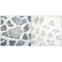
 WB (Western Blot)
(Western blot analysis of extracts of MCF7, using BAK1 antibody. The lane on the left is treated with the antigen-specific peptide.)
WB (Western Blot)
(Western blot analysis of extracts of MCF7, using BAK1 antibody. The lane on the left is treated with the antigen-specific peptide.)
Rabbit BAK1 Polyclonal Antibody | anti-BAK1 antibody
BAK1 Antibody
Gene Names
BAK1; BAK; CDN1; BCL2L7; BAK-LIKE
Reactivity
Human, Mouse, Rat
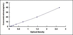
Applications
ELISA, Immunocytochemistry, Immunofluorescence, Western Blot
Purity
The antiserum was purified by peptide affinity chromatography using SulfoLink Coupling Resin.
Synonyms
BAK1, Antibody; BAK1 Antibody; Apoptosis regulator BAK; BAK; BAK like; BAK NT; BAK_HUMAN; BAK1; Bcl 2 homologous antagonist/killer; Bcl 2 like 7 protein; Bcl-2 homologous antagonist/killer; Bcl-2-like protein 7; BCL2 antagonist/killer 1; Bcl2 like 7 Protein; Bcl2-L-7; BCL2L7; CDN1; Cell death inhibitor 1; MGC117255; MGC3887; NBAK; Pro apoptotic protein BAK; anti-BAK1 antibody
Host
Rabbit
Reactivity
Human, Mouse, Rat
Clonality
Polyclonal
Isotype
IgG
Specificity
BAK1 antibody detects endogenous levels of total BAK1
Purity/Purification
The antiserum was purified by peptide affinity chromatography using SulfoLink Coupling Resin.
Form/Format
Liquid
Phosphate buffered saline, pH 7.4, 150mM NaCl, 0.02% sodium azide and 50% glycerol.
Phosphate buffered saline, pH 7.4, 150mM NaCl, 0.02% sodium azide and 50% glycerol.
Concentration
1mg/ml (varies by lot)
Sequence Length
211
Applicable Applications for anti-BAK1 antibody
ELISA, ICC (Immunocytochemistry), IF (Immunofluorescence), WB (Western Blot)
Immunogen
N term-peptide of human BAK1
Subcellular Location
Mitochondrion Membrane.
Tissue Specificity
Expressed in a wide variety of tissues, with highest levels in the heart and skeletal muscle.
Conjugation
Unconjugated
Preparation and Storage
Store at -20 degree C. Stable for 12 months from date of receipt.
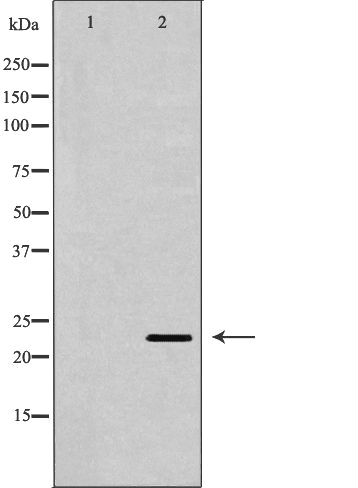
WB (Western Blot)
(Western blot analysis of extracts of MCF7, using BAK1 antibody. The lane on the left is treated with the antigen-specific peptide.)
WB (Western Blot)
(Western blot analysis of extracts of MCF7, using BAK1 antibody. The lane on the left is treated with the antigen-specific peptide.)
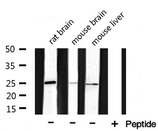
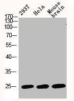
WB (Western Blot)
(Western blot analysis of extracts of various samples, using bak1 Antibody.)
WB (Western Blot)
(Western blot analysis of extracts of various samples, using bak1 Antibody.)
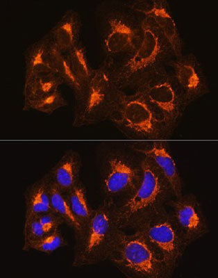
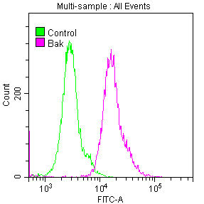
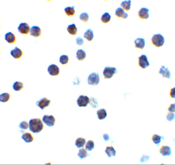
IF (Immunofluorescence)
(AAA325824 staining 293 cells by IF/ICC. The sample were fixed with PFA and permeabilized in 0.1% Triton X-100, then blocked in 10% serum for 45 minutes at 25 degree C. The primary antibody was diluted at 1/200 and incubated with the sample for 1 hour at 37 degree C. An Alexa Fluor 594 conjugated goat anti-rabbit IgG (H+L) antibody, diluted at 1/600, was used as secondary antibody.)
Related Product Information for anti-BAK1 antibody
Description: Bak is a proapoptotic member of the Bcl-2 family (1). This protein is located on the outer membrane of mitochondria and is an essential component for transduction of apoptotic signals through the mitochondrial pathway (2, 3). Upon apoptotic stimulation, an upstream stimulator like truncated BID (tBID) induces conformational changes in Bak to form oligomer channels in the mitochondrial membrane for cytochrome c release. The release of cytochrome c to the cytosol activates the caspase-9 pathway and eventually leads to cell death (4, 5).
Function: Plays a role in the mitochondrial apoptosic process. Upon arrival of cell death signals, promotes mitochondrial outer membrane (MOM) permeabilization by oligomerizing to form pores within the MOM. This releases apoptogenic factors into the cytosol, including cytochrome c, promoting the activation of caspase 9 which in turn processes and activates the effector caspases.
Subunit Structure: Interacts with BCL2A1 (By similarity). Homodimer. Formation of the homodimer is zinc-dependent. Forms heterodimers with BCL2 and BCL2L1 isoform Bcl-X(L). Interacts with RTL10/BOP. Interacts with VDAC1 (PubMed:25296756).
Similarity: Intact BH3 motif is required by BIK, BID, BAK, BAD and BAX for their pro-apoptotic activity and for their interaction with anti-apoptotic members of the Bcl-2 family. Belongs to the Bcl-2 family.
NCBI and Uniprot Product Information
NCBI GI #
NCBI GeneID
NCBI Accession #
NCBI GenBank Nucleotide #
Molecular Weight
Observed: 23 kDa
Predicted: 24 kDa
Predicted: 24 kDa
NCBI Official Full Name
bcl-2 homologous antagonist/killer
NCBI Official Synonym Full Names
BCL2 antagonist/killer 1
NCBI Official Symbol
BAK1
NCBI Official Synonym Symbols
BAK; CDN1; BCL2L7; BAK-LIKE
NCBI Protein Information
bcl-2 homologous antagonist/killer
UniProt Protein Name
Bcl-2 homologous antagonist/killer
UniProt Gene Name
BAK1
UniProt Synonym Gene Names
BAK; BCL2L7; CDN1; Bcl2-L-7
Customer Reviews
Loading reviews...
Share Your Experience
Similar Products


Product Notes
The BAK1 bak1 (Catalog #AAA325824) is an Antibody produced from Rabbit and is intended for research purposes only. The product is available for immediate purchase. The BAK1 Antibody reacts with Human, Mouse, Rat and may cross-react with other species as described in the data sheet. AAA Biotech's BAK1 can be used in a range of immunoassay formats including, but not limited to, ELISA, ICC (Immunocytochemistry), IF (Immunofluorescence), WB (Western Blot). Researchers should empirically determine the suitability of the BAK1 bak1 for an application not listed in the data sheet. Researchers commonly develop new applications and it is an integral, important part of the investigative research process. It is sometimes possible for the material contained within the vial of "BAK1, Polyclonal Antibody" to become dispersed throughout the inside of the vial, particularly around the seal of said vial, during shipment and storage. We always suggest centrifuging these vials to consolidate all of the liquid away from the lid and to the bottom of the vial prior to opening. Please be advised that certain products may require dry ice for shipping and that, if this is the case, an additional dry ice fee may also be required.Precautions
All products in the AAA Biotech catalog are strictly for research-use only, and are absolutely not suitable for use in any sort of medical, therapeutic, prophylactic, in-vivo, or diagnostic capacity. By purchasing a product from AAA Biotech, you are explicitly certifying that said products will be properly tested and used in line with industry standard. AAA Biotech and its authorized distribution partners reserve the right to refuse to fulfill any order if we have any indication that a purchaser may be intending to use a product outside of our accepted criteria.Disclaimer
Though we do strive to guarantee the information represented in this datasheet, AAA Biotech cannot be held responsible for any oversights or imprecisions. AAA Biotech reserves the right to adjust any aspect of this datasheet at any time and without notice. It is the responsibility of the customer to inform AAA Biotech of any product performance issues observed or experienced within 30 days of receipt of said product. To see additional details on this or any of our other policies, please see our Terms & Conditions page.Item has been added to Shopping Cart
If you are ready to order, navigate to Shopping Cart and get ready to checkout.