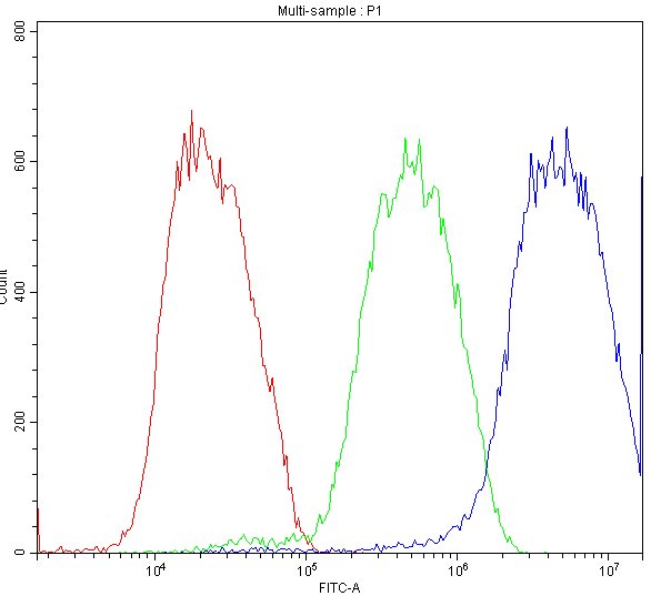
 FCM (Flow Cytometry)
(Figure 8. Flow Cytometry analysis of HepG2 cells using anti-ATF2 antibody (AAA11634).Overlay histogram showing HepG2 cells stained with AAA11634 (Blue line).The cells were blocked with 10% normal goat serum. And then incubated with rabbit anti-ATF2 Antibody (AAA11634,1ug/1x10^6 cells) for 30 min at 20 degree C. DyLight ®488 conjugated goat anti-rabbit IgG (5-10ug/1x10^6 cells) was used as secondary antibody for 30 minutes at 20 degree C. Isotype control antibody (Green line) was rabbit IgG (1ug/1x106) used under the same conditions. Unlabelled sample (Red line) was also used as a control.)
FCM (Flow Cytometry)
(Figure 8. Flow Cytometry analysis of HepG2 cells using anti-ATF2 antibody (AAA11634).Overlay histogram showing HepG2 cells stained with AAA11634 (Blue line).The cells were blocked with 10% normal goat serum. And then incubated with rabbit anti-ATF2 Antibody (AAA11634,1ug/1x10^6 cells) for 30 min at 20 degree C. DyLight ®488 conjugated goat anti-rabbit IgG (5-10ug/1x10^6 cells) was used as secondary antibody for 30 minutes at 20 degree C. Isotype control antibody (Green line) was rabbit IgG (1ug/1x106) used under the same conditions. Unlabelled sample (Red line) was also used as a control.)
Rabbit Cyclic AMP-dependent transcription factor ATF-2 Polyclonal Antibody | anti-ATF2 antibody
Anti-ATF2 Antibody
Each vial contains 5mg BSA, 0.9mg NaCl, 0.2mg Na2HPO4, 0.05mg NaN3.
Immunohistochemistry (Paraffin-embedded Section): Concentration: 0.5-1mug/ml; Tested Species: Human, Mouse, Rat
Immunohistochemistry (Frozen Section): Concentration: 0.5-1mug/ml; Tested Species: Rat
FCM (Flow Cytometry)
(Figure 8. Flow Cytometry analysis of HepG2 cells using anti-ATF2 antibody (AAA11634).Overlay histogram showing HepG2 cells stained with AAA11634 (Blue line).The cells were blocked with 10% normal goat serum. And then incubated with rabbit anti-ATF2 Antibody (AAA11634,1ug/1x10^6 cells) for 30 min at 20 degree C. DyLight ®488 conjugated goat anti-rabbit IgG (5-10ug/1x10^6 cells) was used as secondary antibody for 30 minutes at 20 degree C. Isotype control antibody (Green line) was rabbit IgG (1ug/1x106) used under the same conditions. Unlabelled sample (Red line) was also used as a control.)
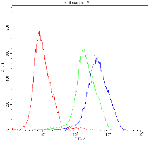
FCM (Flow Cytometry)
(Figure 7. Flow Cytometry analysis of K562 cells using anti-ATF2 antibody (AAA11634).Overlay histogram showing K562 cells stained with AAA11634 (Blue line).The cells were blocked with 10% normal goat serum. And then incubated with rabbit anti-ATF2 Antibody (AAA11634,1ug/1x10^6 cells) for 30 min at 20 degree C. DyLight ®488 conjugated goat anti-rabbit IgG (5-10ug/1x10^6 cells) was used as secondary antibody for 30 minutes at 20 degree C. Isotype control antibody (Green line) was rabbit IgG (1ug/1x106) used under the same conditions. Unlabelled sample (Red line) was also used as a control.)
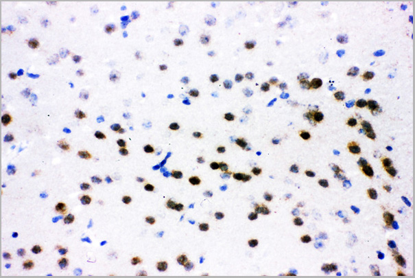
IHC (Immunohistchemistry)
(Figure 6. IHC analysis of ATF2 using anti-ATF2 antibody (AAA11634).ATF2 was detected in frozen section of mouse brain tissue. Heat mediated antigen retrieval was performed in citrate buffer (pH6, epitope retrieval solution) for 20 mins. The tissue section was blocked with 10% goat serum. The tissue section was then incubated with 1ug/ml rabbit anti-ATF2 Antibody (AAA11634) overnight at 4 degree C. Biotinylated goat anti-rabbit IgG was used as secondary antibody and incubated for 30 minutes at 37 degree C. The tissue section was developed using Strepavidin-Biotin-Complex (SABC) with DAB as the chromogen.)
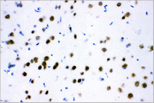
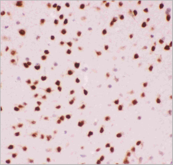
IHC (Immunohistochemistry)
(Figure 5. IHC analysis of ATF2 using anti-ATF2 antibody (AAA11634).ATF2 was detected in frozen section of rat brain tissue. Heat mediated antigen retrieval was performed in citrate buffer (pH6, epitope retrieval solution) for 20 mins. The tissue section was blocked with 10% goat serum. The tissue section was then incubated with 1ug/ml rabbit anti-ATF2 Antibody (AAA11634) overnight at 4 degree C. Biotinylated goat anti-rabbit IgG was used as secondary antibody and incubated for 30 minutes at 37 degree C. The tissue section was developed using Strepavidin-Biotin-Complex (SABC) with DAB as the chromogen.)
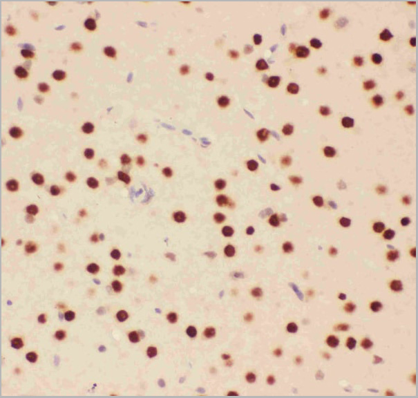
IHC (Immunohistochemistry)
(Figure 4. IHC analysis of ATF2 using anti-ATF2 antibody (AAA11634).ATF2 was detected in paraffin-embedded section of rat brain tissue. Heat mediated antigen retrieval was performed in citrate buffer (pH6, epitope retrieval solution) for 20 mins. The tissue section was blocked with 10% goat serum. The tissue section was then incubated with 1ug/ml rabbit anti-ATF2 Antibody (AAA11634) overnight at 4 degree C. Biotinylated goat anti-rabbit IgG was used as secondary antibody and incubated for 30 minutes at 37 degree C. The tissue section was developed using Strepavidin-Biotin-Complex (SABC) with DAB as the chromogen.)
IHC (Immunohistochemistry)
(Figure 3. IHC analysis of ATF2 using anti-ATF2 antibody (AAA11634).ATF2 was detected in paraffin-embedded section of mouse brain tissue. Heat mediated antigen retrieval was performed in citrate buffer (pH6, epitope retrieval solution) for 20 mins. The tissue section was blocked with 10% goat serum. The tissue section was then incubated with 1ug/ml rabbit anti-ATF2 Antibody (AAA11634) overnight at 4 degree C. Biotinylated goat anti-rabbit IgG was used as secondary antibody and incubated for 30 minutes at 37 degree C. The tissue section was developed using Strepavidin-Biotin-Complex (SABC) with DAB as the chromogen.)
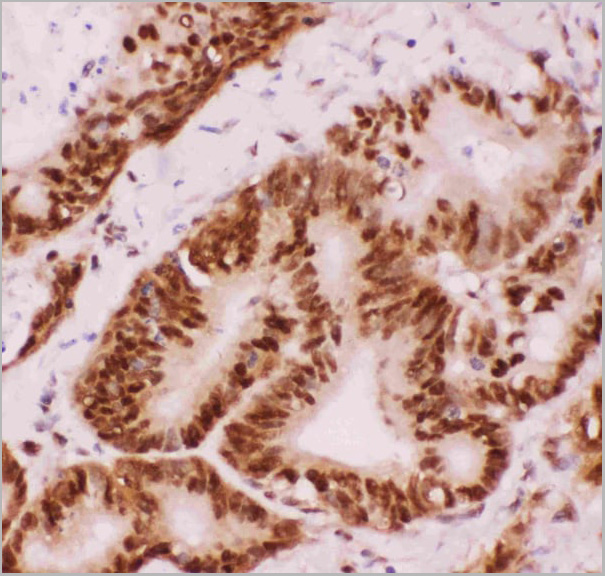
IHC (Immunohistochemistry)
(Figure 2. IHC analysis of ATF2 using anti-ATF2 antibody (AAA11634).ATF2 was detected in paraffin-embedded section of human intestinal cancer tissue. Heat mediated antigen retrieval was performed in citrate buffer (pH6, epitope retrieval solution) for 20 mins. The tissue section was blocked with 10% goat serum. The tissue section was then incubated with 1ug/ml rabbit anti-ATF2 Antibody (AAA11634) overnight at 4 degree C. Biotinylated goat anti-rabbit IgG was used as secondary antibody and incubated for 30 minutes at 37 degree C. The tissue section was developed using Strepavidin-Biotin-Complex (SABC) with DAB as the chromogen.)
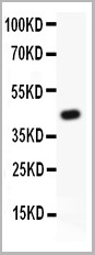
WB (Western Blot)
(Figure 1. Western blot analysis of ATF2 using anti-ATF2 antibody (AAA11634).Electrophoresis was performed on a 5-20% SDS-PAGE gel at 70V (Stacking gel) / 90V (Resolving gel) for 2-3 hours.Lane 1: Recombinant Human ATF2 Protein 0.5ng.After Electrophoresis, proteins were transferred to a Nitrocellulose membrane at 150mA for 50-90 minutes. Blocked the membrane with 5% Non-fat Milk/ TBS for 1.5 hour at RT. The membrane was incubated with rabbit anti-ATF2 antigen affinity purified polyclonal antibody at 0.5ug/mL overnight at 4 degree C, then washed with TBS-0.1%Tween 3 times with 5 minutes each and probed with a goat anti-rabbit IgG-HRP secondary antibody at a dilution of 1:10000 for 1.5 hour at RT. The signal is developed using an Enhanced Chemiluminescent detection (ECL) kit with Tanon 5200 system. A specific band was detected for ATF2 at approximately 49KD. The expected band size for ATF2 is at 49KD.)
Background: ATF2, also known as Activating transcription factor 2, is a protein that in humans is encoded by the ATF2 gene. It is mapped to 2q31.1. This gene encodes a transcription factor that is a member of the leucine zipper family of DNA-binding proteins. This protein binds to the cAMP-responsive element (CRE), an octameric palindrome. The protein forms a homodimer or heterodimer with c-Jun and stimulates CRE-dependent transcription. The protein is also a histone acetyltransferase (HAT) that specifically acetylates histones H2B and H4 in vitro, thus, it may represent a class of sequence-specific factors that activate transcription by direct effects on chromatin components. Additional transcript variants have been identified but their biological validity has not been determined.
NCBI and Uniprot Product Information
Similar Products


Product Notes
The ATF2 atf2 (Catalog #AAA11634) is an Antibody produced from Rabbit and is intended for research purposes only. The product is available for immediate purchase. The Anti-ATF2 Antibody reacts with Human, Mouse, Rat. No cross reactivity with other proteins. and may cross-react with other species as described in the data sheet. AAA Biotech's Cyclic AMP-dependent transcription factor ATF-2 can be used in a range of immunoassay formats including, but not limited to, Western Blot (WB), Immunohistochemistry (IHC-P), Immunohistochemistry (IHC-F). Western Blot: Concentration: 0.1-0.5mug/ml; Tested Species: Human, Rat Immunohistochemistry (Paraffin-embedded Section): Concentration: 0.5-1mug/ml; Tested Species: Human, Mouse, Rat Immunohistochemistry (Frozen Section): Concentration: 0.5-1mug/ml; Tested Species: Rat. Researchers should empirically determine the suitability of the ATF2 atf2 for an application not listed in the data sheet. Researchers commonly develop new applications and it is an integral, important part of the investigative research process. It is sometimes possible for the material contained within the vial of "Cyclic AMP-dependent transcription factor ATF-2, Polyclonal Antibody" to become dispersed throughout the inside of the vial, particularly around the seal of said vial, during shipment and storage. We always suggest centrifuging these vials to consolidate all of the liquid away from the lid and to the bottom of the vial prior to opening. Please be advised that certain products may require dry ice for shipping and that, if this is the case, an additional dry ice fee may also be required.Precautions
All products in the AAA Biotech catalog are strictly for research-use only, and are absolutely not suitable for use in any sort of medical, therapeutic, prophylactic, in-vivo, or diagnostic capacity. By purchasing a product from AAA Biotech, you are explicitly certifying that said products will be properly tested and used in line with industry standard. AAA Biotech and its authorized distribution partners reserve the right to refuse to fulfill any order if we have any indication that a purchaser may be intending to use a product outside of our accepted criteria.Disclaimer
Though we do strive to guarantee the information represented in this datasheet, AAA Biotech cannot be held responsible for any oversights or imprecisions. AAA Biotech reserves the right to adjust any aspect of this datasheet at any time and without notice. It is the responsibility of the customer to inform AAA Biotech of any product performance issues observed or experienced within 30 days of receipt of said product. To see additional details on this or any of our other policies, please see our Terms & Conditions page.Item has been added to Shopping Cart
If you are ready to order, navigate to Shopping Cart and get ready to checkout.