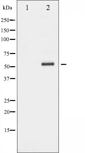
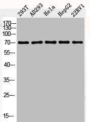
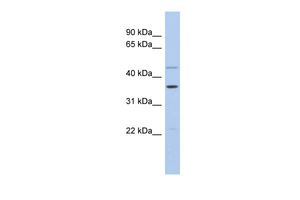
 WB (Western Blot)
(Western blot analysis of extracts from rat brain, using Fos Antibody.)
WB (Western Blot)
(Western blot analysis of extracts from rat brain, using Fos Antibody.)
Rabbit Fos Polyclonal Antibody | anti-Fos antibody
FOS Antibody
Phosphate buffered saline, pH 7.4, 150mM NaCl, 0.02% sodium azide and 50% glycerol.
WB (Western Blot)
(Western blot analysis of extracts from rat brain, using Fos Antibody.)
WB (Western Blot)
(Western blot analysis of extracts from rat brain, using Fos Antibody.)
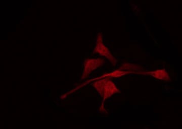
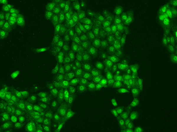
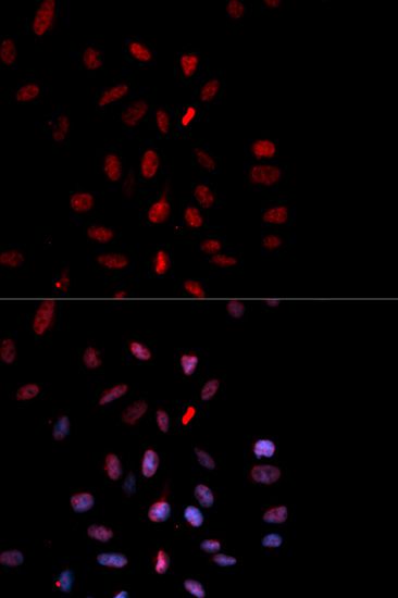
IF (Immunofluorescence)
(AAA322005 staining K562 by IF/ICC. The sample were fixed with PFA and permeabilized in 0.1% Triton X-100, then blocked in 10% serum for 45 minutes at 25 degree C. The primary antibody was diluted at 1/200 and incubated with the sample for 1 hour at 37 degree C. An Alexa Fluor 594 conjugated goat anti-rabbit IgG (H+L) Ab, diluted at 1/600, was used as the secondary antibody.)
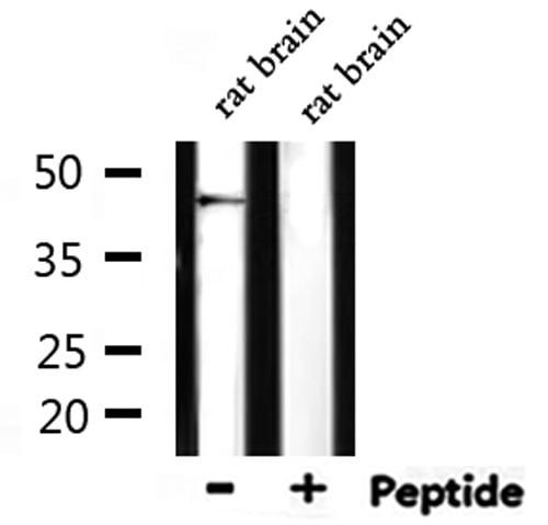
WB (Western Blot)
(Western blot analysis of Fos expression in HepG2 cell extract.The lane on the left is treated with the antigen-specific peptide.)
WB (Western Blot)
(Western blot analysis of Fos expression in HepG2 cell extract.The lane on the left is treated with the antigen-specific peptide.)
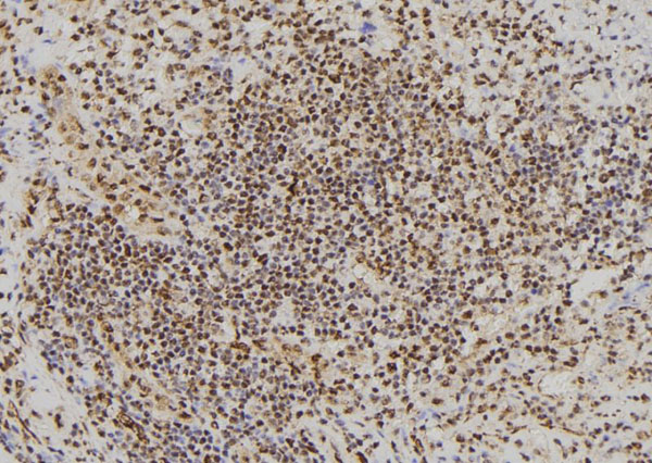
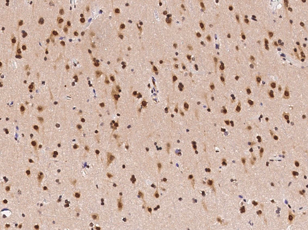
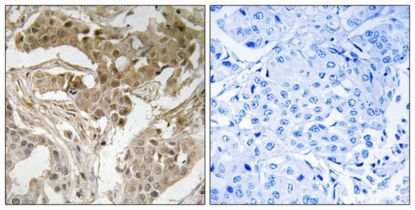
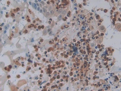
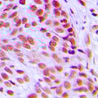
IHC (Immunohistochemistry)
(AAA322005 at 1/100 staining Human spleen tissue by IHC-P. The sample was formaldehyde fixed and a heat mediated antigen retrieval step in citrate buffer was performed. The sample was then blocked and incubated with the antibody for 1.5 hours at 22 degree C. An HRP conjugated goat anti-rabbit antibody was used as the secondary.)
Function: Nuclear phosphoprotein which forms a tight but non-covalently linked complex with the JUN/AP-1 transcription factor. In the heterodimer, FOS and JUN/AP-1 basic regions each seems to interact with symmetrical DNA half sites. On TGF-beta activation, forms a multimeric SMAD3/SMAD4/JUN/FOS complex at the AP1/SMAD-binding site to regulate TGF-beta-mediated signaling. Has a critical function in regulating the development of cells destined to form and maintain the skeleton. It is thought to have an important role in signal transduction, cell proliferation and differentiation. In growing cells, activates phospholipid synthesis, possibly by activating CDS1 and PI4K2A. This activity requires Tyr-dephosphorylation and association with the endoplasmic reticulum.
Subunit Structure: Heterodimer; with JUN (By similarity). Interacts with MAFB (By similarity). Component of the SMAD3/SMAD4/JUN/FOS complex required for synergistic TGF-beta-mediated transcription at the AP1 promoter site. Interacts with SMAD3; the interaction is weak even on TGF-beta activation. Interacts with MAFB. Interacts with DSIPI; this interaction inhibits the binding of active AP1 to its target DNA. Interacts with CDS1 and PI4K2A (By similarity).
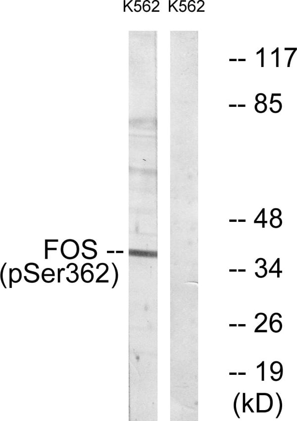
Post-translational Modifications: Phosphorylated in the C-terminal upon stimulation by nerve growth factor (NGF) and epidermal growth factor (EGF). Phosphorylated, in vitro, by MAPK and RSK1. Phosphorylation on both Ser-362 and Ser-374 by MAPK1/2 and RSK1/2 leads to protein stabilization with phosphorylation on Ser-374 being the major site for protein stabilization on NGF stimulation. Phosphorylation on Ser-362 and Ser-374 primes further phosphorylations on Thr-325 and Thr-331 through promoting docking of MAPK to the DEF domain. Phosphorylation on Thr-232, induced by HA-RAS, activates the transcriptional activity and antagonizes sumoylation. Phosphorylation on Ser-362 by RSK2 in osteoblasts contributes to osteoblast transformation (By similarity). Constitutively sumoylated with SUMO1, SUMO2 and SUMO3. Desumoylated by SENP2. Sumoylation requires heterodimerization with JUN and is enhanced by mitogen stimulation. Sumoylation inhibits the AP-1 transcriptional activity and is, itself, inhibited by Ras-activated phosphorylation on Thr-232. In quiescent cells, the small amount of FOS present is phosphorylated at Tyr-10 and Tyr-30 by SRC. This Tyr-phosphorylated form is cytosolic. In growing cells, dephosphorylated by PTPN2. Dephosphorylation leads to the association with endoplasmic reticulum membranes and activation of phospholipid synthesis.
Similarity: Belongs to the bZIP family. Fos subfamily.
NCBI and Uniprot Product Information
Predicted: 41 kDa
Customer Reviews
Loading reviews...
Share Your Experience
Similar Products


Product Notes
The Fos fos (Catalog #AAA322005) is an Antibody produced from Rabbit and is intended for research purposes only. The product is available for immediate purchase. The FOS Antibody reacts with Human, Mouse, Rat and may cross-react with other species as described in the data sheet. AAA Biotech's Fos can be used in a range of immunoassay formats including, but not limited to, ELISA, IP (Immunoprecipitation), ICC (Immunocytochemistry), IF (Immunofluorescence), IHC (Immunohistochemistry), WB (Western Blot). Researchers should empirically determine the suitability of the Fos fos for an application not listed in the data sheet. Researchers commonly develop new applications and it is an integral, important part of the investigative research process. It is sometimes possible for the material contained within the vial of "Fos, Polyclonal Antibody" to become dispersed throughout the inside of the vial, particularly around the seal of said vial, during shipment and storage. We always suggest centrifuging these vials to consolidate all of the liquid away from the lid and to the bottom of the vial prior to opening. Please be advised that certain products may require dry ice for shipping and that, if this is the case, an additional dry ice fee may also be required.Precautions
All products in the AAA Biotech catalog are strictly for research-use only, and are absolutely not suitable for use in any sort of medical, therapeutic, prophylactic, in-vivo, or diagnostic capacity. By purchasing a product from AAA Biotech, you are explicitly certifying that said products will be properly tested and used in line with industry standard. AAA Biotech and its authorized distribution partners reserve the right to refuse to fulfill any order if we have any indication that a purchaser may be intending to use a product outside of our accepted criteria.Disclaimer
Though we do strive to guarantee the information represented in this datasheet, AAA Biotech cannot be held responsible for any oversights or imprecisions. AAA Biotech reserves the right to adjust any aspect of this datasheet at any time and without notice. It is the responsibility of the customer to inform AAA Biotech of any product performance issues observed or experienced within 30 days of receipt of said product. To see additional details on this or any of our other policies, please see our Terms & Conditions page.Item has been added to Shopping Cart
If you are ready to order, navigate to Shopping Cart and get ready to checkout.