


 Application Data
(At 25 degree C. Samples were then incubated with primary Ab(At 37 degree C. An AlexaFluor594 conjugated goat anti-rabbit IgG(H+L) Ab(Red) and an AlexaFluor488 conjugated goat anti-mouse IgG(H+L) Ab(Green) were used as the secondary antibody.The nuclear counter stain is DAPI (blue).)
Application Data
(At 25 degree C. Samples were then incubated with primary Ab(At 37 degree C. An AlexaFluor594 conjugated goat anti-rabbit IgG(H+L) Ab(Red) and an AlexaFluor488 conjugated goat anti-mouse IgG(H+L) Ab(Green) were used as the secondary antibody.The nuclear counter stain is DAPI (blue).)
Rabbit SIRT2 Polyclonal Antibody | anti-SIRT2 antibody
Phospho-SIRT2 (Ser368) Antibody
Tissue Specificity: Isoform 1 is expressed in heart, liver and skeletal muscle, weakly expressed in the cortex. Isoform 2 is strongly expressed in the cortex, weakly expressed in heart and liver. Weakly expressed in several malignancies including breast, liver, brain, kidney and prostate cancers compared to normal tissues. Weakly expressed in glioma cell lines compared to normal brain tissues (at protein level). Widely expressed. Highly expressed in heart, brain and skeletal muscle, while it is weakly expressed in placenta and lung. Down-regulated in many gliomas suggesting that it may act as a tumor suppressor gene in human gliomas possibly through the regulation of microtubule network.
IF/ICC: 1:100-1:500
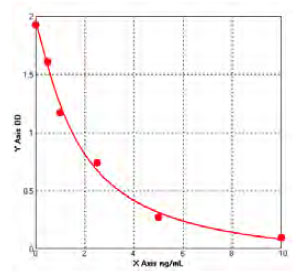
Peptide ELISA: 1:20,000-1:40,000
Note: Deacetylates FOXO3 in the cytoplasm. Colocalizes with PLP1 in internodal regions, at paranodal axoglial junction and Schmidt-Lanterman incisures of myelin sheat. Colocalizes with CDK5R1 in the perikaryon, neurites and growth cone of hippocampal neurons. Colocalizes with alpha-tubulin in neuronal growth cone. Localizes in the cytoplasm and nucleus of germinal vesicle (GV) stage oocytes. Colocalizes with alpha-tubulin on the meiotic spindle as the oocytes enter into metaphase, and also during meiotic anaphase and telophase, especially with the midbody. Colocalizes with PARD3 in internodal region of axons. Colocalizes with acetylated alpha-tubulin in cell projection processes during primary oligodendrocyte precursor (OLP) differentiation (By similarity). Localizes in the cytoplasm during most of the cell cycle except in the G2/M transition and during mitosis, where it is localized in association with chromatin and induces deacetylation of histone at 'Lys-16' (H4K16ac). Colocalizes with KMT5A at mitotic foci. Colocalizes with CDK1 at centrosome during prophase and splindle fibers during metaphase. Colocalizes with Aurora kinase AURKA at centrosome during early prophase and in the centrioles and growing mitotic spindle throughout metaphase. Colocalizes with Aurora kinase AURKB during cytokinesis with the midbody. Colocalizes with microtubules. Detected in perinuclear foci that may be aggresomes containing misfolded, ubiquitinated proteins. Shuttles between the cytoplasm and the nucleus through the CRM1 export pathway. Colocalizes with EP300 in the nucleus. Translocates to the nucleus and chromatin upon bacterium Listeria monocytogenes infection in interphase cells.Cytoplasm. Nucleus.
Note: Predominantly localized in the cytoplasmic.Cytoplasm. Nucleus.
Note: Predominantly localized in the cytoplasmic.Cytoplasm. Nucleus.
Note: Predominantly localized in the nucleus.
Application Data
(At 25 degree C. Samples were then incubated with primary Ab(At 37 degree C. An AlexaFluor594 conjugated goat anti-rabbit IgG(H+L) Ab(Red) and an AlexaFluor488 conjugated goat anti-mouse IgG(H+L) Ab(Green) were used as the secondary antibody.The nuclear counter stain is DAPI (blue).)
IHC (Immunohistochemistry)
(At 1/100 staining Mouse kidney tissue by IHC-P. The sample was formaldehyde fixed and a heat mediated antigen retrieval step in citrate buffer was performed. The sample was then blocked and incubated with the primary antibody at 4 degree C overnight. An HRP conjugated anti-Rabbit antibody was used as the secondary antibody.)
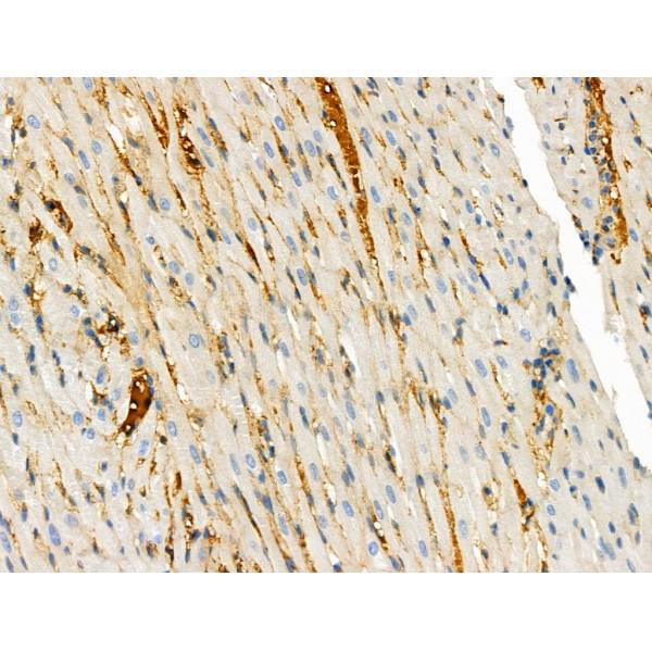
IHC (Immunohistochemistry)
(At 1/100 staining Rat heart tissue by IHC-P. The sample was formaldehyde fixed and a heat mediated antigen retrieval step in citrate buffer was performed. The sample was then blocked and incubated with the primary antibody at 4 degree C overnight. An HRP conjugated anti-Rabbit antibody was used as the secondary antibody.)
IHC (Immunohistochemistry)
(At 1/100 staining Rat liver tissue by IHC-P. The sample was formaldehyde fixed and a heat mediated antigen retrieval step in citrate buffer was performed. The sample was then blocked and incubated with the primary antibody at 4 degree C overnight. An HRP conjugated anti-Rabbit antibody was used as the secondary antibody.)
IHC (Immunohistochemistry)
(Staining Rat lung tissue by IHC-P. The sample was formaldehyde fixed and a heat mediated antigen retrieval step in citrate buffer was performed. The sample was then blocked and incubated with the primary antibody at 4 degree C overnight. An HRP conjugated anti-Rabbit antibody was used as the secondary antibody.)
IHC (Immunohistochemistry)
(At 1/100 staining Rat kidney tissue by IHC-P. The sample was formaldehyde fixed and a heat mediated antigen retrieval step in citrate buffer was performed. The sample was then blocked and incubated with the primary antibody at 4 degree C overnight. An HRP conjugated anti-Rabbit antibody was used as the secondary antibody.)
NCBI and Uniprot Product Information
Similar Products


Product Notes
The SIRT2 sirt2 (Catalog #AAA31286) is an Antibody produced from Rabbit and is intended for research purposes only. The product is available for immediate purchase. The Phospho-SIRT2 (Ser368) Antibody reacts with Human, Mouse, Rat and may cross-react with other species as described in the data sheet. AAA Biotech's SIRT2 can be used in a range of immunoassay formats including, but not limited to, Immunohistochemistry (IHC), Immunofluorescence (IF), Immunocytochemistry (ICC), Peptide ELISA (EIA). IHC: 1:50-1:200 IF/ICC: 1:100-1:500 Peptide ELISA: 1:20,000-1:40,000. Researchers should empirically determine the suitability of the SIRT2 sirt2 for an application not listed in the data sheet. Researchers commonly develop new applications and it is an integral, important part of the investigative research process. It is sometimes possible for the material contained within the vial of "SIRT2, Polyclonal Antibody" to become dispersed throughout the inside of the vial, particularly around the seal of said vial, during shipment and storage. We always suggest centrifuging these vials to consolidate all of the liquid away from the lid and to the bottom of the vial prior to opening. Please be advised that certain products may require dry ice for shipping and that, if this is the case, an additional dry ice fee may also be required.Precautions
All products in the AAA Biotech catalog are strictly for research-use only, and are absolutely not suitable for use in any sort of medical, therapeutic, prophylactic, in-vivo, or diagnostic capacity. By purchasing a product from AAA Biotech, you are explicitly certifying that said products will be properly tested and used in line with industry standard. AAA Biotech and its authorized distribution partners reserve the right to refuse to fulfill any order if we have any indication that a purchaser may be intending to use a product outside of our accepted criteria.Disclaimer
Though we do strive to guarantee the information represented in this datasheet, AAA Biotech cannot be held responsible for any oversights or imprecisions. AAA Biotech reserves the right to adjust any aspect of this datasheet at any time and without notice. It is the responsibility of the customer to inform AAA Biotech of any product performance issues observed or experienced within 30 days of receipt of said product. To see additional details on this or any of our other policies, please see our Terms & Conditions page.Item has been added to Shopping Cart
If you are ready to order, navigate to Shopping Cart and get ready to checkout.