 Application Data
Rabbit anti-Mouse Lymphocytes Antibody
Rabbit Anti-Mouse Lymphocyte Serum
Reactivity
Mouse
Synonyms
Lymphocytes; N/A; Rabbit Anti-Mouse Lymphocyte Serum; Lymphocytes, Lyophilized (Polyclonal) (rabbit serum); anti-Lymphocytes antibody
Host
Rabbit
Reactivity
Mouse
Presentation
1.0 ml, 5.0 ml lyophilized
Sterility
This reagent is not sold as sterile, but can be sterilized by filtration if necessary. To minimize loss of volume during filtration, dilute to the final working concentration in the appropriate medium before filtration and filter through a 0.22um filter.
Specifications
Immunizing Strain: BALB/c
Heat Inactivation: 60 minutes at 56°C
Absorption: Mouse erythrocyes/hepatocytes
Heat Inactivation: 60 minutes at 56°C
Absorption: Mouse erythrocyes/hepatocytes
Lot Specifications
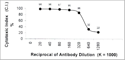
Antiserum Titration:
Cell Source: Spleen, Thymus
Donors: BALB/c
Cell Concentration: 1.1x10⁶ cells per ml
Complement: Low-Tox-M Rabbit Complement
Complement Concentration: 1:10
Procedure: Two stage cytotoxicity as described on Recommended Method for Determining Percent Cytotoxicity with Anti-Mouse Lymphocyte Serum Plus Complement.
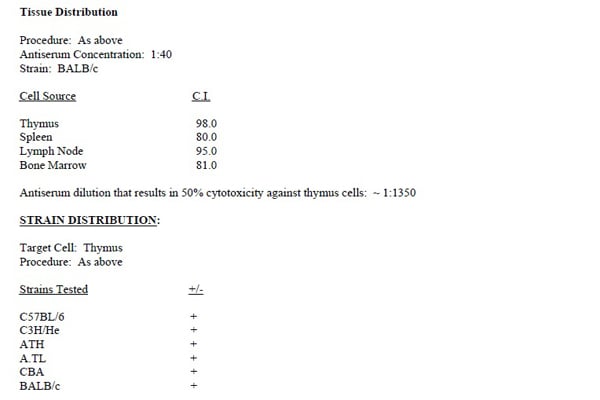
Tissue Distribution
Procedure: As above
Antiserum Concentration: 1:40
Strain: BALB/c
Antiserum Concentration: 1:40
Strain: BALB/c
Cell Source/C.I.
Thymus: 100.0
Spleen: 50.0
Lymph Node: 95.0
Bone Marrow: 81.0
Antiserum dilution that results in 50% cytotoxicity against thymus cells: ~1:1350
Spleen: 50.0
Lymph Node: 95.0
Bone Marrow: 81.0
Antiserum dilution that results in 50% cytotoxicity against thymus cells: ~1:1350
Strain Distribution
Target Cell: Thymus
Procedure: As above
Procedure: As above
Strains Tested (+/-)
C57BL/6: -
C3H/He: -
ATH: -
A.TL: -
CBA: -
BALB/c: +
C3H/He: -
ATH: -
A.TL: -
CBA: -
BALB/c: +
Functional Testing
Cell Source: Splenocytes and Thymocytes
Cell Concentration: 1x10⁶ cells/ml.
Antiserum Concentration: 1:10
Complement: Low-Tox-M at 1:10
Donors: BALB/c
Cell Concentration: 1x10⁶ cells/ml.
Antiserum Concentration: 1:10
Complement: Low-Tox-M at 1:10
Donors: BALB/c
Procedure
Cells were treated as described in "Recommended Method for Depleting A Cell Population of Mouse Lymphocytes." The remaining viable cells were exposed to the mitogens Concanavalin A (CON A), Phytohaemagglutinin (PHA), and Lipopolysaccharide (LPS). Cell depletionwith Anti-Mouse Lymphocyte Serum was found to inhibit the CON A, PHA,and LPS responses. Treatment of mouse splenocytes with Anti-Mouse Lymphocyte Serum plus complement essentially eliminated in vitro T effector cell function.
Recommended Method For Depleting A Cell Population Of Mouse Lymphocytes
1. Prepare a cell suspension from the appropriate tissue in Cytotoxicity Medium or equivalent. Remove red cells and dead cells (where necessary) by purification of viable lymphocytes on Lympholyte-M2 density cell separation medium. After washing, adjust the cell concentration to 1x10⁶ cells per ml in Cytotoxicity Medium.
2. Add the antiserum to a final concentration of 1:40 and mix.
3. Incubate for 60 minutes at 4°C.
4. Centrifuge to pellet the cells and discard the supernatant.
5. Resuspend to the original volume in Low-Tox-M3 Rabbit Complement, diluted to the appropriate concentration in Cyotoxicity Medium. (Recommended concentration included with each batch of Low-Tox-M Complement)
6. Incubate for 60 minutes at 37°C.
7. Monitor for percent cytotoxicity at this stage, before further processing. For this purpose remove a small sample from each tube, dilute 1:10 with medium, and add 1/10 volume of 1% Trypan Blue. After 3-5 minutes, score live versus dead cells in a hemacytometer.
8. For functional studies, remove the dead cells from the treated groups before further processing, particularly if the treated cells are to be cultured. Layering the suspension on cell separation medium and centrifuging at room temperature as per the instructions provided can do this. Live cells will form a layer at the interface, while dead cells pellet.The interface can then be collected and washed in Cytotoxicity Medium before being resuspended in the appropriate medium for further processing.
Alternatively, the cells can then be washed and resuspended in the appropriate medium for further processing immediately after Step #6, provided that the dead cells will not interfere with subsequent assays.
Recommended Method For Determining Percent Cytotoxicity With Anti-Mouse Lymphocyte Serum Plus Complement
1. Prepare a cell suspension from the appropriate tissue in Cytotoxicity Medium or equivalent. Remove red cells and dead cells (where necessary) by purification of viable lymphocytes on Lympholyteo-M2 density cell separation medium. After washing, adjust the cell concentration to 1.1 x 10⁶ cells per ml in Cytotoxicity Medium.
2. Add the antiserum to a final concentration of 1:40 and mix.
3. Incubate for 60 minutes at 4°C.
4. Centrifuge to pellet the cells and discard the supernatant.
5. Resuspend to the original volume in Low-Tox-M Rabbit Complement3 diluted to the appropriate concentration in Cyotoxicity Medium. (Recommended concentration included with each batch of Low-Tox-M Rabbit Complement ~ 1:10 - 1:25)
6. Incubate for 60 minutes at 37°C.
7. Place on ice.
8. Add Trypan Blue. 10% by volume of 1%Trypan blue (w/v) added 3-5 minutes before scoring works well. Score live versus dead cells in a hemacytometer.
Cyotoxic index (C.I.) can be calculated as follows:
C.I. = % cyt (antibody + complement) - % cvt (complement alone)/100% - % cyt (complement alone) x 100
Preparation and Storage
Lyophilized form stable at 4°C or -20°C. Reconstitute with 1.0 ml or 5.0 ml distilled water.
After reconstitution, aliquot and freeze unused portions at -70°C in volumes appropriate for single usage.
Avoid repeated freeze/thaw cycles.
After reconstitution, aliquot and freeze unused portions at -70°C in volumes appropriate for single usage.
Avoid repeated freeze/thaw cycles.
Application Data
Application Data
Related Product Information for anti-Lymphocytes antibody
Description: Anti-Mouse Lymphocyte Serum is prepared by immunizing rabbits with mouse thymus, spleen and lymph node cells followed by absorption with mouse erythrocytes and hepatocytes. This antiserum is strongly cytotoxic to all mouse lymphocytes.
Notes: 1. Cytotoxicity Medium is RPMI-1640 with 25mM Hepes buffer and 0.3% bovine serum albumin (BSA). BSA is substituted for the conventionally used fetal calf serum (FCS) because we have found that many batches of FCS contain complement dependent cytotoxins to mouse lymphocytes, thus increasing the background killing in the presence ofcomplement. We recommend that cells not be exposed to FCS prior to or during exposure to antibody and complement. Some batches of BSA als ocontain complement dependent cytotoxins, resulting in the same problem. We screen for batches of BSA giving low background in the presence ofcomplement and use the selected BSA for preparing Cytotoxicity Medium.
2. Lympholyte-M cell separation medium is density cell separation medium designed specifically for the isolation of viable mouse lymphocytes. This separation medium provides a high and non-selective recovery of viable mouse lymphocytes, removing red cells and dead cells. The density of this medium is 1.0865-1.0885. Isolation of mouse lymphocytes on cell separation medium of density 1.077 will result in high and selective loss of lymphocytes and should be avoided.
3. Rabbit serum provides the most potent source of complement for use with antibodies to mouse cell surface antigens. However, rabbit serum itself is very toxic to murine lymphocytes. Low-Tox-M Rabbit Complement is absorbed to remove toxicity to mouse lymphocytes, while maintaining its high complement activity. When used in conjunction with Cytotoxicity Medium, this reagent provides a highly potent source of complement with minimal background toxicity.
Customer Reviews
Loading reviews...
Share Your Experience
Similar Products


Product Notes
The Lymphocytes (Catalog #AAA14281) is an Antibody produced from Rabbit and is intended for research purposes only. The product is available for immediate purchase. The Rabbit Anti-Mouse Lymphocyte Serum reacts with Mouse and may cross-react with other species as described in the data sheet. It is sometimes possible for the material contained within the vial of "Lymphocytes, Antibody" to become dispersed throughout the inside of the vial, particularly around the seal of said vial, during shipment and storage. We always suggest centrifuging these vials to consolidate all of the liquid away from the lid and to the bottom of the vial prior to opening. Please be advised that certain products may require dry ice for shipping and that, if this is the case, an additional dry ice fee may also be required.Precautions
All products in the AAA Biotech catalog are strictly for research-use only, and are absolutely not suitable for use in any sort of medical, therapeutic, prophylactic, in-vivo, or diagnostic capacity. By purchasing a product from AAA Biotech, you are explicitly certifying that said products will be properly tested and used in line with industry standard. AAA Biotech and its authorized distribution partners reserve the right to refuse to fulfill any order if we have any indication that a purchaser may be intending to use a product outside of our accepted criteria.Disclaimer
Though we do strive to guarantee the information represented in this datasheet, AAA Biotech cannot be held responsible for any oversights or imprecisions. AAA Biotech reserves the right to adjust any aspect of this datasheet at any time and without notice. It is the responsibility of the customer to inform AAA Biotech of any product performance issues observed or experienced within 30 days of receipt of said product. To see additional details on this or any of our other policies, please see our Terms & Conditions page.Item has been added to Shopping Cart
If you are ready to order, navigate to Shopping Cart and get ready to checkout.