
Filters
Clonality
Type
Reactivity
Gene Name
Isotype
Host
Application
Clone
10000 results for "Recombinant Proteins" - showing 1-50
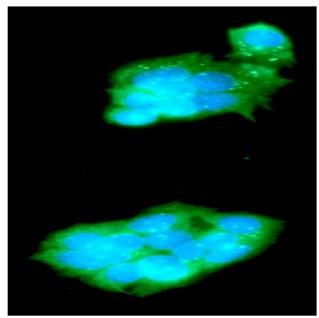

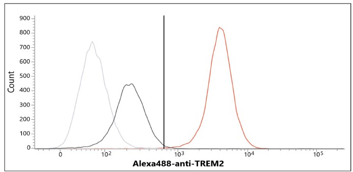
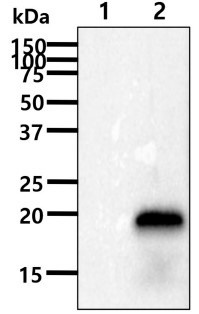 Western Blot (WB)
(The cell lysates(40ug) were resolved by SDS-PAGE, transferred to PVDF membrane and probed with anti-human TREM2 antibody (1:1000). Proteins were visualized using a goat anti-mouse secondary antibody conjugated to HRP and an ECL detection system.Lane 1 : 293T cell lysateLane 2 : TREM2 19-161aa Transfected 293T cell lysate)
Western Blot (WB)
(The cell lysates(40ug) were resolved by SDS-PAGE, transferred to PVDF membrane and probed with anti-human TREM2 antibody (1:1000). Proteins were visualized using a goat anti-mouse secondary antibody conjugated to HRP and an ECL detection system.Lane 1 : 293T cell lysateLane 2 : TREM2 19-161aa Transfected 293T cell lysate)
TREM2, Monoclonal Antibody (Cat# AAA332953)
Full Name
Human TREM2 antibody
Reactivity
Human
Applications
Western Blot, Immunocytochemistry, Flow Cytometry
Purity
By protein-A affinity chromatography
Pricing


 Immunohistochemistry (IHC)-Formalin
(Formalin-fixed, paraffin-embedded human cardiac muscle stained with Dystrophin Mouse Monoclonal Antibody (DMD/3677).)
Immunohistochemistry (IHC)-Formalin
(Formalin-fixed, paraffin-embedded human cardiac muscle stained with Dystrophin Mouse Monoclonal Antibody (DMD/3677).)
Dystrophin (DMD), Monoclonal Antibody (Cat# AAA333002)
Full Name
Dystrophin (DMD) (Marker of Duchenne and Becker Muscular Dystrophy)
Gene Names
DMD; BMD; CMD3B; MRX85; DXS142; DXS164; DXS206; DXS230; DXS239; DXS268; DXS269; DXS270; DXS272
Reactivity
Human
Applications
EIA, IHC
Purity
Purified Ab with BSA and Azide at 200ug/ml or Purified Ab with BSA and Azide at 200ug/ml or Purified Ab WITHOUT BSA and Azide at 1.0mg/ml
Pricing


 IP (Immunoprecipitation)
(Immunoprecipitation analysis of 200ug extracts of 293T cells using 1ug HIF1AN antibody. Western blot was performed from the immunoprecipitate using HIF1AN antibody at a dilition of 1:1000.)
IP (Immunoprecipitation)
(Immunoprecipitation analysis of 200ug extracts of 293T cells using 1ug HIF1AN antibody. Western blot was performed from the immunoprecipitate using HIF1AN antibody at a dilition of 1:1000.)
HIF1AN, Polyclonal Antibody (Cat# AAA28174)
Full Name
HIF1AN Polyclonal Antibody
Gene Names
HIF1AN; FIH1
Reactivity
Human, Mouse, Rat
Applications
Western Blot, Immunohistochemistry, Immunofluorescence, Immunoprecipitation, Chromatin Immunoprecipitation
Purity
Affinity Purification
Pricing
 IF (Immunofluorescence)
(Figure 9. IF analysis of ATP5D/ATP5F1D using anti-ATP5D/ATP5F1D antibody (AAA20015).ATP5D/ATP5F1D was detected in an immunocytochemical section of HELA cells. Enzyme antigen retrieval was performed using IHC enzyme antigen retrieval reagent ( epitope retrieval solution). The tissue section was blocked with 10% goat serum. The tissue section was then incubated with 2ug/ml rabbit anti-ATP5D/ATP5F1D Antibody (AAA20015) overnight at 4 degree C. Peroxidase Conjugated Goat Anti-rabbit IgG was used as secondary antibody and incubated for 30 minutes at 37 degree C. The tissue section was developed using HRP Conjugated Rabbit IgG Super Vision Assay Kit ( epitope retrieval solution). The tissue section was blocked with 10% goat serum. The tissue section was then incubated with 2ug/ml rabbit anti-ATP5D/ATP5F1D Antibody (AAA20015) overnight at 4 degree C. Peroxidase Conjugated Goat Anti-rabbit IgG was used as secondary antibody and incubated for 30 minutes at 37 degree C. The tissue section was developed using HRP Conjugated Rabbit IgG Super Vision Assay Kit ( epitope retrieval solution). The tissue section was blocked with 10% goat serum. The tissue section was then incubated with 2ug/ml rabbit anti-ATP5D/ATP5F1D Antibody (AAA20015) overnight at 4 degree C. Peroxidase Conjugated Goat Anti-rabbit IgG was used as secondary antibody and incubated for 30 minutes at 37 degree C. The tissue section was developed using HRP Conjugated Rabbit IgG Super Vision Assay Kit ( epitope retrieval solution). The tissue section was blocked with 10% goat serum. The tissue section was then incubated with 2ug/ml rabbit anti-ATP5D/ATP5F1D Antibody (AAA20015) overnight at 4 degree C. Peroxidase Conjugated Goat Anti-rabbit IgG was used as secondary antibody and incubated for 30 minutes at 37 degree C. The tissue section was developed using HRP Conjugated Rabbit IgG Super Vision Assay Kit ( epitope retrieval solution). The tissue section was blocked with 10% goat serum. The tissue section was then incubated with 2ug/ml rabbit anti-ATP5D/ATP5F1D Antibody (AAA20015) overnight at 4 degree C. Peroxidase Conjugated Goat Anti-rabbit IgG was used as secondary antibody and incubated for 30 minutes at 37 degree C. The tissue section was developed using HRP Conjugated Rabbit IgG Super Vision Assay Kit ( epitope retrieval solution). The tissue section was blocked with 10% goat serum. The tissue section was then incubated with 2ug/ml rabbit anti-ATP5D/ATP5F1D Antibody (AAA20015) overnight at 4 degree C. Peroxidase Conjugated Goat Anti-rabbit IgG was used as secondary antibody and incubated for 30 minutes at 37 degree C. The tissue section was developed using HRP Conjugated Rabbit IgG Super Vision Assay Kit ( epitope retrieval solution). The tissue section was blocked with 10% goat serum. The tissue section was then incubated with 2ug/ml rabbit anti-ATP5D/ATP5F1D Antibody (AAA20015) overnight at 4 degree C. Peroxidase Conjugated Goat Anti-rabbit IgG was used as secondary antibody and incubated for 30 minutes at 37 degree C. The tissue section was developed using HRP Conjugated Rabbit IgG Super Vision Assay Kit (Lane 2: human HepG2 whole cell lysates,Lane 3: human MCF-7 whole cell lysates,Lane 4: human Raji whole cell lysates,Lane 5: rat liver tissue lysates,Lane 6: rat kidney tissue lysates,Lane 7: mouse liver tissue lysates.After electrophoresis, proteins were transferred to a nitrocellulose membrane at 150 mA for 50-90 minutes. Blocked the membrane with 5% non-fat milk/TBS for 1.5 hour at RT. The membrane was incubated with rabbit anti-ATP5D/ATP5F1D antigen affinity purified polyclonal antibody (#AAA20015) at 0.5ug/mL overnight at 4 degree C, then washed with TBS-0.1%Tween 3 times with 5 minutes each and probed with a goat anti-rabbit IgG-HRP secondary antibody at a dilution of 1:5000 for 1.5 hour at RT. The signal is developed using an Enhanced Chemiluminescent detection (ECL) kit with Tanon 5200 system. A specific band was detected for ATP5D/ATP5F1D at approximately 17 kDa. The expected band size for ATP5D/ATP5F1D is at 17 kDa.)
IF (Immunofluorescence)
(Figure 9. IF analysis of ATP5D/ATP5F1D using anti-ATP5D/ATP5F1D antibody (AAA20015).ATP5D/ATP5F1D was detected in an immunocytochemical section of HELA cells. Enzyme antigen retrieval was performed using IHC enzyme antigen retrieval reagent ( epitope retrieval solution). The tissue section was blocked with 10% goat serum. The tissue section was then incubated with 2ug/ml rabbit anti-ATP5D/ATP5F1D Antibody (AAA20015) overnight at 4 degree C. Peroxidase Conjugated Goat Anti-rabbit IgG was used as secondary antibody and incubated for 30 minutes at 37 degree C. The tissue section was developed using HRP Conjugated Rabbit IgG Super Vision Assay Kit ( epitope retrieval solution). The tissue section was blocked with 10% goat serum. The tissue section was then incubated with 2ug/ml rabbit anti-ATP5D/ATP5F1D Antibody (AAA20015) overnight at 4 degree C. Peroxidase Conjugated Goat Anti-rabbit IgG was used as secondary antibody and incubated for 30 minutes at 37 degree C. The tissue section was developed using HRP Conjugated Rabbit IgG Super Vision Assay Kit ( epitope retrieval solution). The tissue section was blocked with 10% goat serum. The tissue section was then incubated with 2ug/ml rabbit anti-ATP5D/ATP5F1D Antibody (AAA20015) overnight at 4 degree C. Peroxidase Conjugated Goat Anti-rabbit IgG was used as secondary antibody and incubated for 30 minutes at 37 degree C. The tissue section was developed using HRP Conjugated Rabbit IgG Super Vision Assay Kit ( epitope retrieval solution). The tissue section was blocked with 10% goat serum. The tissue section was then incubated with 2ug/ml rabbit anti-ATP5D/ATP5F1D Antibody (AAA20015) overnight at 4 degree C. Peroxidase Conjugated Goat Anti-rabbit IgG was used as secondary antibody and incubated for 30 minutes at 37 degree C. The tissue section was developed using HRP Conjugated Rabbit IgG Super Vision Assay Kit ( epitope retrieval solution). The tissue section was blocked with 10% goat serum. The tissue section was then incubated with 2ug/ml rabbit anti-ATP5D/ATP5F1D Antibody (AAA20015) overnight at 4 degree C. Peroxidase Conjugated Goat Anti-rabbit IgG was used as secondary antibody and incubated for 30 minutes at 37 degree C. The tissue section was developed using HRP Conjugated Rabbit IgG Super Vision Assay Kit ( epitope retrieval solution). The tissue section was blocked with 10% goat serum. The tissue section was then incubated with 2ug/ml rabbit anti-ATP5D/ATP5F1D Antibody (AAA20015) overnight at 4 degree C. Peroxidase Conjugated Goat Anti-rabbit IgG was used as secondary antibody and incubated for 30 minutes at 37 degree C. The tissue section was developed using HRP Conjugated Rabbit IgG Super Vision Assay Kit ( epitope retrieval solution). The tissue section was blocked with 10% goat serum. The tissue section was then incubated with 2ug/ml rabbit anti-ATP5D/ATP5F1D Antibody (AAA20015) overnight at 4 degree C. Peroxidase Conjugated Goat Anti-rabbit IgG was used as secondary antibody and incubated for 30 minutes at 37 degree C. The tissue section was developed using HRP Conjugated Rabbit IgG Super Vision Assay Kit (Lane 2: human HepG2 whole cell lysates,Lane 3: human MCF-7 whole cell lysates,Lane 4: human Raji whole cell lysates,Lane 5: rat liver tissue lysates,Lane 6: rat kidney tissue lysates,Lane 7: mouse liver tissue lysates.After electrophoresis, proteins were transferred to a nitrocellulose membrane at 150 mA for 50-90 minutes. Blocked the membrane with 5% non-fat milk/TBS for 1.5 hour at RT. The membrane was incubated with rabbit anti-ATP5D/ATP5F1D antigen affinity purified polyclonal antibody (#AAA20015) at 0.5ug/mL overnight at 4 degree C, then washed with TBS-0.1%Tween 3 times with 5 minutes each and probed with a goat anti-rabbit IgG-HRP secondary antibody at a dilution of 1:5000 for 1.5 hour at RT. The signal is developed using an Enhanced Chemiluminescent detection (ECL) kit with Tanon 5200 system. A specific band was detected for ATP5D/ATP5F1D at approximately 17 kDa. The expected band size for ATP5D/ATP5F1D is at 17 kDa.)
ATP5F1D, Polyclonal Antibody (Cat# AAA20015)
Full Name
Anti-ATP5F1D Antibody Picoband
Reactivity
Human, Mouse, Rat
Applications
Western Blot, Immunohistochemistry, Immunocytochemistry, Immunofluorescence
Purity
Immunogen affinity purified.
Pricing
Retinoblastoma, Recombinant Protein (Cat# AAA10852)
Full Name
Recombinant Human Retinoblastoma Associated Protein
Gene Names
RB1; RB; pRb; OSRC; pp110; p105-Rb; PPP1R130
Purity
Greater than 95.0% as determined by: (a) Analysis by RP-HPLC. (b) Analysis by SDS-PAGE.
Pricing


 IF (Immunofluorescence)
(Immunofluorescence analysis of PC-12 cells using Bcl-2 Mouse mAb at dilution of 1:100 (40x lens). Blue: DAPI for nuclear staining.)
IF (Immunofluorescence)
(Immunofluorescence analysis of PC-12 cells using Bcl-2 Mouse mAb at dilution of 1:100 (40x lens). Blue: DAPI for nuclear staining.)
Bcl-2, Polyclonal Antibody (Cat# AAA28424)
Full Name
Bcl-2 Mouse mAb
Gene Names
VEGFA; VPF; VEGF; MVCD1
Reactivity
Human, Mouse, Rat
Applications
Western Blot, Immunofluorescence, Immunocytochemistry
Purity
Affinity purification
Pricing
 IHC (Immunohistochemistry)
(Figure 7. IHC analysis of GOLGA7 using anti-GOLGA7 antibody (AAA19952).GOLGA7 was detected in a paraffin-embedded section of human ovarian cancer tissue. Heat mediated antigen retrieval was performed in EDTA buffer (pH 8.0, epitope retrieval solution). The tissue section was blocked with 10% goat serum. The tissue section was then incubated with 2ug/ml rabbit anti-GOLGA7 Antibody (AAA19952) overnight at 4 degree C. Peroxidase Conjugated Goat Anti-rabbit IgG was used as secondary antibody and incubated for 30 minutes at 37 degree C. The tissue section was developed using HRP Conjugated Rabbit IgG Super Vision Assay Kit ( epitope retrieval solution). The tissue section was blocked with 10% goat serum. The tissue section was then incubated with 2ug/ml rabbit anti-GOLGA7 Antibody (AAA19952) overnight at 4 degree C. Peroxidase Conjugated Goat Anti-rabbit IgG was used as secondary antibody and incubated for 30 minutes at 37 degree C. The tissue section was developed using HRP Conjugated Rabbit IgG Super Vision Assay Kit ( epitope retrieval solution). The tissue section was blocked with 10% goat serum. The tissue section was then incubated with 2ug/ml rabbit anti-GOLGA7 Antibody (AAA19952) overnight at 4 degree C. Peroxidase Conjugated Goat Anti-rabbit IgG was used as secondary antibody and incubated for 30 minutes at 37 degree C. The tissue section was developed using HRP Conjugated Rabbit IgG Super Vision Assay Kit ( epitope retrieval solution). The tissue section was blocked with 10% goat serum. The tissue section was then incubated with 2ug/ml rabbit anti-GOLGA7 Antibody (AAA19952) overnight at 4 degree C. Peroxidase Conjugated Goat Anti-rabbit IgG was used as secondary antibody and incubated for 30 minutes at 37 degree C. The tissue section was developed using HRP Conjugated Rabbit IgG Super Vision Assay Kit ( epitope retrieval solution). The tissue section was blocked with 10% goat serum. The tissue section was then incubated with 2ug/ml rabbit anti-GOLGA7 Antibody (AAA19952) overnight at 4 degree C. Peroxidase Conjugated Goat Anti-rabbit IgG was used as secondary antibody and incubated for 30 minutes at 37 degree C. The tissue section was developed using HRP Conjugated Rabbit IgG Super Vision Assay Kit ( epitope retrieval solution). The tissue section was blocked with 10% goat serum. The tissue section was then incubated with 2ug/ml rabbit anti-GOLGA7 Antibody (AAA19952) overnight at 4 degree C. Peroxidase Conjugated Goat Anti-rabbit IgG was used as secondary antibody and incubated for 30 minutes at 37 degree C. The tissue section was developed using HRP Conjugated Rabbit IgG Super Vision Assay Kit ( epitope retrieval solution). The tissue section was blocked with 10% goat serum. The tissue section was then incubated with 2ug/ml rabbit anti-GOLGA7 Antibody (AAA19952) overnight at 4 degree C. Peroxidase Conjugated Goat Anti-rabbit IgG was used as secondary antibody and incubated for 30 minutes at 37 degree C. The tissue section was developed using HRP Conjugated Rabbit IgG Super Vision Assay Kit with DAB as the chromogen.)
IHC (Immunohistochemistry)
(Figure 7. IHC analysis of GOLGA7 using anti-GOLGA7 antibody (AAA19952).GOLGA7 was detected in a paraffin-embedded section of human ovarian cancer tissue. Heat mediated antigen retrieval was performed in EDTA buffer (pH 8.0, epitope retrieval solution). The tissue section was blocked with 10% goat serum. The tissue section was then incubated with 2ug/ml rabbit anti-GOLGA7 Antibody (AAA19952) overnight at 4 degree C. Peroxidase Conjugated Goat Anti-rabbit IgG was used as secondary antibody and incubated for 30 minutes at 37 degree C. The tissue section was developed using HRP Conjugated Rabbit IgG Super Vision Assay Kit ( epitope retrieval solution). The tissue section was blocked with 10% goat serum. The tissue section was then incubated with 2ug/ml rabbit anti-GOLGA7 Antibody (AAA19952) overnight at 4 degree C. Peroxidase Conjugated Goat Anti-rabbit IgG was used as secondary antibody and incubated for 30 minutes at 37 degree C. The tissue section was developed using HRP Conjugated Rabbit IgG Super Vision Assay Kit ( epitope retrieval solution). The tissue section was blocked with 10% goat serum. The tissue section was then incubated with 2ug/ml rabbit anti-GOLGA7 Antibody (AAA19952) overnight at 4 degree C. Peroxidase Conjugated Goat Anti-rabbit IgG was used as secondary antibody and incubated for 30 minutes at 37 degree C. The tissue section was developed using HRP Conjugated Rabbit IgG Super Vision Assay Kit ( epitope retrieval solution). The tissue section was blocked with 10% goat serum. The tissue section was then incubated with 2ug/ml rabbit anti-GOLGA7 Antibody (AAA19952) overnight at 4 degree C. Peroxidase Conjugated Goat Anti-rabbit IgG was used as secondary antibody and incubated for 30 minutes at 37 degree C. The tissue section was developed using HRP Conjugated Rabbit IgG Super Vision Assay Kit ( epitope retrieval solution). The tissue section was blocked with 10% goat serum. The tissue section was then incubated with 2ug/ml rabbit anti-GOLGA7 Antibody (AAA19952) overnight at 4 degree C. Peroxidase Conjugated Goat Anti-rabbit IgG was used as secondary antibody and incubated for 30 minutes at 37 degree C. The tissue section was developed using HRP Conjugated Rabbit IgG Super Vision Assay Kit ( epitope retrieval solution). The tissue section was blocked with 10% goat serum. The tissue section was then incubated with 2ug/ml rabbit anti-GOLGA7 Antibody (AAA19952) overnight at 4 degree C. Peroxidase Conjugated Goat Anti-rabbit IgG was used as secondary antibody and incubated for 30 minutes at 37 degree C. The tissue section was developed using HRP Conjugated Rabbit IgG Super Vision Assay Kit ( epitope retrieval solution). The tissue section was blocked with 10% goat serum. The tissue section was then incubated with 2ug/ml rabbit anti-GOLGA7 Antibody (AAA19952) overnight at 4 degree C. Peroxidase Conjugated Goat Anti-rabbit IgG was used as secondary antibody and incubated for 30 minutes at 37 degree C. The tissue section was developed using HRP Conjugated Rabbit IgG Super Vision Assay Kit with DAB as the chromogen.)
GOLGA7, Polyclonal Antibody (Cat# AAA19952)
Full Name
Anti-GOLGA7 Antibody
Gene Names
GOLGA7; GCP16; GOLGA7A; HSPC041; GOLGA3AP1
Reactivity
Human
Applications
Immunohistochemistry
Purity
Immunogen affinity purified.
Pricing
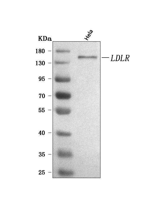
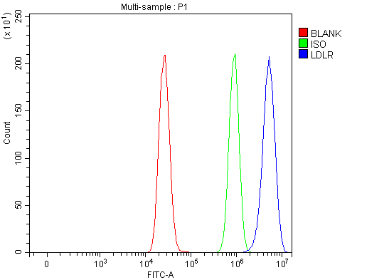 FCM/FACS (Flow Cytometry)
(Figure 2. Flow Cytometry analysis of MCF-7 cells using anti-LDLR antibody (AAA126965).Overlay histogram showing MCF-7 cells stained with AAA126965 (Blue line). To facilitate intracellular staining, cells were fixed with 4% paraformaldehyde and permeabilized with permeabilization buffer. The cells were blocked with 10% normal goat serum. And then incubated with rabbit anti-LDLR Antibody (AAA126965, 1ug/1x106 cells) for 30 min at 20 degree C. DyLight488 conjugated goat anti-rabbit IgG was used as secondary antibody for 30 minutes at 20 degree C. Isotype control antibody (Green line) was rabbit IgG (1ug/1x106) used under the same conditions. Unlabelled sample (Red line) was also used as a control.)
FCM/FACS (Flow Cytometry)
(Figure 2. Flow Cytometry analysis of MCF-7 cells using anti-LDLR antibody (AAA126965).Overlay histogram showing MCF-7 cells stained with AAA126965 (Blue line). To facilitate intracellular staining, cells were fixed with 4% paraformaldehyde and permeabilized with permeabilization buffer. The cells were blocked with 10% normal goat serum. And then incubated with rabbit anti-LDLR Antibody (AAA126965, 1ug/1x106 cells) for 30 min at 20 degree C. DyLight488 conjugated goat anti-rabbit IgG was used as secondary antibody for 30 minutes at 20 degree C. Isotype control antibody (Green line) was rabbit IgG (1ug/1x106) used under the same conditions. Unlabelled sample (Red line) was also used as a control.)
LDLR, Polyclonal Antibody (Cat# AAA126965)
Full Name
Anti-LDLR Antibody Picoband
Gene Names
LDLR; FH; FHC; LDLCQ2
Reactivity
Human
Applications
Flow Cytometry, Western Blot
Purity
Immunogen affinity purified.
Pricing


 WB (Western Blot)
(Figure 1. Western blot analysis of FOXK1 using anti-FOXK1 antibody.)
WB (Western Blot)
(Figure 1. Western blot analysis of FOXK1 using anti-FOXK1 antibody.)
FOXK1/MNF, Polyclonal Antibody (Cat# AAA19209)
Full Name
Anti-FOXK1/MNF Antibody
Gene Names
FOXK1; FOXK1L
Reactivity
Human, Mouse, Rat
Applications
Western Blot, Immunohistochemistry, Immunocytochemistry, Immunofluorescence, Flow Cytometry, Direct ELISA
Purity
Immunogen affinity purified.
Pricing


 Application Data
(Detection limit for recombinant GST tagged PTBP1 is ~0.1ng/ml as a capture antibody.)
Application Data
(Detection limit for recombinant GST tagged PTBP1 is ~0.1ng/ml as a capture antibody.)
PTBP1, Monoclonal Antibody (Cat# AAA25809)
Full Name
PTBP1 (Polypyrimidine Tract-binding Protein 1, PTB, Heterogeneous Nuclear Ribonucleoprotein I, hnRNP I, 57kD RNA-binding Protein PPTB-1, MGC10830, MGC8461) (PE)
Gene Names
PTBP1; PTB; PTB2; PTB3; PTB4; pPTB; HNRPI; PTB-1; PTB-T; HNRNPI; HNRNP-I
Reactivity
Human
Applications
Immunofluorescence, Immunohistochemistry, Immunoprecipitation, Western Blot
Purity
Purified by Protein A Affinity Chromatography.
Pricing


 IF (Immunofluorescence)
(Immunofluorescence analysis of U2OS cells using BAG1 antibody. Blue: DAPI for nuclear staining.)
IF (Immunofluorescence)
(Immunofluorescence analysis of U2OS cells using BAG1 antibody. Blue: DAPI for nuclear staining.)
BAG1, Polyclonal Antibody (Cat# AAA10702)
Full Name
BAG1 Polyclonal Antibody
Gene Names
BAG1; HAP; BAG-1; RAP46
Reactivity
Human, Mouse
Applications
Western Blot, Immunohistochemistry, Immunofluorescence
Purity
Affinity Purification
Pricing


 IP (Immunoprecipitation)
(Immunoprecipitation analysis of 150ug extracts of A549 cells using 3ug TP53 antibody. Western blot was performed from the immunoprecipitate using TP53 antibody at a dilition of 1:500.)
IP (Immunoprecipitation)
(Immunoprecipitation analysis of 150ug extracts of A549 cells using 3ug TP53 antibody. Western blot was performed from the immunoprecipitate using TP53 antibody at a dilition of 1:500.)
TP53, Polyclonal Antibody (Cat# AAA28134)
Full Name
TP53 Polyclonal Antibody
Gene Names
Trp53; bbl; bfy; bhy; p44; p53; Tp53
Reactivity
Human, Mouse, Rat
Applications
Western Blot, Immunohistochemistry, Immunofluorescence
Purity
Affinity Purification
Pricing


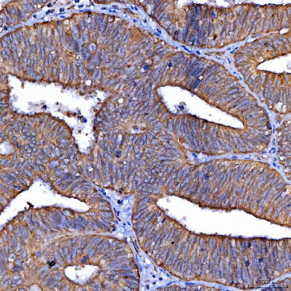


 FCM (Flow Cytometry)
(Figure 7. Flow Cytometry analysis of U87 cells using anti-Scavenging Receptor SRB2/SCARB2 antibody (AAA19535).Overlay histogram showing U87 cells stained with AAA19535 (Blue line). The cells were blocked with 10% normal goat serum. And then incubated with rabbit anti-Scavenging Receptor SRB2/SCARB2 Antibody (AAA19535, 1 ug/1x10^6 cells) for 30 min at 20 degree C. DyLight488 conjugated goat anti-rabbit IgG was used as secondary antibody for 30 minutes at 20 degree C. Isotype control antibody (Green line) was rabbit IgG (1 ug/1x10^6) used under the same conditions. Unlabelled sample (Red line) was also used as a control.)
FCM (Flow Cytometry)
(Figure 7. Flow Cytometry analysis of U87 cells using anti-Scavenging Receptor SRB2/SCARB2 antibody (AAA19535).Overlay histogram showing U87 cells stained with AAA19535 (Blue line). The cells were blocked with 10% normal goat serum. And then incubated with rabbit anti-Scavenging Receptor SRB2/SCARB2 Antibody (AAA19535, 1 ug/1x10^6 cells) for 30 min at 20 degree C. DyLight488 conjugated goat anti-rabbit IgG was used as secondary antibody for 30 minutes at 20 degree C. Isotype control antibody (Green line) was rabbit IgG (1 ug/1x10^6) used under the same conditions. Unlabelled sample (Red line) was also used as a control.)
Scavenging Receptor SRB2/SCARB2, Polyclonal Antibody (Cat# AAA19535)
Full Name
Anti-Scavenging Receptor SRB2/SCARB2 Antibody Picoband
Gene Names
SCARB2; AMRF; EPM4; LGP85; CD36L2; HLGP85; LIMP-2; LIMPII; SR-BII
Reactivity
Human
Applications
Western Blot, Immunohistochemistry, Flow Cytometry
Purity
Immunogen affinity purified.
Pricing


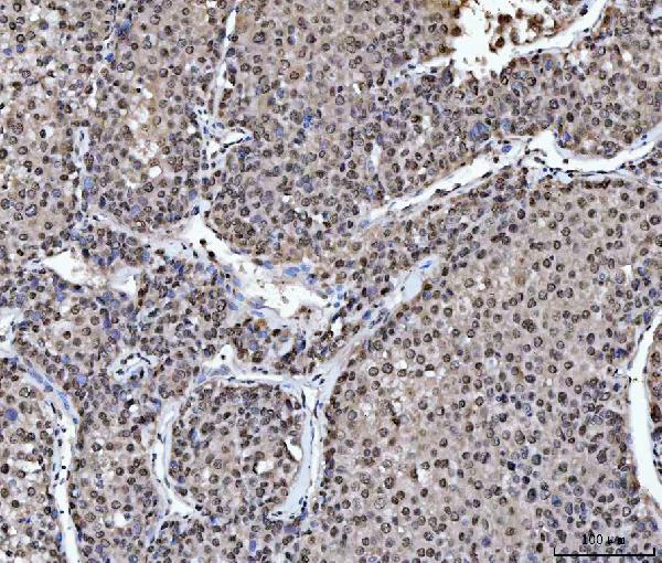


 FCM (Flow Cytometry)
(Figure 14. Flow Cytometry analysis of HEL cells using anti-hnRNP L/HNRNPL antibody (AAA19499).Overlay histogram showing HEL cells stained with AAA19499 (Blue line). The cells were blocked with 10% normal goat serum. And then incubated with rabbit anti-hnRNP L/HNRNPL Antibody (AAA19499, 1 ug/1x106 cells) for 30 min at 20 degree C. DyLight488 conjugated goat anti-rabbit IgG (BA1127, 5-10 ug/1x106 cells) was used as secondary antibody for 30 minutes at 20 degree C. Isotype control antibody (Green line) was rabbit IgG (1 ug/1x106) used under the same conditions. Unlabelled sample (Red line) was also used as a control.)
FCM (Flow Cytometry)
(Figure 14. Flow Cytometry analysis of HEL cells using anti-hnRNP L/HNRNPL antibody (AAA19499).Overlay histogram showing HEL cells stained with AAA19499 (Blue line). The cells were blocked with 10% normal goat serum. And then incubated with rabbit anti-hnRNP L/HNRNPL Antibody (AAA19499, 1 ug/1x106 cells) for 30 min at 20 degree C. DyLight488 conjugated goat anti-rabbit IgG (BA1127, 5-10 ug/1x106 cells) was used as secondary antibody for 30 minutes at 20 degree C. Isotype control antibody (Green line) was rabbit IgG (1 ug/1x106) used under the same conditions. Unlabelled sample (Red line) was also used as a control.)
hnRNP L/HNRNPL, Polyclonal Antibody (Cat# AAA19499)
Full Name
Anti-hnRNP L/HNRNPL Antibody Picoband
Gene Names
HNRNPL; HNRPL; hnRNP-L; P/OKcl.14
Reactivity
Human, Mouse, Rat
Applications
Western Blot, Immunohistochemistry, Immunocytochemistry, Immunofluorescence, Flow Cytometry
Purity
Immunogen affinity purified.
Pricing

 FCM (Flow Cytometry)
(Figure 9. Flow Cytometry analysis of MCF-7 cells using anti-MCCC2 antibody (AAA19873).Overlay histogram showing MCF-7 cells stained with AAA19873 (Blue line). To facilitate intracellular staining, cells were fixed with 4% paraformaldehyde and permeabilized with permeabilization buffer. The cells were blocked with 10% normal goat serum. And then incubated with rabbit anti-MCCC2 Antibody (AAA19873, 1ug/1x106 cells) for 30 min at 20 degree C. DyLight488 conjugated goat anti-rabbit IgG was used as secondary antibody for 30 minutes at 20 degree C. Isotype control antibody (Green line) was rabbit IgG (1ug/1x106) used under the same conditions. Unlabelled sample (Red line) was also used as a control.)
FCM (Flow Cytometry)
(Figure 9. Flow Cytometry analysis of MCF-7 cells using anti-MCCC2 antibody (AAA19873).Overlay histogram showing MCF-7 cells stained with AAA19873 (Blue line). To facilitate intracellular staining, cells were fixed with 4% paraformaldehyde and permeabilized with permeabilization buffer. The cells were blocked with 10% normal goat serum. And then incubated with rabbit anti-MCCC2 Antibody (AAA19873, 1ug/1x106 cells) for 30 min at 20 degree C. DyLight488 conjugated goat anti-rabbit IgG was used as secondary antibody for 30 minutes at 20 degree C. Isotype control antibody (Green line) was rabbit IgG (1ug/1x106) used under the same conditions. Unlabelled sample (Red line) was also used as a control.)
MCCC2, Polyclonal Antibody (Cat# AAA19873)
Full Name
Anti-MCCC2 Antibody Picoband
Gene Names
MCCC2; MCCB
Reactivity
Human, Mouse, Rat
Applications
Western Blot, Immunohistochemistry, Flow Cytometry
Purity
Immunogen affinity purified.
Pricing

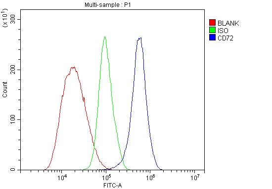


 IF (Immunofluorescence)
(Figure 6. IF analysis of CD72 using anti- CD72 antibody (AAA19329).CD72 was detected in immunocytochemical section of HEPA1-6 cells. Enzyme antigen retrieval was performed using IHC enzyme antigen retrieval reagent for 15 mins. The cells were blocked with 10% goat serum. And then incubated with 5μg/mL rabbit anti- CD72 Antibody (AAA19329) overnight at 4 degree C. DyLight®488 Conjugated Goat Anti-Rabbit IgG was used as secondary antibody at 1:100 dilution and incubated for 30 minutes at 37 degree C. The section was counterstained with DAPI. Visualize using a fluorescence microscope and filter sets appropriate for the label used.)
IF (Immunofluorescence)
(Figure 6. IF analysis of CD72 using anti- CD72 antibody (AAA19329).CD72 was detected in immunocytochemical section of HEPA1-6 cells. Enzyme antigen retrieval was performed using IHC enzyme antigen retrieval reagent for 15 mins. The cells were blocked with 10% goat serum. And then incubated with 5μg/mL rabbit anti- CD72 Antibody (AAA19329) overnight at 4 degree C. DyLight®488 Conjugated Goat Anti-Rabbit IgG was used as secondary antibody at 1:100 dilution and incubated for 30 minutes at 37 degree C. The section was counterstained with DAPI. Visualize using a fluorescence microscope and filter sets appropriate for the label used.)
CD72, Polyclonal Antibody (Cat# AAA19329)
Full Name
Anti-CD72 Antibody
Gene Names
Cd72; CD72c; Ly-19; Ly-32; Lyb-2; Ly-m19
Reactivity
Mouse, Rat
Applications
Western Blot, Immunohistochemistry, Immunocytochemistry, Immunofluorescence, Flow Cytometry, Direct ELISA
Purity
Immunogen affinity purified.
Pricing


 IF (Immunofluorescence)
(Immunofluorescence analysis of HeLa cells using VAPB antibody. Blue: DAPI for nuclear staining.)
IF (Immunofluorescence)
(Immunofluorescence analysis of HeLa cells using VAPB antibody. Blue: DAPI for nuclear staining.)
VAPB, Polyclonal Antibody (Cat# AAA28162)
Full Name
VAPB Polyclonal Antibody
Gene Names
VAPB; ALS8; VAP-B; VAMP-B
Reactivity
Human, Mouse, Rat
Applications
Western Blot, Immunohistochemistry, Immunofluorescence
Purity
Affinity Purification
Pricing
High-Mobility Group Box 1, Recombinant Protein (Cat# AAA10851)
Full Name
Recombinant Human High-Mobility Group Box 1
Gene Names
HMGB1; HMG1; HMG3; SBP-1
Purity
Greater than 95.0% as determined by: (a) Analysis by RP-HPLC. (b) Analysis by SDS-PAGE.
Pricing
Centromere Protein-B, Recombinant Protein (Cat# AAA10838)
Full Name
Recombinant Human Centromere Protein-B
Applications
Western Blot
Purity
Greater than 95% as determined by SDS-PAGE.
Pricing


 IHC (Immunohistchemistry)
(Immunohistochemistry of paraffin-embedded rat brain using SCAF4 Rabbit pAb at dilution of 1:50 (40x lens).Perform high pressure antigen retrieval with 10 mM citrate buffer pH 6.0 before commencing with IHC staining protocol.)
IHC (Immunohistchemistry)
(Immunohistochemistry of paraffin-embedded rat brain using SCAF4 Rabbit pAb at dilution of 1:50 (40x lens).Perform high pressure antigen retrieval with 10 mM citrate buffer pH 6.0 before commencing with IHC staining protocol.)
SCAF4, Polyclonal Antibody (Cat# AAA28418)
Full Name
SCAF4 Rabbit pAb
Reactivity
Human, Mouse, Rat
Applications
Western Blot, Immunohistochemistry
Purity
Affinity purification
Pricing


 Application Data
(Detection limit for recombinant GST tagged S100A6 is ~0.03ng/ml as a capture antibody.)
Application Data
(Detection limit for recombinant GST tagged S100A6 is ~0.03ng/ml as a capture antibody.)
S100A6, Monoclonal Antibody (Cat# AAA25829)
Full Name
S100A6 (Protein S100-A6, S100 Calcium-binding Protein A6, Calcyclin, Prolactin Receptor-associated Protein, PRA, Growth Factor-inducible Protein 2A9, MLN 4, CACY) (PE)
Gene Names
S100A6; 2A9; PRA; 5B10; CABP; CACY
Reactivity
Human
Applications
Immunofluorescence, Immunohistochemistry, Western Blot
Purity
Purified by Protein A Affinity Chromatography.
Pricing
Cullin-1, Recombinant Protein (Cat# AAA10882)
Full Name
Recombinant Human Cullin-1
Purity
Greater than 95% as determined by SDS-PAGE.
Pricing


 SDS-PAGE
Procollagen III N-Terminal Propeptide, Recombinant Protein (Cat# AAA21046)
Full Name
Recombinant Procollagen III N-Terminal Propeptide (PIIINP)
Reactivity
Homo sapiens (Human)
Applications
Western Blot
Purity
> 90%
Pricing


 SDS-PAGE
Inhibin Beta E (INHbE), Recombinant Protein (Cat# AAA20279)
Full Name
Recombinant Inhibin Beta E (INHbE)
Reactivity
Homo sapiens (Human)
Applications
Western Blot
Purity
> 90%
Pricing

 SDS-PAGE
Thrombin/Antithrombin Complex (TAT), Recombinant Protein (Cat# AAA21174)
Full Name
Recombinant Thrombin/Antithrombin Complex (TAT)
Reactivity
Homo sapiens (Human)
Applications
Western Blot
Purity
> 80%
Pricing

 SDS-PAGE
Plasmin/Antiplasmin Complex (PAP), Recombinant Protein (Cat# AAA21146)
Full Name
Recombinant Plasmin/Antiplasmin Complex (PAP)
Reactivity
Homo sapiens (Human)
Applications
Western Blot
Purity
> 95%
Pricing

 FCM (Flow Cytometry)
(Figure 6. Flow Cytometry analysis of 293T cells using anti-ABR antibody (AAA19758).Overlay histogram showing 293T cells stained with AAA19758 (Blue line). To facilitate intracellular staining, cells were fixed with 4% paraformaldehyde and permeabilized with permeabilization buffer. The cells were blocked with 10% normal goat serum. And then incubated with rabbit anti-ABR Antibody (AAA19758, 1ug/1x106 cells) for 30 min at 20 degree C. DyLight488 conjugated goat anti-rabbit IgG was used as secondary antibody for 30 minutes at 20 degree C. Isotype control antibody (Green line) was rabbit IgG (1ug/1x106) used under the same conditions. Unlabelled sample without incubation with primary antibody and secondary antibody (Red line) was used as a blank control.)
FCM (Flow Cytometry)
(Figure 6. Flow Cytometry analysis of 293T cells using anti-ABR antibody (AAA19758).Overlay histogram showing 293T cells stained with AAA19758 (Blue line). To facilitate intracellular staining, cells were fixed with 4% paraformaldehyde and permeabilized with permeabilization buffer. The cells were blocked with 10% normal goat serum. And then incubated with rabbit anti-ABR Antibody (AAA19758, 1ug/1x106 cells) for 30 min at 20 degree C. DyLight488 conjugated goat anti-rabbit IgG was used as secondary antibody for 30 minutes at 20 degree C. Isotype control antibody (Green line) was rabbit IgG (1ug/1x106) used under the same conditions. Unlabelled sample without incubation with primary antibody and secondary antibody (Red line) was used as a blank control.)
ABR, Polyclonal Antibody (Cat# AAA19758)
Full Name
Anti-ABR Antibody Picoband
Gene Names
ABR; MDB
Reactivity
Human, Mouse, Rat
Applications
Western Blot, Immunohistochemistry, Immunocytochemistry, Immunofluorescence, Flow Cytometry
Purity
Immunogen affinity purified.
Pricing

 FCM (Flow Cytometry)
(Figure 6. Flow Cytometry analysis of MCF-7 cells using anti-NUBPL antibody (AAA19948).Overlay histogram showing MCF-7 cells stained with AAA19948 (Blue line). To facilitate intracellular staining, cells were fixed with 4% paraformaldehyde and permeabilized with permeabilization buffer. The cells were blocked with 10% normal goat serum. And then incubated with rabbit anti-NUBPL Antibody (AAA19948, 1ug/1x106 cells) for 30 min at 20 degree C. DyLight488 conjugated goat anti-rabbit IgG was used as secondary antibody for 30 minutes at 20 degree C. Isotype control antibody (Green line) was rabbit IgG (1ug/1x106) used under the same conditions. Unlabelled sample (Red line) was also used as a control.)
FCM (Flow Cytometry)
(Figure 6. Flow Cytometry analysis of MCF-7 cells using anti-NUBPL antibody (AAA19948).Overlay histogram showing MCF-7 cells stained with AAA19948 (Blue line). To facilitate intracellular staining, cells were fixed with 4% paraformaldehyde and permeabilized with permeabilization buffer. The cells were blocked with 10% normal goat serum. And then incubated with rabbit anti-NUBPL Antibody (AAA19948, 1ug/1x106 cells) for 30 min at 20 degree C. DyLight488 conjugated goat anti-rabbit IgG was used as secondary antibody for 30 minutes at 20 degree C. Isotype control antibody (Green line) was rabbit IgG (1ug/1x106) used under the same conditions. Unlabelled sample (Red line) was also used as a control.)
NUBPL, Polyclonal Antibody (Cat# AAA19948)
Full Name
Anti-NUBPL Antibody Picoband
Gene Names
NUBPL; IND1; huInd1; C14orf127
Reactivity
Human, Mouse
Applications
Western Blot, Immunohistochemistry, Immunocytochemistry, Immunofluorescence, Flow Cytometry
Purity
Immunogen affinity purified.
Pricing
AIG2-Like Domain 1, Recombinant Protein (Cat# AAA10884)
Full Name
Recombinant Human AIG2-Like Domain 1
Gene Names
GGACT; A2LD1
Purity
Greater than 95.0% as determined by SDS-PAGE.
Pricing


 IF (Immunofluorescence)
(Figure 6. IF analysis of MIRO1/RHOT1 using anti-MIRO1/RHOT1 antibody (AAA19560).MIRO1/RHOT1 was detected in an immunocytochemical section of T47D cells. Enzyme antigen retrieval was performed using IHC enzyme antigen retrieval reagent (AR0022) for 15 mins. The cells were blocked with 10% goat serum. And then incubated with 5 ug/mL rabbit anti-MIRO1/RHOT1 Antibody (AAA19560) overnight at 4 degree C. DyLight488 Conjugated Goat Anti-Rabbit IgG was used as secondary antibody at 1:100 dilution and incubated for 30 minutes at 37 degree C. The section was counterstained with DAPI. Visualize using a fluorescence microscope and filter sets appropriate for the label used.)
IF (Immunofluorescence)
(Figure 6. IF analysis of MIRO1/RHOT1 using anti-MIRO1/RHOT1 antibody (AAA19560).MIRO1/RHOT1 was detected in an immunocytochemical section of T47D cells. Enzyme antigen retrieval was performed using IHC enzyme antigen retrieval reagent (AR0022) for 15 mins. The cells were blocked with 10% goat serum. And then incubated with 5 ug/mL rabbit anti-MIRO1/RHOT1 Antibody (AAA19560) overnight at 4 degree C. DyLight488 Conjugated Goat Anti-Rabbit IgG was used as secondary antibody at 1:100 dilution and incubated for 30 minutes at 37 degree C. The section was counterstained with DAPI. Visualize using a fluorescence microscope and filter sets appropriate for the label used.)
MIRO1/RHOT1, Polyclonal Antibody (Cat# AAA19560)
Full Name
Anti-MIRO1/RHOT1 Antibody Picoband
Gene Names
RHOT1; ARHT1; MIRO1; MIRO-1
Reactivity
Human, Mouse, Rat
Applications
Western Blot, Immunohistochemistry, Immunocytochemistry, Immunofluorescence
Purity
Immunogen affinity purified.
Pricing
Der P1, Recombinant Protein (Cat# AAA10807)
Full Name
Recombinant Der P1 Protein
Purity
Protein is >95% pure as determined by 10% SDS-PAGE (coomassie staining)
Purified by proprietary chromatographic technique.
Purified by proprietary chromatographic technique.
Pricing


 Application Data
Prostate Stem Cell Antigen (PSCA), Recombinant Protein (Cat# AAA20218)
Full Name
Recombinant Prostate Stem Cell Antigen (PSCA)
Reactivity
Homo sapiens (Human)
Applications
Western Blot, Immunoprecipitation
Purity
>98%
Pricing


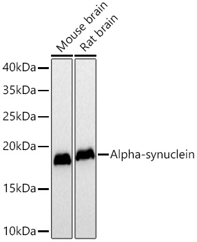
 ICC (Immunocytochemistry)
(Confocal imaging of paraffin-embedded Rat brain tissue using Alpha-synuclein Mouse mAb (AAA28601, dilution 1:200) followed by a further incubation with Cy3 Goat Anti-Mouse IgG (H+L) (AS008, dilution 1:500) (Red). DAPI was used for nuclear staining (Blue). Microwave antigen retrieval performed with 0.01M Citrate Buffer (pH 6.0) prior to IF staining. Objective: 40x.)
ICC (Immunocytochemistry)
(Confocal imaging of paraffin-embedded Rat brain tissue using Alpha-synuclein Mouse mAb (AAA28601, dilution 1:200) followed by a further incubation with Cy3 Goat Anti-Mouse IgG (H+L) (AS008, dilution 1:500) (Red). DAPI was used for nuclear staining (Blue). Microwave antigen retrieval performed with 0.01M Citrate Buffer (pH 6.0) prior to IF staining. Objective: 40x.)
Alpha-synuclein, Monoclonal Antibody (Cat# AAA28601)
Full Name
Alpha-synuclein Mouse mAb
Reactivity
Human
Applications
Western Blot, Immunohistochemistry, Immunofluorescence, Immunocytochemistry
Purity
Affinity purification
Pricing


 SDS-PAGE
Microtubule Associated Protein 2 (MAP2), Recombinant Protein (Cat# AAA20281)
Full Name
Recombinant Microtubule Associated Protein 2 (MAP2)
Gene Names
MAP2; MAP2A; MAP2B; MAP2C
Reactivity
Homo sapiens (Human)
Applications
Western Blot
Purity
> 97%
Pricing
 WB (Western Blot)
Pru P 1, Recombinant Protein (Cat# AAA24012)
Full Name
Pru P 1 protein
Applications
Western Blot
Purity
> 95% by SDS-PAGE
Pricing


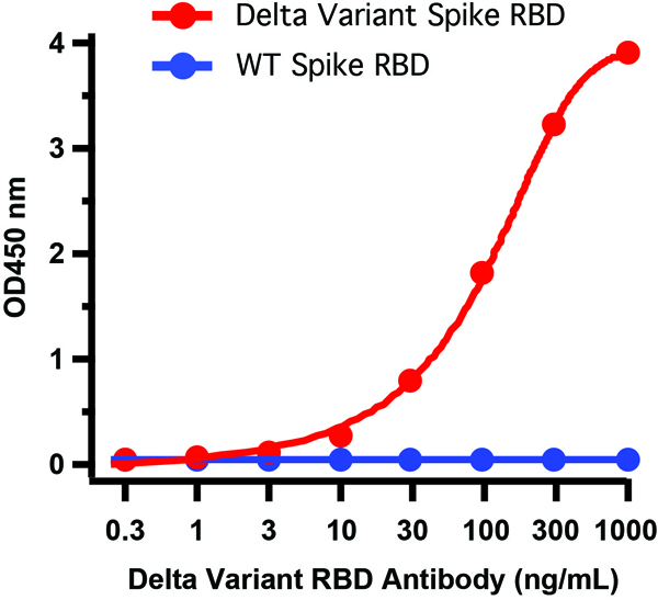


 WB (Western Blot)
(Figure 6 WB Validation of Delta Variant Spike RBD Protein with SARS-CoV-2 Spike RBD Antibodies Loading: 20 ng of SARS-CoV-2 spike RBD proteins, including WT and Delta variant (B.1.671.2).Detection Antibodies: SARS-CoV-2 Spike RBD antibody, 1 μg/mL, incubate at RT for 1 hr.Secondary Antibodies: Goat anti-rabbit HRP at 1:20,000, incubate at RT for 1 hr.)
WB (Western Blot)
(Figure 6 WB Validation of Delta Variant Spike RBD Protein with SARS-CoV-2 Spike RBD Antibodies Loading: 20 ng of SARS-CoV-2 spike RBD proteins, including WT and Delta variant (B.1.671.2).Detection Antibodies: SARS-CoV-2 Spike RBD antibody, 1 μg/mL, incubate at RT for 1 hr.Secondary Antibodies: Goat anti-rabbit HRP at 1:20,000, incubate at RT for 1 hr.)
COVID 19 Delta Variant Spike RBD (His-Avi Tag) Coronavirus, Recombinant Protein (Cat# AAA11039)
Full Name
SARS-CoV-2 (COVID-19) Delta Variant Spike RBD (His-Avi Tag) Recombinant Protein
Applications
Western Blot
Purity
> 95% as determined by Bis-Tris PAGE
Pricing
 SDS-PAGE
(SDS-PAGE (12%):purified E2 (HCV) protein (aa 383-663), subtype 1a)
SDS-PAGE
(SDS-PAGE (12%):purified E2 (HCV) protein (aa 383-663), subtype 1a)
HCV E2 protein, Recombinant Protein (Cat# AAA13770)
Full Name
HCV E2 protein (aa 383-663), Subtype 1a
Applications
Western Blot
Purity
>= 95% purity
Pricing
Mammaglobin-A, Recombinant Protein (Cat# AAA10805)
Full Name
Recombinant Human Mammaglobin-A, HEK
Gene Names
SCGB2A2; MGB1; UGB2
Purity
Greater than 95.0% as determined by SDS-PAGE
Pricing


 SDS-PAGE
Carbohydrate Antigen 125 (CA125), Recombinant Protein (Cat# AAA20282)
Full Name
Recombinant Carbohydrate Antigen 125 (CA125)
Gene Names
MUC16; CA125
Reactivity
Homo sapiens (Human)
Applications
Western Blot
Purity
> 95%
Pricing


 ELISA
(Figure 6 ELISA Validation of Delta Variant Spike S1 Protein with SARS-CoV-2 Spike S1 Antibodies Coating Antigen: SARS-CoV-2 spike S1 proteins, including WT and Delta variant (B.1.671.2), 5 μg/mL, incubated at 4 ˚C overnight.Detection Antibodies: SARS-CoV-2 Spike S1 antibody, dilution: 0.3-1000 ng/mL, incubate at RT for 1 hr.Secondary Antibodies: Goat anti-rabbit HRP at 1:20,000, incubate at RT for 1 hr.Antibody 9083 does not distinguish between WT and Delta.)
ELISA
(Figure 6 ELISA Validation of Delta Variant Spike S1 Protein with SARS-CoV-2 Spike S1 Antibodies Coating Antigen: SARS-CoV-2 spike S1 proteins, including WT and Delta variant (B.1.671.2), 5 μg/mL, incubated at 4 ˚C overnight.Detection Antibodies: SARS-CoV-2 Spike S1 antibody, dilution: 0.3-1000 ng/mL, incubate at RT for 1 hr.Secondary Antibodies: Goat anti-rabbit HRP at 1:20,000, incubate at RT for 1 hr.Antibody 9083 does not distinguish between WT and Delta.)
COVID 19 Delta Variant Spike S1 (His-Avi Tag) Coronavirus, Recombinant Protein (Cat# AAA11040)
Full Name
SARS-CoV-2 (COVID-19) Delta Variant Spike S1 (His-Avi Tag) Recombinant Protein
Applications
Western Blot
Purity
> 90% as determined by Bis-Tris PAGE
Pricing


 SDS-PAGE
Fibroblast Growth Factor 10 (FGF10), Recombinant Protein (Cat# AAA20261)
Full Name
Recombinant Fibroblast Growth Factor 10 (FGF10)
Gene Names
Fgf10; AEY17; Fgf-10; BB213776; Gsfaey17
Reactivity
Mus musculus (Mouse)
Applications
Western Blot
Purity
> 97%
Pricing


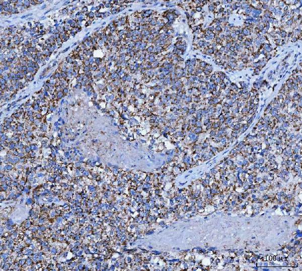


 IF (Immunofluorescence)
(Figure 8. IF analysis of TRK fused gene/TFG using anti-TRK fused gene/TFG antibody (AAA19472).TRK fused gene/TFG was detected in an immunocytochemical section of U87 cells. Enzyme antigen retrieval was performed using IHC enzyme antigen retrieval reagent (AR0022) for 15 mins. The cells were blocked with 10% goat serum. And then incubated with 5 ug/mL rabbit anti-TRK fused gene/TFG Antibody (AAA19472) overnight at 4 degree C. DyLight488 Conjugated Goat Anti-Rabbit IgG was used as secondary antibody at 1:100 dilution and incubated for 30 minutes at 37 degree C. The section was counterstained with DAPI. Visualize using a fluorescence microscope and filter sets appropriate for the label used.)
IF (Immunofluorescence)
(Figure 8. IF analysis of TRK fused gene/TFG using anti-TRK fused gene/TFG antibody (AAA19472).TRK fused gene/TFG was detected in an immunocytochemical section of U87 cells. Enzyme antigen retrieval was performed using IHC enzyme antigen retrieval reagent (AR0022) for 15 mins. The cells were blocked with 10% goat serum. And then incubated with 5 ug/mL rabbit anti-TRK fused gene/TFG Antibody (AAA19472) overnight at 4 degree C. DyLight488 Conjugated Goat Anti-Rabbit IgG was used as secondary antibody at 1:100 dilution and incubated for 30 minutes at 37 degree C. The section was counterstained with DAPI. Visualize using a fluorescence microscope and filter sets appropriate for the label used.)
TRK fused gene/TFG, Polyclonal Antibody (Cat# AAA19472)
Full Name
Anti-TRK fused gene/TFG Antibody Picoband
Gene Names
TFG; TF6; HMSNP; SPG57; TRKT3
Reactivity
Human, Mouse, Rat
Applications
Western Blot, Immunohistochemistry, Immunocytochemistry, Immunofluorescence
Purity
Immunogen affinity purified.
Pricing


 IHC (Immunohistochemistry)
(Immunohistochemistry analysis of paraffin-embedded Human tonsil using von Willebrand factor (VWF) Rabbit mAb (AAA28546) at dilution of 1:800 (40x lens). High pressure antigen retrieval performed with 0.01M Citrate Bufferr (pH 6.0) prior to IHC staining.)
IHC (Immunohistochemistry)
(Immunohistochemistry analysis of paraffin-embedded Human tonsil using von Willebrand factor (VWF) Rabbit mAb (AAA28546) at dilution of 1:800 (40x lens). High pressure antigen retrieval performed with 0.01M Citrate Bufferr (pH 6.0) prior to IHC staining.)
von Willebrand factor (VWF), Monoclonal Antibody (Cat# AAA28546)
Full Name
von Willebrand factor (VWF) Rabbit mAb
Reactivity
Human
Applications
Immunohistochemistry
Purity
Affinity purification
Pricing


 SDS-PAGE
Myosin Heavy Chain 6, Cardiac Muscle, Alpha (MYH6), Recombinant Protein (Cat# AAA20287)
Full Name
Recombinant Myosin Heavy Chain 6, Cardiac Muscle, Alpha (MYH6)
Gene Names
Myh6; Myhca
Reactivity
Rattus norvegicus (Rat)
Applications
Western Blot
Purity
> 95%
Pricing

 SDS-PAGE
Mucin 1 (MUC1), Recombinant Protein (Cat# AAA20263)
Full Name
Recombinant Mucin 1 (MUC1)
Gene Names
Muc1; EMA; CD227; Muc-1
Reactivity
Mus musculus (Mouse)
Applications
Western Blot
Purity
> 97%
Pricing
 Application Data
(SDS-PAGE: purified HA (B/Florida/4/2006)(aa 16-546) proteinA, reducing condition; B, non-reducing condition)
Application Data
(SDS-PAGE: purified HA (B/Florida/4/2006)(aa 16-546) proteinA, reducing condition; B, non-reducing condition)
HA (B/Florida/4/2006), Recombinant Protein (Cat# AAA13746)
Full Name
HA (aa 16-546)(B/Florida/4/2006)
Applications
Western Blot
Purity
> 95% purity (SDS-PAGE)
Pricing


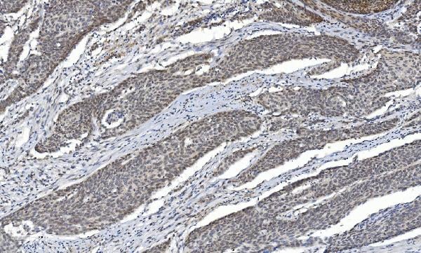


 FCM (Flow Cytometry)
(Figure 10. Flow Cytometry analysis of THP-1 cells using anti-HDAC5 antibody (AAA19236).Overlay histogram showing THP-1 cells stained with AAA19236 (Blue line). The cells were blocked with 10% normal goat serum. And then incubated with rabbit anti-HDAC5 Antibody (AAA19236, 1μg/1x106 cells) for 30 min at 20 degree C. DyLight®488 conjugated goat anti-rabbit IgG (5-10μg/1x106 cells) was used as secondary antibody for 30 minutes at 20 degree C. Isotype control antibody (Green line) was rabbit IgG (1μg/1x106) used under the same conditions. Unlabelled sample (Red line) was also used as a control.)
FCM (Flow Cytometry)
(Figure 10. Flow Cytometry analysis of THP-1 cells using anti-HDAC5 antibody (AAA19236).Overlay histogram showing THP-1 cells stained with AAA19236 (Blue line). The cells were blocked with 10% normal goat serum. And then incubated with rabbit anti-HDAC5 Antibody (AAA19236, 1μg/1x106 cells) for 30 min at 20 degree C. DyLight®488 conjugated goat anti-rabbit IgG (5-10μg/1x106 cells) was used as secondary antibody for 30 minutes at 20 degree C. Isotype control antibody (Green line) was rabbit IgG (1μg/1x106) used under the same conditions. Unlabelled sample (Red line) was also used as a control.)
HDAC5, Polyclonal Antibody (Cat# AAA19236)
Full Name
Anti-HDAC5 Antibody
Gene Names
HDAC5; HD5; NY-CO-9
Reactivity
Human, Mouse, Rat
Applications
Western Blot, Immunohistochemistry, Immunocytochemistry, Immunofluorescence, Flow Cytometry, Direct ELISA
Purity
Immunogen affinity purified.
Pricing


 Bioactivity
(Detects Camrelizumab in indirect ELISAs.)
Bioactivity
(Detects Camrelizumab in indirect ELISAs.)
CD279/PDCD1/PD1, Recombinant Protein (Cat# AAA18907)
Full Name
Recombinant Human CD279/PDCD1/PD1, C-His
Gene Names
PDCD1; PD1; PD-1; CD279; SLEB2; hPD-1; hPD-l; hSLE1
Applications
Western Blot
Purity
>90% as determined by SDS-PAGE
Pricing


 SDS-PAGE
Procollagen I N-Terminal Propeptide (PINP), Recombinant Protein (Cat# AAA21111)
Full Name
Recombinant Procollagen I N-Terminal Propeptide (PINP)
Gene Names
COL1A1; OI1; OI2; OI3; OI4; EDSC; EDSARTH1
Reactivity
Homo sapiens (Human)
Applications
Western Blot
Purity
> 90%
Pricing
Heat Shock Protein 90 Alpha, Recombinant Protein (Cat# AAA10829)
Full Name
Recombinant Human Heat Shock Protein 90 Alpha
Gene Names
HSP90AA1; EL52; HSPN; LAP2; HSP86; HSPC1; HSPCA; Hsp89; Hsp90; LAP-2; HSP89A; HSP90A; HSP90N; HSPCAL1; HSPCAL4
Purity
Greater than 90.0% as determined by SDS-PAGE.
Pricing