
Filters
Clonality
Type
Reactivity
Gene Name
Isotype
Host
Application
Clone
10000 results for "Recombinant Proteins" - showing 5750-5800
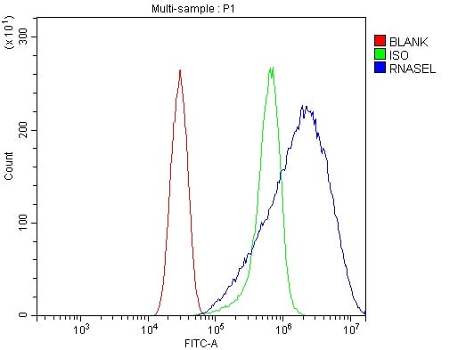
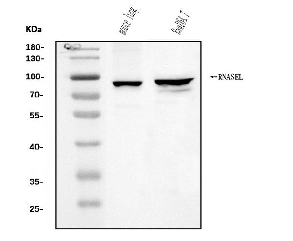
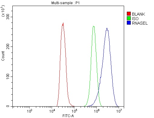 FCM/FACS (Flow Cytometry)
(Figure 3. Flow Cytometry analysis of NRK cells using anti-RNase L/Rnasel antibody (AAA125662).Overlay histogram showing NRK cells stained with AAA125662 (Blue line). The cells were blocked with 10% normal goat serum. And then incubated with rabbit anti-RNase L/Rnasel Antibody (AAA125662, 1μg/1x106 cells) for 30 min at 20 degree C. DyLight®488 conjugated goat anti-rabbit IgG (5-10μg/1x106 cells) was used as secondary antibody for 30 minutes at 20 degree C. Isotype control antibody (Green line) was rabbit IgG (1μg/1x106) used under the same conditions. Unlabelled sample (Red line) was also used as a control.)
FCM/FACS (Flow Cytometry)
(Figure 3. Flow Cytometry analysis of NRK cells using anti-RNase L/Rnasel antibody (AAA125662).Overlay histogram showing NRK cells stained with AAA125662 (Blue line). The cells were blocked with 10% normal goat serum. And then incubated with rabbit anti-RNase L/Rnasel Antibody (AAA125662, 1μg/1x106 cells) for 30 min at 20 degree C. DyLight®488 conjugated goat anti-rabbit IgG (5-10μg/1x106 cells) was used as secondary antibody for 30 minutes at 20 degree C. Isotype control antibody (Green line) was rabbit IgG (1μg/1x106) used under the same conditions. Unlabelled sample (Red line) was also used as a control.)
RNase L/Rnasel, Polyclonal Antibody (Cat# AAA125662)
Full Name
Anti-RNase L/Rnasel Antibody
Gene Names
Rnasel; E230029I04Rik
Reactivity
Mouse, Rat
Applications
Direct ELISA, Flow Cytometry, Western Blot
Purity
Immunogen affinity purified.
Pricing
 Bioactivity
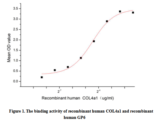
(Collagen Type IV Alpha 1 (COL4a1), a secreted glycoprotein,is a member of the type IV collagen family. COL4a1 is the major structural component of glomerular basement membranes (GBM), forming a 'chicken-wire' meshwork together with laminins, proteoglycans and entactin/nidogen. It can inhibit angiogenesis and tumor formation, and specifically inhibit endothelial cell proliferation, migration and tubule formation. COL4A1 mutations can remain asymptomatic or cause devastating disease. Neonates and children may present with porencephaly, intracerebral hemorrhage, or hemiparesis, whereas adults tend to develop intracranial aneurysms or retinal arteriolar tortuosities. Glycoprotein VI (GP6) has been identified as an interactor of COL4a1, thus a functional binding ELISA assay was conducted to detect the interaction of recombinant human COL4a1 and recombinant human GP6. Briefly, COL4a1 was diluted serially in PBS with 0.01% BSA (pH 7.4). Duplicate samples of 100 ul were then transferred to GP6-coated microtiter wells and incubated for 1h at 37 degree C. Wells were washed with PBST and incubated for 1h with anti-COL4a1 pAb, then aspirated and washed 3 times. After incubation with HRP labelled secondary antibody for 1h at 37 degree C, wells were aspirated and washed 5 times. With the addition of substrate solution, wells were incubated 15-25 minutes at 37 degree C. Finally, add 50 uL stop solution to the wells and read at 450/630 nm immediately. The binding activity of recombinant human COL4a1 and recombinant human GP6 was shown in Figure 1, the EC50 for this effect is 0.05 ug/mL.)
Bioactivity
(Collagen Type IV Alpha 1 (COL4a1), a secreted glycoprotein,is a member of the type IV collagen family. COL4a1 is the major structural component of glomerular basement membranes (GBM), forming a 'chicken-wire' meshwork together with laminins, proteoglycans and entactin/nidogen. It can inhibit angiogenesis and tumor formation, and specifically inhibit endothelial cell proliferation, migration and tubule formation. COL4A1 mutations can remain asymptomatic or cause devastating disease. Neonates and children may present with porencephaly, intracerebral hemorrhage, or hemiparesis, whereas adults tend to develop intracranial aneurysms or retinal arteriolar tortuosities. Glycoprotein VI (GP6) has been identified as an interactor of COL4a1, thus a functional binding ELISA assay was conducted to detect the interaction of recombinant human COL4a1 and recombinant human GP6. Briefly, COL4a1 was diluted serially in PBS with 0.01% BSA (pH 7.4). Duplicate samples of 100 ul were then transferred to GP6-coated microtiter wells and incubated for 1h at 37 degree C. Wells were washed with PBST and incubated for 1h with anti-COL4a1 pAb, then aspirated and washed 3 times. After incubation with HRP labelled secondary antibody for 1h at 37 degree C, wells were aspirated and washed 5 times. With the addition of substrate solution, wells were incubated 15-25 minutes at 37 degree C. Finally, add 50 uL stop solution to the wells and read at 450/630 nm immediately. The binding activity of recombinant human COL4a1 and recombinant human GP6 was shown in Figure 1, the EC50 for this effect is 0.05 ug/mL.)
Collagen Type IV Alpha 1 (COL4a1), Active Protein (Cat# AAA161709)
Full Name
Active Collagen Type IV Alpha 1 (COL4a1)
Gene Names
COL4A1; ICH; HANAC; POREN1; arresten
Applications
Activity Assay, Cell Culture
Purity
Greater than 80% by SDS-PAGE
Pricing
 FCM/FACS (Flow Cytometry)
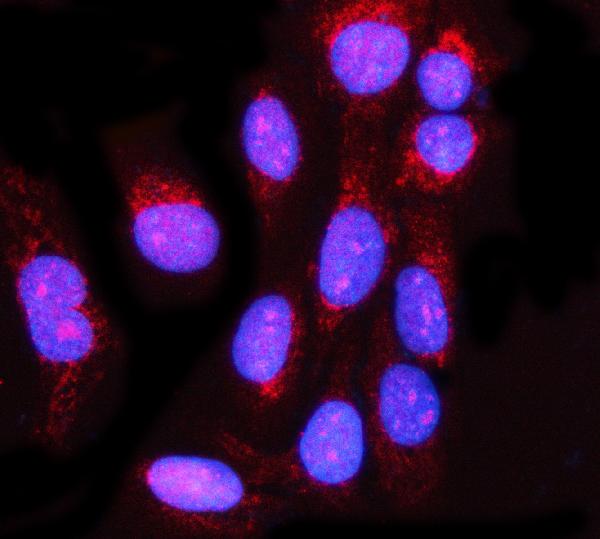
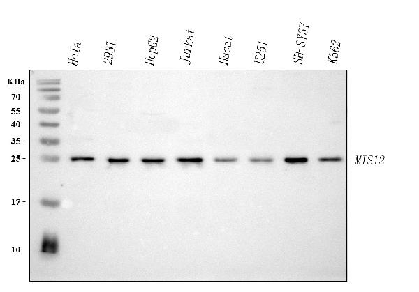
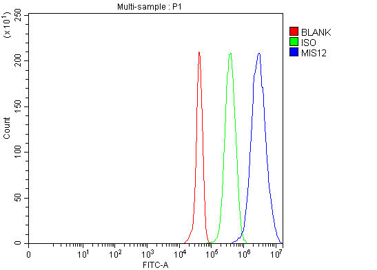
(Figure 3. Flow Cytometry analysis of JK cells using anti-MIS12 antibody (AAA127408).Overlay histogram showing JK cells stained with AAA127408 (Blue line). To facilitate intracellular staining, cells were fixed with 4% paraformaldehyde and permeabilized with permeabilization buffer. The cells were blocked with 10% normal goat serum. And then incubated with rabbit anti-MIS12 Antibody (AAA127408, 1ug/1x106 cells) for 30 min at 20 degree C. DyLight488 conjugated goat anti-rabbit IgG was used as secondary antibody for 30 minutes at 20 degree C. Isotype control antibody (Green line) was rabbit IgG (1ug/1x106) used under the same conditions. Unlabelled sample (Red line) was also used as a control.)
FCM/FACS (Flow Cytometry)
(Figure 3. Flow Cytometry analysis of JK cells using anti-MIS12 antibody (AAA127408).Overlay histogram showing JK cells stained with AAA127408 (Blue line). To facilitate intracellular staining, cells were fixed with 4% paraformaldehyde and permeabilized with permeabilization buffer. The cells were blocked with 10% normal goat serum. And then incubated with rabbit anti-MIS12 Antibody (AAA127408, 1ug/1x106 cells) for 30 min at 20 degree C. DyLight488 conjugated goat anti-rabbit IgG was used as secondary antibody for 30 minutes at 20 degree C. Isotype control antibody (Green line) was rabbit IgG (1ug/1x106) used under the same conditions. Unlabelled sample (Red line) was also used as a control.)
MIS12, Polyclonal Antibody (Cat# AAA127408)
Full Name
Anti-MIS12 Antibody Picoband
Gene Names
MIS12; MTW1; hMis12; KNTC2AP; 2510025F08Rik
Reactivity
Human
Applications
Flow Cytometry, Immunofluorescence, Immunocytochemistry, Western Blot
Purity
Immunogen affinity purified.
Pricing
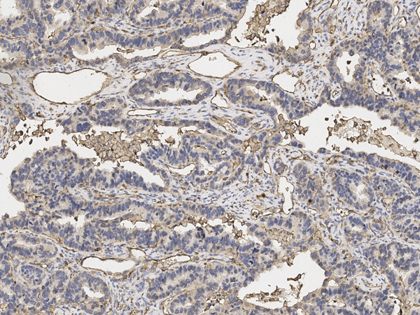
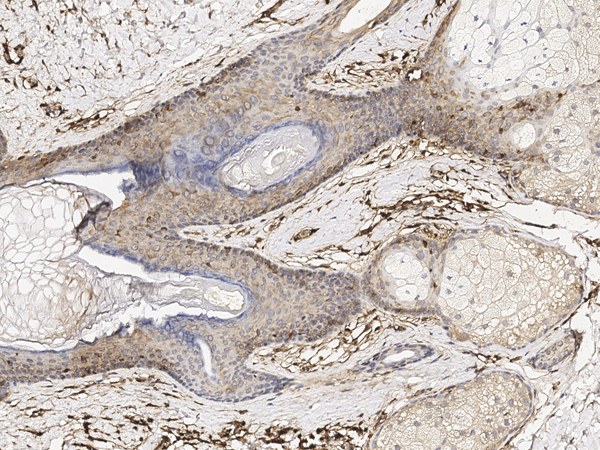
 IHC (Immunohiostchemistry)
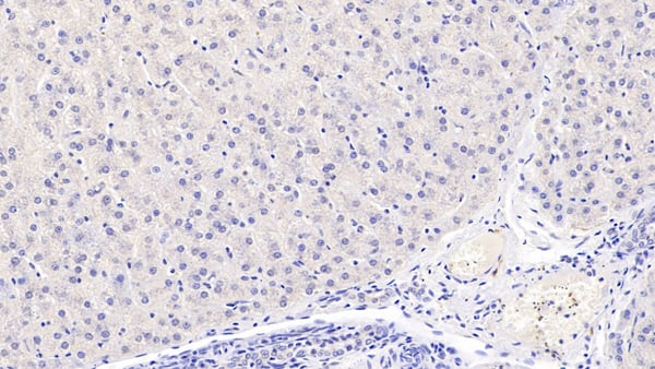
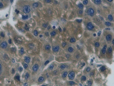
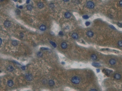
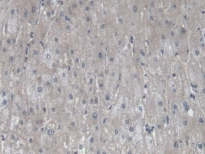
(DAB staining on IHCP; Samples: Human Liver Tissue; Primary Ab: 20ug/ml Rabbit AntiHuman CFB Antibody Second Ab: 2ug/mL HRPLinked Caprine AntiRabbit IgG Polyclonal Antibody (Catalog: SAA544Rb19))
IHC (Immunohiostchemistry)
(DAB staining on IHCP; Samples: Human Liver Tissue; Primary Ab: 20ug/ml Rabbit AntiHuman CFB Antibody Second Ab: 2ug/mL HRPLinked Caprine AntiRabbit IgG Polyclonal Antibody (Catalog: SAA544Rb19))
Complement Factor B (CFB), Polyclonal Antibody (Cat# AAA151957)
Full Name
Polyclonal Antibody to Complement Factor B (CFB)
Gene Names
CFB; BF; FB; BFD; GBG; CFAB; PBF2; AHUS4; FBI12; H2-Bf
Reactivity
Human, Pig
Applications
Immunoprecipitation, Immunocytochemistry, Immunohistochemistry, Western Blot
Purity
Affinity Chromatography
Pricing
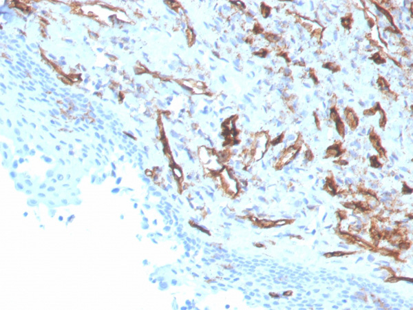
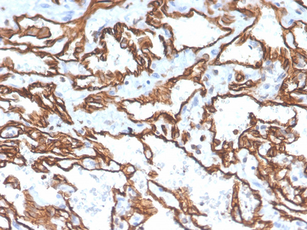
 Application Data
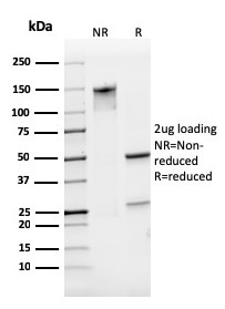
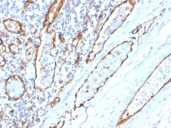
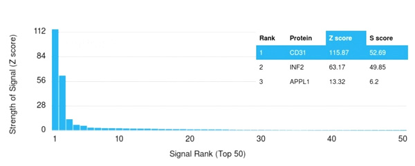
(Analysis of Protein Array containing more than 19,000 full-length human proteins using CD31 Mouse Monoclonal Antibody (PECAM1/3529) Z- and S- Score: The Z-score represents the strength of a signal that a monoclonal antibody (MAb) (in combination with a fluorescently-tagged anti-IgG secondary antibody) produces when binding to a particular protein on the HuProtTM array. Z-scores are described in units of standard deviations (SD’s) above the mean value of all signals generated on that array. If targets on HuProtTM are arranged in descending order of the Z-score, the S-score is the difference (also in units of SD’s) between the Z-score. S-score therefore represents the relative target specificity of a MAb to its intended target. A MAb is considered to specific to its intended target, if the MAb has an S-score of at least 2.5. For example, if a MAb binds to protein X with a Z-score of 43 and to protein Y with a Z-score of 14, then the S-score for the binding of that MAb to protein X is equal to 29.)
Application Data
(Analysis of Protein Array containing more than 19,000 full-length human proteins using CD31 Mouse Monoclonal Antibody (PECAM1/3529) Z- and S- Score: The Z-score represents the strength of a signal that a monoclonal antibody (MAb) (in combination with a fluorescently-tagged anti-IgG secondary antibody) produces when binding to a particular protein on the HuProtTM array. Z-scores are described in units of standard deviations (SD’s) above the mean value of all signals generated on that array. If targets on HuProtTM are arranged in descending order of the Z-score, the S-score is the difference (also in units of SD’s) between the Z-score. S-score therefore represents the relative target specificity of a MAb to its intended target. A MAb is considered to specific to its intended target, if the MAb has an S-score of at least 2.5. For example, if a MAb binds to protein X with a Z-score of 43 and to protein Y with a Z-score of 14, then the S-score for the binding of that MAb to protein X is equal to 29.)
CD31/PECAM-1, Monoclonal Antibody (Cat# AAA215132)
Full Name
CD31/PECAM-1 (Endothelial Cell Marker)
Gene Names
PECAM1; CD31; PECA1; GPIIA'; PECAM-1; endoCAM; CD31/EndoCAM
Reactivity
Human
Applications
Immunohistochemistry
Purity
Purified Ab with BSA and Azide at 200ug/ml OR Purified Ab WITHOUT BSA and Azide at 1.0mg/ml
Pricing
 Application Data
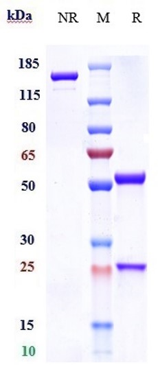
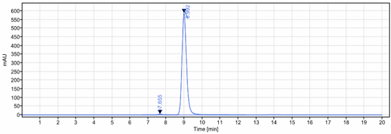
(The purity of Anti-CD11 Reference Antibody (Rovelizumab)is more than 99.22% ,determined by SEC-HPLC.)
Application Data
(The purity of Anti-CD11 Reference Antibody (Rovelizumab)is more than 99.22% ,determined by SEC-HPLC.)
CD11, Monoclonal Recombinant Antibody (Cat# AAA297150)
Full Name
Anti-CD11 Reference Antibody (Rovelizumab)
Gene Names
CDK11A; CDC2L2; CDC2L3; p58GTA; PITSLRE; CDK11-p46; CDK11-p58; CDK11-p110
Reactivity
Human
Applications
Functional Assay, Flow Cytometry
Pricing
 WB (Western Blot)
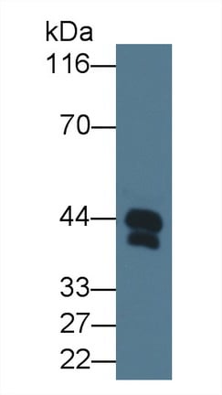
(Western Blot; Sample: Human Serum; Primary Ab: 3ug/ml Mouse AntiHuman Hpt Antibody Second Ab: 0.2ug/mL HRPLinked Caprine AntiMouse IgG Polyclonal Antibody (Catalog: SAA544Mu19))
WB (Western Blot)
(Western Blot; Sample: Human Serum; Primary Ab: 3ug/ml Mouse AntiHuman Hpt Antibody Second Ab: 0.2ug/mL HRPLinked Caprine AntiMouse IgG Polyclonal Antibody (Catalog: SAA544Mu19))
Haptoglobin (Hpt), Monoclonal Antibody (Cat# AAA151630)
Full Name
Monoclonal Antibody to Haptoglobin (Hpt)
Gene Names
HP; BP; HPA1S; HP2ALPHA2
Reactivity
Human
Applications
Immunoprecipitation, Immunocytochemistry, Immunohistochemistry, Western Blot
Purity
Protein A + Protein G affinity chromatography
Pricing
 IHC (Immunohiostchemistry)
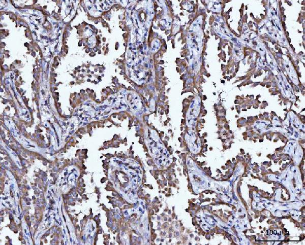
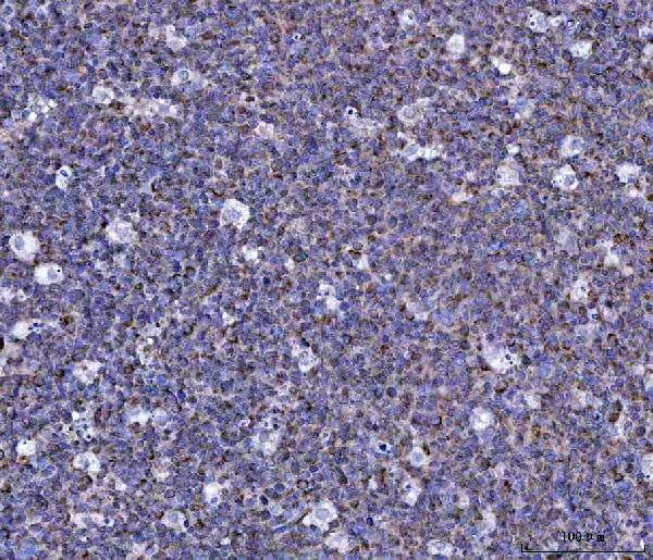
(Figure 2. IHC analysis of CCL15 using anti-CCL15 antibody (AAA127309).CCL15 was detected in a paraffin-embedded section of human tonsil tissue. Heat mediated antigen retrieval was performed in EDTA buffer (pH 8.0, epitope retrieval solution). The tissue section was blocked with 10% goat serum. The tissue section was then incubated with 2ug/ml rabbit anti-CCL15 Antibody (AAA127309) overnight at 4 degree C. Peroxidase Conjugated Goat Anti-rabbit IgG was used as secondary antibody and incubated for 30 minutes at 37 degree C. The tissue section was developed using HRP Conjugated Rabbit IgG Super Vision Assay Kit with DAB as the chromogen.)
IHC (Immunohiostchemistry)
(Figure 2. IHC analysis of CCL15 using anti-CCL15 antibody (AAA127309).CCL15 was detected in a paraffin-embedded section of human tonsil tissue. Heat mediated antigen retrieval was performed in EDTA buffer (pH 8.0, epitope retrieval solution). The tissue section was blocked with 10% goat serum. The tissue section was then incubated with 2ug/ml rabbit anti-CCL15 Antibody (AAA127309) overnight at 4 degree C. Peroxidase Conjugated Goat Anti-rabbit IgG was used as secondary antibody and incubated for 30 minutes at 37 degree C. The tissue section was developed using HRP Conjugated Rabbit IgG Super Vision Assay Kit with DAB as the chromogen.)
CCL15, Polyclonal Antibody (Cat# AAA127309)
Full Name
Anti-CCL15 Antibody
Gene Names
CCL15; LKN1; NCC3; SY15; HCC-2; LKN-1; MIP-5; NCC-3; SCYL3; MIP-1D; MRP-2B; SCYA15; HMRP-2B; MIP-1 delta
Reactivity
Human
Applications
Immunohistochemistry
Purity
Immunogen affinity purified.
Pricing
 Application Data
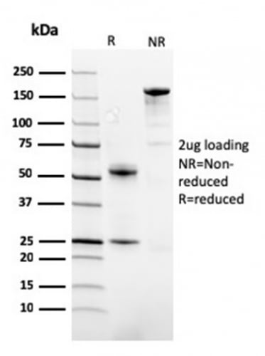
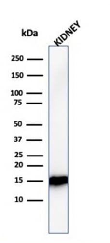
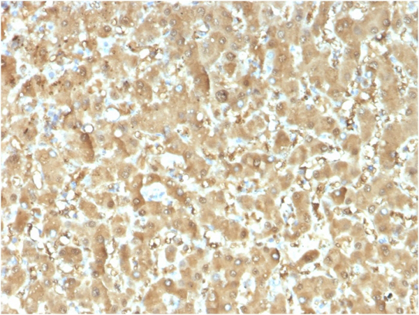
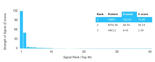
(Analysis of Protein Array containing more than 19,000 full-length human proteins using FABP1 Mouse Monoclonal Antibody (FABP1/3482). Z- and S- Score: The Z-score represents the strength of a signal that a monoclonal antibody (MAb) (in combination with a fluorescently-tagged anti-IgG secondary antibody) produces when binding to a particular protein on the HuProtTM array. Z-scores are described in units of standard deviations (SD's) above the mean value of all signals generated on that array. If targets on HuProtTM are arranged in descending order of the Z-score, the S-score is the difference (also in units of SD's) between the Z-score. S-score therefore represents the relative target specificity of a MAb to its intended target. A MAb is considered to specific to its intended target, if the MAb has an S-score of at least 2.5. For example, if a MAb binds to protein X with a Z-score of 43 and to protein Y with a Z-score of 14, then the S-score for the binding of that MAb to protein X is equal to 29.)
Application Data
(Analysis of Protein Array containing more than 19,000 full-length human proteins using FABP1 Mouse Monoclonal Antibody (FABP1/3482). Z- and S- Score: The Z-score represents the strength of a signal that a monoclonal antibody (MAb) (in combination with a fluorescently-tagged anti-IgG secondary antibody) produces when binding to a particular protein on the HuProtTM array. Z-scores are described in units of standard deviations (SD's) above the mean value of all signals generated on that array. If targets on HuProtTM are arranged in descending order of the Z-score, the S-score is the difference (also in units of SD's) between the Z-score. S-score therefore represents the relative target specificity of a MAb to its intended target. A MAb is considered to specific to its intended target, if the MAb has an S-score of at least 2.5. For example, if a MAb binds to protein X with a Z-score of 43 and to protein Y with a Z-score of 14, then the S-score for the binding of that MAb to protein X is equal to 29.)
Fatty Acid Binding Protein/FABP1, Monoclonal Antibody (Cat# AAA215360)
Full Name
Fatty Acid Binding Protein (Liver)/FABP1
Gene Names
FABP1; FABPL; L-FABP
Reactivity
Human
Applications
Western Blot, Immunohistochemistry
Purity
Purified Ab with BSA and Azide at 200ug/ml or Purified Ab with BSA and Azide at 200ug/ml or Purified Ab WITHOUT BSA and Azide at 1.0mg/ml
Pricing
 WB (Western Blot)
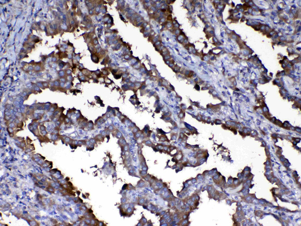
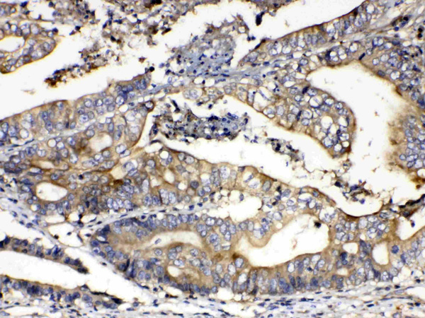
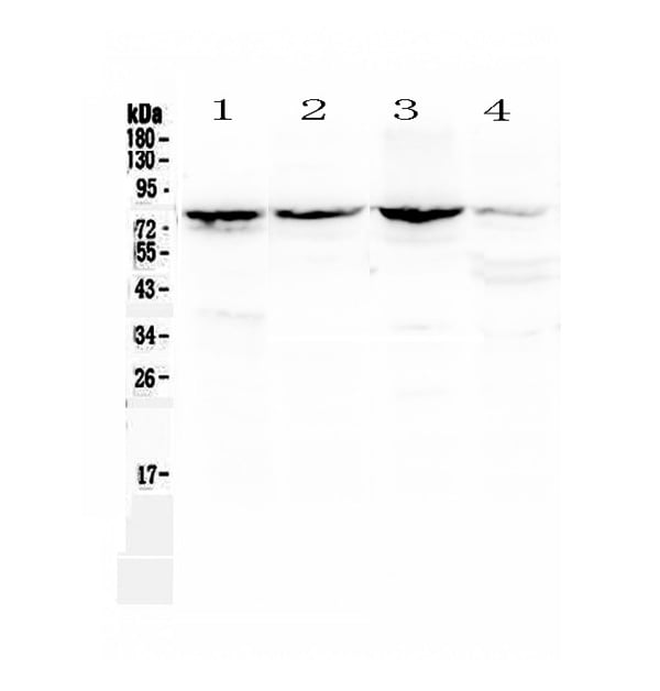
(Figure 1. Western blot analysis of Calpain 2 using anti-Calpain 2 antibody (AAA124708).Electrophoresis was performed on a 5-20% SDS-PAGE gel at 70V (Stacking gel) / 90V (Resolving gel) for 2-3 hours. The sample well of each lane was loaded with 50ug of sample under reducing conditions.Lane 1: rat kidney tissue lysate,Lane 2: mouse kidney tissue lysate,Lane 3: mouse lung tissue lysate,Lane 4: human MCF-7 whole Cell lysate.After Electrophoresis, proteins were transferred to a Nitrocellulose membrane at 150mA for 50-90 minutes. Blocked the membrane with 5% Non-fat Milk/ TBS for 1.5 hour at RT. The membrane was incubated with rabbit anti-Calpain 2 antigen affinity purified polyclonal antibody at 0.5ug/mL overnight at 4 degree C, then washed with TBS-0.1%Tween 3 times with 5 minutes each and probed with a goat anti-rabbit IgG-HRP secondary antibody at a dilution of 1:10000 for 1.5 hour at RT. The signal is developed using an Enhanced Chemiluminescent detection (ECL) kit with Tanon 5200 system. A specific band was detected for Calpain 2 at approximately 80KD. The expected band size for Calpain 2 is at 80KD.)
WB (Western Blot)
(Figure 1. Western blot analysis of Calpain 2 using anti-Calpain 2 antibody (AAA124708).Electrophoresis was performed on a 5-20% SDS-PAGE gel at 70V (Stacking gel) / 90V (Resolving gel) for 2-3 hours. The sample well of each lane was loaded with 50ug of sample under reducing conditions.Lane 1: rat kidney tissue lysate,Lane 2: mouse kidney tissue lysate,Lane 3: mouse lung tissue lysate,Lane 4: human MCF-7 whole Cell lysate.After Electrophoresis, proteins were transferred to a Nitrocellulose membrane at 150mA for 50-90 minutes. Blocked the membrane with 5% Non-fat Milk/ TBS for 1.5 hour at RT. The membrane was incubated with rabbit anti-Calpain 2 antigen affinity purified polyclonal antibody at 0.5ug/mL overnight at 4 degree C, then washed with TBS-0.1%Tween 3 times with 5 minutes each and probed with a goat anti-rabbit IgG-HRP secondary antibody at a dilution of 1:10000 for 1.5 hour at RT. The signal is developed using an Enhanced Chemiluminescent detection (ECL) kit with Tanon 5200 system. A specific band was detected for Calpain 2 at approximately 80KD. The expected band size for Calpain 2 is at 80KD.)
Calpain 2, Polyclonal Antibody (Cat# AAA124708)
Full Name
Anti-Calpain 2 Picoband Antibody
Gene Names
CAPN2; CANP2; mCANP; CANPL2; CANPml
Reactivity
Human, Mouse, Rat
No cross reactivity with other proteins.
No cross reactivity with other proteins.
Applications
Western Blot, Immunohistochemistry
Purity
Immunogen affinity purified
Pricing


 Application Data
(Analysis of Protein Array containing more than 19,000 full-length human proteins using Mouse Vinculin Monoclonal Antibody (VCL/2573). Z- and S- Score: The Z-score represents the strength of a signal that a monoclonal antibody (MAb) (in combination with a fluorescently-tagged anti-IgG secondary antibody) produces when binding to a particular protein on the HuProtTM array. Z-scores are described in units of standard deviations (SD's) above the mean value of all signals generated on that array. If targets on HuProtTM are arranged in descending order of the Z-score, the S-score is the difference (also in units of SD's) between the Z-score. S-score therefore represents the relative target specificity of a MAb to its intended target. A MAb is considered to specific to its intended target, if the MAb has an S-score of at least 2.5. For example, if a MAb binds to protein X with a Z-score of 43 and to protein Y with a Z-score of 14, then the S-score for the binding of that MAb to protein X is equal to 29.)
Application Data
(Analysis of Protein Array containing more than 19,000 full-length human proteins using Mouse Vinculin Monoclonal Antibody (VCL/2573). Z- and S- Score: The Z-score represents the strength of a signal that a monoclonal antibody (MAb) (in combination with a fluorescently-tagged anti-IgG secondary antibody) produces when binding to a particular protein on the HuProtTM array. Z-scores are described in units of standard deviations (SD's) above the mean value of all signals generated on that array. If targets on HuProtTM are arranged in descending order of the Z-score, the S-score is the difference (also in units of SD's) between the Z-score. S-score therefore represents the relative target specificity of a MAb to its intended target. A MAb is considered to specific to its intended target, if the MAb has an S-score of at least 2.5. For example, if a MAb binds to protein X with a Z-score of 43 and to protein Y with a Z-score of 14, then the S-score for the binding of that MAb to protein X is equal to 29.)
Vinculin, Monoclonal Antibody (Cat# AAA214829)
Full Name
Vinculin (Marker of Age-related Macular Degeneration)
Gene Names
VCL; MV; MVCL; CMD1W; CMH15; HEL114
Reactivity
Human. Others not known.
Applications
Immunohistochemistry
Purity
Purified Ab with BSA and Azide at 200ug/ml OR Purified Ab WITHOUT BSA and Azide at 1.0mg/ml
Pricing
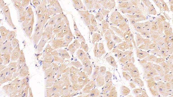 IHC (Immunohistochemisry)
(DAB staining on IHCP;Sample: Human Cardiac Muscle Tissue; Primary Ab: 40ug/ml Mouse AntiHuman FSTL1 AntibodySecond Ab: 2ug/mL HRPLinked Caprine AntiMouse IgG Polyclonal Antibody(Catalog: SAA544Mu19))
IHC (Immunohistochemisry)
(DAB staining on IHCP;Sample: Human Cardiac Muscle Tissue; Primary Ab: 40ug/ml Mouse AntiHuman FSTL1 AntibodySecond Ab: 2ug/mL HRPLinked Caprine AntiMouse IgG Polyclonal Antibody(Catalog: SAA544Mu19))
Follistatin Like Protein 1 (FSTL1), Monoclonal Antibody (Cat# AAA151889)
Full Name
Monoclonal Antibody to Follistatin Like Protein 1 (FSTL1)
Gene Names
FSTL1; FRP; FSL1; MIR198
Reactivity
Human, Pig
Applications
Immunoprecipitation, Immunocytochemistry, Immunohistochemistry, Western Blot
Purity
Protein A + Protein G affinity chromatography
Pricing
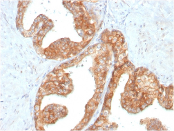
 Application Data
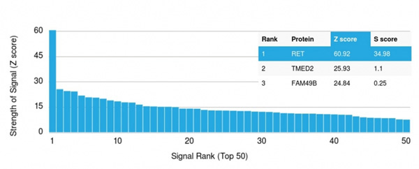
(Analysis of Protein Array containing more than 19,000 full-length human proteins using RET Mouse Monoclonal Antibody (RET/2795). Z- and S- Score: The Z-score represents the strength of a signal that a monoclonal antibody (MAb) (in combination with a fluorescently-tagged anti-IgG secondary antibody) produces when binding to a particular protein on the HuProtTM array. Z-scores are described in units of standard deviations (SD's) above the mean value of all signals generated on that array. If targets on HuProtTM are arranged in descending order of the Z-score, the S-score is the difference (also in units of SD's) between the Z-score. S-score therefore represents the relative target specificity of a MAb to its intended target. A MAb is considered to specific to its intended target, if the MAb has an S-score of at least 2.5. For example, if a MAb binds to protein X with a Z-score of 43 and to protein Y with a Z-score of 14, then the S-score for the binding of that MAb to protein X is equal to 29.)
Application Data
(Analysis of Protein Array containing more than 19,000 full-length human proteins using RET Mouse Monoclonal Antibody (RET/2795). Z- and S- Score: The Z-score represents the strength of a signal that a monoclonal antibody (MAb) (in combination with a fluorescently-tagged anti-IgG secondary antibody) produces when binding to a particular protein on the HuProtTM array. Z-scores are described in units of standard deviations (SD's) above the mean value of all signals generated on that array. If targets on HuProtTM are arranged in descending order of the Z-score, the S-score is the difference (also in units of SD's) between the Z-score. S-score therefore represents the relative target specificity of a MAb to its intended target. A MAb is considered to specific to its intended target, if the MAb has an S-score of at least 2.5. For example, if a MAb binds to protein X with a Z-score of 43 and to protein Y with a Z-score of 14, then the S-score for the binding of that MAb to protein X is equal to 29.)
RET Proto-oncogene, Monoclonal Antibody (Cat# AAA215182)
Full Name
RET Proto-oncogene
Gene Names
RET; PTC; MTC1; HSCR1; MEN2A; MEN2B; CDHF12; CDHR16; RET-ELE1
Reactivity
Human
Applications
Immunohistochemistry
Purity
Purified Ab with BSA and Azide at 200ug/ml OR Purified Ab WITHOUT BSA and Azide at 1.0mg/ml
Pricing
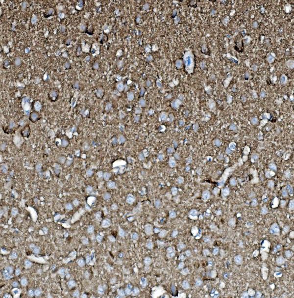
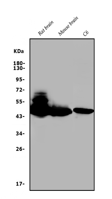
 FCM/FACS (Flow Cytometry)
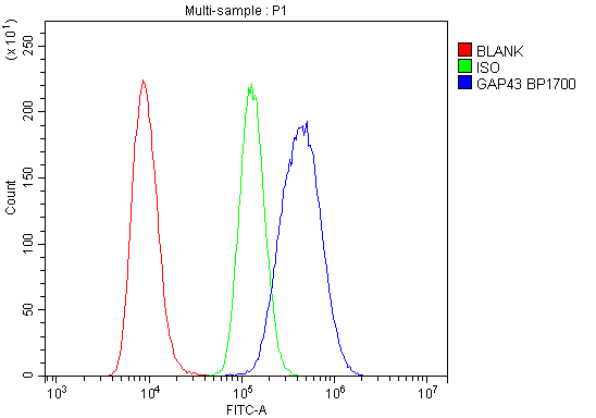
(Figure 3. Flow Cytometry analysis of Raji cells using anti-GAP43 antibody (AAA125626).Overlay histogram showing Raji cells stained with AAA125626 (Blue line). The cells were blocked with 10% normal goat serum. And then incubated with rabbit anti-GAP43 Antibody (AAA125626,1μg/1x106 cells) for 30 min at 20 degree C. DyLight®488 conjugated goat anti-rabbit IgG (5-10μg/1x106 cells) was used as secondary antibody for 30 minutes at 20 degree C. Isotype control antibody (Green line) was rabbit IgG (1μg/1x106) used under the same conditions. Unlabelled sample (Red line) was also used as a control.)
FCM/FACS (Flow Cytometry)
(Figure 3. Flow Cytometry analysis of Raji cells using anti-GAP43 antibody (AAA125626).Overlay histogram showing Raji cells stained with AAA125626 (Blue line). The cells were blocked with 10% normal goat serum. And then incubated with rabbit anti-GAP43 Antibody (AAA125626,1μg/1x106 cells) for 30 min at 20 degree C. DyLight®488 conjugated goat anti-rabbit IgG (5-10μg/1x106 cells) was used as secondary antibody for 30 minutes at 20 degree C. Isotype control antibody (Green line) was rabbit IgG (1μg/1x106) used under the same conditions. Unlabelled sample (Red line) was also used as a control.)
GAP43, Polyclonal Antibody (Cat# AAA125626)
Full Name
Anti-GAP43 Antibody
Gene Names
Gap43; B-50; Basp2; GAP-43
Reactivity
Human, Mouse, Rat
Applications
Direct ELISA, Flow Cytometry, Immunohistochemistry, Western Blot
Purity
Immunogen affinity purified.
Pricing
 FCM/FACS (Flow Cytometry)
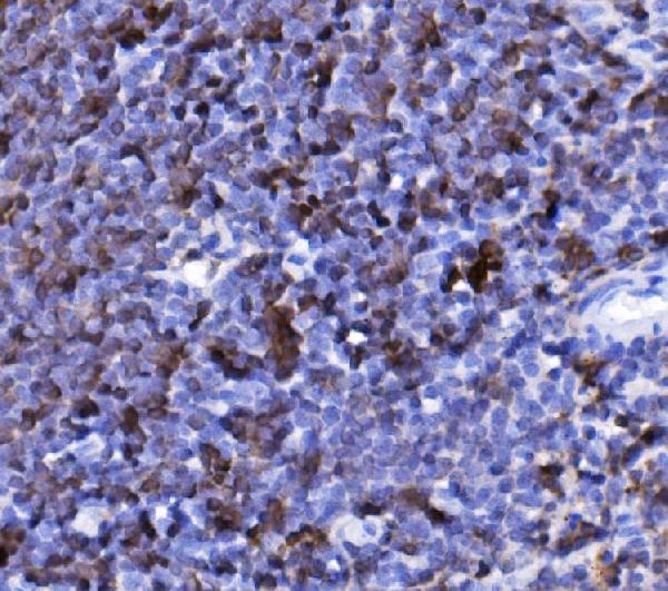
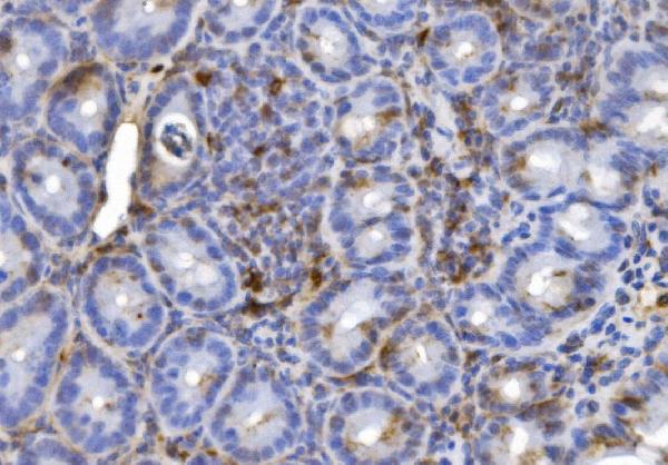
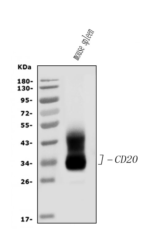
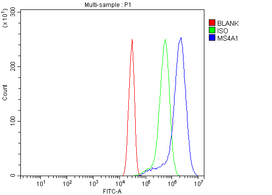
(Figure 4. Flow Cytometry analysis of HEPA1-6 cells using anti-CD20/MS4A1 antibody (AAA125704).Overlay histogram showing HEPA1-6 cells stained with AAA125704 (Blue line). The cells were blocked with 10% normal goat serum. And then incubated with rabbit anti-CD20/MS4A1 Antibody (AAA125704, 1μg/1x106 cells) for 30 min at 20 degree C. DyLight®488 conjugated goat anti-rabbit IgG (5-10μg/1x106 cells) was used as secondary antibody for 30 minutes at 20 degree C. Isotype control antibody (Green line) was rabbit IgG (1μg/1x106) used under the same conditions. Unlabelled sample (Red line) was also used as a control.)
FCM/FACS (Flow Cytometry)
(Figure 4. Flow Cytometry analysis of HEPA1-6 cells using anti-CD20/MS4A1 antibody (AAA125704).Overlay histogram showing HEPA1-6 cells stained with AAA125704 (Blue line). The cells were blocked with 10% normal goat serum. And then incubated with rabbit anti-CD20/MS4A1 Antibody (AAA125704, 1μg/1x106 cells) for 30 min at 20 degree C. DyLight®488 conjugated goat anti-rabbit IgG (5-10μg/1x106 cells) was used as secondary antibody for 30 minutes at 20 degree C. Isotype control antibody (Green line) was rabbit IgG (1μg/1x106) used under the same conditions. Unlabelled sample (Red line) was also used as a control.)
CD20/MS4A1, Polyclonal Antibody (Cat# AAA125704)
Full Name
Anti-CD20/MS4A1 Antibody
Gene Names
MS4A1; B1; S7; Bp35; CD20; CVID5; MS4A2; LEU-16
Reactivity
Mouse
Applications
Direct ELISA, Flow Cytometry, Immunohistochemistry, Western Blot
Purity
Immunogen affinity purified.
Pricing
 Standard Curve (Sample)
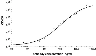
(Standard Curve for Human Serum Albumin (Analyte: Human Serum Albumin protein ); using Capture Antibody Mouse monoclonal [1A8-C5-B4] to Human Serum Albumin at 8 ug/ml and Detector Antibody Rabbit polyclonal antiserum to Human Serum Albumin.)
Standard Curve (Sample)
(Standard Curve for Human Serum Albumin (Analyte: Human Serum Albumin protein ); using Capture Antibody Mouse monoclonal [1A8-C5-B4] to Human Serum Albumin at 8 ug/ml and Detector Antibody Rabbit polyclonal antiserum to Human Serum Albumin.)
Serum Albumin, Monoclonal Antibody (Cat# AAA320318)
Full Name
Human Serum Albumin Mouse mAb
Gene Names
ALB; PRO0883; PRO0903; PRO1341
Reactivity
Human
Applications
Western Blot
Purity
The antibody was affinity-purified from mouse ascites by affinity-chromatography using epitope-specific immunogen.
Pricing
 Application Data
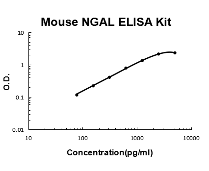
Lipocalin-2/NGAL, ELISA Kit (Cat# AAA125878)
Full Name
Mouse Lipocalin-2/NGAL Quick ELISA Kit
Gene Names
Lcn2; 24p3; Sip24; AW212229
Reactivity
Mouse
Pricing
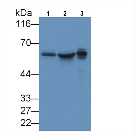 WB (Western Blot)
(Western Blot; Sample: Lane1: U2OS cell lysate; Lane2: Jurkat cell lysate; Lane3: 293T cell lysate Primary Ab: 3ug/ml Mouse AntiHuman KPNa2 Antibody Second Ab: 0.2ug/mL HRPLinked Caprine AntiMouse IgG Polyclonal Antibody (Catalog: SAA544Mu19))
WB (Western Blot)
(Western Blot; Sample: Lane1: U2OS cell lysate; Lane2: Jurkat cell lysate; Lane3: 293T cell lysate Primary Ab: 3ug/ml Mouse AntiHuman KPNa2 Antibody Second Ab: 0.2ug/mL HRPLinked Caprine AntiMouse IgG Polyclonal Antibody (Catalog: SAA544Mu19))
Karyopherin Alpha 2 (KPNa2), Monoclonal Antibody (Cat# AAA151858)
Full Name
Monoclonal Antibody to Karyopherin Alpha 2 (KPNa2)
Gene Names
KPNA2; QIP2; RCH1; IPOA1; SRP1alpha
Reactivity
Human, Pig
Applications
Immunoprecipitation, Immunocytochemistry, Immunohistochemistry, Western Blot
Purity
Protein A + Protein G affinity chromatography
Pricing
 WB (Western Blot)
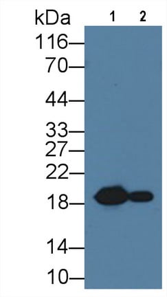
(Western Blot; Sample: Lane1: Rat Heart lysate; Lane1: Rat Skeletal muscle lysate Primary Ab: 1ug/ml Mouse AntiRat HSPb6 Antibody Second Ab: 0.2ug/mL HRPLinked Caprine AntiMouse IgG Polyclonal Antibody (Catalog: SAA544Mu19))
WB (Western Blot)
(Western Blot; Sample: Lane1: Rat Heart lysate; Lane1: Rat Skeletal muscle lysate Primary Ab: 1ug/ml Mouse AntiRat HSPb6 Antibody Second Ab: 0.2ug/mL HRPLinked Caprine AntiMouse IgG Polyclonal Antibody (Catalog: SAA544Mu19))
Heat Shock Protein Beta 6 (HSPb6), Monoclonal Antibody (Cat# AAA151867)
Full Name
Monoclonal Antibody to Heat Shock Protein Beta 6 (HSPb6)
Gene Names
Hspb6; p20; Hsp20
Reactivity
Rat
Applications
Immunoprecipitation, Immunocytochemistry, Immunohistochemistry, Western Blot
Purity
Protein A + Protein G affinity chromatography
Pricing
 WB (Western Blot)
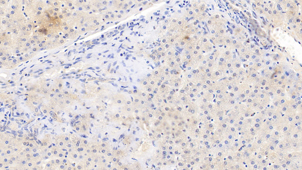
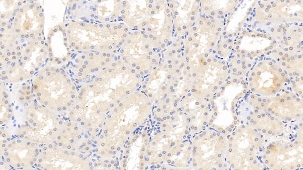
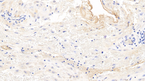
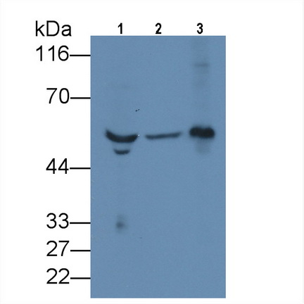
(Western Blot; Sample: Lane1: Human Serum; Lane2: Human Urine; Lane3: Human Placenta lysate Primary Ab: 3ug/ml Mouse AntiHuman FGb Antibody Second Ab: 0.2ug/mL HRPLinked Caprine AntiMouse IgG Polyclonal Antibody (Catalog: SAA544Mu19))
WB (Western Blot)
(Western Blot; Sample: Lane1: Human Serum; Lane2: Human Urine; Lane3: Human Placenta lysate Primary Ab: 3ug/ml Mouse AntiHuman FGb Antibody Second Ab: 0.2ug/mL HRPLinked Caprine AntiMouse IgG Polyclonal Antibody (Catalog: SAA544Mu19))
Fibrinogen Beta Chain (FGB), Monoclonal Antibody (Cat# AAA151693)
Full Name
Monoclonal Antibody to Fibrinogen Beta Chain (FGB)
Reactivity
Human, Pig
Applications
Immunoprecipitation, Immunocytochemistry, Immunohistochemistry, Western Blot
Purity
Protein A + Protein G affinity chromatography
Pricing
 IHC (Immunohistochemisry)
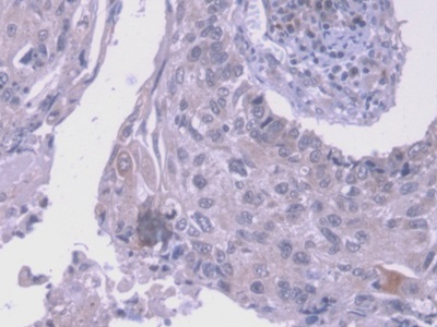
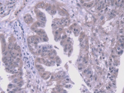
(DAB staining on IHCP;Sample: Human Liver cancer Tissue; Primary Ab: 20ug/ml Mouse AntiHuman VEGF121 AntibodySecond Ab: 2ug/mL HRPLinked Caprine AntiMouse IgG Polyclonal Antibody(Catalog: SAA544Mu19))
IHC (Immunohistochemisry)
(DAB staining on IHCP;Sample: Human Liver cancer Tissue; Primary Ab: 20ug/ml Mouse AntiHuman VEGF121 AntibodySecond Ab: 2ug/mL HRPLinked Caprine AntiMouse IgG Polyclonal Antibody(Catalog: SAA544Mu19))
Vascular Endothelial Growth Factor 121 (VEGF121), Monoclonal Antibody (Cat# AAA151757)
Full Name
Monoclonal Antibody to Vascular Endothelial Growth Factor 121 (VEGF121)
Gene Names
VEGFA; VPF; VEGF; MVCD1
Reactivity
Human
Applications
Immunoprecipitation, Immunocytochemistry, Immunohistochemistry, Western Blot
Purity
Protein A + Protein G affinity chromatography
Pricing
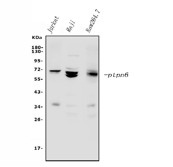
 FCM/FACS (Flow Cytometry)
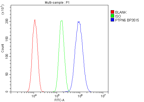
(Figure 3. Flow Cytometry analysis of A431 cells using anti-SHP1/PTPN6 antibody (AAA125581).Overlay histogram showing A431 cells stained with AAA125581 (Blue line). The cells were blocked with 10% normal goat serum. And then incubated with rabbit anti-SHP1/PTPN6 Antibody (AAA125581, 1μg/1x106 cells) for 30 min at 20 degree C. DyLight®488 conjugated goat anti-rabbit IgG (5-10μg/1x106 cells) was used as secondary antibody for 30 minutes at 20 degree C. Isotype control antibody (Green line) was rabbit IgG (1μg/1x106) used under the same conditions. Unlabelled sample (Red line) was also used as a control.)
FCM/FACS (Flow Cytometry)
(Figure 3. Flow Cytometry analysis of A431 cells using anti-SHP1/PTPN6 antibody (AAA125581).Overlay histogram showing A431 cells stained with AAA125581 (Blue line). The cells were blocked with 10% normal goat serum. And then incubated with rabbit anti-SHP1/PTPN6 Antibody (AAA125581, 1μg/1x106 cells) for 30 min at 20 degree C. DyLight®488 conjugated goat anti-rabbit IgG (5-10μg/1x106 cells) was used as secondary antibody for 30 minutes at 20 degree C. Isotype control antibody (Green line) was rabbit IgG (1μg/1x106) used under the same conditions. Unlabelled sample (Red line) was also used as a control.)
SHP1/PTPN6, Polyclonal Antibody (Cat# AAA125581)
Full Name
Anti-SHP1/PTPN6 Antibody
Gene Names
PTPN6; HCP; HCPH; SHP1; SHP-1; HPTP1C; PTP-1C; SHP-1L; SH-PTP1
Reactivity
Human, Mouse
Applications
Direct ELISA, Flow Cytometry, Immunofluorescence, Immunocytochemistry, Western Blot
Purity
Immunogen affinity purified.
Pricing
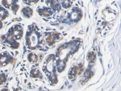
 WB (Western Blot)
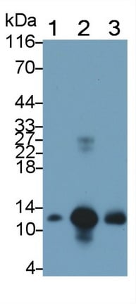
(Western Blot; Sample: Lane1: Human Saliva; Lane2: Human Leukocyte lysate; Lane3: Human Lymphocyte lysate Primary Ab: 0.1ug/ml Mouse AntiHuman S100A8 Antibody Second Ab: 0.2ug/mL HRPLinked Caprine AntiMouse IgG Polyclonal Antibody (Catalog: SAA544Mu19))
WB (Western Blot)
(Western Blot; Sample: Lane1: Human Saliva; Lane2: Human Leukocyte lysate; Lane3: Human Lymphocyte lysate Primary Ab: 0.1ug/ml Mouse AntiHuman S100A8 Antibody Second Ab: 0.2ug/mL HRPLinked Caprine AntiMouse IgG Polyclonal Antibody (Catalog: SAA544Mu19))
S100 Calcium Binding Protein A8 (S100A8), Monoclonal Antibody (Cat# AAA151746)
Full Name
Monoclonal Antibody to S100 Calcium Binding Protein A8 (S100A8)
Reactivity
Human
Applications
Immunoprecipitation, Immunocytochemistry, Immunohistochemistry, Western Blot
Purity
Protein A + Protein G affinity chromatography
Pricing
 WB (Western Blot)
(Western Blot; Sample: Rat Heart lysatePrimary Ab: 3ug/ml Mouse AntiHuman Hpt AntibodySecond Ab: 0.2ug/mL HRPLinked Caprine AntiMouse IgG Polyclonal Antibody(Catalog: SAA544Mu19))
WB (Western Blot)
(Western Blot; Sample: Rat Heart lysatePrimary Ab: 3ug/ml Mouse AntiHuman Hpt AntibodySecond Ab: 0.2ug/mL HRPLinked Caprine AntiMouse IgG Polyclonal Antibody(Catalog: SAA544Mu19))
Haptoglobin (Hpt), Monoclonal Antibody (Cat# AAA151629)
Full Name
Monoclonal Antibody to Haptoglobin (Hpt)
Gene Names
HP; BP; HPA1S; HP2ALPHA2
Reactivity
Human, Rat
Applications
Immunoprecipitation, Immunocytochemistry, Immunohistochemistry, Western Blot
Purity
Protein A + Protein G affinity chromatography
Pricing
 IHC (Immunohistochemisry)
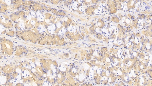
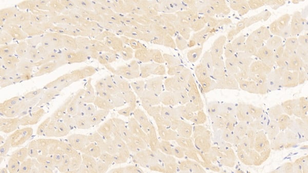
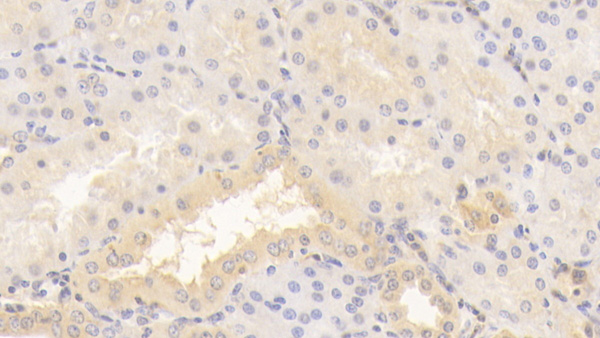
(DAB staining on IHCP;Sample: Human Kidney Tissue; Primary Ab: 20ug/ml Mouse AntiHuman TNC AntibodySecond Ab: 2ug/mL HRPLinked Caprine AntiMouse IgG Polyclonal Antibody(Catalog: SAA544Mu19))
IHC (Immunohistochemisry)
(DAB staining on IHCP;Sample: Human Kidney Tissue; Primary Ab: 20ug/ml Mouse AntiHuman TNC AntibodySecond Ab: 2ug/mL HRPLinked Caprine AntiMouse IgG Polyclonal Antibody(Catalog: SAA544Mu19))
Tenascin C (TNC), Monoclonal Antibody (Cat# AAA151773)
Full Name
Monoclonal Antibody to Tenascin C (TNC)
Gene Names
TNC; GP; JI; TN; HXB; GMEM; TN-C; 150-225
Reactivity
Human, Pig
Applications
Immunoprecipitation, Immunocytochemistry, Immunohistochemistry, Western Blot
Purity
Protein A + Protein G affinity chromatography
Pricing
 Application Data
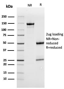
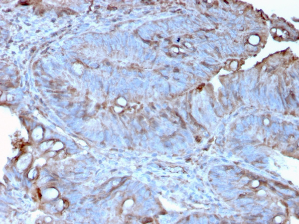
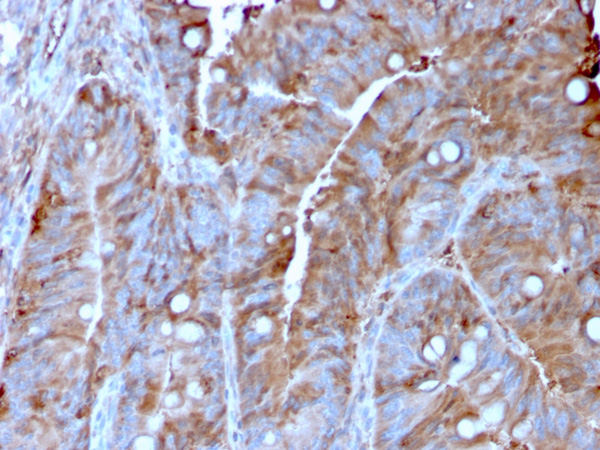
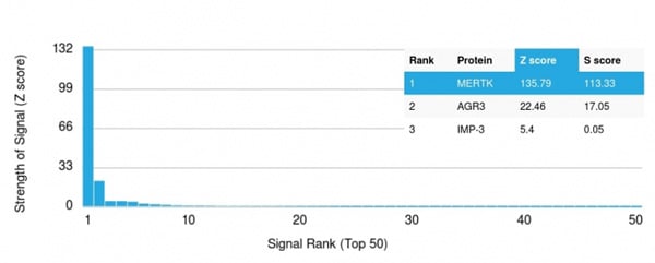
(Analysis of Protein Array containing more than 19,000 full-length human proteins using MerTK Mouse Monoclonal Antibody (MERTK/3022). Z- and S- Score: The Z-score represents the strength of a signal that a monoclonal antibody (MAb) (in combination with a fluorescently-tagged anti-IgG secondary antibody) produces when binding to a particular protein on the HuProtTM array. Z-scores are described in units of standard deviations (SD's) above the mean value of all signals generated on that array. If targets on HuProtTM are arranged in descending order of the Z-score, the S-score is the difference (also in units of SD's) between the Z-score. S-score therefore represents the relative target specificity of a MAb to its intended target. A MAb is considered to specific to its intended target, if the MAb has an S-score of at least 2.5. For example, if a MAb binds to protein X with a Z-score of 43 and to protein Y with a Z-score of 14, then the S-score for the binding of that MAb to protein X is equal to 29.)
Application Data
(Analysis of Protein Array containing more than 19,000 full-length human proteins using MerTK Mouse Monoclonal Antibody (MERTK/3022). Z- and S- Score: The Z-score represents the strength of a signal that a monoclonal antibody (MAb) (in combination with a fluorescently-tagged anti-IgG secondary antibody) produces when binding to a particular protein on the HuProtTM array. Z-scores are described in units of standard deviations (SD's) above the mean value of all signals generated on that array. If targets on HuProtTM are arranged in descending order of the Z-score, the S-score is the difference (also in units of SD's) between the Z-score. S-score therefore represents the relative target specificity of a MAb to its intended target. A MAb is considered to specific to its intended target, if the MAb has an S-score of at least 2.5. For example, if a MAb binds to protein X with a Z-score of 43 and to protein Y with a Z-score of 14, then the S-score for the binding of that MAb to protein X is equal to 29.)
MerTK, Monoclonal Antibody (Cat# AAA214882)
Full Name
MerTK (Innate Immune Checkpoint)
Gene Names
MERTK; MER; RP38; c-Eyk; c-mer; Tyro12
Reactivity
Human
Applications
Immunohistochemistry
Purity
Purified Ab with BSA and Azide at 200ug/ml OR Purified Ab WITHOUT BSA and Azide at 1.0mg/ml
Pricing
 FCM/FACS (Flow Cytometry)
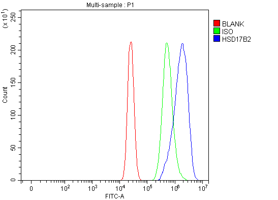
(Figure 3. Flow Cytometry analysis of MCF-7 cells using anti-HSD17B2 antibody (AAA127123).Overlay histogram showing MCF-7 cells stained with AAA127123 (Blue line). To facilitate intracellular staining, cells were fixed with 4% paraformaldehyde and permeabilized with permeabilization buffer. The cells were blocked with 10% normal goat serum. And then incubated with rabbit anti-HSD17B2 Antibody (AAA127123, 1ug/1x106 cells) for 30 min at 20 degree C. DyLight488 conjugated goat anti-rabbit IgG was used as secondary antibody for 30 minutes at 20 degree C. Isotype control antibody (Green line) was rabbit IgG (1ug/1x106) used under the same conditions. Unlabelled sample (Red line) was also used as a control.)
FCM/FACS (Flow Cytometry)
(Figure 3. Flow Cytometry analysis of MCF-7 cells using anti-HSD17B2 antibody (AAA127123).Overlay histogram showing MCF-7 cells stained with AAA127123 (Blue line). To facilitate intracellular staining, cells were fixed with 4% paraformaldehyde and permeabilized with permeabilization buffer. The cells were blocked with 10% normal goat serum. And then incubated with rabbit anti-HSD17B2 Antibody (AAA127123, 1ug/1x106 cells) for 30 min at 20 degree C. DyLight488 conjugated goat anti-rabbit IgG was used as secondary antibody for 30 minutes at 20 degree C. Isotype control antibody (Green line) was rabbit IgG (1ug/1x106) used under the same conditions. Unlabelled sample (Red line) was also used as a control.)
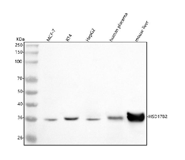
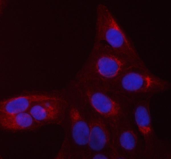
HSD17B2, Polyclonal Antibody (Cat# AAA127123)
Full Name
Anti-HSD17B2 Antibody Picoband
Gene Names
HSD17B2; HSD17; SDR9C2; EDH17B2
Reactivity
Human, Mouse
Applications
Flow Cytometry, Western Blot, Immunofluorescence, Immunocytochemistry
Purity
Immunogen affinity purified.
Pricing
 FCM/FACS (Flow Cytometry)
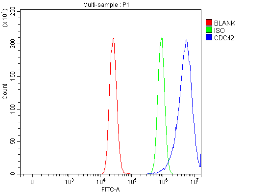
(Figure 3. Flow Cytometry analysis of MCF-7 cells using anti-CDC42 antibody (AAA126967).Overlay histogram showing MCF-7 cells stained with AAA126967 (Blue line). To facilitate intracellular staining, cells were fixed with 4% paraformaldehyde and permeabilized with permeabilization buffer. The cells were blocked with 10% normal goat serum. And then incubated with rabbit anti-CDC42 Antibody (AAA126967, 1ug/1x106 cells) for 30 min at 20 degree C. DyLight488 conjugated goat anti-rabbit IgG was used as secondary antibody for 30 minutes at 20 degree C. Isotype control antibody (Green line) was rabbit IgG (1ug/1x106) used under the same conditions. Unlabelled sample (Red line) was also used as a control.)
FCM/FACS (Flow Cytometry)
(Figure 3. Flow Cytometry analysis of MCF-7 cells using anti-CDC42 antibody (AAA126967).Overlay histogram showing MCF-7 cells stained with AAA126967 (Blue line). To facilitate intracellular staining, cells were fixed with 4% paraformaldehyde and permeabilized with permeabilization buffer. The cells were blocked with 10% normal goat serum. And then incubated with rabbit anti-CDC42 Antibody (AAA126967, 1ug/1x106 cells) for 30 min at 20 degree C. DyLight488 conjugated goat anti-rabbit IgG was used as secondary antibody for 30 minutes at 20 degree C. Isotype control antibody (Green line) was rabbit IgG (1ug/1x106) used under the same conditions. Unlabelled sample (Red line) was also used as a control.)
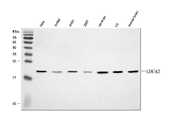
CDC42, Polyclonal Antibody (Cat# AAA126967)
Full Name
Anti-CDC42 Antibody Picoband
Gene Names
CDC42; G25K; CDC42Hs
Reactivity
Human, Mouse, Rat
Applications
Flow Cytometry, Immunofluorescence, Immunocytochemistry, Western Blot
Purity
Immunogen affinity purified.
Pricing
 IP (Immunoprecipitation)
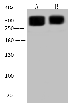
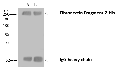
(Fibronectin Fragment 2-His was immunoprecipitated using:Lane A:0.5 mg NIH3T3 Whole Cell LysateLane B:0.5 mg HepG2 Whole Cell Lysate4 uL anti-Fibronectin Fragment 2-His rabbit polyclonal antibody and 60 ug of Immunomagnetic beads Protein A/G.Primary antibody:Anti-Fibronectin Fragment 2-His rabbit polyclonal antibody,at 1:100 dilutionSecondary antibody:Goat Anti-Rabbit IgG (H+L)/HRP at 1/10000 dilutionDeveloped using the ECL technique.Performed under reducing conditions.Predicted band size: 300 kDaObserved band size :300 kDa)
IP (Immunoprecipitation)
(Fibronectin Fragment 2-His was immunoprecipitated using:Lane A:0.5 mg NIH3T3 Whole Cell LysateLane B:0.5 mg HepG2 Whole Cell Lysate4 uL anti-Fibronectin Fragment 2-His rabbit polyclonal antibody and 60 ug of Immunomagnetic beads Protein A/G.Primary antibody:Anti-Fibronectin Fragment 2-His rabbit polyclonal antibody,at 1:100 dilutionSecondary antibody:Goat Anti-Rabbit IgG (H+L)/HRP at 1/10000 dilutionDeveloped using the ECL technique.Performed under reducing conditions.Predicted band size: 300 kDaObserved band size :300 kDa)
Fibronectin, Polyclonal Antibody (Cat# AAA258791)
Full Name
Anti-Fibronectin Antibody, Rabbit Polyclonal
Reactivity
Human
Applications
Immunoprecipitation, Immunofluorescence, Immunocytochemistry, Western Blot
Purity
Protein A & Antigen Affinity
Pricing
 IHC (Immunohistochemistry)
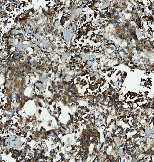
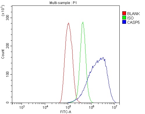
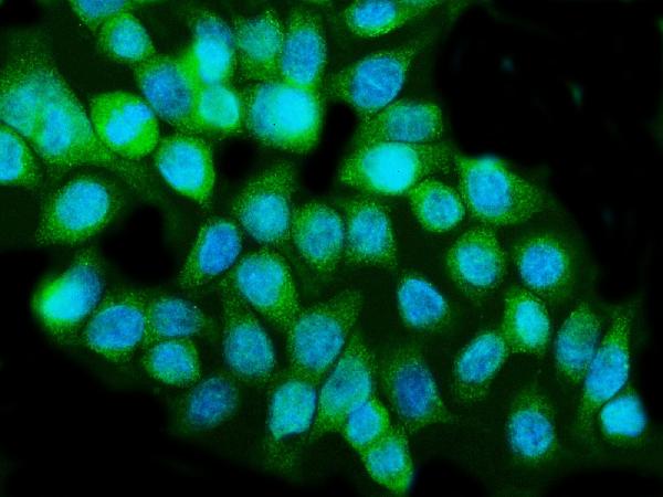
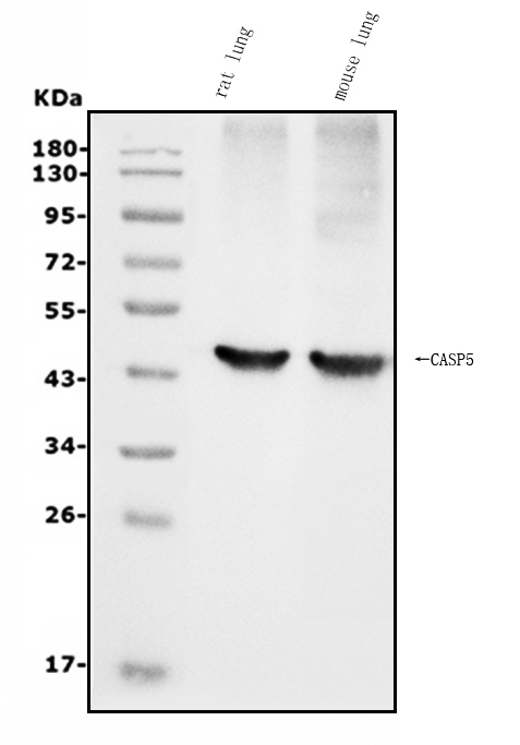
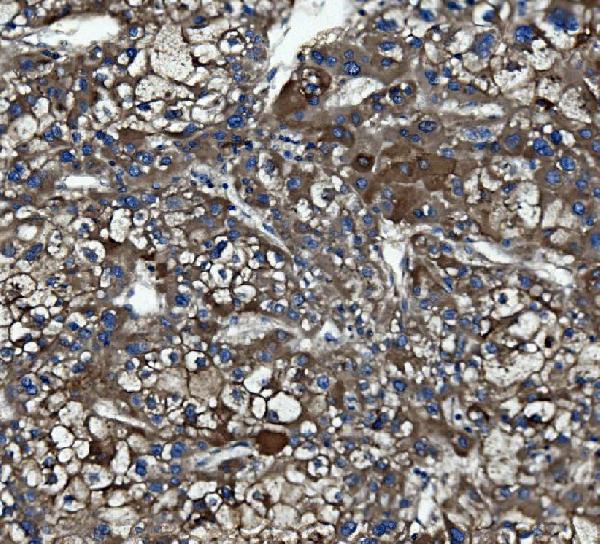
(Figure 5. IHC analysis of Caspase-5/CASP5 using anti-Caspase-5/CASP5 antibody (AAA125743).Caspase-5/CASP5 was detected in paraffin-embedded section of human liver cancer tissue. Heat mediated antigen retrieval was performed in EDTA buffer (pH8. 0, epitope retrieval solution). The tissue section was blocked with 10% goat serum. The tissue section was then incubated with 2μg/ml rabbit anti-Caspase-5/CASP5 Antibody (AAA125743) overnight at 4 degree C. Biotinylated goat anti-rabbit IgG was used as secondary antibody and incubated for 30 minutes at 37 degree C. The tissue section was developed using Strepavidin-Biotin-Complex (SABC) with DAB as the chromogen.)
IHC (Immunohistochemistry)
(Figure 5. IHC analysis of Caspase-5/CASP5 using anti-Caspase-5/CASP5 antibody (AAA125743).Caspase-5/CASP5 was detected in paraffin-embedded section of human liver cancer tissue. Heat mediated antigen retrieval was performed in EDTA buffer (pH8. 0, epitope retrieval solution). The tissue section was blocked with 10% goat serum. The tissue section was then incubated with 2μg/ml rabbit anti-Caspase-5/CASP5 Antibody (AAA125743) overnight at 4 degree C. Biotinylated goat anti-rabbit IgG was used as secondary antibody and incubated for 30 minutes at 37 degree C. The tissue section was developed using Strepavidin-Biotin-Complex (SABC) with DAB as the chromogen.)
Caspase-5/CASP5, Polyclonal Antibody (Cat# AAA125743)
Full Name
Anti-Caspase-5/CASP5 Antibody
Gene Names
CASP5; ICH-3; ICEREL-III; ICE(rel)III
Reactivity
Human, Mouse, Rat
Applications
Direct ELISA, Flow Cytometry, Immunofluorescence, Immunocytochemistry, Immunohistochemistry, Western Blot
Purity
Immunogen affinity purified.
Pricing
 WB (Western Blot)
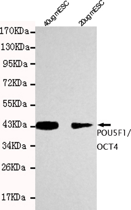
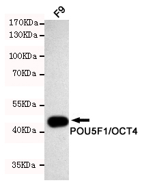
(Western blot detection of POU5F1/OCT4 in F9 cell lysates using POU5F1/OCT4 mouse mAb (1:1000 diluted).Predicted band size: 45KDa.Observed band size: 45KDa.)
WB (Western Blot)
(Western blot detection of POU5F1/OCT4 in F9 cell lysates using POU5F1/OCT4 mouse mAb (1:1000 diluted).Predicted band size: 45KDa.Observed band size: 45KDa.)
POU5F1/OCT4, Monoclonal Antibody (Cat# AAA320299)
Full Name
POU5F1/OCT4 Mouse mAb
Gene Names
POU5F1; OCT3; OCT4; OTF3; OTF4; OTF-3; Oct-3; Oct-4
Reactivity
Mouse
Applications
Western Blot
Purity
The antibody was affinity-purified from mouse ascites by affinity-chromatography using epitope-specific immunogen.
Pricing
 IP (Immunoprecipitation)
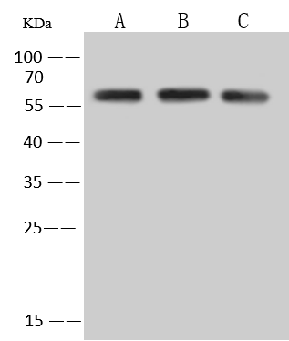
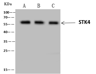
(STK4 was immunoprecipitated using:; Lane A:0.5 mg Jurkat Whole Cell Lysate; Lane B:0.5 mg HeLa Whole Cell Lysate; Lane C:0.5 mg HepG2 Whole Cell Lysate; 4 uL anti-STK4 rabbit polyclonal antibody and 60 ug of Immunomagnetic beads Protein A/G.; Primary antibody:; Anti-STK4 rabbit polyclonal antibody,at 1:100 dilution; Secondary antibody:; Clean-Blot IP Detection Reagent (HRP) at 1:1000dilution; Developed using the ECL technique.; Performed under reducing conditions.; Predicted band size: 58 kDa; Observed band size :58 kDa)
IP (Immunoprecipitation)
(STK4 was immunoprecipitated using:; Lane A:0.5 mg Jurkat Whole Cell Lysate; Lane B:0.5 mg HeLa Whole Cell Lysate; Lane C:0.5 mg HepG2 Whole Cell Lysate; 4 uL anti-STK4 rabbit polyclonal antibody and 60 ug of Immunomagnetic beads Protein A/G.; Primary antibody:; Anti-STK4 rabbit polyclonal antibody,at 1:100 dilution; Secondary antibody:; Clean-Blot IP Detection Reagent (HRP) at 1:1000dilution; Developed using the ECL technique.; Performed under reducing conditions.; Predicted band size: 58 kDa; Observed band size :58 kDa)
MST1, Polyclonal Antibody (Cat# AAA258632)
Full Name
MST1 Antibody, Rabbit PAb, Antigen Affinity Purified
Gene Names
STK4; KRS2; MST1; YSK3
Reactivity
Human
Applications
Immunoprecipitation, Immunofluorescence, Immunocytochemistry, Western Blot
Pricing
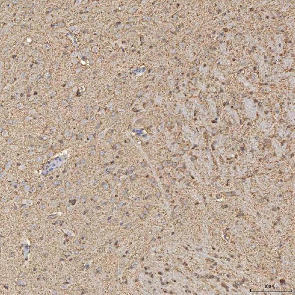 IHC (Immunohistochemisry)
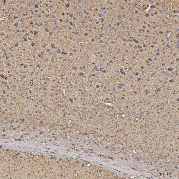
(Figure 3. IHC analysis of Synaptogyrin 3/SYNGR3 using anti-Synaptogyrin 3/SYNGR3 antibody (AAA127845).Synaptogyrin 3/SYNGR3 was detected in a paraffin-embedded section of rat brain tissue. Heat mediated antigen retrieval was performed in EDTA buffer (pH 8.0, epitope retrieval solution). The tissue section was blocked with 10% goat serum. The tissue section was then incubated with 2ug/ml rabbit anti-Synaptogyrin 3/SYNGR3 Antibody (AAA127845) overnight at 4 degree C. Peroxidase Conjugated Goat Anti-rabbit IgG was used as secondary antibody and incubated for 30 minutes at 37 degree C. The tissue section was developed using HRP Conjugated Rabbit IgG Super Vision Assay Kit with DAB as the chromogen.)
IHC (Immunohistochemisry)
(Figure 3. IHC analysis of Synaptogyrin 3/SYNGR3 using anti-Synaptogyrin 3/SYNGR3 antibody (AAA127845).Synaptogyrin 3/SYNGR3 was detected in a paraffin-embedded section of rat brain tissue. Heat mediated antigen retrieval was performed in EDTA buffer (pH 8.0, epitope retrieval solution). The tissue section was blocked with 10% goat serum. The tissue section was then incubated with 2ug/ml rabbit anti-Synaptogyrin 3/SYNGR3 Antibody (AAA127845) overnight at 4 degree C. Peroxidase Conjugated Goat Anti-rabbit IgG was used as secondary antibody and incubated for 30 minutes at 37 degree C. The tissue section was developed using HRP Conjugated Rabbit IgG Super Vision Assay Kit with DAB as the chromogen.)
Synaptogyrin 3/SYNGR3, Polyclonal Antibody (Cat# AAA127845)
Full Name
Anti-Synaptogyrin 3/SYNGR3 Antibody Picoband
Reactivity
Human, Mouse, Rat
Applications
Immunohistochemistry, Western Blot
Purity
Immunogen affinity purified.
Pricing
 IF (Immunofluorescence)
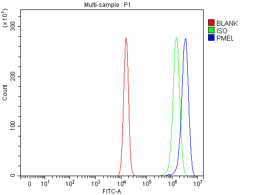
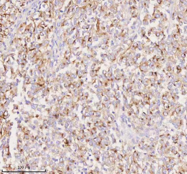
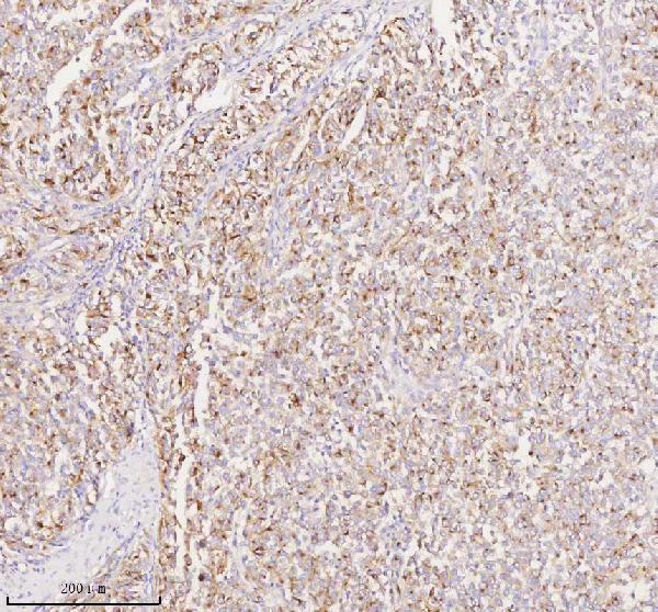
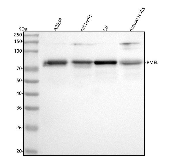
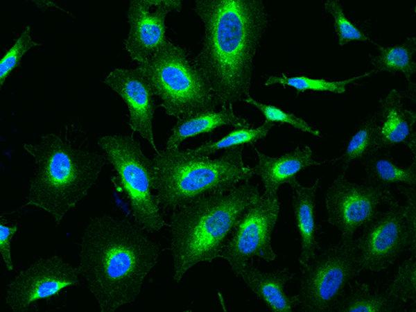
(Figure 5. IF analysis of SILV/PMEL using anti-SILV/PMEL antibody (AAA127046).SILV/PMEL was detected in a paraffin-embedded section of human melanoma tissue. Heat mediated antigen retrieval was performed in EDTA buffer (pH 8.0, epitope retrieval solution). The tissue section was blocked with 10% goat serum. The tissue section was then incubated with 5ug/mL rabbit anti-SILV/PMEL Antibody (AAA127046) overnight at 4 degree C. DyLight594 Conjugated Goat Anti-Rabbit IgG (BA1142) was used as secondary antibody at 1:500 dilution and incubated for 30 minutes at 37 degree C. The section was counterstained with DAPI. Visualize using a fluorescence microscope and filter sets appropriate for the label used.)
IF (Immunofluorescence)
(Figure 5. IF analysis of SILV/PMEL using anti-SILV/PMEL antibody (AAA127046).SILV/PMEL was detected in a paraffin-embedded section of human melanoma tissue. Heat mediated antigen retrieval was performed in EDTA buffer (pH 8.0, epitope retrieval solution). The tissue section was blocked with 10% goat serum. The tissue section was then incubated with 5ug/mL rabbit anti-SILV/PMEL Antibody (AAA127046) overnight at 4 degree C. DyLight594 Conjugated Goat Anti-Rabbit IgG (BA1142) was used as secondary antibody at 1:500 dilution and incubated for 30 minutes at 37 degree C. The section was counterstained with DAPI. Visualize using a fluorescence microscope and filter sets appropriate for the label used.)
SILV/PMEL, Polyclonal Antibody (Cat# AAA127046)
Full Name
Anti-SILV/PMEL Antibody Picoband
Gene Names
PMEL; P1; SI; SIL; ME20; P100; SILV; ME20M; gp100; ME20-M; PMEL17; D12S53E
Reactivity
Human, Mouse, Rat
Applications
Western Blot, Immunohistochemistry, Immunofluorescence, Flow Cytometry
Purity
Immunogen affinity purified.
Pricing
 Bioactivity
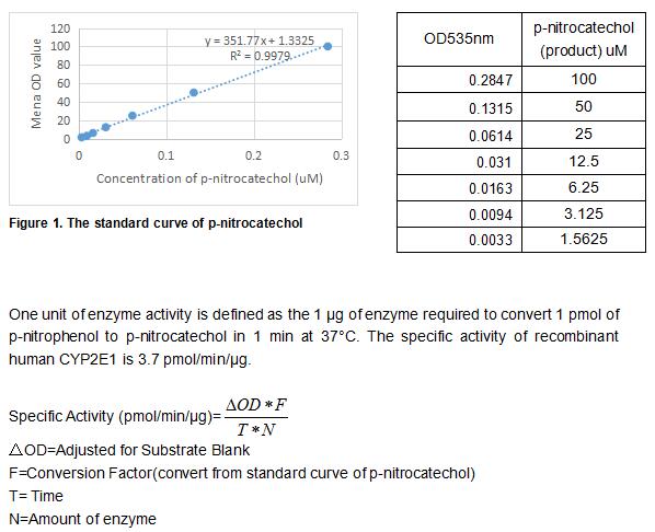
(The cytochrome P450 enzyme CYP2E1 catalyzes the oxidative metabolism of many solvents and other small organic molecules. CYP2E1 is expressed in adult and fetal human liver in addition to extrahepatic tissues such as lung and placenta. Treatment of primary cultures of human hepatocytes with ethanol induces CYP2E1 protein, and this is consistent with the finding that hepatic CYP2E1 protein and mRNA levels are increased in individuals with alcoholism. Although only a few drugs (e.g., acetaminophen have been identified as substrates for CYP2E1, many low molecular weight procarcinogens are activated by this cytochrome P450 (P450). Chlorzoxazone 6-hydroxylation, N-nitrosodimethylamine N-demethylation and p-nitrophenol hydroxylation can be used to measure the catalytic activity of CYP2E1. Thus, the recombinant human CYP2E1 activity was measured by its ability to hydroxylate p-nitrophenol to p-nitrocatechol. The reaction was performed in 50 mM potassium phosphate, pH 7.4 (Assay Buffer), initiated by addition 20 uL of 500 ug/ml CYP2E1 to 10 uL of 5 mM substrate p-nitrophenol and 30 ul of 26 mM NADPH in a total volume of 500 ul. Incubated at 37 degree C for 30min, then read at a wavelength of 535 nm after acidification of the reaction mixture with trichloroacetic acid followed by neutralization using 2 M NaOH.)
Bioactivity
(The cytochrome P450 enzyme CYP2E1 catalyzes the oxidative metabolism of many solvents and other small organic molecules. CYP2E1 is expressed in adult and fetal human liver in addition to extrahepatic tissues such as lung and placenta. Treatment of primary cultures of human hepatocytes with ethanol induces CYP2E1 protein, and this is consistent with the finding that hepatic CYP2E1 protein and mRNA levels are increased in individuals with alcoholism. Although only a few drugs (e.g., acetaminophen have been identified as substrates for CYP2E1, many low molecular weight procarcinogens are activated by this cytochrome P450 (P450). Chlorzoxazone 6-hydroxylation, N-nitrosodimethylamine N-demethylation and p-nitrophenol hydroxylation can be used to measure the catalytic activity of CYP2E1. Thus, the recombinant human CYP2E1 activity was measured by its ability to hydroxylate p-nitrophenol to p-nitrocatechol. The reaction was performed in 50 mM potassium phosphate, pH 7.4 (Assay Buffer), initiated by addition 20 uL of 500 ug/ml CYP2E1 to 10 uL of 5 mM substrate p-nitrophenol and 30 ul of 26 mM NADPH in a total volume of 500 ul. Incubated at 37 degree C for 30min, then read at a wavelength of 535 nm after acidification of the reaction mixture with trichloroacetic acid followed by neutralization using 2 M NaOH.)
Cytochrome P450 2E1 (CYP2E1), Active Protein (Cat# AAA161816)
Full Name
Active Cytochrome P450 2E1 (CYP2E1)
Gene Names
CYP2E1; CPE1; CYP2E; P450-J; P450C2E
Applications
Activity Assay, Cell Culture
Purity
Greater than 90% by SDS-PAGE
Pricing
 IP (Immunoprecipitation)
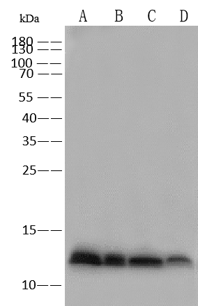
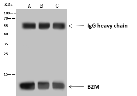
(B2M was immunoprecipitated using:; Lane A:0.5 mg A431 Whole Cell Lysate; Lane B:0.5 mg Raji Whole Cell Lysate; Lane C:0.5 mg Hela Whole Cell Lysate; 4 uL anti-B2M rabbit polyclonal antibody and 60 ug of Immunomagnetic beads Protein A/G.; Primary antibody:; Anti-B2M rabbit polyclonal antibody,at 1:100 dilution; Secondary antibody:; Goat Anti-Rabbit IgG (H+L)/HRP at 1/10000 dilution; Developed using the ECL technique.; Performed under reducing conditions.; Predicted band size: 12 kDa; Observed band size :12 kDa)
IP (Immunoprecipitation)
(B2M was immunoprecipitated using:; Lane A:0.5 mg A431 Whole Cell Lysate; Lane B:0.5 mg Raji Whole Cell Lysate; Lane C:0.5 mg Hela Whole Cell Lysate; 4 uL anti-B2M rabbit polyclonal antibody and 60 ug of Immunomagnetic beads Protein A/G.; Primary antibody:; Anti-B2M rabbit polyclonal antibody,at 1:100 dilution; Secondary antibody:; Goat Anti-Rabbit IgG (H+L)/HRP at 1/10000 dilution; Developed using the ECL technique.; Performed under reducing conditions.; Predicted band size: 12 kDa; Observed band size :12 kDa)
Beta-2 microglobulin, Polyclonal Antibody (Cat# AAA258635)
Full Name
Beta-2 microglobulin Antibody, Rabbit PAb, Antigen Affinity Purified
Reactivity
Human
Applications
Immunoprecipitation, Immunofluorescence, Immunocytochemistry, Immunohistochemistry, Western Blot
Pricing
 IP (Immunoprecipitation)
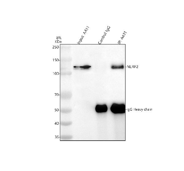
(Figure 4. Immunoprecipitating NLRP2 in A431 whole cell lysate .Western blot analysis of NLRP2 using anti-NLRP2 antibody (AAA127253).Lane 1: A431 whole cell lysates (30ug)Lane 2: Rabbit control IgG instead of anti-NLRP2 antibody in A431 whole cell lysate.Lane 3: anti-NLRP2 antibody (2ug) + A431 whole cell lysate (500ug)After electrophoresis, proteins were transferred to a membrane. Then the membrane was incubated with rabbit anti-NLRP2 antigen affinity purified polyclonal antibody (AAA127253) at a dilution of 0.5ug/mL and probed with a goat anti-rabbit IgG-HRP secondary antibody . The signal is developed using ECL Plus Western Blotting Substrate . A specific band was detected for NLRP2 at approximately 121 kDa. The expected band size for NLRP2 is at 121 kDa.)
IP (Immunoprecipitation)
(Figure 4. Immunoprecipitating NLRP2 in A431 whole cell lysate .Western blot analysis of NLRP2 using anti-NLRP2 antibody (AAA127253).Lane 1: A431 whole cell lysates (30ug)Lane 2: Rabbit control IgG instead of anti-NLRP2 antibody in A431 whole cell lysate.Lane 3: anti-NLRP2 antibody (2ug) + A431 whole cell lysate (500ug)After electrophoresis, proteins were transferred to a membrane. Then the membrane was incubated with rabbit anti-NLRP2 antigen affinity purified polyclonal antibody (AAA127253) at a dilution of 0.5ug/mL and probed with a goat anti-rabbit IgG-HRP secondary antibody . The signal is developed using ECL Plus Western Blotting Substrate . A specific band was detected for NLRP2 at approximately 121 kDa. The expected band size for NLRP2 is at 121 kDa.)
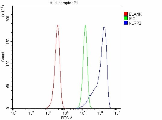
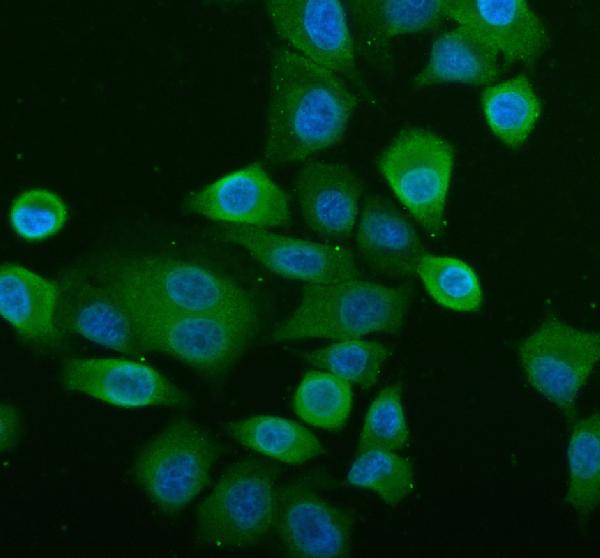
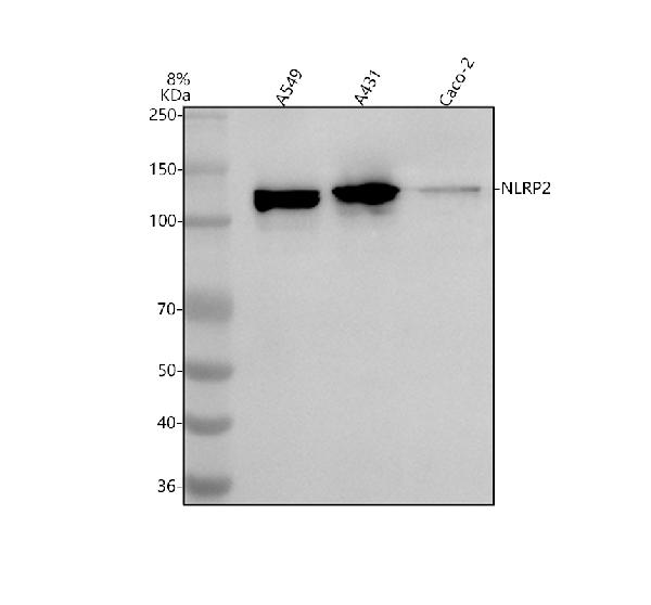
NLRP2, Polyclonal Antibody (Cat# AAA127253)
Full Name
Anti-NLRP2 Antibody Picoband
Gene Names
NLRP2; NBS1; PAN1; NALP2; PYPAF2; CLR19.9
Reactivity
Human
Applications
Western Blot, Immunofluorescence, Immunocytochemistry, Immunoprecipitation, Flow Cytometry
Purity
Immunogen affinity purified.
Pricing
 IF (Immunofluorescence)
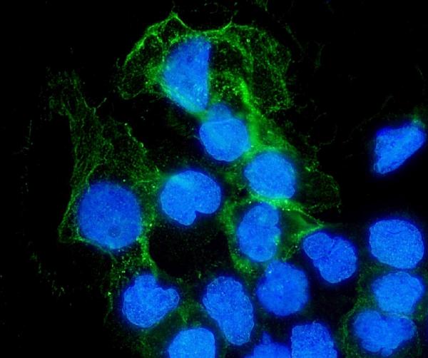
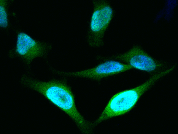
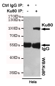
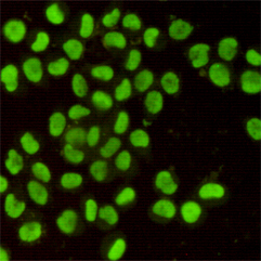
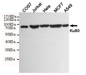
(Immunofluorescent analysis of Hela cells using Ku80 mouse mAb (1:400).)
IF (Immunofluorescence)
(Immunofluorescent analysis of Hela cells using Ku80 mouse mAb (1:400).)
Ku80, Monoclonal Antibody (Cat# AAA320384)
Full Name
Ku80 Mouse mAb
Gene Names
XRCC5; KU80; KUB2; Ku86; NFIV; KARP1; KARP-1
Reactivity
Human, Monkey
Applications
Immunoprecipitation, Immunocytochemistry, Immunofluorescence, Western Blot
Purity
The antibody was affinity-purified from mouse ascites by affinity-chromatography using epitope-specific immunogen.
Pricing
 FCM/FACS (Flow Cytometry)
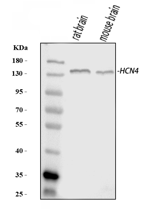
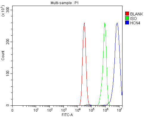
(Figure 2. Flow Cytometry analysis of MCF-7 cells using anti-HCN4 antibody (AAA126208).Overlay histogram showing MCF-7 cells stained with AAA126208 (Blue line). The cells were blocked with 10% normal goat serum. And then incubated with rabbit anti-HCN4 Antibody (AAA126208, 1 ug/1x10^6 cells) for 30 min at 20 degree C. DyLight488 conjugated goat anti-rabbit IgG was used as secondary antibody for 30 minutes at 20 degree C. Isotype control antibody (Green line) was rabbit IgG (1 ug/1x10^6) used under the same conditions. Unlabelled sample (Red line) was also used as a control.)
FCM/FACS (Flow Cytometry)
(Figure 2. Flow Cytometry analysis of MCF-7 cells using anti-HCN4 antibody (AAA126208).Overlay histogram showing MCF-7 cells stained with AAA126208 (Blue line). The cells were blocked with 10% normal goat serum. And then incubated with rabbit anti-HCN4 Antibody (AAA126208, 1 ug/1x10^6 cells) for 30 min at 20 degree C. DyLight488 conjugated goat anti-rabbit IgG was used as secondary antibody for 30 minutes at 20 degree C. Isotype control antibody (Green line) was rabbit IgG (1 ug/1x10^6) used under the same conditions. Unlabelled sample (Red line) was also used as a control.)
HCN4, Polyclonal Antibody (Cat# AAA126208)
Full Name
Anti-HCN4 Antibody Picoband
Gene Names
HCN4; SSS2
Reactivity
Human, Mouse, Rat
Applications
Flow Cytometry, Western Blot
Purity
Immunogen affinity purified.
Pricing
 FCM/FACS (Flow Cytometry)
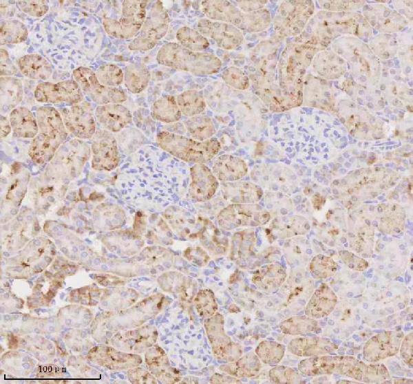
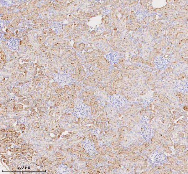
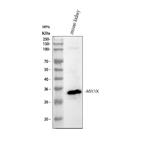
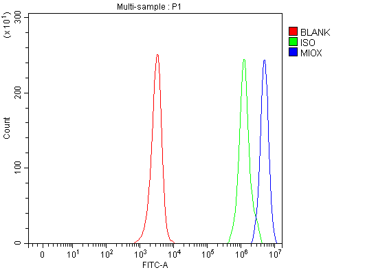
(Figure 5. Flow Cytometry analysis of JK cells using anti-MIOX antibody (AAA127413).Overlay histogram showing JK cells stained with AAA127413 (Blue line). To facilitate intracellular staining, cells were fixed with 4% paraformaldehyde and permeabilized with permeabilization buffer. The cells were blocked with 10% normal goat serum. And then incubated with rabbit anti-MIOX Antibody (AAA127413, 1ug/1x106 cells) for 30 min at 20 degree C. DyLight488 conjugated goat anti-rabbit IgG was used as secondary antibody for 30 minutes at 20 degree C. Isotype control antibody (Green line) was rabbit IgG (1ug/1x106) used under the same conditions. Unlabelled sample (Red line) was also used as a control.)
FCM/FACS (Flow Cytometry)
(Figure 5. Flow Cytometry analysis of JK cells using anti-MIOX antibody (AAA127413).Overlay histogram showing JK cells stained with AAA127413 (Blue line). To facilitate intracellular staining, cells were fixed with 4% paraformaldehyde and permeabilized with permeabilization buffer. The cells were blocked with 10% normal goat serum. And then incubated with rabbit anti-MIOX Antibody (AAA127413, 1ug/1x106 cells) for 30 min at 20 degree C. DyLight488 conjugated goat anti-rabbit IgG was used as secondary antibody for 30 minutes at 20 degree C. Isotype control antibody (Green line) was rabbit IgG (1ug/1x106) used under the same conditions. Unlabelled sample (Red line) was also used as a control.)
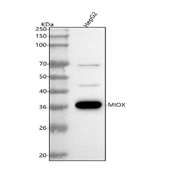
MIOX, Polyclonal Antibody (Cat# AAA127413)
Full Name
Anti-MIOX Antibody Picoband
Gene Names
MIOX; ALDRL6
Reactivity
Human, Mouse, Rat
Applications
Flow Cytometry, Immunohistochemistry, Western Blot
Purity
Immunogen affinity purified.
Pricing
 IF (Immunofluorescence)
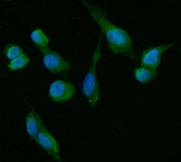
(Figure 2. IF analysis of HSD3B1/B2 using anti-HSD3B1/B2 antibody (AAA126920).HSD3B1/B2 was detected in an immunocytochemical section of U2OS cells. Enzyme antigen retrieval was performed using IHC enzyme antigen retrieval reagent (AR0022) for 15 mins. The cells were blocked with 10% goat serum. And then incubated with 5 ug/mL mouse anti-HSD3B1/B2 Antibody (AAA126920) overnight at 4 degree C. DyLight488 Conjugated Goat Anti-Mouse IgG was used as secondary antibody at 1:100 dilution and incubated for 30 minutes at 37 degree C. The section was counterstained with DAPI. Visualize using a fluorescence microscope and filter sets appropriate for the label used.)
IF (Immunofluorescence)
(Figure 2. IF analysis of HSD3B1/B2 using anti-HSD3B1/B2 antibody (AAA126920).HSD3B1/B2 was detected in an immunocytochemical section of U2OS cells. Enzyme antigen retrieval was performed using IHC enzyme antigen retrieval reagent (AR0022) for 15 mins. The cells were blocked with 10% goat serum. And then incubated with 5 ug/mL mouse anti-HSD3B1/B2 Antibody (AAA126920) overnight at 4 degree C. DyLight488 Conjugated Goat Anti-Mouse IgG was used as secondary antibody at 1:100 dilution and incubated for 30 minutes at 37 degree C. The section was counterstained with DAPI. Visualize using a fluorescence microscope and filter sets appropriate for the label used.)
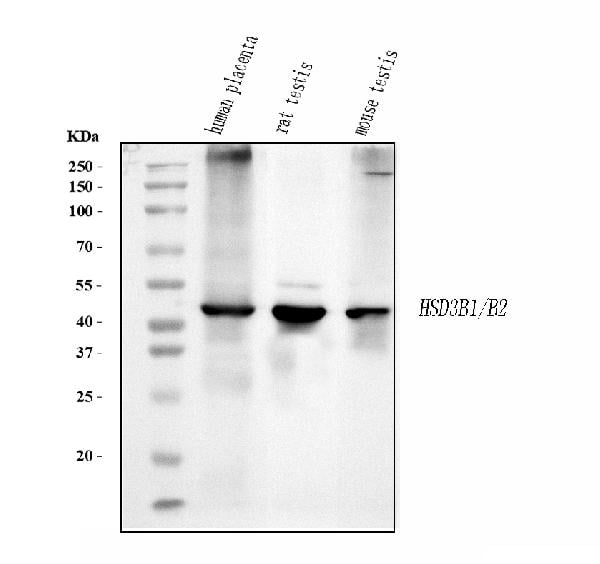
HSD3B1/B2, Monoclonal Antibody (Cat# AAA126920)
Full Name
Anti-HSD3B1/B2 Antibody Picoband (monoclonal, 3F3C/7F9C8)
Gene Names
HSD3B1; I; HSD3B; HSDB3; HSDB3A; SDR11E1; 3BETAHSD
Reactivity
Human, Mouse, Rat
Applications
Immunofluorescence, Immunocytochemistry, Western Blot
Purity
Immunogen affinity purified.
Pricing
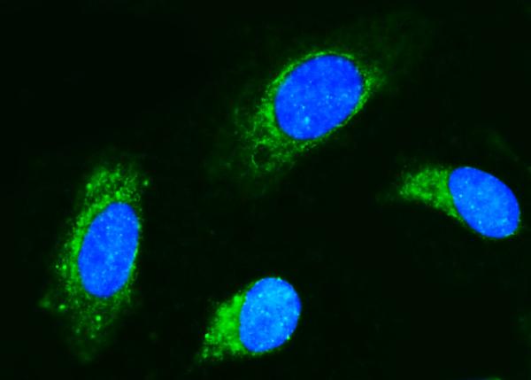
 WB (Western Blot)
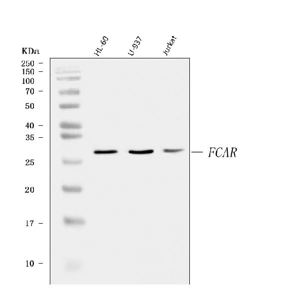
(Figure 1. Western blot analysis of CD89/FCAR using anti-CD89/FCAR antibody (AAA126497).Electrophoresis was performed on a 5-20% SDS-PAGE gel at 70V (Stacking gel)/90V (Resolving gel) for 2-3 hours. The sample well of each lane was loaded with 30 ug of sample under reducing conditions.Lane 1: human HL-60 whole cell lysates,Lane 2: human U-937 whole cell lysates,Lane 3: human Jurkat whole cell lysates.After electrophoresis, proteins were transferred to a nitrocellulose membrane at 150 mA for 50-90 minutes. Blocked the membrane with 5% non-fat milk/TBS for 1.5 hour at RT. The membrane was incubated with rabbit anti-CD89/FCAR antigen affinity purified polyclonal antibody (#AAA126497) at 0.25 ug/mL overnight at 4 degree C, then washed with TBS-0.1%Tween 3 times with 5 minutes each and probed with a goat anti-rabbit IgG-HRP secondary antibody at a dilution of 1:5000 for 1.5 hour at RT. The signal is developed using an Enhanced Chemiluminescent detection (ECL) kit with Tanon 5200 system. A specific band was detected for CD89/FCAR at approximately 32 kDa. The expected band size for CD89/FCAR is at 32 kDa.)
WB (Western Blot)
(Figure 1. Western blot analysis of CD89/FCAR using anti-CD89/FCAR antibody (AAA126497).Electrophoresis was performed on a 5-20% SDS-PAGE gel at 70V (Stacking gel)/90V (Resolving gel) for 2-3 hours. The sample well of each lane was loaded with 30 ug of sample under reducing conditions.Lane 1: human HL-60 whole cell lysates,Lane 2: human U-937 whole cell lysates,Lane 3: human Jurkat whole cell lysates.After electrophoresis, proteins were transferred to a nitrocellulose membrane at 150 mA for 50-90 minutes. Blocked the membrane with 5% non-fat milk/TBS for 1.5 hour at RT. The membrane was incubated with rabbit anti-CD89/FCAR antigen affinity purified polyclonal antibody (#AAA126497) at 0.25 ug/mL overnight at 4 degree C, then washed with TBS-0.1%Tween 3 times with 5 minutes each and probed with a goat anti-rabbit IgG-HRP secondary antibody at a dilution of 1:5000 for 1.5 hour at RT. The signal is developed using an Enhanced Chemiluminescent detection (ECL) kit with Tanon 5200 system. A specific band was detected for CD89/FCAR at approximately 32 kDa. The expected band size for CD89/FCAR is at 32 kDa.)
CD89/FCAR, Polyclonal Antibody (Cat# AAA126497)
Full Name
Anti-CD89/FCAR Antibody Picoband
Gene Names
FCAR; CD89; FcalphaRI; CTB-61M7.2
Reactivity
Human
Applications
Flow Cytometry, Immunofluorescence, Immunocytochemistry, Western Blot
Purity
Immunogen affinity purified.
Pricing
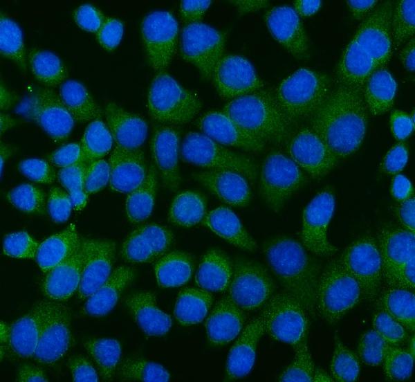
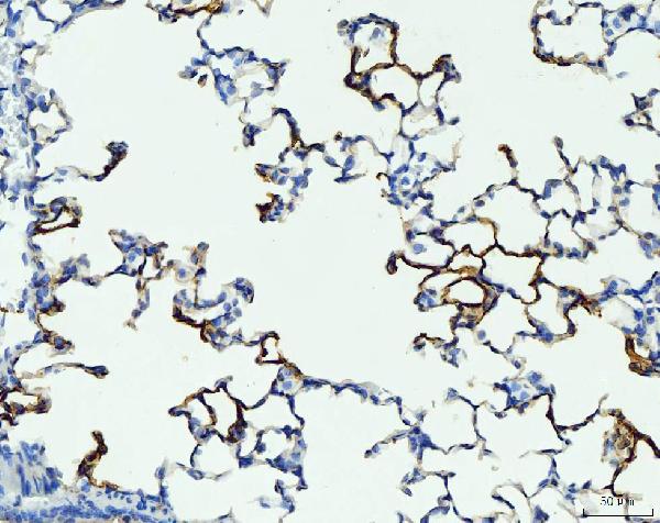
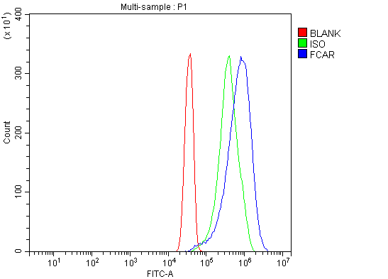
 FCM/FACS (Flow Cytometry)
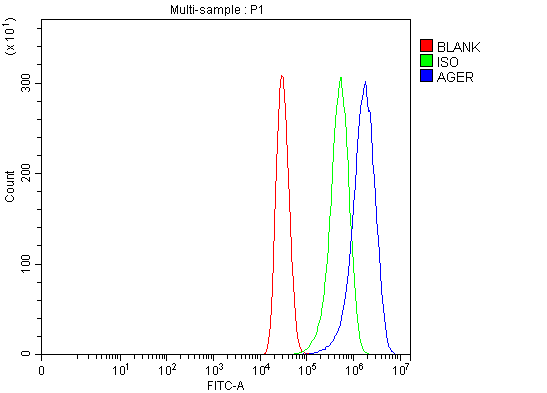
(Figure 4. Flow Cytometry analysis of MCF-7 cells using anti-RAGE/AGER antibody (AAA126307).Overlay histogram showing MCF-7 cells stained with AAA126307 (Blue line). The cells were blocked with 10% normal goat serum. And then incubated with rabbit anti-RAGE/AGER Antibody (AAA126307, 1 ug/1x10^6 cells) for 30 min at 20 degree C. DyLight488 conjugated goat anti-rabbit IgG was used as secondary antibody for 30 minutes at 20 degree C. Isotype control antibody (Green line) was rabbit IgG (1 ug/1x10^6) used under the same conditions. Unlabelled sample (Red line) was also used as a control.)
FCM/FACS (Flow Cytometry)
(Figure 4. Flow Cytometry analysis of MCF-7 cells using anti-RAGE/AGER antibody (AAA126307).Overlay histogram showing MCF-7 cells stained with AAA126307 (Blue line). The cells were blocked with 10% normal goat serum. And then incubated with rabbit anti-RAGE/AGER Antibody (AAA126307, 1 ug/1x10^6 cells) for 30 min at 20 degree C. DyLight488 conjugated goat anti-rabbit IgG was used as secondary antibody for 30 minutes at 20 degree C. Isotype control antibody (Green line) was rabbit IgG (1 ug/1x10^6) used under the same conditions. Unlabelled sample (Red line) was also used as a control.)
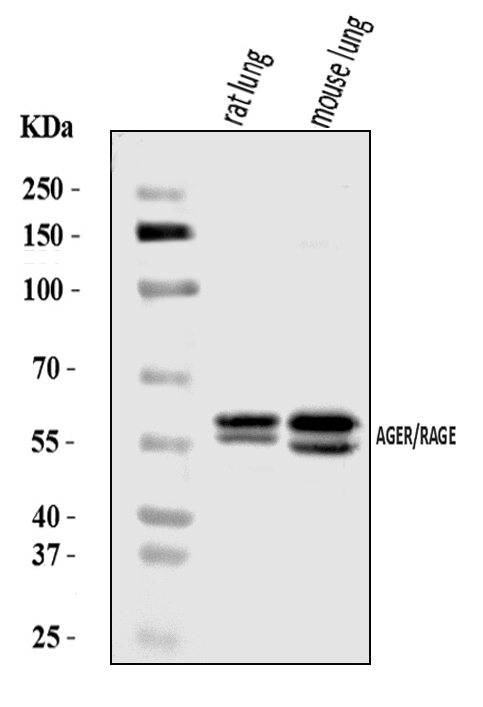
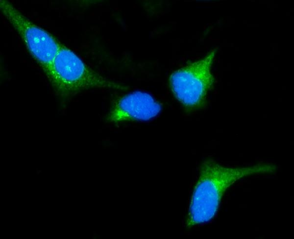
RAGE/AGER, Polyclonal Antibody (Cat# AAA126307)
Full Name
Anti-RAGE/AGER Antibody Picoband
Gene Names
AGER; RAGE
Reactivity
Human, Mouse, Rat
Applications
Western Blot, Immunocytochemistry, Immunohistochemistry, Immunofluorescence, Flow Cytometry
Purity
Immunogen affinity purified.
Pricing
 FCM/FACS (Flow Cytometry)
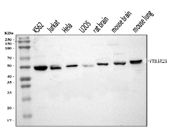
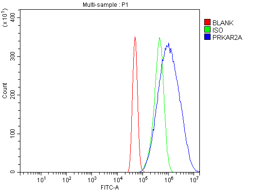
(Figure 3. Flow Cytometry analysis of A431 cells using anti-PKA R2/PKR2/PRKAR2A antibody (AAA126342).Overlay histogram showing A431 cells stained with AAA126342 (Blue line). The cells were blocked with 10% normal goat serum. And then incubated with rabbit anti-PKA R2/PKR2/PRKAR2A Antibody (AAA126342, 1 ug/1x10^6 cells) for 30 min at 20 degree C. DyLight488 conjugated goat anti-rabbit IgG was used as secondary antibody for 30 minutes at 20 degree C. Isotype control antibody (Green line) was rabbit IgG (1 ug/1x10^6) used under the same conditions. Unlabelled sample (Red line) was also used as a control.)
FCM/FACS (Flow Cytometry)
(Figure 3. Flow Cytometry analysis of A431 cells using anti-PKA R2/PKR2/PRKAR2A antibody (AAA126342).Overlay histogram showing A431 cells stained with AAA126342 (Blue line). The cells were blocked with 10% normal goat serum. And then incubated with rabbit anti-PKA R2/PKR2/PRKAR2A Antibody (AAA126342, 1 ug/1x10^6 cells) for 30 min at 20 degree C. DyLight488 conjugated goat anti-rabbit IgG was used as secondary antibody for 30 minutes at 20 degree C. Isotype control antibody (Green line) was rabbit IgG (1 ug/1x10^6) used under the same conditions. Unlabelled sample (Red line) was also used as a control.)
PKA R2/PKR2/PRKAR2A, Polyclonal Antibody (Cat# AAA126342)
Full Name
Anti-PKA R2/PKR2/PRKAR2A Antibody Picoband
Gene Names
PRKAR2A; PKR2; PRKAR2
Reactivity
Human, Mouse, Rat
Applications
Western Blot, Immunocytochemistry, Immunofluorescence, Flow Cytometry
Purity
Immunogen affinity purified.
Pricing
 FCM/FACS (Flow Cytometry)
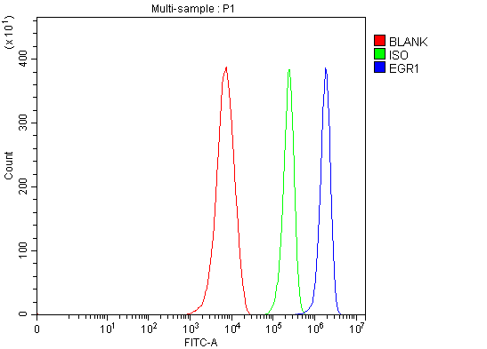
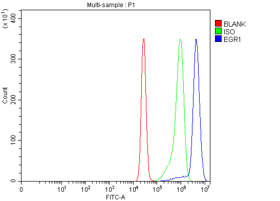
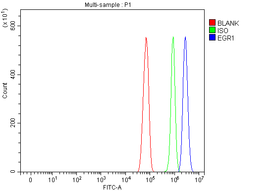
(Figure 4. Flow Cytometry analysis of RH35 cells using anti-EGR1 antibody (AAA126057).Overlay histogram showing RH35 cells stained with AAA126057 (Blue line). The cells were blocked with 10% normal goat serum. And then incubated with rabbit anti-EGR1 Antibody (AAA126057, 1 ug/1x10^6 cells) for 30 min at 20 degree C. DyLight488 conjugated goat anti-rabbit IgG was used as secondary antibody for 30 minutes at 20 degree C. Isotype control antibody (Green line) was rabbit IgG (1 ug/1x10^6) used under the same conditions. Unlabelled sample (Red line) was also used as a control.)
FCM/FACS (Flow Cytometry)
(Figure 4. Flow Cytometry analysis of RH35 cells using anti-EGR1 antibody (AAA126057).Overlay histogram showing RH35 cells stained with AAA126057 (Blue line). The cells were blocked with 10% normal goat serum. And then incubated with rabbit anti-EGR1 Antibody (AAA126057, 1 ug/1x10^6 cells) for 30 min at 20 degree C. DyLight488 conjugated goat anti-rabbit IgG was used as secondary antibody for 30 minutes at 20 degree C. Isotype control antibody (Green line) was rabbit IgG (1 ug/1x10^6) used under the same conditions. Unlabelled sample (Red line) was also used as a control.)
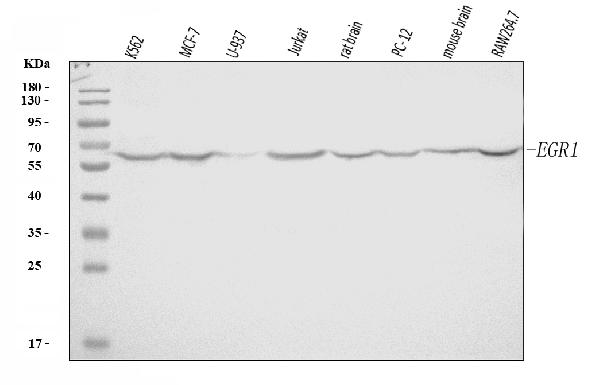
EGR1, Polyclonal Antibody (Cat# AAA126057)
Full Name
Anti-EGR1 Antibody Picoband
Gene Names
EGR1; TIS8; AT225; G0S30; NGFI-A; ZNF225; KROX-24; ZIF-268
Reactivity
Human, Mouse, Rat
Applications
Western Blot, Flow Cytometry
Purity
Immunogen affinity purified.
Pricing
 WB (Western Blot)
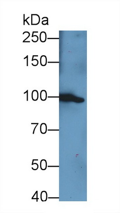
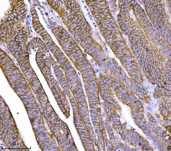
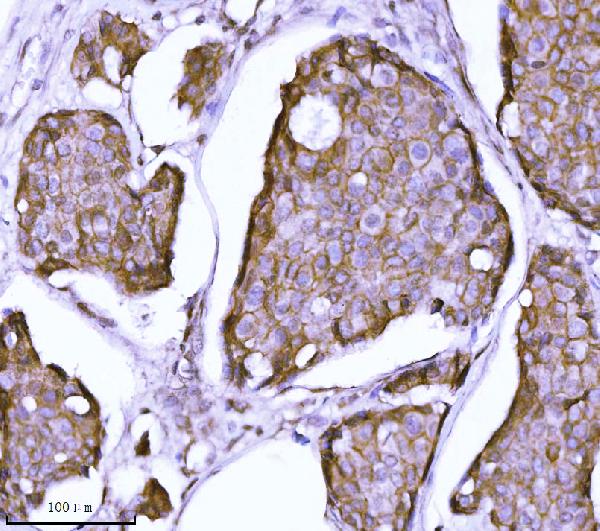
(Figure 1. Western blot analysis of CTNND1 using anti-CTNND1 antibody (AAA126908).Electrophoresis was performed on a 5-20% SDS-PAGE gel at 70V (Stacking gel)/90V (Resolving gel) for 2-3 hours. The sample well of each lane was loaded with 30 ug of sample under reducing conditions.Lane 1: human A549 whole cell lysates,Lane 2: human Hacat whole cell lysates,Lane 3: human RT4 whole cell lysates,Lane 4: human T47D whole cell lysates,Lane 5: rat PC-12 whole cell lysates,Lane 6: rat C6 whole cell lysates.After electrophoresis, proteins were transferred to a nitrocellulose membrane at 150 mA for 50-90 minutes. Blocked the membrane with 5% non-fat milk/TBS for 1.5 hour at RT. The membrane was incubated with mouse anti-CTNND1 antigen affinity purified monoclonal antibody (#AAA126908) at 0.5 ug/mL overnight at 4 degree C, then washed with TBS-0.1%Tween 3 times with 5 minutes each and probed with a goat anti-mouse IgG-HRP secondary antibody at a dilution of 1:10000 for 1.5 hour at RT. The signal is developed using an Enhanced Chemiluminescent detection (ECL) kit with Tanon 5200 system. A specific band was detected for CTNND1 at approximately 100 kDa. The expected band size for CTNND1 is at 108 kDa.)
WB (Western Blot)
(Figure 1. Western blot analysis of CTNND1 using anti-CTNND1 antibody (AAA126908).Electrophoresis was performed on a 5-20% SDS-PAGE gel at 70V (Stacking gel)/90V (Resolving gel) for 2-3 hours. The sample well of each lane was loaded with 30 ug of sample under reducing conditions.Lane 1: human A549 whole cell lysates,Lane 2: human Hacat whole cell lysates,Lane 3: human RT4 whole cell lysates,Lane 4: human T47D whole cell lysates,Lane 5: rat PC-12 whole cell lysates,Lane 6: rat C6 whole cell lysates.After electrophoresis, proteins were transferred to a nitrocellulose membrane at 150 mA for 50-90 minutes. Blocked the membrane with 5% non-fat milk/TBS for 1.5 hour at RT. The membrane was incubated with mouse anti-CTNND1 antigen affinity purified monoclonal antibody (#AAA126908) at 0.5 ug/mL overnight at 4 degree C, then washed with TBS-0.1%Tween 3 times with 5 minutes each and probed with a goat anti-mouse IgG-HRP secondary antibody at a dilution of 1:10000 for 1.5 hour at RT. The signal is developed using an Enhanced Chemiluminescent detection (ECL) kit with Tanon 5200 system. A specific band was detected for CTNND1 at approximately 100 kDa. The expected band size for CTNND1 is at 108 kDa.)
delta 1 Catenin/CAS/CTNND1, Monoclonal Antibody (Cat# AAA126908)
Full Name
Anti-delta 1 Catenin/CAS/CTNND1 Antibody Picoband (monoclonal, 8G7E4)
Gene Names
CTNND1; CAS; p120; CTNND; P120CAS; P120CTN; p120(CAS); p120(CTN)
Reactivity
Human, Mouse, Rat
Applications
Immunofluorescence, Immunocytochemistry, Immunohistochemistry, Western Blot
Purity
Immunogen affinity purified.
Pricing
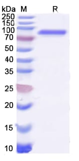
 SDS-PAGE
 SDS-PAGE
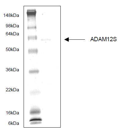
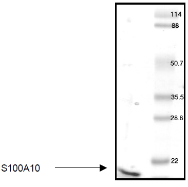
S100A10, Recombinant Protein (Cat# AAA58764)
Full Name
S100A10
Gene Names
S100A10; 42C; P11; p10; GP11; ANX2L; CAL1L; CLP11; Ca[1]; ANX2LG
Purity
>95% by SDS-PAGE
Pricing
 SDS-PAGE
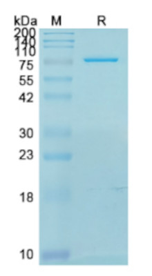
(SDS-PAGE for Recombinant Human NSF Protein)
SDS-PAGE
(SDS-PAGE for Recombinant Human NSF Protein)
NSF, Recombinant Protein (Cat# AAA120775)
Full Name
Recombinant Human NSF Protein, N-His
Gene Names
NSF; SKD2
Applications
Western Blot
Purity
>90% as determined by SDS-PAGE.
Pricing
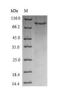 SDS-PAGE
Rab proteins geranylgeranyltransferase component A 1, Recombinant Protein (Cat# AAA113679)
Full Name
Recombinant Human Rab proteins geranylgeranyltransferase component A 1
Gene Names
CHM; TCD; GGTA; REP-1; DXS540; HSD-32
Purity
Greater or equal to 85% purity as determined by SDS-PAGE.
Pricing