
Filters
Clonality
Type
Reactivity
Gene Name
Isotype
Host
Application
Clone
10000 results for "Recombinant Proteins" - showing 5900-5950
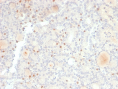
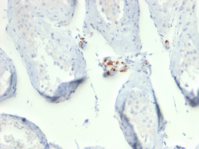
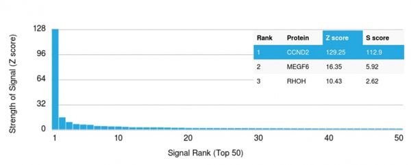 Application Data
(Analysis of Protein Array containing more than 19,000 full-length human proteins using Cyclin D2 Mouse Recombinant Monoclonal Antibody (CCND2/2620). Z- and S- Score: The Z-score represents the strength of a signal that a monoclonal antibody (MAb) (in combination with a fluorescently-tagged anti-IgG secondary antibody) produces when binding to a particular protein on the HuProtTM array. Z-scores are described in units of standard deviations (SD's) above the mean value of all signals generated on that array. If targets on HuProtTM are arranged in descending order of the Z-score, the S-score is the difference (also in units of SD's) between the Z-score. S-score therefore represents the relative target specificity of a MAb to its intended target. A MAb is considered to specific to its intended target, if the MAb has an S-score of at least 2.5. For example, if a MAb binds to protein X with a Z-score of 43 and to protein Y with a Z-score of 14, then the S-score for the binding of that MAb to protein X is equal to 29.)
Application Data
(Analysis of Protein Array containing more than 19,000 full-length human proteins using Cyclin D2 Mouse Recombinant Monoclonal Antibody (CCND2/2620). Z- and S- Score: The Z-score represents the strength of a signal that a monoclonal antibody (MAb) (in combination with a fluorescently-tagged anti-IgG secondary antibody) produces when binding to a particular protein on the HuProtTM array. Z-scores are described in units of standard deviations (SD's) above the mean value of all signals generated on that array. If targets on HuProtTM are arranged in descending order of the Z-score, the S-score is the difference (also in units of SD's) between the Z-score. S-score therefore represents the relative target specificity of a MAb to its intended target. A MAb is considered to specific to its intended target, if the MAb has an S-score of at least 2.5. For example, if a MAb binds to protein X with a Z-score of 43 and to protein Y with a Z-score of 14, then the S-score for the binding of that MAb to protein X is equal to 29.)
Cyclin D2, Monoclonal Antibody (Cat# AAA214845)
Full Name
Cyclin D2
Gene Names
CCND2; MPPH3; KIAK0002
Reactivity
Human. Others not known.
Applications
Immunohistochemistry
Purity
Purified Ab with BSA and Azide at 200ug/ml OR Purified Ab WITHOUT BSA and Azide at 1.0mg/ml
Pricing
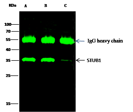
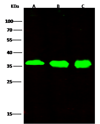
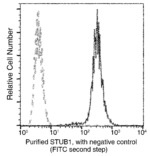 FCM/FACS (Flow Cytometry)
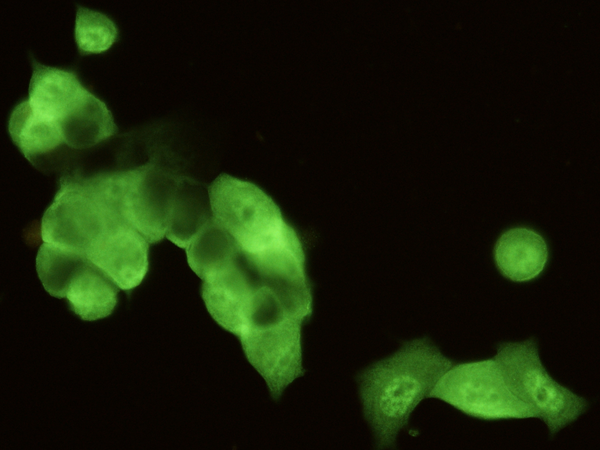
(Flow cytometric analysis of Human STUB1 expression on Hela cells. The cells were treated according to manufacturer's manual (BD Pharmingen'), stained with purified anti-Human STUB1, then a FITC-conjugated second step antibody. The fluorescence histograms were derived from gated events with the forward and side light-scatter characteristics of intact cells.)
FCM/FACS (Flow Cytometry)
(Flow cytometric analysis of Human STUB1 expression on Hela cells. The cells were treated according to manufacturer's manual (BD Pharmingen'), stained with purified anti-Human STUB1, then a FITC-conjugated second step antibody. The fluorescence histograms were derived from gated events with the forward and side light-scatter characteristics of intact cells.)
STUB1, Monoclonal Antibody (Cat# AAA255588)
Full Name
Recombinant Anti-STUB1 Antibody, Rabbit Monoclonal
Reactivity
Human
Applications
Immunoprecipitation, Immunofluorescence, Immunocytochemistry, Flow Cytometry, Western Blot
Purity
Protein A
Pricing


 WB (Western Blot)
(Western Blot; Sample: Rat Liver lysate; Primary Ab: 2ug/ml Mouse AntiHuman Ubiquitin Antibody Second Ab: 0.2ug/mL HRPLinked Caprine AntiMouse IgG Polyclonal Antibody (Catalog: SAA544Mu19))
WB (Western Blot)
(Western Blot; Sample: Rat Liver lysate; Primary Ab: 2ug/ml Mouse AntiHuman Ubiquitin Antibody Second Ab: 0.2ug/mL HRPLinked Caprine AntiMouse IgG Polyclonal Antibody (Catalog: SAA544Mu19))
Ubiquitin (Ub), Monoclonal Antibody (Cat# AAA151499)
Full Name
Monoclonal Antibody to Ubiquitin (Ub)
Reactivity
Human, Rat, Pig
Applications
Immunoprecipitation, Immunocytochemistry, Immunohistochemistry, Western Blot
Purity
Protein A + Protein G affinity chromatography
Pricing
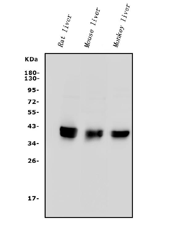
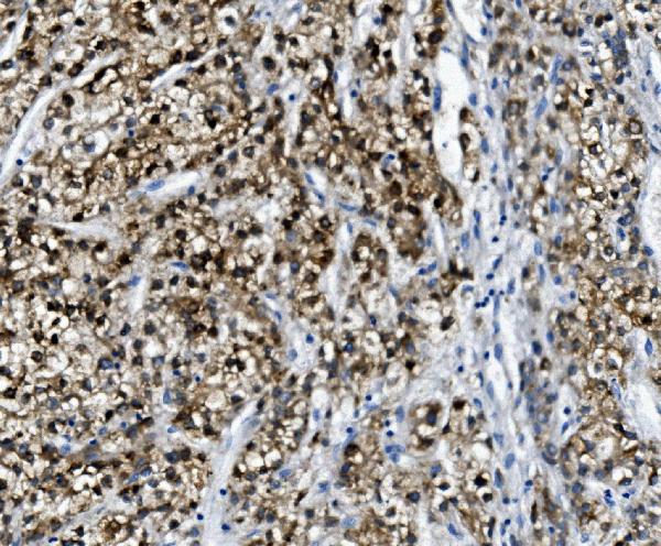 IHC (Immunohiostchemistry)
(Figure 2. IHC analysis of PON1 using anti-PON1 antibody (AAA125554).PON1 was detected in paraffin-embedded section of human liver cancer tissue. Heat mediated antigen retrieval was performed in EDTA buffer (pH8. 0, epitope retrieval solution). The tissue section was blocked with 10% goat serum. The tissue section was then incubated with 2μg/ml rabbit anti-PON1 Antibody (AAA125554) overnight at 4 degree C. Biotinylated goat anti-rabbit IgG was used as secondary antibody and incubated for 30 minutes at 37 degree C. The tissue section was developed using Strepavidin-Biotin-Complex (SABC) with DAB as the chromogen.)
IHC (Immunohiostchemistry)
(Figure 2. IHC analysis of PON1 using anti-PON1 antibody (AAA125554).PON1 was detected in paraffin-embedded section of human liver cancer tissue. Heat mediated antigen retrieval was performed in EDTA buffer (pH8. 0, epitope retrieval solution). The tissue section was blocked with 10% goat serum. The tissue section was then incubated with 2μg/ml rabbit anti-PON1 Antibody (AAA125554) overnight at 4 degree C. Biotinylated goat anti-rabbit IgG was used as secondary antibody and incubated for 30 minutes at 37 degree C. The tissue section was developed using Strepavidin-Biotin-Complex (SABC) with DAB as the chromogen.)
PON1, Polyclonal Antibody (Cat# AAA125554)
Full Name
Anti-PON1 Antibody
Gene Names
PON1; ESA; PON; MVCD5
Reactivity
Human, Mouse, Rat, Monkey
Applications
Direct ELISA, Immunohistochemistry, Western Blot
Purity
Immunogen affinity purified.
Pricing
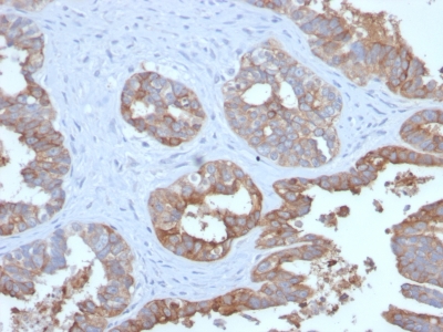
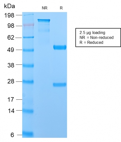 SDS-PAGE
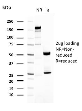
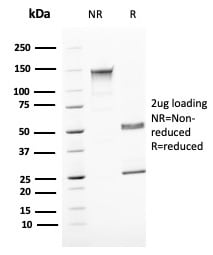
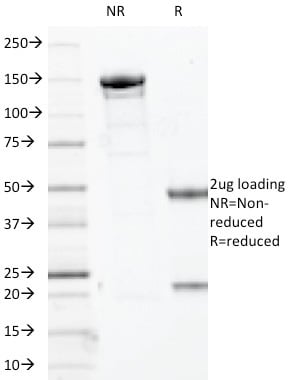
(SDS-PAGE Analysis Purified MUC1 Recombinant Rabbit Monoclonal Antibody (MUC1/2729R). Confirmation of Purity and Integrity of Antibody.)
SDS-PAGE
(SDS-PAGE Analysis Purified MUC1 Recombinant Rabbit Monoclonal Antibody (MUC1/2729R). Confirmation of Purity and Integrity of Antibody.)
MUC1/CA15-3/EMA/CD227, Monoclonal Antibody (Cat# AAA214782)
Full Name
MUC1/CA15-3/EMA/CD227 (Epithelial Marker)
Gene Names
MUC1; EMA; MCD; PEM; PUM; KL-6; MAM6; MCKD; PEMT; CD227; H23AG; MCKD1; MUC-1; ADMCKD; ADMCKD1; CA 15-3; MUC-1/X; MUC1/ZD; MUC-1/SEC
Reactivity
Human. Others not known.
Applications
Immunohistochemistry
Purity
Purified Ab with BSA and Azide at 200ug/ml OR Purified Ab WITHOUT BSA and Azide at 1.0mg/ml
Pricing
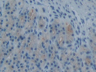
 IHC (Immunohiostchemistry)
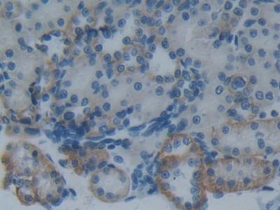
(DAB staining on IHCP;Sample: Rat Kidney Tissue; Primary Ab: 30ug/ml Mouse AntiRat PAI2 AntibodySecond Ab: 2ug/mL HRPLinked Caprine AntiMouse IgG Polyclonal Antibody(Catalog: SAA544Mu19))
IHC (Immunohiostchemistry)
(DAB staining on IHCP;Sample: Rat Kidney Tissue; Primary Ab: 30ug/ml Mouse AntiRat PAI2 AntibodySecond Ab: 2ug/mL HRPLinked Caprine AntiMouse IgG Polyclonal Antibody(Catalog: SAA544Mu19))
Plasminogen Activator Inhibitor 2 (PAI2), Monoclonal Antibody (Cat# AAA151553)
Full Name
Monoclonal Antibody to Plasminogen Activator Inhibitor 2 (PAI2)
Gene Names
Serpinb2; Pai2a
Reactivity
Rat
Applications
Immunoprecipitation, Immunocytochemistry, Immunohistochemistry, Western Blot
Purity
Protein A + Protein G affinity chromatography
Pricing
 Application Data
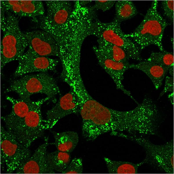
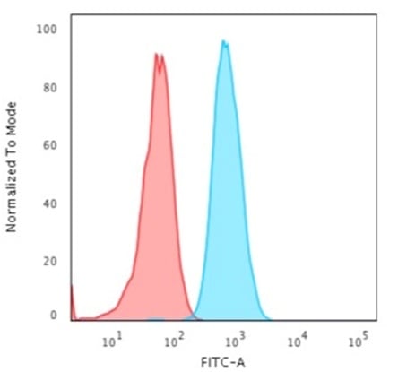
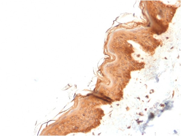
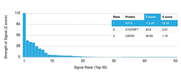
(Analysis of Protein Array containing more than 19,000 full-length human proteins using Cytokeratin 4 Mouse Monoclonal Antibody (KRT4/2804). Z- and S- Score: The Z-score represents the strength of a signal that a monoclonal antibody (MAb) (in combination with a fluorescently-tagged anti-IgG secondary antibody) produces when binding to a particular protein on the HuProtTM array. Z-scores are described in units of standard deviations (SD's) above the mean value of all signals generated on that array. If targets on HuProtTM are arranged in descending order of the Z-score, the S-score is the difference (also in units of SD's) between the Z-score. S-score therefore represents the relative target specificity of a MAb to its intended target. A MAb is considered to specific to its intended target, if the MAb has an S-score of at least 2.5. For example, if a MAb binds to protein X with a Z-score of 43 and to protein Y with a Z-score of 14, then the S-score for the binding of that MAb to protein X is equal to 29.)
Application Data
(Analysis of Protein Array containing more than 19,000 full-length human proteins using Cytokeratin 4 Mouse Monoclonal Antibody (KRT4/2804). Z- and S- Score: The Z-score represents the strength of a signal that a monoclonal antibody (MAb) (in combination with a fluorescently-tagged anti-IgG secondary antibody) produces when binding to a particular protein on the HuProtTM array. Z-scores are described in units of standard deviations (SD's) above the mean value of all signals generated on that array. If targets on HuProtTM are arranged in descending order of the Z-score, the S-score is the difference (also in units of SD's) between the Z-score. S-score therefore represents the relative target specificity of a MAb to its intended target. A MAb is considered to specific to its intended target, if the MAb has an S-score of at least 2.5. For example, if a MAb binds to protein X with a Z-score of 43 and to protein Y with a Z-score of 14, then the S-score for the binding of that MAb to protein X is equal to 29.)
Cytokeratin 4 (KRT4), Monoclonal Antibody (Cat# AAA215056)
Full Name
Cytokeratin 4 (KRT4)
Gene Names
KRT4; K4; CK4; CK-4; CYK4; WSN1
Reactivity
Human
Applications
Immunohistochemistry, Immunofluorescence, Flow Cytometry
Purity
Purified Ab with BSA and Azide at 200ug/ml OR Purified Ab WITHOUT BSA and Azide at 1.0mg/ml
Pricing
 Bioactivity
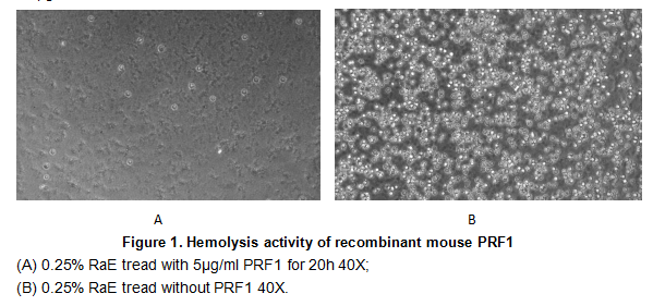
(Perforin 1 (PRF1) is a pore forming cytolytic protein found in the granules of cytotoxic T lymphocytes (CTLs) and NK cells. Upon degranulation, perforin binds to the target cell's plasma membrane, and oligomerises in a Ca2 dependent manner to form pores on the target cell. The pore formed allows for the passive diffusion of a family of pro-apoptotic proteases, known as the granzymes, into the target cell. The activity of recombinant PRF1 was measured by lysis of erythrocytes using a hemolysis assay. A general procedure is as fllows: two-fold dilute the recombinant mouse PRF1 with 0.9% NaCl, add 50ul a serial dilution of PRF1, 10ul 0.1M CaCl2 to each well, then add 50ul 0.25% rabbit erythrocyte (RaE) to each well and mixed gently. Add 50ul 0.9% NaCl to replace PRF1 in control wells. The plate is incubated for 20 hours at 37 degree C, 5% CO2. The results are shown in Figure 1. It was obvious that the minimal effective concentration of PRF1 is 5ug/ml.)
Bioactivity
(Perforin 1 (PRF1) is a pore forming cytolytic protein found in the granules of cytotoxic T lymphocytes (CTLs) and NK cells. Upon degranulation, perforin binds to the target cell's plasma membrane, and oligomerises in a Ca2 dependent manner to form pores on the target cell. The pore formed allows for the passive diffusion of a family of pro-apoptotic proteases, known as the granzymes, into the target cell. The activity of recombinant PRF1 was measured by lysis of erythrocytes using a hemolysis assay. A general procedure is as fllows: two-fold dilute the recombinant mouse PRF1 with 0.9% NaCl, add 50ul a serial dilution of PRF1, 10ul 0.1M CaCl2 to each well, then add 50ul 0.25% rabbit erythrocyte (RaE) to each well and mixed gently. Add 50ul 0.9% NaCl to replace PRF1 in control wells. The plate is incubated for 20 hours at 37 degree C, 5% CO2. The results are shown in Figure 1. It was obvious that the minimal effective concentration of PRF1 is 5ug/ml.)
Perforin 1 (PRF1), Active Protein (Cat# AAA152181)
Full Name
Active Perforin 1 (PRF1)
Gene Names
Prf1; Pfn; Pfp; Prf-1
Applications
Activity Assay, Cell Culture
Purity
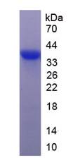
Greater than 90% by SDS-PAGE
Pricing
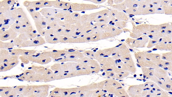 IHC (Immunohiostchemistry)
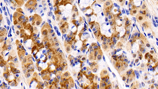
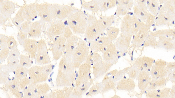
(DAB staining on IHCP;Sample: Human Cardiac Muscle Tissue; Primary Ab: 30ug/ml Mouse AntiHuman NTProANP AntibodySecond Ab: 2ug/mL HRPLinked Caprine AntiMouse IgG Polyclonal Antibody(Catalog: SAA544Mu19))
IHC (Immunohiostchemistry)
(DAB staining on IHCP;Sample: Human Cardiac Muscle Tissue; Primary Ab: 30ug/ml Mouse AntiHuman NTProANP AntibodySecond Ab: 2ug/mL HRPLinked Caprine AntiMouse IgG Polyclonal Antibody(Catalog: SAA544Mu19))
NTerminal ProAtrial Natriuretic Peptide (NTProANP), Monoclonal Antibody (Cat# AAA151545)
Full Name
Monoclonal Antibody to NTerminal ProAtrial Natriuretic Peptide (NTProANP)
Gene Names
NPPA; ANF; ANP; PND; ATFB6; CDD-ANF
Reactivity
Human, Pig
Applications
Immunoprecipitation, Immunocytochemistry, Immunohistochemistry, Western Blot
Purity
Protein A + Protein G affinity chromatography
Pricing
 FCM/FACS (Flow Cytometry)
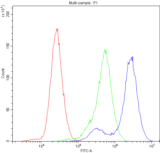
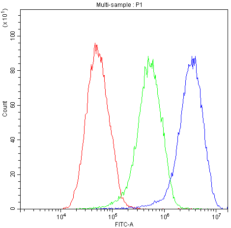
(Figure 3. Flow Cytometry analysis of HEPG2 cells using anti-Factor I antibody (AAA46611).Overlay histogram showing HEPG2 cells stained with AAA46611 (Blue line).The cells were blocked with 10% normal goat serum. And then incubated with rabbit anti-Factor I Antibody (AAA46611,1ug/1x10^6 cells) for 30 min at 20 degree C. DyLight®488 conjugated goat anti-rabbit IgG (5-10ug/1x10^6 cells) was used as secondary antibody for 30 minutes at 20 degree C. Isotype control antibody (Green line) was rabbit IgG (1ug/1x106) used under the same conditions. Unlabelled sample (Red line) was also used as a control.)
FCM/FACS (Flow Cytometry)
(Figure 3. Flow Cytometry analysis of HEPG2 cells using anti-Factor I antibody (AAA46611).Overlay histogram showing HEPG2 cells stained with AAA46611 (Blue line).The cells were blocked with 10% normal goat serum. And then incubated with rabbit anti-Factor I Antibody (AAA46611,1ug/1x10^6 cells) for 30 min at 20 degree C. DyLight®488 conjugated goat anti-rabbit IgG (5-10ug/1x10^6 cells) was used as secondary antibody for 30 minutes at 20 degree C. Isotype control antibody (Green line) was rabbit IgG (1ug/1x106) used under the same conditions. Unlabelled sample (Red line) was also used as a control.)
Factor I, Polyclonal Antibody (Cat# AAA46611)
Full Name
Anti-Factor I Antibody
Gene Names
CFI; FI; IF; KAF; AHUS3; ARMD13; C3BINA; C3b-INA
Reactivity
Human, Rat
No cross reactivity with other proteins.
No cross reactivity with other proteins.
Applications
Western Blot
Purity
Immunogen affinity purified.
Pricing
 IHC (Immunohistochemistry)
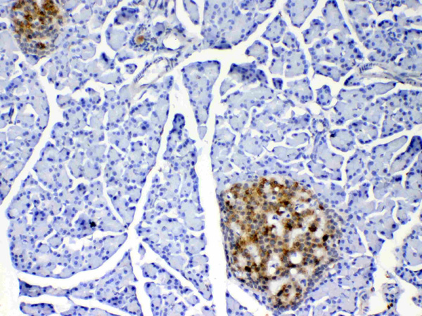
(Figure 4. IHC analysis of IL17 using anti-IL17 antibody (AAA124572).IL17 was detected in paraffin-embedded section of rat pancreas tissue. Heat mediated antigen retrieval was performed in citrate buffer (pH6, epitope retrieval solution) for 20 mins. The tissue section was blocked with 10% goat serum. The tissue section was then incubated with 1ug/ml rabbit anti-IL17 Antibody (AAA124572) overnight at 4 degree C. Biotinylated goat anti-rabbit IgG was used as secondary antibody and incubated for 30 minutes at 37 degree C. The tissue section was developed using Strepavidin-Biotin-Complex (SABC) with DAB as the chromogen.)
IHC (Immunohistochemistry)
(Figure 4. IHC analysis of IL17 using anti-IL17 antibody (AAA124572).IL17 was detected in paraffin-embedded section of rat pancreas tissue. Heat mediated antigen retrieval was performed in citrate buffer (pH6, epitope retrieval solution) for 20 mins. The tissue section was blocked with 10% goat serum. The tissue section was then incubated with 1ug/ml rabbit anti-IL17 Antibody (AAA124572) overnight at 4 degree C. Biotinylated goat anti-rabbit IgG was used as secondary antibody and incubated for 30 minutes at 37 degree C. The tissue section was developed using Strepavidin-Biotin-Complex (SABC) with DAB as the chromogen.)
IL17, Polyclonal Antibody (Cat# AAA124572)
Full Name
Anti-IL17 Picoband Antibody
Gene Names
Il17a; Il17; Ctla8; IL-17; Ctla-8; IL-17A
Reactivity
Human, Mouse, Rat
No cross reactivity with other proteins.
No cross reactivity with other proteins.
Applications
Western Blot, Immunohistochemistry
Purity
Immunogen affinity purified
Pricing
 Application Data
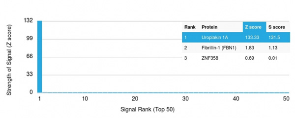
(Analysis of Protein Array containing more than 19,000 full-length human proteins using Uroplakin 1A Mouse Monoclonal Antibody (UPK1A/2921) Z- and S- Score: The Z-score represents the strength of a signal that a monoclonal antibody (MAb) (in combination with a fluorescently-tagged anti-IgG secondary antibody) produces when binding to a particular protein on the HuProtTM array. Z-scores are described in units of standard deviations (SD's) above the mean value of all signals generated on that array. If targets on HuProtTM are arranged in descending order of the Z-score, the S-score is the difference (also in units of SD's) between the Z-score. S-score therefore represents the relative target specificity of a MAb to its intended target. A MAb is considered to specific to its intended target, if the MAb has an S-score of at least 2.5. For example, if a MAb binds to protein X with a Z-score of 43 and to protein Y with a Z-score of 14, then the S-score for the binding of that MAb to protein X is equal to 29.)
Application Data
(Analysis of Protein Array containing more than 19,000 full-length human proteins using Uroplakin 1A Mouse Monoclonal Antibody (UPK1A/2921) Z- and S- Score: The Z-score represents the strength of a signal that a monoclonal antibody (MAb) (in combination with a fluorescently-tagged anti-IgG secondary antibody) produces when binding to a particular protein on the HuProtTM array. Z-scores are described in units of standard deviations (SD's) above the mean value of all signals generated on that array. If targets on HuProtTM are arranged in descending order of the Z-score, the S-score is the difference (also in units of SD's) between the Z-score. S-score therefore represents the relative target specificity of a MAb to its intended target. A MAb is considered to specific to its intended target, if the MAb has an S-score of at least 2.5. For example, if a MAb binds to protein X with a Z-score of 43 and to protein Y with a Z-score of 14, then the S-score for the binding of that MAb to protein X is equal to 29.)
Uroplakin 1A, Monoclonal Antibody (Cat# AAA214887)
Full Name
Uroplakin 1A (Urothelial Differentiation Marker)
Gene Names
UPK1A; UP1A; UPIA; UPKA; TSPAN21
Reactivity
Human
Applications
Immunohistochemistry
Purity
Purified Ab with BSA and Azide at 200ug/ml OR Purified Ab WITHOUT BSA and Azide at 1.0mg/ml
Pricing
 Application Data
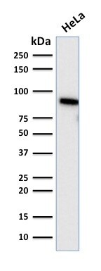
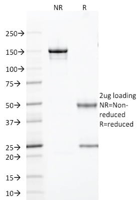
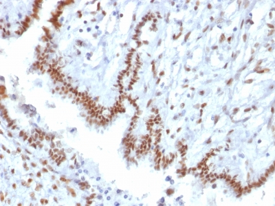
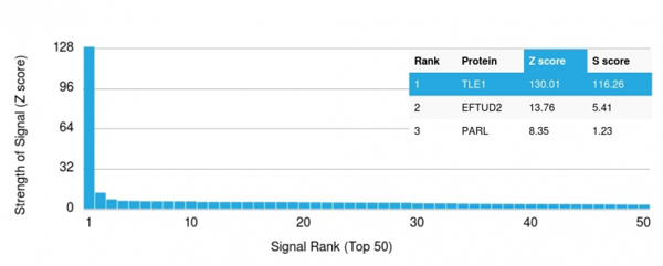
(Analysis of Protein Array containing more than 19, 000 full-length human proteins using TLE1 Mouse Monoclonal Antibody (TLE1/2085).Z- and S- Score: The Z-score represents the strength of a signal that a monoclonal antibody (MAb) (in combination with a fluorescently-tagged anti-IgG secondary antibody) produces when binding to a particular protein on the HuProtTM array. Z-scores are described in units of standard deviations (SD's) above the mean value of all signals generated on that array. If targets on HuProtTM are arranged in descending order of the Z-score, the S-score is the difference (also in units of SD's) between the Z-score. S-score therefore represents the relative target specificity of a MAb to its intended target. A MAb is considered to specific to its intended target, if the MAb has an S-score of at least 2.5. For example, if a MAb binds to protein X with a Z-score of 43 and to protein Y with a Z-score of 14, then the S-score for the binding of that MAb to protein X is equal to 29.)
Application Data
(Analysis of Protein Array containing more than 19, 000 full-length human proteins using TLE1 Mouse Monoclonal Antibody (TLE1/2085).Z- and S- Score: The Z-score represents the strength of a signal that a monoclonal antibody (MAb) (in combination with a fluorescently-tagged anti-IgG secondary antibody) produces when binding to a particular protein on the HuProtTM array. Z-scores are described in units of standard deviations (SD's) above the mean value of all signals generated on that array. If targets on HuProtTM are arranged in descending order of the Z-score, the S-score is the difference (also in units of SD's) between the Z-score. S-score therefore represents the relative target specificity of a MAb to its intended target. A MAb is considered to specific to its intended target, if the MAb has an S-score of at least 2.5. For example, if a MAb binds to protein X with a Z-score of 43 and to protein Y with a Z-score of 14, then the S-score for the binding of that MAb to protein X is equal to 29.)
TLE1, Monoclonal Antibody (Cat# AAA214701)
Full Name
TLE1 (Synovial Sarcoma Marker)
Gene Names
TLE1; ESG; ESG1; GRG1
Reactivity
Human. Others not known.
Applications
Immunohistochemistry
Pricing


 Application Data
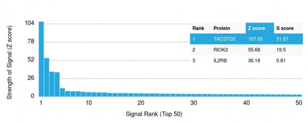
(Analysis of Protein Array containing >19, 000 full-length human proteins using TACSTD2 Mouse Monoclonal Antibody (TACSTD2/2151) Z- and S- Score: The Z-score represents the strength of a signal that a monoclonal antibody (MAb) (in combination with a fluorescently-tagged anti-IgG secondary antibody) produces when binding to a particular protein on the HuProtTM array. Z-scores are described in units of standard deviations (SD's) above the mean value of all signals generated on that array. If targets on HuProtTM are arranged in descending order of the Z-score, the S-score is the difference (also in units of SD's) between the Z-score. S-score therefore represents the relative target specificity of a MAb to its intended target. A MAb is considered to specific to its intended target, if the MAb has an S-score of at least 2.5. For example, if a MAb binds to protein X with a Z-score of 43 and to protein Y with a Z-score of 14, then the S-score for the binding of that MAb to protein X is equal to 29.)
Application Data
(Analysis of Protein Array containing >19, 000 full-length human proteins using TACSTD2 Mouse Monoclonal Antibody (TACSTD2/2151) Z- and S- Score: The Z-score represents the strength of a signal that a monoclonal antibody (MAb) (in combination with a fluorescently-tagged anti-IgG secondary antibody) produces when binding to a particular protein on the HuProtTM array. Z-scores are described in units of standard deviations (SD's) above the mean value of all signals generated on that array. If targets on HuProtTM are arranged in descending order of the Z-score, the S-score is the difference (also in units of SD's) between the Z-score. S-score therefore represents the relative target specificity of a MAb to its intended target. A MAb is considered to specific to its intended target, if the MAb has an S-score of at least 2.5. For example, if a MAb binds to protein X with a Z-score of 43 and to protein Y with a Z-score of 14, then the S-score for the binding of that MAb to protein X is equal to 29.)
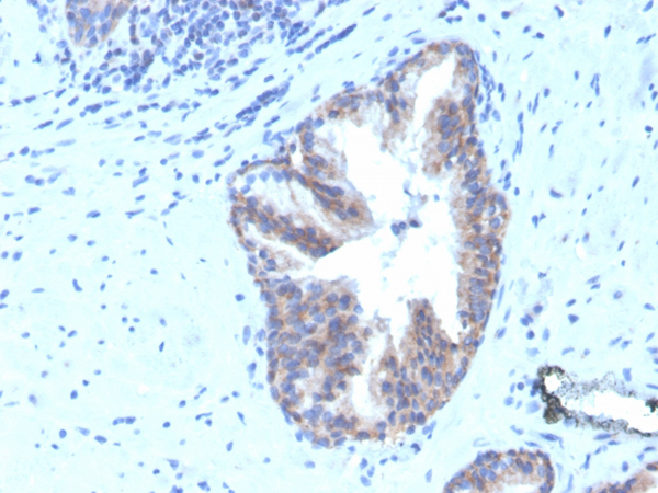
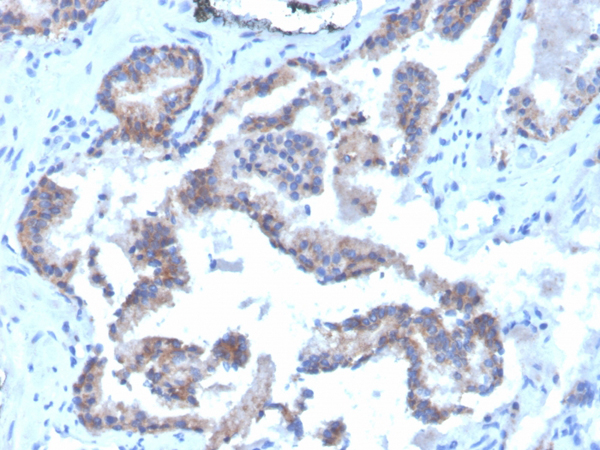
TACSTD2/TROP2, Monoclonal Antibody (Cat# AAA214662)
Full Name
TACSTD2/TROP2 (Epithelial Marker)
Gene Names
TACSTD2; EGP1; GP50; M1S1; EGP-1; TROP2; GA7331; GA733-1
Reactivity
Human. Others not known.
Applications
Immunohistochemistry, Western Blot
Pricing
 Application Data
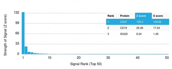
(Analysis of Protein Array containing more than 19,000 full-length human proteins using CD47 Mouse Monoclonal Antibody (CD47/3019) Z- and S- Score: The Z-score represents the strength of a signal that a monoclonal antibody (MAb) (in combination with a fluorescently-tagged anti-IgG secondary antibody) produces when binding to a particular protein on the HuProtTM array. Z-scores are described in units of standard deviations (SD's) above the mean value of all signals generated on that array. If targets on HuProtTM are arranged in descending order of the Z-score, the S-score is the difference (also in units of SD's) between the Z-score. S-score therefore represents the relative target specificity of a MAb to its intended target. A MAb is considered to specific to its intended target, if the MAb has an S-score of at least 2.5. For example, if a MAb binds to protein X with a Z-score of 43 and to protein Y with a Z-score of 14, then the S-score for the binding of that MAb to protein X is equal to 29.)
Application Data
(Analysis of Protein Array containing more than 19,000 full-length human proteins using CD47 Mouse Monoclonal Antibody (CD47/3019) Z- and S- Score: The Z-score represents the strength of a signal that a monoclonal antibody (MAb) (in combination with a fluorescently-tagged anti-IgG secondary antibody) produces when binding to a particular protein on the HuProtTM array. Z-scores are described in units of standard deviations (SD's) above the mean value of all signals generated on that array. If targets on HuProtTM are arranged in descending order of the Z-score, the S-score is the difference (also in units of SD's) between the Z-score. S-score therefore represents the relative target specificity of a MAb to its intended target. A MAb is considered to specific to its intended target, if the MAb has an S-score of at least 2.5. For example, if a MAb binds to protein X with a Z-score of 43 and to protein Y with a Z-score of 14, then the S-score for the binding of that MAb to protein X is equal to 29.)
CD47/IAP, Monoclonal Antibody (Cat# AAA215494)
Full Name
CD47/IAP (Integrin Associated Protein)
Gene Names
CD47; IAP; OA3; MER6
Reactivity
Human
Applications
Immunohistochemistry, Immunofluorescence, Flow Cytometry
Purity
Purified Ab conjugated to Biotin
Pricing
 Application Data
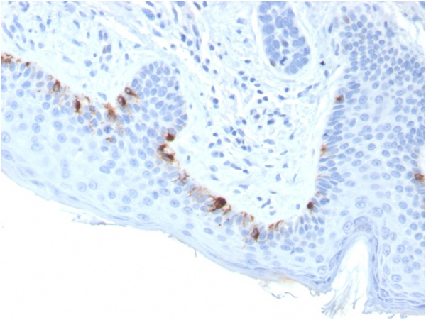
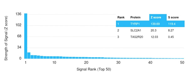
(Analysis of Protein Array containing more than 19,000 full-length human proteins using TYRP1-Monospecific Mouse Monoclonal Antibody (TYRP1/3283) Z- and S- Score: The Z-score represents the strength of a signal that a monoclonal antibody (Monoclonal Antibody) (in combination with a fluorescently-tagged anti-IgG secondary antibody) produces when binding to a particular protein on the HuProtTM array. Z-scores are described in units of standard deviations (SD’s) above the mean value of all signals generated on that array. If targets on HuProtTM are arranged in descending order of the Z-score, the S-score is the difference (also in units of SD’s) between the Z-score. S-score therefore represents the relative target specificity of a Monoclonal Antibody to its intended target. A Monoclonal Antibody is considered to specific to its intended target, if the Monoclonal Antibody has an S-score of at least 2.5. For example, if a Monoclonal Antibody binds to protein X with a Z-score of 43 and to protein Y with a Z-score of 14, then the S-score for the binding of that Monoclonal Antibody to protein X is equal to 29.)
Application Data
(Analysis of Protein Array containing more than 19,000 full-length human proteins using TYRP1-Monospecific Mouse Monoclonal Antibody (TYRP1/3283) Z- and S- Score: The Z-score represents the strength of a signal that a monoclonal antibody (Monoclonal Antibody) (in combination with a fluorescently-tagged anti-IgG secondary antibody) produces when binding to a particular protein on the HuProtTM array. Z-scores are described in units of standard deviations (SD’s) above the mean value of all signals generated on that array. If targets on HuProtTM are arranged in descending order of the Z-score, the S-score is the difference (also in units of SD’s) between the Z-score. S-score therefore represents the relative target specificity of a Monoclonal Antibody to its intended target. A Monoclonal Antibody is considered to specific to its intended target, if the Monoclonal Antibody has an S-score of at least 2.5. For example, if a Monoclonal Antibody binds to protein X with a Z-score of 43 and to protein Y with a Z-score of 14, then the S-score for the binding of that Monoclonal Antibody to protein X is equal to 29.)
Tyrosinase-Related Protein-1 (TYRP-1), Monoclonal Antibody (Cat# AAA215244)
Full Name
Tyrosinase-Related Protein-1 (TYRP-1) (Melanoma Marker)
Gene Names
TYRP1; TRP; CAS2; CATB; GP75; OCA3; TRP1; TYRP; b-PROTEIN
Reactivity
Human
Applications
Immunohistochemistry
Purity
Purified Ab with BSA and Azide at 200ug/ml OR Purified Ab WITHOUT BSA and Azide at 1.0mg/ml
Pricing
 IF (Immunofluorescence)
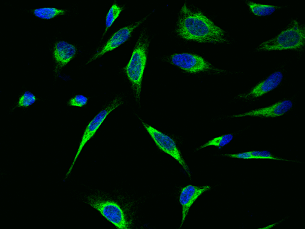
(Immunofluorescence staining of VIM-His in HeLa cells. Cells were fixed with 4% PFA, permeabilzed with 0.3% Triton X-100 in PBS,blocked with 10% serum, and incubated with rabbit anti-Human VIM-His polyclonal antibody (dilution ratio 1:100) at 4? overnight. Then cells were stained with the Alexa Fluor488-conjugated Goat Anti-rabbit IgG secondary antibody (green) and counterstained with DAPI (blue).Positive staining was localized to Cytoplasm.)
IF (Immunofluorescence)
(Immunofluorescence staining of VIM-His in HeLa cells. Cells were fixed with 4% PFA, permeabilzed with 0.3% Triton X-100 in PBS,blocked with 10% serum, and incubated with rabbit anti-Human VIM-His polyclonal antibody (dilution ratio 1:100) at 4? overnight. Then cells were stained with the Alexa Fluor488-conjugated Goat Anti-rabbit IgG secondary antibody (green) and counterstained with DAPI (blue).Positive staining was localized to Cytoplasm.)
Vimentin, Polyclonal Antibody (Cat# AAA258713)
Full Name
Anti-Vimentin Antibody, Rabbit Polyclonal
Reactivity
Human
Applications
Immunoprecipitation, Immunofluorescence, Immunocytochemistry, Immunohistochemistry, Western Blot
Purity
Protein A & Antigen Affinity
Pricing
 Bioactivity
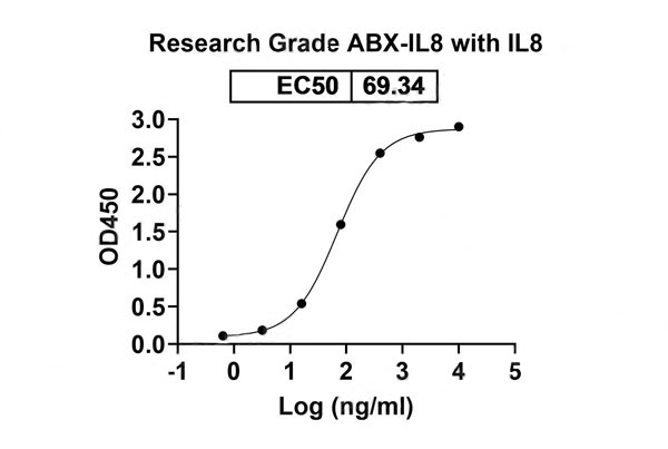
(Detects ABX-IL8 in indirect ELISAs.)
Bioactivity
(Detects ABX-IL8 in indirect ELISAs.)
IL8/CXCL8, Recombinant Protein (Cat# AAA120521)
Full Name
Recombinant Human IL8/CXCL8, C-His
Gene Names
IL8; NAF; GCP1; LECT; LUCT; NAP1; CXCL8; GCP-1; LYNAP; MDNCF; MONAP; NAP-1
Applications
Western Blot
Purity
>90% as determined by SDS-PAGE
Pricing
 Application Data
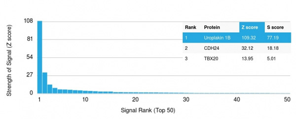
(Analysis of Protein Array containing more than 19,000 full-length human proteins using Uroplakin 1B Mouse Monoclonal Antibody (UPK1B/3081) Z- and S- Score: The Z-score represents the strength of a signal that a monoclonal antibody (MAb) (in combination with a fluorescently-tagged anti-IgG secondary antibody) produces when binding to a particular protein on the HuProtTM array. Z-scores are described in units of standard deviations (SD's) above the mean value of all signals generated on that array. If targets on HuProtTM are arranged in descending order of the Z-score, the S-score is the difference (also in units of SD's) between the Z-score. S-score therefore represents the relative target specificity of a MAb to its intended target. A MAb is considered to specific to its intended target, if the MAb has an S-score of at least 2.5. For example, if a MAb binds to protein X with a Z-score of 43 and to protein Y with a Z-score of 14, then the S-score for the binding of that MAb to protein X is equal to 29.)
Application Data
(Analysis of Protein Array containing more than 19,000 full-length human proteins using Uroplakin 1B Mouse Monoclonal Antibody (UPK1B/3081) Z- and S- Score: The Z-score represents the strength of a signal that a monoclonal antibody (MAb) (in combination with a fluorescently-tagged anti-IgG secondary antibody) produces when binding to a particular protein on the HuProtTM array. Z-scores are described in units of standard deviations (SD's) above the mean value of all signals generated on that array. If targets on HuProtTM are arranged in descending order of the Z-score, the S-score is the difference (also in units of SD's) between the Z-score. S-score therefore represents the relative target specificity of a MAb to its intended target. A MAb is considered to specific to its intended target, if the MAb has an S-score of at least 2.5. For example, if a MAb binds to protein X with a Z-score of 43 and to protein Y with a Z-score of 14, then the S-score for the binding of that MAb to protein X is equal to 29.)
Uroplakin 1B, Monoclonal Antibody (Cat# AAA215248)
Full Name
Uroplakin 1B (Urothelial Differentiation Marker)
Gene Names
UPK1B; UPIB; UPK1; TSPAN20
Reactivity
Human
Applications
Immunohistochemistry
Purity
Purified Ab with BSA and Azide at 200ug/ml OR Purified Ab WITHOUT BSA and Azide at 1.0mg/ml
Pricing
 Amino Acid Info
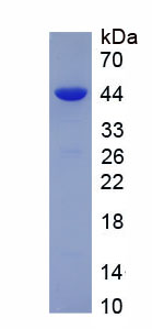
Eukaryotic C-Type Lectin Domain Family 2, Member C (CLEC2C), Protein (Cat# AAA153204)
Full Name
Eukaryotic C-Type Lectin Domain Family 2, Member C (CLEC2C)
Gene Names
CD69; AIM; EA1; MLR-3; CLEC2C; GP32/28; BL-AC/P26
Applications
Western Blot
Purity
Greater than 90% by SDS-PAGE
Pricing
 WB (Western Blot)
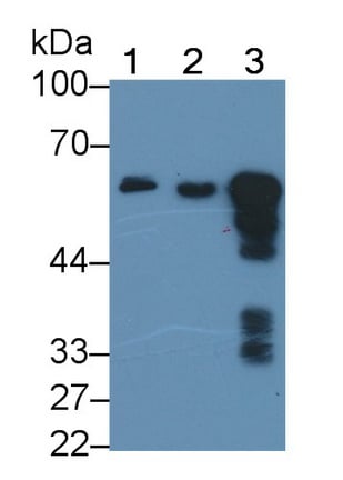
(Western Blot; Sample: Lane1: Human Saliva; Lane2: Rat Pancreas lysate; Lane3: Mouse Pancreas lysate Primary Ab: 0.3ug/ml Mouse AntiHuman AMY1 Antibody Second Ab: 0.2ug/mL HRPLinked Caprine AntiMouse IgG Polyclonal Antibody (Catalog: SAA544Mu19))
WB (Western Blot)
(Western Blot; Sample: Lane1: Human Saliva; Lane2: Rat Pancreas lysate; Lane3: Mouse Pancreas lysate Primary Ab: 0.3ug/ml Mouse AntiHuman AMY1 Antibody Second Ab: 0.2ug/mL HRPLinked Caprine AntiMouse IgG Polyclonal Antibody (Catalog: SAA544Mu19))
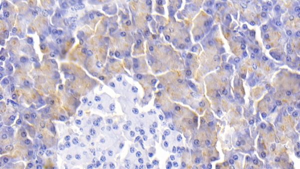
Salivary Alpha Amylase (AMY1A), Monoclonal Antibody (Cat# AAA151719)
Full Name
Monoclonal Antibody to Salivary Alpha Amylase (AMY1A)
Gene Names
AMY1A; AMY1
Reactivity
Human, Mouse, Rat, Pig
Applications
Immunoprecipitation, Immunocytochemistry, Immunohistochemistry, Western Blot
Purity
Protein A + Protein G affinity chromatography
Pricing
 Application Data
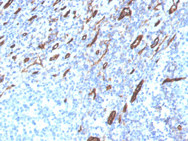
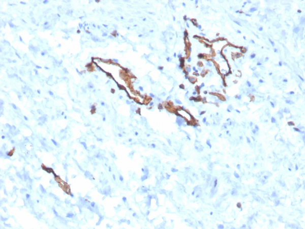
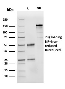
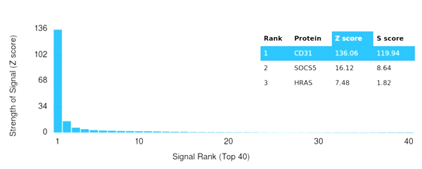
(Analysis of Protein Array containing more than 19,000 full-length human proteins using CD31 Mouse Monoclonal Antibody (PECAM1/3525) Z- and S- Score: The Z-score represents the strength of a signal that a monoclonal antibody (MAb) (in combination with a fluorescently-tagged anti-IgG secondary antibody) produces when binding to a particular protein on the HuProtTM array. Z-scores are described in units of standard deviations (SD's) above the mean value of all signals generated on that array. If targets on HuProtTM are arranged in descending order of the Z-score, the S-score is the difference (also in units of SD's) between the Z-score. S-score therefore represents the relative target specificity of a MAb to its intended target. A MAb is considered to specific to its intended target, if the MAb has an S-score of at least 2.5. For example, if a MAb binds to protein X with a Z-score of 43 and to protein Y with a Z-score of 14, then the S-score for the binding of that MAb to protein X is equal to 29.)
Application Data
(Analysis of Protein Array containing more than 19,000 full-length human proteins using CD31 Mouse Monoclonal Antibody (PECAM1/3525) Z- and S- Score: The Z-score represents the strength of a signal that a monoclonal antibody (MAb) (in combination with a fluorescently-tagged anti-IgG secondary antibody) produces when binding to a particular protein on the HuProtTM array. Z-scores are described in units of standard deviations (SD's) above the mean value of all signals generated on that array. If targets on HuProtTM are arranged in descending order of the Z-score, the S-score is the difference (also in units of SD's) between the Z-score. S-score therefore represents the relative target specificity of a MAb to its intended target. A MAb is considered to specific to its intended target, if the MAb has an S-score of at least 2.5. For example, if a MAb binds to protein X with a Z-score of 43 and to protein Y with a Z-score of 14, then the S-score for the binding of that MAb to protein X is equal to 29.)
CD31/PECAM-1, Monoclonal Antibody (Cat# AAA215379)
Full Name
CD31/PECAM-1 (Endothelial Cell Marker)
Gene Names
PECAM1; CD31; PECA1; GPIIA'; PECAM-1; endoCAM; CD31/EndoCAM
Reactivity
Human
Applications
Immunohistochemistry
Purity
Purified Ab with BSA and Azide at 200ug/ml or Purified Ab with BSA and Azide at 200ug/ml or Purified Ab WITHOUT BSA and Azide at 1.0mg/ml
Pricing
 Application Data
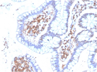
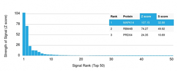
(Analysis of Protein Array containing more than 19,000 full-length human proteins using MAPK14 Mouse Monoclonal Antibody (CPTC-MAPK14-1). Z- and S- Score: The Z-score represents the strength of a signal that a monoclonal antibody (MAb) (in combination with a fluorescently-tagged anti-IgG secondary antibody) produces when binding to a particular protein on the HuProtTM array. Z-scores are described in units of standard deviations (SD's) above the mean value of all signals generated on that array. If targets on HuProtTM are arranged in descending order of the Z-score, the S-score is the difference (also in units of SD's) between the Z-score. S-score therefore represents the relative target specificity of a MAb to its intended target. A MAb is considered to specific to its intended target, if the MAb has an S-score of at least 2.5. For example, if a MAb binds to protein X with a Z-score of 43 and to protein Y with a Z-score of 14, then the S-score for the binding of that MAb to protein X is equal to 29.)
Application Data
(Analysis of Protein Array containing more than 19,000 full-length human proteins using MAPK14 Mouse Monoclonal Antibody (CPTC-MAPK14-1). Z- and S- Score: The Z-score represents the strength of a signal that a monoclonal antibody (MAb) (in combination with a fluorescently-tagged anti-IgG secondary antibody) produces when binding to a particular protein on the HuProtTM array. Z-scores are described in units of standard deviations (SD's) above the mean value of all signals generated on that array. If targets on HuProtTM are arranged in descending order of the Z-score, the S-score is the difference (also in units of SD's) between the Z-score. S-score therefore represents the relative target specificity of a MAb to its intended target. A MAb is considered to specific to its intended target, if the MAb has an S-score of at least 2.5. For example, if a MAb binds to protein X with a Z-score of 43 and to protein Y with a Z-score of 14, then the S-score for the binding of that MAb to protein X is equal to 29.)
MAPK14, Monoclonal Antibody (Cat# AAA214728)
Full Name
MAPK14
Gene Names
MAPK14; RK; p38; CSBP; EXIP; Mxi2; CSBP1; CSBP2; CSPB1; PRKM14; PRKM15; SAPK2A; p38ALPHA
Reactivity
Human. Others not tested.
Applications
Western Blot
Purity
Purified Ab with BSA and Azide at 200ug/ml OR Purified Ab WITHOUT BSA and Azide at 1.0mg/ml
Pricing
 FCM/FACS (Flow Cytometry)
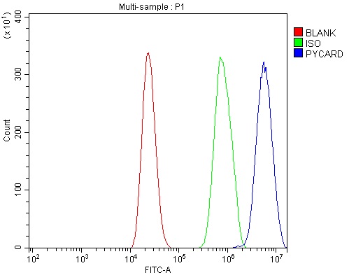
(Figure 3. Flow Cytometry analysis of RAW264. 7 cells using anti-TMS1/ASC/PYCARD antibody (AAA125544).Overlay histogram showing RAW264. 7 cells stained with AAA125544 (Blue line). The cells were blocked with 10% normal goat serum. And then incubated with rabbit anti-TMS1/ASC/PYCARD Antibody (AAA125544, 1μg/1x106 cells) for 30 min at 20 degree C. DyLight®488 conjugated goat anti-rabbit IgG (5-10μg/1x106 cells) was used as secondary antibody for 30 minutes at 20 degree C. Isotype control antibody (Green line) was rabbit IgG (1μg/1x106) used under the same conditions. Unlabelled sample (Red line) was also used as a control.)
FCM/FACS (Flow Cytometry)
(Figure 3. Flow Cytometry analysis of RAW264. 7 cells using anti-TMS1/ASC/PYCARD antibody (AAA125544).Overlay histogram showing RAW264. 7 cells stained with AAA125544 (Blue line). The cells were blocked with 10% normal goat serum. And then incubated with rabbit anti-TMS1/ASC/PYCARD Antibody (AAA125544, 1μg/1x106 cells) for 30 min at 20 degree C. DyLight®488 conjugated goat anti-rabbit IgG (5-10μg/1x106 cells) was used as secondary antibody for 30 minutes at 20 degree C. Isotype control antibody (Green line) was rabbit IgG (1μg/1x106) used under the same conditions. Unlabelled sample (Red line) was also used as a control.)
TMS1/ASC/Pycard, Polyclonal Antibody (Cat# AAA125544)
Full Name
Anti-TMS1/ASC/Pycard Antibody
Gene Names
Pycard; Asc; TNS1; masc; CARD5; TMS-1; 9130417A21Rik
Reactivity
Mouse, Rat
Applications
Direct ELISA, Flow Cytometry, Immunohistochemistry, Western Blot
Purity
Immunogen affinity purified.
Pricing
 IHC (Immunohiostchemistry)
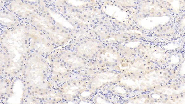
(DAB staining on IHCP;Sample: Human Kidney Tissue; Primary Ab: 20ug/ml Mouse AntiHuman HBEGF AntibodySecond Ab: 2ug/mL HRPLinked Caprine AntiMouse IgG Polyclonal Antibody(Catalog: SAA544Mu19))
IHC (Immunohiostchemistry)
(DAB staining on IHCP;Sample: Human Kidney Tissue; Primary Ab: 20ug/ml Mouse AntiHuman HBEGF AntibodySecond Ab: 2ug/mL HRPLinked Caprine AntiMouse IgG Polyclonal Antibody(Catalog: SAA544Mu19))
Heparin Binding Epidermal Growth Factor Like Growth Factor (HBEGF), Monoclonal Antibody (Cat# AAA151716)
Full Name
Monoclonal Antibody to Heparin Binding Epidermal Growth Factor Like Growth Factor (HBEGF)
Gene Names
HBEGF; DTR; DTS; DTSF; HEGFL
Reactivity
Human, Pig
Applications
Immunoprecipitation, Immunocytochemistry, Immunohistochemistry, Western Blot
Purity
Protein A + Protein G affinity chromatography
Pricing
 Application Data
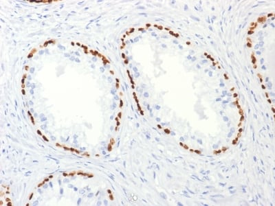
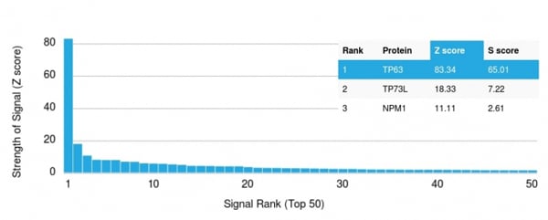
(Analysis of Protein Array containing more than 19, 000 full-length human proteins using p63 Mouse Monoclonal Antibody (TP63/2427).Z- and S- Score: The Z-score represents the strength of a signal that a monoclonal antibody (MAb) (in combination with a fluorescently-tagged anti-IgG secondary antibody) produces when binding to a particular protein on the HuProtTM array. Z-scores are described in units of standard deviations (SD's) above the mean value of all signals generated on that array. If targets on HuProtTM are arranged in descending order of the Z-score, the S-score is the difference (also in units of SD's) between the Z-score. S-score therefore represents the relative target specificity of a MAb to its intended target. A MAb is considered to specific to its intended target, if the MAb has an S-score of at least 2.5. For example, if a MAb binds to protein X with a Z-score of 43 and to protein Y with a Z-score of 14, then the S-score for the binding of that MAb to protein X is equal to 29.)
Application Data
(Analysis of Protein Array containing more than 19, 000 full-length human proteins using p63 Mouse Monoclonal Antibody (TP63/2427).Z- and S- Score: The Z-score represents the strength of a signal that a monoclonal antibody (MAb) (in combination with a fluorescently-tagged anti-IgG secondary antibody) produces when binding to a particular protein on the HuProtTM array. Z-scores are described in units of standard deviations (SD's) above the mean value of all signals generated on that array. If targets on HuProtTM are arranged in descending order of the Z-score, the S-score is the difference (also in units of SD's) between the Z-score. S-score therefore represents the relative target specificity of a MAb to its intended target. A MAb is considered to specific to its intended target, if the MAb has an S-score of at least 2.5. For example, if a MAb binds to protein X with a Z-score of 43 and to protein Y with a Z-score of 14, then the S-score for the binding of that MAb to protein X is equal to 29.)
p63, Monoclonal Antibody (Cat# AAA214710)
Full Name
p63 (Squamous, Basal & Myoepithelial Cell Marker)
Gene Names
TP63; AIS; KET; LMS; NBP; RHS; p40; p51; p63; EEC3; OFC8; p73H; p73L; SHFM4; TP53L; TP73L; p53CP; TP53CP; B(p51A); B(p51B)
Reactivity
Human. Others not known.
Applications
Immunohistochemistry
Pricing
 Application Data
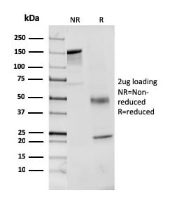
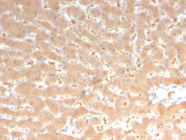
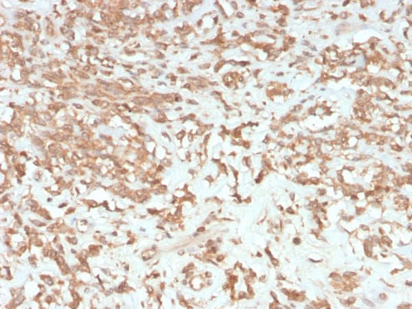
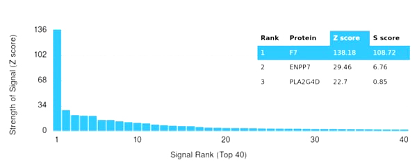
(Analysis of Protein Array containing more than 19,000 full-length human proteins using Coagulation Factor VII Mouse Monoclonal Antibody (F7/3513). Z- and S- Score: The Z-score represents the strength of a signal that a monoclonal antibody (MAb) (in combination with a fluorescently-tagged anti-IgG secondary antibody) produces when binding to a particular protein on the HuProtTM array. Z-scores are described in units of standard deviations (SD's) above the mean value of all signals generated on that array. If targets on HuProtTM are arranged in descending order of the Z-score, the S-score is the difference (also in units of SD's) between the Z-score. S-score therefore represents the relative target specificity of a MAb to its intended target. A MAb is considered to specific to its intended target, if the MAb has an S-score of at least 2.5. For example, if a MAb binds to protein X with a Z-score of 43 and to protein Y with a Z-score of 14, then the S-score for the binding of that MAb to protein X is equal to 29.)
Application Data
(Analysis of Protein Array containing more than 19,000 full-length human proteins using Coagulation Factor VII Mouse Monoclonal Antibody (F7/3513). Z- and S- Score: The Z-score represents the strength of a signal that a monoclonal antibody (MAb) (in combination with a fluorescently-tagged anti-IgG secondary antibody) produces when binding to a particular protein on the HuProtTM array. Z-scores are described in units of standard deviations (SD's) above the mean value of all signals generated on that array. If targets on HuProtTM are arranged in descending order of the Z-score, the S-score is the difference (also in units of SD's) between the Z-score. S-score therefore represents the relative target specificity of a MAb to its intended target. A MAb is considered to specific to its intended target, if the MAb has an S-score of at least 2.5. For example, if a MAb binds to protein X with a Z-score of 43 and to protein Y with a Z-score of 14, then the S-score for the binding of that MAb to protein X is equal to 29.)
VII/F7, Monoclonal Antibody (Cat# AAA215446)
Full Name
Coagulation Factor VII/F7
Gene Names
F7; SPCA
Reactivity
Human
Applications
Immunohistochemistry
Purity
Purified Ab with BSA and Azide at 200ug/ml or Purified Ab with BSA and Azide at 200ug/ml or Purified Ab WITHOUT BSA and Azide at 1.0mg/ml
Pricing
 FCM/FACS (Flow Cytometry)
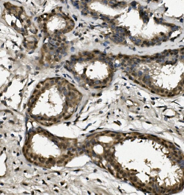
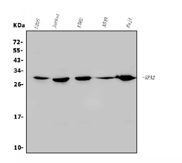
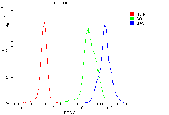
(Figure 5. Flow Cytometry analysis of U937 cells using anti-RPA32/RPA2 antibody (AAA125632).Overlay histogram showing U937 cells stained with AAA125632 (Blue line). The cells were blocked with 10% normal goat serum. And then incubated with rabbit anti-RPA32/RPA2 Antibody (AAA125632, 1μg/1x106 cells) for 30 min at 20 degree C. DyLight®488 conjugated goat anti-rabbit IgG (5-10μg/1x106 cells) was used as secondary antibody for 30 minutes at 20 degree C. Isotype control antibody (Green line) was rabbit IgG (1μg/1x106) used under the same conditions. Unlabelled sample (Red line) was also used as a control.)
FCM/FACS (Flow Cytometry)
(Figure 5. Flow Cytometry analysis of U937 cells using anti-RPA32/RPA2 antibody (AAA125632).Overlay histogram showing U937 cells stained with AAA125632 (Blue line). The cells were blocked with 10% normal goat serum. And then incubated with rabbit anti-RPA32/RPA2 Antibody (AAA125632, 1μg/1x106 cells) for 30 min at 20 degree C. DyLight®488 conjugated goat anti-rabbit IgG (5-10μg/1x106 cells) was used as secondary antibody for 30 minutes at 20 degree C. Isotype control antibody (Green line) was rabbit IgG (1μg/1x106) used under the same conditions. Unlabelled sample (Red line) was also used as a control.)
RPA32/RPA2, Polyclonal Antibody (Cat# AAA125632)
Full Name
Anti-RPA32/RPA2 Antibody
Gene Names
RPA2; REPA2; RPA32
Reactivity
Human
Applications
Direct ELISA, Flow Cytometry, Immunofluorescence, Immunocytochemistry, Immunohistochemistry, Western Blot
Purity
Immunogen affinity purified.
Pricing
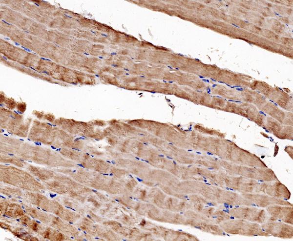
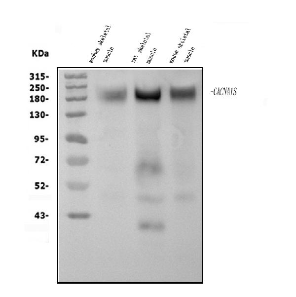
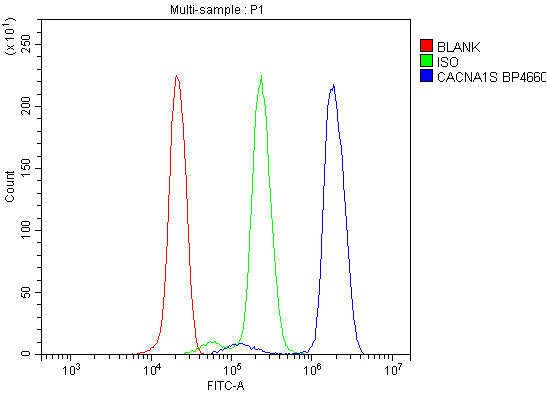 FCM/FACS (Flow Cytometry)
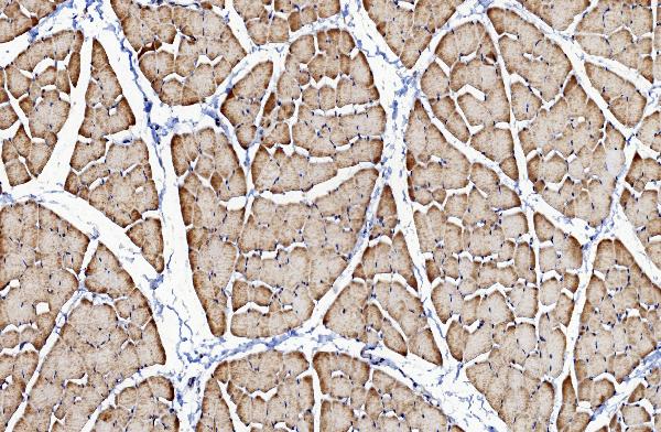
(Figure 4. Flow Cytometry analysis of U937 cells using anti-CACNA1S antibody (AAA125682).Overlay histogram showing U937 cells stained with AAA125682 (Blue line). The cells were blocked with 10% normal goat serum. And then incubated with rabbit anti-CACNA1S Antibody (AAA125682, 1μg/1x106 cells) for 30 min at 20 degree C. DyLight®488 conjugated goat anti-rabbit IgG (5-10μg/1x106 cells) was used as secondary antibody for 30 minutes at 20 degree C. Isotype control antibody (Green line) was rabbit IgG (1μg/1x106) used under the same conditions. Unlabelled sample (Red line) was also used as a control.)
FCM/FACS (Flow Cytometry)
(Figure 4. Flow Cytometry analysis of U937 cells using anti-CACNA1S antibody (AAA125682).Overlay histogram showing U937 cells stained with AAA125682 (Blue line). The cells were blocked with 10% normal goat serum. And then incubated with rabbit anti-CACNA1S Antibody (AAA125682, 1μg/1x106 cells) for 30 min at 20 degree C. DyLight®488 conjugated goat anti-rabbit IgG (5-10μg/1x106 cells) was used as secondary antibody for 30 minutes at 20 degree C. Isotype control antibody (Green line) was rabbit IgG (1μg/1x106) used under the same conditions. Unlabelled sample (Red line) was also used as a control.)
CACNA1S, Polyclonal Antibody (Cat# AAA125682)
Full Name
Anti-CACNA1S Antibody
Gene Names
CACNA1S; MHS5; HOKPP; TTPP1; Cav1.1; HOKPP1; hypoPP; CCHL1A3; CACNL1A3
Reactivity
Human, Mouse, Rat, Monkey
Applications
Direct ELISA, Flow Cytometry, Immunohistochemistry, Western Blot
Purity
Immunogen affinity purified.
Pricing
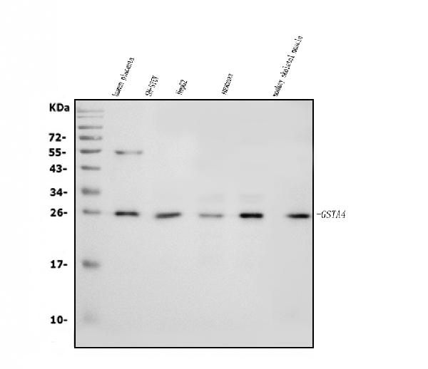
 FCM/FACS (Flow Cytometry)
(Figure 3. Flow Cytometry analysis of U20S cells using anti-GSTA4 antibody (AAA125700).Overlay histogram showing U20S cells stained with AAA125700 (Blue line). The cells were blocked with 10% normal goat serum. And then incubated with rabbit anti-GSTA4 Antibody (AAA125700, 1μg/1x106 cells) for 30 min at 20 degree C. DyLight®488 conjugated goat anti-rabbit IgG (5-10μg/1x106 cells) was used as secondary antibody for 30 minutes at 20 degree C. Isotype control antibody (Green line) was rabbit IgG (1μg/1x106) used under the same conditions. Unlabelled sample (Red line) was also used as a control.)
FCM/FACS (Flow Cytometry)
(Figure 3. Flow Cytometry analysis of U20S cells using anti-GSTA4 antibody (AAA125700).Overlay histogram showing U20S cells stained with AAA125700 (Blue line). The cells were blocked with 10% normal goat serum. And then incubated with rabbit anti-GSTA4 Antibody (AAA125700, 1μg/1x106 cells) for 30 min at 20 degree C. DyLight®488 conjugated goat anti-rabbit IgG (5-10μg/1x106 cells) was used as secondary antibody for 30 minutes at 20 degree C. Isotype control antibody (Green line) was rabbit IgG (1μg/1x106) used under the same conditions. Unlabelled sample (Red line) was also used as a control.)
GSTA4, Polyclonal Antibody (Cat# AAA125700)
Full Name
Anti-GSTA4 Antibody
Gene Names
GSTA4; GTA4; GSTA4-4
Reactivity
Human, Monkey
Applications
Direct ELISA, Flow Cytometry, Immunohistochemistry, Western Blot
Purity
Immunogen affinity purified.
Pricing
 IHC (Immunohiostchemistry)
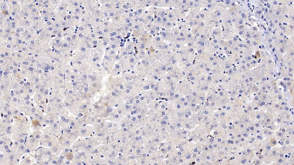
(DAB staining on IHCP;Sample: Human Liver Tissue; Primary Ab: 10ug/ml Rabbit AntiHuman CYP1A2 AntibodySecond Ab: 2ug/mL HRPLinked Caprine AntiRabbit IgG Polyclonal Antibody(Catalog: SAA544Rb19))
IHC (Immunohiostchemistry)
(DAB staining on IHCP;Sample: Human Liver Tissue; Primary Ab: 10ug/ml Rabbit AntiHuman CYP1A2 AntibodySecond Ab: 2ug/mL HRPLinked Caprine AntiRabbit IgG Polyclonal Antibody(Catalog: SAA544Rb19))
Cytochrome P450 1A2 (CYP1A2), Polyclonal Antibody (Cat# AAA151965)
Full Name
Polyclonal Antibody to Cytochrome P450 1A2 (CYP1A2)
Gene Names
CYP1A2; CP12; P3-450; P450(PA)
Reactivity
Human, Pig
Applications
Immunoprecipitation, Immunocytochemistry, Immunohistochemistry, Western Blot
Purity
Antigenspecific affinity chromatography followed by Protein A affinity chromatography
Pricing
 FCM/FACS (Flow Cytometry)
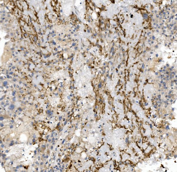
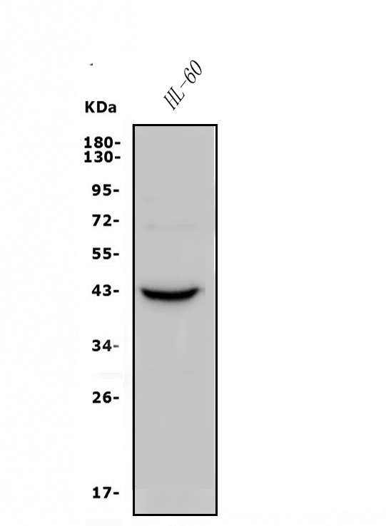
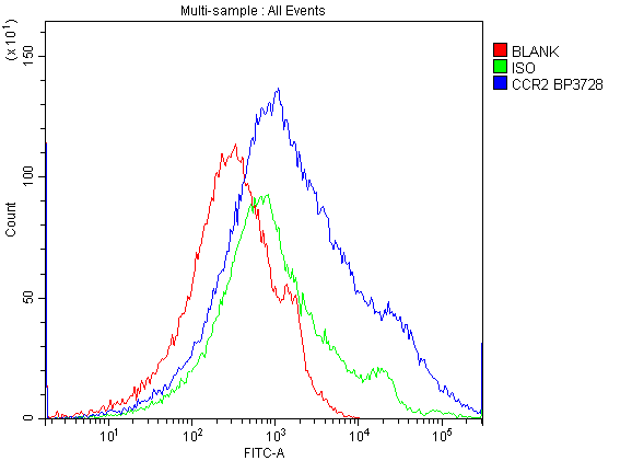
(Figure 4. Flow Cytometry analysis of h. PBMC cells using anti-CCR2 antibody (AAA125520).Overlay histogram showing h. PBMC cells stained with AAA125520 (Blue line). The cells were blocked with 10% normal goat serum. And then incubated with rabbit anti-CCR2 Antibody (AAA125520, 1μg/1x106 cells) for 30 min at 20 degree C. DyLight®488 conjugated goat anti-rabbit IgG (5-10μg/1x106 cells) was used as secondary antibody for 30 minutes at 20 degree C. Isotype control antibody (Green line) was rabbit IgG (1μg/1x106) used under the same conditions. Unlabelled sample (Red line) was also used as a control.)
FCM/FACS (Flow Cytometry)
(Figure 4. Flow Cytometry analysis of h. PBMC cells using anti-CCR2 antibody (AAA125520).Overlay histogram showing h. PBMC cells stained with AAA125520 (Blue line). The cells were blocked with 10% normal goat serum. And then incubated with rabbit anti-CCR2 Antibody (AAA125520, 1μg/1x106 cells) for 30 min at 20 degree C. DyLight®488 conjugated goat anti-rabbit IgG (5-10μg/1x106 cells) was used as secondary antibody for 30 minutes at 20 degree C. Isotype control antibody (Green line) was rabbit IgG (1μg/1x106) used under the same conditions. Unlabelled sample (Red line) was also used as a control.)
CCR2, Polyclonal Antibody (Cat# AAA125520)
Full Name
Anti-CCR2 Antibody
Gene Names
CCR2; CKR2; CCR-2; CCR2A; CCR2B; CD192; CKR2A; CKR2B; CMKBR2; MCP-1-R; CC-CKR-2
Reactivity
Human
Applications
Direct ELISA, Flow Cytometry, Immunohistochemistry, Western Blot
Purity
Immunogen affinity purified.
Pricing
 WB (Western Blot)
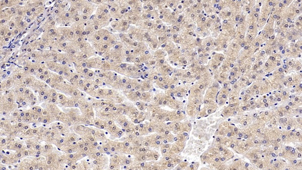
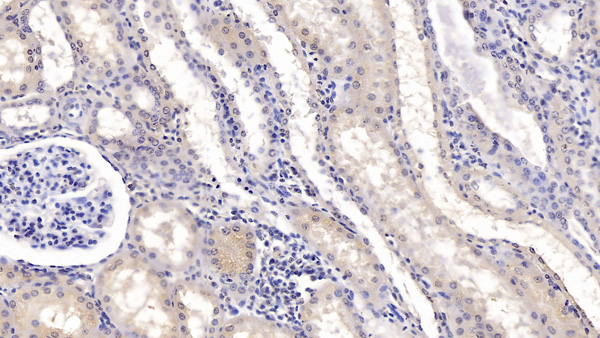
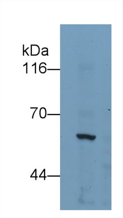
(Western Blot; Sample: 293T cell lysate Primary Ab: 3ug/ml Mouse AntiHuman KPNa2 Antibody Second Ab: 0.2ug/mL HRPLinked Caprine AntiMouse IgG Polyclonal Antibody (Catalog: SAA544Mu19))
WB (Western Blot)
(Western Blot; Sample: 293T cell lysate Primary Ab: 3ug/ml Mouse AntiHuman KPNa2 Antibody Second Ab: 0.2ug/mL HRPLinked Caprine AntiMouse IgG Polyclonal Antibody (Catalog: SAA544Mu19))
Karyopherin Alpha 2 (KPNa2), Monoclonal Antibody (Cat# AAA151857)
Full Name
Monoclonal Antibody to Karyopherin Alpha 2 (KPNa2)
Gene Names
KPNA2; QIP2; RCH1; IPOA1; SRP1alpha
Reactivity
Human, Pig
Applications
Immunoprecipitation, Immunocytochemistry, Immunohistochemistry, Western Blot
Purity
Protein A + Protein G affinity chromatography
Pricing
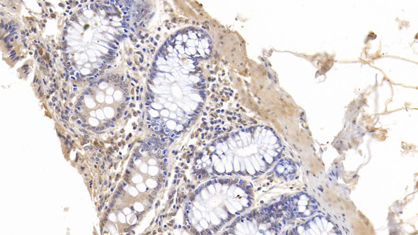
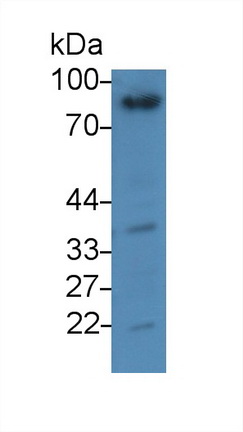 WB (Western Blot)
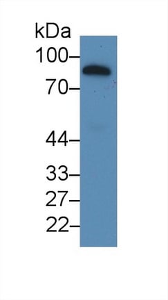
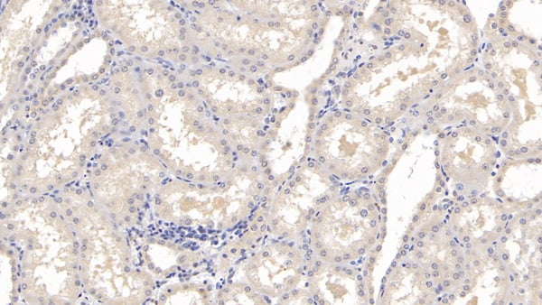
(Western Blot; Sample: Human Lung lysatePrimary Ab: 3ug/ml Mouse AntiHuman GSN AntibodySecond Ab: 0.2ug/mL HRPLinked Caprine AntiMouse IgG Polyclonal Antibody(Catalog: SAA544Mu19))
WB (Western Blot)
(Western Blot; Sample: Human Lung lysatePrimary Ab: 3ug/ml Mouse AntiHuman GSN AntibodySecond Ab: 0.2ug/mL HRPLinked Caprine AntiMouse IgG Polyclonal Antibody(Catalog: SAA544Mu19))
Gelsolin (GSN), Monoclonal Antibody (Cat# AAA151524)
Full Name
Monoclonal Antibody to Gelsolin (GSN)
Gene Names
GSN; ADF; AGEL
Reactivity
Human, Pig
Applications
Immunoprecipitation, Immunocytochemistry, Immunohistochemistry, Western Blot
Purity
Protein A + Protein G affinity chromatography
Pricing
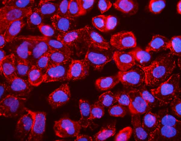
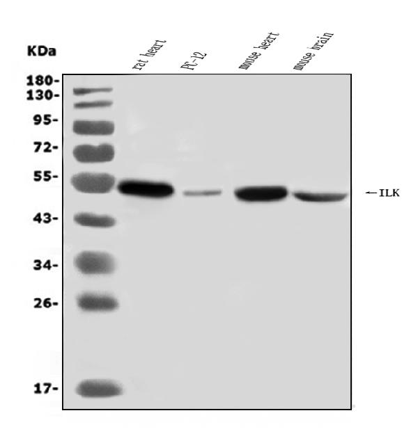
 FCM/FACS (Flow Cytometry)
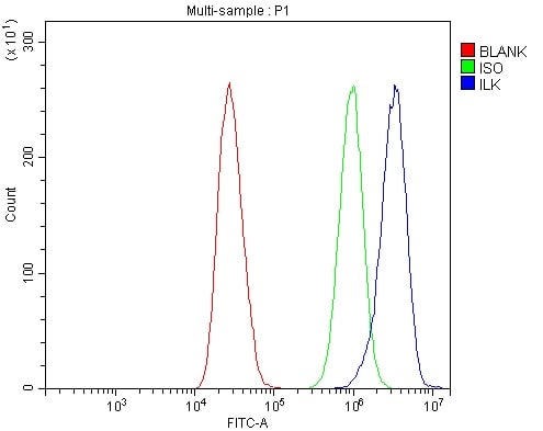
(Figure 3. Flow Cytometry analysis of MCF-7 cells using anti-Integrin linked ILK antibody (AAA125679).Overlay histogram showing MCF-7 cells stained with AAA125679 (Blue line). The cells were blocked with 10% normal goat serum. And then incubated with rabbit anti-Integrin linked ILK Antibody (AAA125679, 1μg/1x106 cells) for 30 min at 20 degree C. DyLight®488 conjugated goat anti-rabbit IgG (5-10μg/1x106 cells) was used as secondary antibody for 30 minutes at 20 degree C. Isotype control antibody (Green line) was rabbit IgG (1μg/1x106) used under the same conditions. Unlabelled sample (Red line) was also used as a control.)
FCM/FACS (Flow Cytometry)
(Figure 3. Flow Cytometry analysis of MCF-7 cells using anti-Integrin linked ILK antibody (AAA125679).Overlay histogram showing MCF-7 cells stained with AAA125679 (Blue line). The cells were blocked with 10% normal goat serum. And then incubated with rabbit anti-Integrin linked ILK Antibody (AAA125679, 1μg/1x106 cells) for 30 min at 20 degree C. DyLight®488 conjugated goat anti-rabbit IgG (5-10μg/1x106 cells) was used as secondary antibody for 30 minutes at 20 degree C. Isotype control antibody (Green line) was rabbit IgG (1μg/1x106) used under the same conditions. Unlabelled sample (Red line) was also used as a control.)
Integrin linked ILK, Polyclonal Antibody (Cat# AAA125679)
Full Name
Anti-Integrin linked ILK Antibody
Gene Names
ILK; P59; ILK-1; ILK-2; p59ILK
Reactivity
Human, Mouse, Rat
Applications
Direct ELISA, Flow Cytometry, Immunofluorescence, Immunocytochemistry, Western Blot
Purity
Immunogen affinity purified.
Pricing


 FCM/FACS (Flow Cytometry)
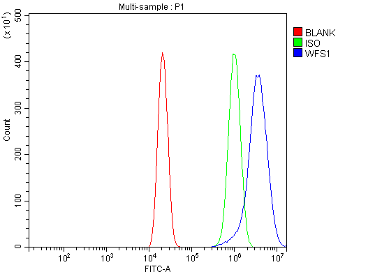
(Figure 4. Flow Cytometry analysis of U20S cells using anti-WFS1 antibody (AAA125604).Overlay histogram showing U20S cells stained with AAA125604 (Blue line). The cells were blocked with 10% normal goat serum. And then incubated with rabbit anti-WFS1 Antibody (AAA125604, 1μg/1x106 cells) for 30 min at 20 degree C. DyLight®488 conjugated goat anti-rabbit IgG (5-10μg/1x106 cells) was used as secondary antibody for 30 minutes at 20 degree C. Isotype control antibody (Green line) was rabbit IgG (1μg/1x106) used under the same conditions. Unlabelled sample (Red line) was also used as a control.)
FCM/FACS (Flow Cytometry)
(Figure 4. Flow Cytometry analysis of U20S cells using anti-WFS1 antibody (AAA125604).Overlay histogram showing U20S cells stained with AAA125604 (Blue line). The cells were blocked with 10% normal goat serum. And then incubated with rabbit anti-WFS1 Antibody (AAA125604, 1μg/1x106 cells) for 30 min at 20 degree C. DyLight®488 conjugated goat anti-rabbit IgG (5-10μg/1x106 cells) was used as secondary antibody for 30 minutes at 20 degree C. Isotype control antibody (Green line) was rabbit IgG (1μg/1x106) used under the same conditions. Unlabelled sample (Red line) was also used as a control.)
WFS1, Polyclonal Antibody (Cat# AAA125604)
Full Name
Anti-WFS1 Antibody
Gene Names
WFS1; WFS; WFRS; WFSL; CTRCT41
Reactivity
Human, Monkey
Applications
Direct ELISA, Flow Cytometry, Immunofluorescence, Immunocytochemistry, Immunohistochemistry, Western Blot
Purity
Immunogen affinity purified.
Pricing
 FCM/FACS (Flow Cytometry)
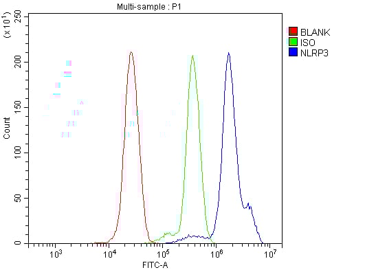
(Figure 2. Flow Cytometry analysis of THP-1 cells using anti-NLRP3 antibody (AAA125512).Overlay histogram showing THP-1 cells stained with AAA125512 (Blue line). The cells were blocked with 10% normal goat serum. And then incubated with rabbit anti-NLRP3 Antibody (AAA125512, 1μg/1x106 cells) for 30 min at 20 degree C. DyLight®488 conjugated goat anti-rabbit IgG (5-10μg/1x106 cells) was used as secondary antibody for 30 minutes at 20 degree C. Isotype control antibody (Green line) was rabbit IgG (1μg/1x106) used under the same conditions. Unlabelled sample (Red line) was also used as a control.)
FCM/FACS (Flow Cytometry)
(Figure 2. Flow Cytometry analysis of THP-1 cells using anti-NLRP3 antibody (AAA125512).Overlay histogram showing THP-1 cells stained with AAA125512 (Blue line). The cells were blocked with 10% normal goat serum. And then incubated with rabbit anti-NLRP3 Antibody (AAA125512, 1μg/1x106 cells) for 30 min at 20 degree C. DyLight®488 conjugated goat anti-rabbit IgG (5-10μg/1x106 cells) was used as secondary antibody for 30 minutes at 20 degree C. Isotype control antibody (Green line) was rabbit IgG (1μg/1x106) used under the same conditions. Unlabelled sample (Red line) was also used as a control.)
CIAS1/NALP3/NLRP3, Polyclonal Antibody (Cat# AAA125512)
Full Name
Anti-CIAS1/NALP3/NLRP3 Antibody
Gene Names
NLRP3; AII; AVP; FCU; MWS; FCAS; CIAS1; FCAS1; NALP3; C1orf7; CLR1.1; PYPAF1; AGTAVPRL
Reactivity
Human, Mouse, Rat
Applications
Direct ELISA, Flow Cytometry, Western Blot
Purity
Immunogen affinity purified.
Pricing
 FCM/FACS (Flow Cytometry)
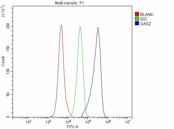
(Figure 2. Flow Cytometry analysis of Raw264.7 cells using anti-OAS2 antibody (AAA127085).Overlay histogram showing Raw264.7 cells stained with AAA127085 (Blue line). The cells were fixed with 4% paraformaldehyde and blocked with 10% normal goat serum. And then incubated with rabbit anti-OAS2 Antibody (AAA127085, 1ug/1x106 cells) for 30 min at 20 degree C. DyLight488 conjugated goat anti-rabbit IgG was used as secondary antibody for 30 minutes at 20 degree C. Isotype control antibody (Green line) was rabbit IgG (1ug/1x106) used under the same conditions. Unlabelled sample without incubation with primary antibody and secondary antibody (Red line) was used as a blank control.)
FCM/FACS (Flow Cytometry)
(Figure 2. Flow Cytometry analysis of Raw264.7 cells using anti-OAS2 antibody (AAA127085).Overlay histogram showing Raw264.7 cells stained with AAA127085 (Blue line). The cells were fixed with 4% paraformaldehyde and blocked with 10% normal goat serum. And then incubated with rabbit anti-OAS2 Antibody (AAA127085, 1ug/1x106 cells) for 30 min at 20 degree C. DyLight488 conjugated goat anti-rabbit IgG was used as secondary antibody for 30 minutes at 20 degree C. Isotype control antibody (Green line) was rabbit IgG (1ug/1x106) used under the same conditions. Unlabelled sample without incubation with primary antibody and secondary antibody (Red line) was used as a blank control.)
OAS2, Polyclonal Antibody (Cat# AAA127085)
Full Name
Anti-OAS2 Antibody Picoband
Gene Names
Oas2; Oasl11
Reactivity
Mouse, Rat
Applications
Flow Cytometry, Western Blot
Purity
Immunogen affinity purified.
Pricing
 IHC (Immunohistochemisry)
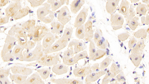
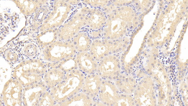
(DAB staining on IHCP;Sample: Porcine Kidney Tissue; Primary Ab: 20ug/ml Mouse AntiHuman HBEGF AntibodySecond Ab: 2ug/mL HRPLinked Caprine AntiMouse IgG Polyclonal Antibody(Catalog: SAA544Mu19))
IHC (Immunohistochemisry)
(DAB staining on IHCP;Sample: Porcine Kidney Tissue; Primary Ab: 20ug/ml Mouse AntiHuman HBEGF AntibodySecond Ab: 2ug/mL HRPLinked Caprine AntiMouse IgG Polyclonal Antibody(Catalog: SAA544Mu19))
Heparin Binding Epidermal Growth Factor Like Growth Factor (HBEGF), Monoclonal Antibody (Cat# AAA151717)
Full Name
Monoclonal Antibody to Heparin Binding Epidermal Growth Factor Like Growth Factor (HBEGF)
Gene Names
HBEGF; DTR; DTS; DTSF; HEGFL
Reactivity
Human, Pig
Applications
Immunoprecipitation, Immunocytochemistry, Immunohistochemistry, Western Blot
Purity
Protein A + Protein G affinity chromatography
Pricing
 Application Data
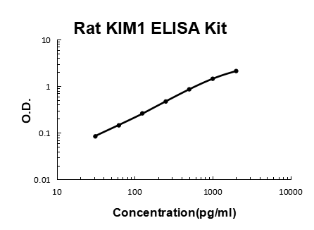
KIM1, ELISA Kit (Cat# AAA125882)
Full Name
Rat KIM1 Quick ELISA Kit
Gene Names
Havcr1; Kim1; KIM-1
Reactivity
Rat
Pricing
 IHC (Immunohistochemistry)
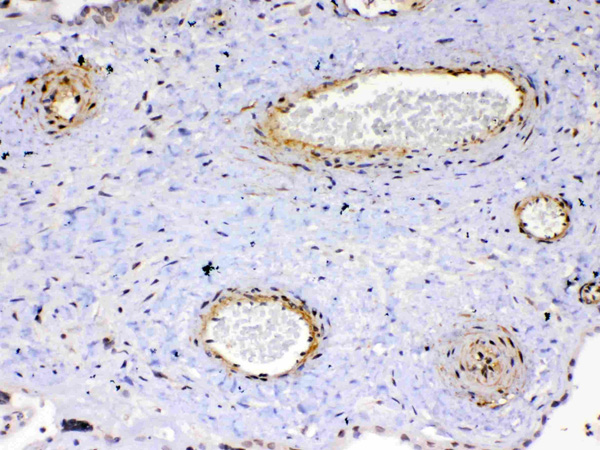
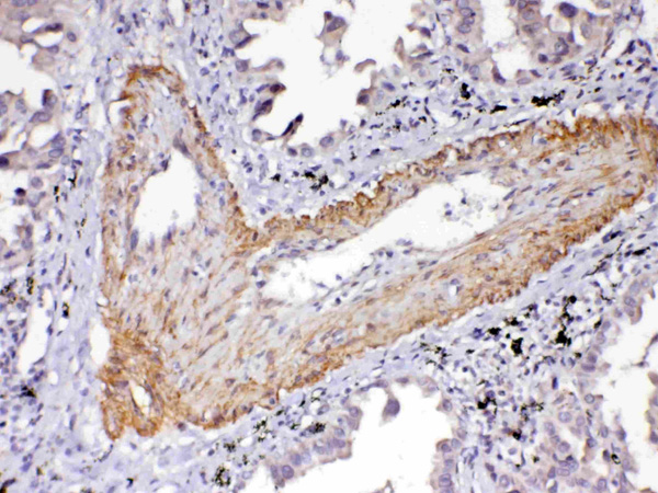
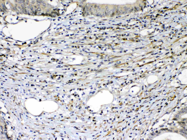
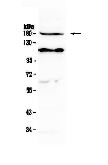
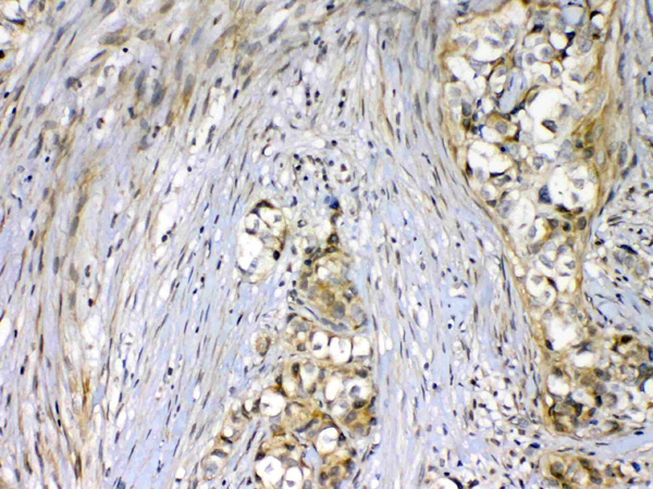
(Figure 5. IHC analysis of VEGF Receptor 2 using anti-VEGF Receptor 2 antibody (AAA124600).VEGF Receptor 2 was detected in paraffin-embedded section of human mammary cancer tissue. Heat mediated antigen retrieval was performed in citrate buffer (pH6, epitope retrieval solution) for 20 mins. The tissue section was blocked with 10% goat serum. The tissue section was then incubated with 1ug/ml rabbit anti-VEGF Receptor 2 Antibody (AAA124600) overnight at 4 degree C. Biotinylated goat anti-rabbit IgG was used as secondary antibody and incubated for 30 minutes at 37 degree C. The tissue section was developed using Strepavidin-Biotin-Complex (SABC) with DAB as the chromogen.)
IHC (Immunohistochemistry)
(Figure 5. IHC analysis of VEGF Receptor 2 using anti-VEGF Receptor 2 antibody (AAA124600).VEGF Receptor 2 was detected in paraffin-embedded section of human mammary cancer tissue. Heat mediated antigen retrieval was performed in citrate buffer (pH6, epitope retrieval solution) for 20 mins. The tissue section was blocked with 10% goat serum. The tissue section was then incubated with 1ug/ml rabbit anti-VEGF Receptor 2 Antibody (AAA124600) overnight at 4 degree C. Biotinylated goat anti-rabbit IgG was used as secondary antibody and incubated for 30 minutes at 37 degree C. The tissue section was developed using Strepavidin-Biotin-Complex (SABC) with DAB as the chromogen.)
VEGF Receptor 2, Polyclonal Antibody (Cat# AAA124600)
Full Name
Anti-VEGF Receptor 2 Picoband Antibody
Gene Names
KDR; FLK1; CD309; VEGFR; VEGFR2
Reactivity
Human
Applications
Flow Cytometry, Western Blot, Immunocytochemistry, Immunohistochemistry
Purity
Immunogen affinity purified
Pricing
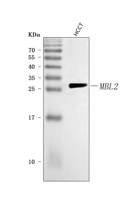
 FCM/FACS (Flow Cytometry)
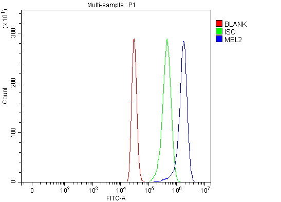
(Figure 2. Flow Cytometry analysis of HepG2 cells using anti-MBL2 antibody (AAA127027).Overlay histogram showing HepG2 cells stained with AAA127027 (Blue line). The cells were fixed with 4% paraformaldehyde and blocked with 10% normal goat serum. And then incubated with rabbit anti-MBL2 Antibody (AAA127027, 1ug/1x106 cells) for 30 min at 20 degree C. DyLight488 conjugated goat anti-rabbit IgG was used as secondary antibody for 30 minutes at 20 degree C. Isotype control antibody (Green line) was rabbit IgG (1ug/1x106) used under the same conditions. Unlabelled sample (Red line) was also used as a control.)
FCM/FACS (Flow Cytometry)
(Figure 2. Flow Cytometry analysis of HepG2 cells using anti-MBL2 antibody (AAA127027).Overlay histogram showing HepG2 cells stained with AAA127027 (Blue line). The cells were fixed with 4% paraformaldehyde and blocked with 10% normal goat serum. And then incubated with rabbit anti-MBL2 Antibody (AAA127027, 1ug/1x106 cells) for 30 min at 20 degree C. DyLight488 conjugated goat anti-rabbit IgG was used as secondary antibody for 30 minutes at 20 degree C. Isotype control antibody (Green line) was rabbit IgG (1ug/1x106) used under the same conditions. Unlabelled sample (Red line) was also used as a control.)
MBL2, Polyclonal Antibody (Cat# AAA127027)
Full Name
Anti-MBL2 Antibody Picoband
Gene Names
MBL2; MBL; MBP; MBP1; MBPD; MBL2D; MBP-C; COLEC1; HSMBPC
Reactivity
Human
Applications
Flow Cytometry, Western Blot
Purity
Immunogen affinity purified.
Pricing
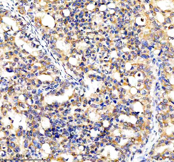
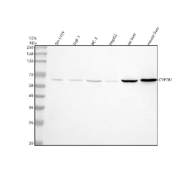
 FCM/FACS (Flow Cytometry)
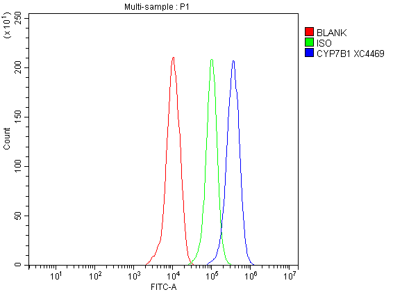
(Figure 3. Flow Cytometry analysis of THP-1 cells using anti-CYP7B1 antibody (AAA128209).Overlay histogram showing THP-1 cells stained with AAA128209 (Blue line). To facilitate intracellular staining, cells were fixed with 4% paraformaldehyde and permeabilized with permeabilization buffer. The cells were blocked with 10% normal goat serum. And then incubated with rabbit anti-CYP7B1 Antibody (AAA128209, 1ug/1x106 cells) for 30 min at 20 degree C. DyLight®488 conjugated goat anti-rabbit IgG was used as secondary antibody for 30 minutes at 20 degree C. Isotype control antibody (Green line) was rabbit IgG (1ug/1x106) used under the same conditions. Unlabelled sample without incubation with primary antibody and secondary antibody (Red line) was used as a blank control.)
FCM/FACS (Flow Cytometry)
(Figure 3. Flow Cytometry analysis of THP-1 cells using anti-CYP7B1 antibody (AAA128209).Overlay histogram showing THP-1 cells stained with AAA128209 (Blue line). To facilitate intracellular staining, cells were fixed with 4% paraformaldehyde and permeabilized with permeabilization buffer. The cells were blocked with 10% normal goat serum. And then incubated with rabbit anti-CYP7B1 Antibody (AAA128209, 1ug/1x106 cells) for 30 min at 20 degree C. DyLight®488 conjugated goat anti-rabbit IgG was used as secondary antibody for 30 minutes at 20 degree C. Isotype control antibody (Green line) was rabbit IgG (1ug/1x106) used under the same conditions. Unlabelled sample without incubation with primary antibody and secondary antibody (Red line) was used as a blank control.)
CYP7B1, Polyclonal Antibody (Cat# AAA128209)
Full Name
Anti-CYP7B1 Antibody Picoband
Gene Names
CYP7B1; CP7B; CBAS3; SPG5A
Reactivity
Human, Mouse, Rat
Applications
Flow Cytometry, Immunohistochemistry, Western Blot
Purity
Immunogen affinity purified.
Pricing
 FCM/FACS (Flow Cytometry)
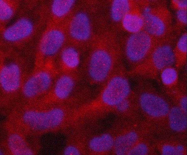
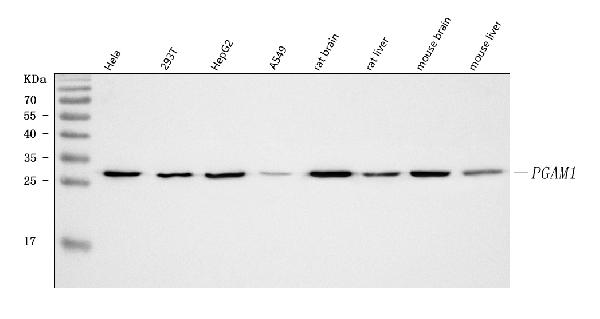
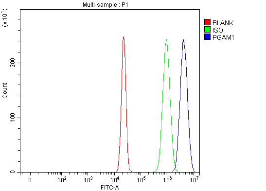
(Figure 3. Flow Cytometry analysis of U20S cells using anti-PGAM1 antibody (AAA127232).Overlay histogram showing U20S cells stained with AAA127232 (Blue line). To facilitate intracellular staining, cells were fixed with 4% paraformaldehyde and permeabilized with permeabilization buffer. The cells were blocked with 10% normal goat serum. And then incubated with rabbit anti-PGAM1 Antibody (AAA127232, 1ug/1x106 cells) for 30 min at 20 degree C. DyLight488 conjugated goat anti-rabbit IgG was used as secondary antibody for 30 minutes at 20 degree C. Isotype control antibody (Green line) was rabbit IgG (1ug/1x106) used under the same conditions. Unlabelled sample without incubation with primary antibody and secondary antibody (Red line) was used as a blank control.)
FCM/FACS (Flow Cytometry)
(Figure 3. Flow Cytometry analysis of U20S cells using anti-PGAM1 antibody (AAA127232).Overlay histogram showing U20S cells stained with AAA127232 (Blue line). To facilitate intracellular staining, cells were fixed with 4% paraformaldehyde and permeabilized with permeabilization buffer. The cells were blocked with 10% normal goat serum. And then incubated with rabbit anti-PGAM1 Antibody (AAA127232, 1ug/1x106 cells) for 30 min at 20 degree C. DyLight488 conjugated goat anti-rabbit IgG was used as secondary antibody for 30 minutes at 20 degree C. Isotype control antibody (Green line) was rabbit IgG (1ug/1x106) used under the same conditions. Unlabelled sample without incubation with primary antibody and secondary antibody (Red line) was used as a blank control.)
PGAM1, Polyclonal Antibody (Cat# AAA127232)
Full Name
Anti-PGAM1 Antibody Picoband
Gene Names
PGAM1; PGAMA; PGAM-B; HEL-S-35
Reactivity
Human, Mouse, Rat
Applications
Flow Cytometry, Immunofluorescence, Immunocytochemistry, Western Blot
Purity
Immunogen affinity purified.
Pricing
 IHC (Immunohistochemistry)
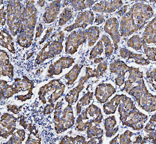
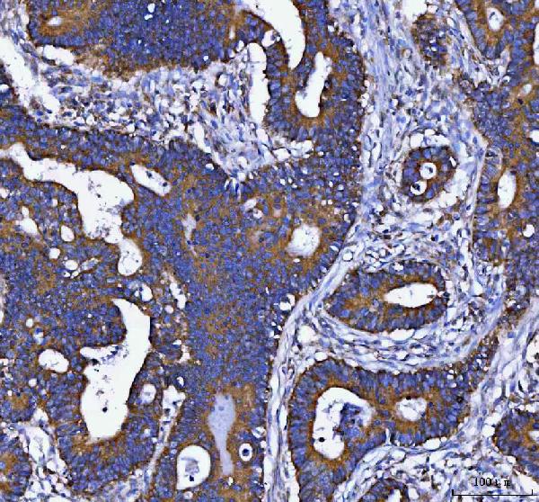
(Figure 4. IHC analysis of DAPK1 using anti-DAPK1 antibody (AAA127041).DAPK1 was detected in a paraffin-embedded section of human lung cancer tissue. Heat mediated antigen retrieval was performed in EDTA buffer (pH 8.0, epitope retrieval solution). The tissue section was blocked with 10% goat serum. The tissue section was then incubated with 2ug/ml rabbit anti-DAPK1 Antibody (AAA127041) overnight at 4 degree C. Peroxidase Conjugated Goat Anti-rabbit IgG was used as secondary antibody and incubated for 30 minutes at 37 degree C. The tissue section was developed using HRP Conjugated Rabbit IgG Super Vision Assay Kit with DAB as the chromogen.)
IHC (Immunohistochemistry)
(Figure 4. IHC analysis of DAPK1 using anti-DAPK1 antibody (AAA127041).DAPK1 was detected in a paraffin-embedded section of human lung cancer tissue. Heat mediated antigen retrieval was performed in EDTA buffer (pH 8.0, epitope retrieval solution). The tissue section was blocked with 10% goat serum. The tissue section was then incubated with 2ug/ml rabbit anti-DAPK1 Antibody (AAA127041) overnight at 4 degree C. Peroxidase Conjugated Goat Anti-rabbit IgG was used as secondary antibody and incubated for 30 minutes at 37 degree C. The tissue section was developed using HRP Conjugated Rabbit IgG Super Vision Assay Kit with DAB as the chromogen.)
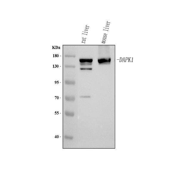
DAPK1, Polyclonal Antibody (Cat# AAA127041)
Full Name
Anti-DAPK1 Antibody Picoband
Gene Names
DAPK1; DAPK
Reactivity
Human, Mouse, Rat
Applications
Immunohistochemistry, Western Blot
Purity
Immunogen affinity purified.
Pricing
 FCM/FACS (Flow Cytometry)
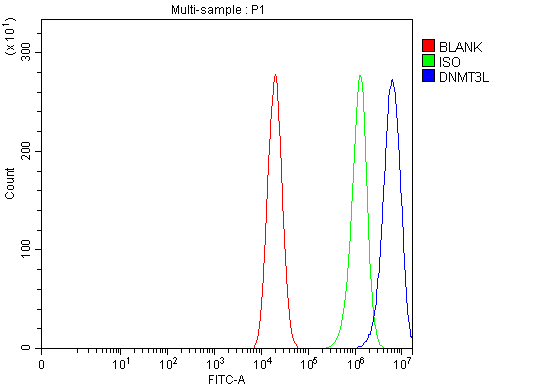
(Figure 2. Flow Cytometry analysis of Caco-2 cells using anti-DNMT3L antibody (AAA126367).Overlay histogram showing Caco-2 cells stained with AAA126367 (Blue line). The cells were blocked with 10% normal goat serum. And then incubated with rabbit anti-DNMT3L Antibody (AAA126367, 1 ug/1x10^6 cells) for 30 min at 20 degree C. DyLight488 conjugated goat anti-rabbit IgG was used as secondary antibody for 30 minutes at 20 degree C. Isotype control antibody (Green line) was rabbit IgG (1 ug/1x10^6) used under the same conditions. Unlabelled sample (Red line) was also used as a control.)
FCM/FACS (Flow Cytometry)
(Figure 2. Flow Cytometry analysis of Caco-2 cells using anti-DNMT3L antibody (AAA126367).Overlay histogram showing Caco-2 cells stained with AAA126367 (Blue line). The cells were blocked with 10% normal goat serum. And then incubated with rabbit anti-DNMT3L Antibody (AAA126367, 1 ug/1x10^6 cells) for 30 min at 20 degree C. DyLight488 conjugated goat anti-rabbit IgG was used as secondary antibody for 30 minutes at 20 degree C. Isotype control antibody (Green line) was rabbit IgG (1 ug/1x10^6) used under the same conditions. Unlabelled sample (Red line) was also used as a control.)
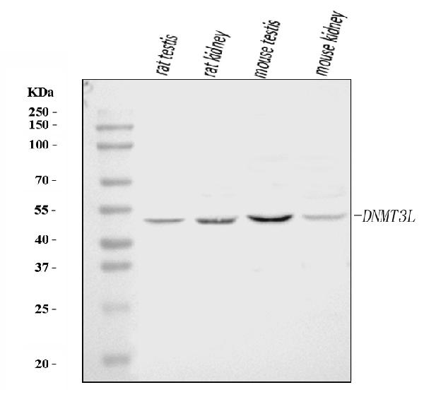
DNMT3L, Polyclonal Antibody (Cat# AAA126367)
Full Name
Anti-DNMT3L Antibody Picoband
Reactivity
Human, Mouse, Rat
Applications
Western Blot, Flow Cytometry
Purity
Immunogen affinity purified.
Pricing
 IHC (Immunohiostchemistry)
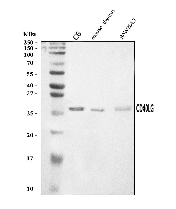
(Figure 2. IHC analysis of TRAP/CD40L/Cd40lg using anti-TRAP/CD40L/Cd40lg antibody (AAA126109).TRAP/CD40L/Cd40lg was detected in a paraffin-embedded section of rat spleen tissue. Heat mediated antigen retrieval was performed in EDTA buffer (pH 8.0, epitope retrieval solution). The tissue section was blocked with 10% goat serum. The tissue section was then incubated with 2 ug/ml rabbit anti-TRAP/CD40L/Cd40lg Antibody (AAA126109) overnight at 4 degree C. Biotinylated goat anti-rabbit IgG was used as secondary antibody and incubated for 30 minutes at 37 degree C. The tissue section was developed using Strepavidin-Biotin-Complex (SABC) with DAB as the chromogen.)
IHC (Immunohiostchemistry)
(Figure 2. IHC analysis of TRAP/CD40L/Cd40lg using anti-TRAP/CD40L/Cd40lg antibody (AAA126109).TRAP/CD40L/Cd40lg was detected in a paraffin-embedded section of rat spleen tissue. Heat mediated antigen retrieval was performed in EDTA buffer (pH 8.0, epitope retrieval solution). The tissue section was blocked with 10% goat serum. The tissue section was then incubated with 2 ug/ml rabbit anti-TRAP/CD40L/Cd40lg Antibody (AAA126109) overnight at 4 degree C. Biotinylated goat anti-rabbit IgG was used as secondary antibody and incubated for 30 minutes at 37 degree C. The tissue section was developed using Strepavidin-Biotin-Complex (SABC) with DAB as the chromogen.)
TRAP/CD40L/Cd40lg, Polyclonal Antibody (Cat# AAA126109)
Full Name
Anti-TRAP/CD40L/Cd40lg Antibody Picoband
Gene Names
Cd40lg; Cd40l; Tnfsf5
Reactivity
Mouse, Rat
Applications
Immunohistochemistry, Western Blot
Purity
Immunogen affinity purified.
Pricing
 IF (Immunofluorescence)
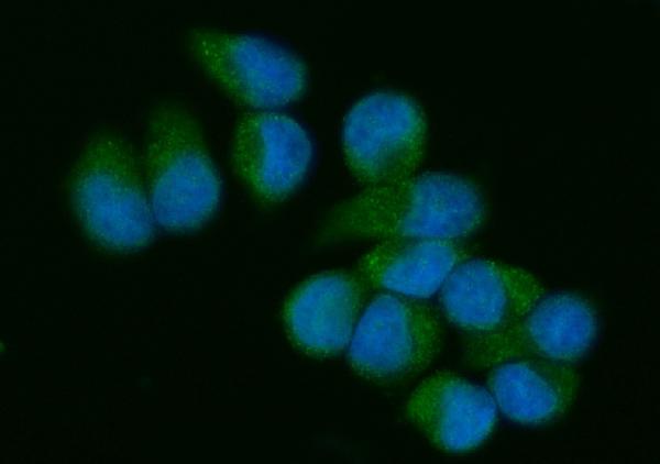
(Figure 2. IF analysis of VAV3 using anti-VAV3 antibody (AAA126290).VAV3 was detected in an immunocytochemical section of T-47D cells. Enzyme antigen retrieval was performed using IHC enzyme antigen retrieval reagent (AR0022) for 15 mins. The cells were blocked with 10% goat serum. And then incubated with 5 ug/mL rabbit anti-VAV3 Antibody (AAA126290) overnight at 4 degree C. DyLight488 Conjugated Goat Anti-Rabbit IgG was used as secondary antibody at 1:100 dilution and incubated for 30 minutes at 37 degree C. The section was counterstained with DAPI. Visualize using a fluorescence microscope and filter sets appropriate for the label used.)
IF (Immunofluorescence)
(Figure 2. IF analysis of VAV3 using anti-VAV3 antibody (AAA126290).VAV3 was detected in an immunocytochemical section of T-47D cells. Enzyme antigen retrieval was performed using IHC enzyme antigen retrieval reagent (AR0022) for 15 mins. The cells were blocked with 10% goat serum. And then incubated with 5 ug/mL rabbit anti-VAV3 Antibody (AAA126290) overnight at 4 degree C. DyLight488 Conjugated Goat Anti-Rabbit IgG was used as secondary antibody at 1:100 dilution and incubated for 30 minutes at 37 degree C. The section was counterstained with DAPI. Visualize using a fluorescence microscope and filter sets appropriate for the label used.)
VAV3, Polyclonal Antibody (Cat# AAA126290)
Full Name
Anti-VAV3 Antibody Picoband
Reactivity
Human
Applications
Western Blot, Immunocytochemistry, Immunofluorescence
Purity
Immunogen affinity purified.
Pricing
 FCM/FACS (Flow Cytometry)
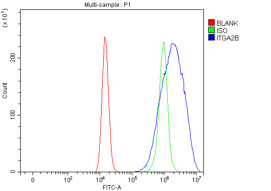
(Figure 3. Flow Cytometry analysis of HEL cells using anti-CD41/Integrin Alpha 2B/ITGA2B antibody (AAA127035).Overlay histogram showing HEL cells stained with AAA127035 (Blue line). The cells were fixed with 4% paraformaldehyde and blocked with 10% normal goat serum. And then incubated with rabbit anti-CD41/Integrin Alpha 2B/ITGA2B Antibody (AAA127035, 1ug/1x106 cells) for 30 min at 20 degree C. DyLight488 conjugated goat anti-rabbit IgG was used as secondary antibody for 30 minutes at 20 degree C. Isotype control antibody (Green line) was rabbit IgG (1ug/1x106) used under the same conditions. Unlabelled sample without incubation with primary antibody and secondary antibody (Red line) was used as a blank control.)
FCM/FACS (Flow Cytometry)
(Figure 3. Flow Cytometry analysis of HEL cells using anti-CD41/Integrin Alpha 2B/ITGA2B antibody (AAA127035).Overlay histogram showing HEL cells stained with AAA127035 (Blue line). The cells were fixed with 4% paraformaldehyde and blocked with 10% normal goat serum. And then incubated with rabbit anti-CD41/Integrin Alpha 2B/ITGA2B Antibody (AAA127035, 1ug/1x106 cells) for 30 min at 20 degree C. DyLight488 conjugated goat anti-rabbit IgG was used as secondary antibody for 30 minutes at 20 degree C. Isotype control antibody (Green line) was rabbit IgG (1ug/1x106) used under the same conditions. Unlabelled sample without incubation with primary antibody and secondary antibody (Red line) was used as a blank control.)
CD41/Integrin Alpha 2B/ITGA2B, Polyclonal Antibody (Cat# AAA127035)
Full Name
Anti-CD41/Integrin Alpha 2B/ITGA2B Antibody Picoband
Gene Names
ITGA2B; GT; GTA; CD41; GP2B; HPA3; CD41B; GPIIb; BDPLT2; BDPLT16
Reactivity
Human
Applications
Flow Cytometry, Immunohistochemistry, Western Blot
Purity
Immunogen affinity purified.
Pricing
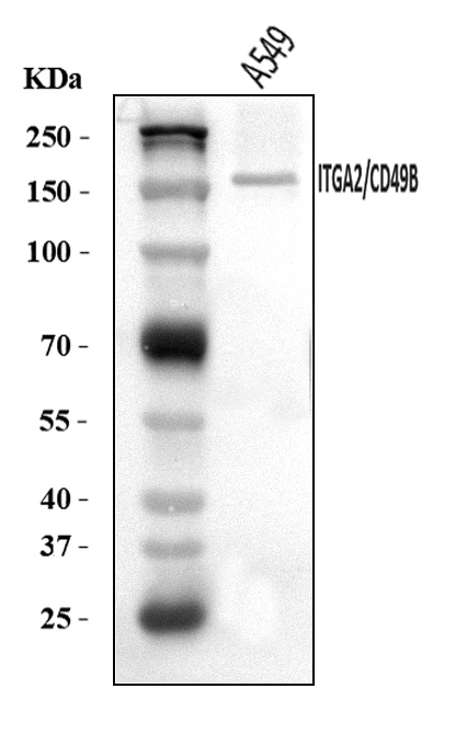
 FCM/FACS (Flow Cytometry)
(Figure 2. Flow Cytometry analysis of A375 cells using anti-Integrin Alpha 2/ITGA2 antibody (AAA126190).Overlay histogram showing A375 cells stained with AAA126190 (Blue line). The cells were blocked with 10% normal goat serum. And then incubated with rabbit anti-Integrin Alpha 2/ITGA2 Antibody (AAA126190, 1 ug/1x10^6 cells) for 30 min at 20 degree C. DyLight488 conjugated goat anti-rabbit IgG was used as secondary antibody for 30 minutes at 20 degree C. Isotype control antibody (Green line) was rabbit IgG (1 ug/1x10^6) used under the same conditions. Unlabelled sample (Red line) was also used as a control.)
FCM/FACS (Flow Cytometry)
(Figure 2. Flow Cytometry analysis of A375 cells using anti-Integrin Alpha 2/ITGA2 antibody (AAA126190).Overlay histogram showing A375 cells stained with AAA126190 (Blue line). The cells were blocked with 10% normal goat serum. And then incubated with rabbit anti-Integrin Alpha 2/ITGA2 Antibody (AAA126190, 1 ug/1x10^6 cells) for 30 min at 20 degree C. DyLight488 conjugated goat anti-rabbit IgG was used as secondary antibody for 30 minutes at 20 degree C. Isotype control antibody (Green line) was rabbit IgG (1 ug/1x10^6) used under the same conditions. Unlabelled sample (Red line) was also used as a control.)
Integrin alpha 2/ITGA2, Polyclonal Antibody (Cat# AAA126190)
Full Name
Anti-Integrin alpha 2/ITGA2 Antibody Picoband
Gene Names
ITGA2; BR; GPIa; CD49B; HPA-5; VLA-2; VLAA2
Reactivity
Human
Applications
Western Blot, Flow Cytometry
Purity
Immunogen affinity purified.
Pricing