Filters
Clonality
Type
Reactivity
Gene Name
Isotype
Host
Application
Clone
6878 results for "ELISA Kit" - showing 6850-6878


 FCM (Flow Cytometry)
(Figure 10. Flow Cytometry analysis of U20S cells using anti-PDIA5 antibody (AAA19331).Overlay histogram showing U20S cells stained with AAA19331 (Blue line). The cells were blocked with 10% normal goat serum. And then incubated with rabbit anti-PDIA5 Antibody (AAA19331, 1μg/1x106 cells) for 30 min at 20 degree C. DyLight®488 conjugated goat anti-rabbit IgG (5-10μg/1x106 cells) was used as secondary antibody for 30 minutes at 20 degree C. Isotype control antibody (Green line) was rabbit IgG (1μg/1x106) used under the same conditions. Unlabelled sample (Red line) was also used as a control.)
FCM (Flow Cytometry)
(Figure 10. Flow Cytometry analysis of U20S cells using anti-PDIA5 antibody (AAA19331).Overlay histogram showing U20S cells stained with AAA19331 (Blue line). The cells were blocked with 10% normal goat serum. And then incubated with rabbit anti-PDIA5 Antibody (AAA19331, 1μg/1x106 cells) for 30 min at 20 degree C. DyLight®488 conjugated goat anti-rabbit IgG (5-10μg/1x106 cells) was used as secondary antibody for 30 minutes at 20 degree C. Isotype control antibody (Green line) was rabbit IgG (1μg/1x106) used under the same conditions. Unlabelled sample (Red line) was also used as a control.)
PDIA5, Polyclonal Antibody (Cat# AAA19331)
Full Name
Anti-PDIA5 Antibody
Gene Names
PDIA5; PDIR
Reactivity
Human, Mouse, Rat
Applications
WB, IHC-P, ICC, IF, FC/FACS/FCM, EIA
Purity
Immunogen affinity purified.
Pricing


 FCM (Flow Cytometry)
(Figure 12. Flow Cytometry analysis of HEPA1-6 cells using anti-LSM5 antibody (AAA19332).Overlay histogram showing HEPA1-6 cells stained with AAA19332 (Blue line). The cells were blocked with 10% normal goat serum. And then incubated with rabbit anti-LSM5 Antibody (AAA19332, 1μg/1x106 cells) for 30 min at 20 degree C. DyLight®488 conjugated goat anti-rabbit IgG (5-10μg/1x106 cells) was used as secondary antibody for 30 minutes at 20 degree C. Isotype control antibody (Green line) was rabbit IgG (1μg/1x106) used under the same conditions. Unlabelled sample (Red line) was also used as a control.)
FCM (Flow Cytometry)
(Figure 12. Flow Cytometry analysis of HEPA1-6 cells using anti-LSM5 antibody (AAA19332).Overlay histogram showing HEPA1-6 cells stained with AAA19332 (Blue line). The cells were blocked with 10% normal goat serum. And then incubated with rabbit anti-LSM5 Antibody (AAA19332, 1μg/1x106 cells) for 30 min at 20 degree C. DyLight®488 conjugated goat anti-rabbit IgG (5-10μg/1x106 cells) was used as secondary antibody for 30 minutes at 20 degree C. Isotype control antibody (Green line) was rabbit IgG (1μg/1x106) used under the same conditions. Unlabelled sample (Red line) was also used as a control.)
LSM5, Polyclonal Antibody (Cat# AAA19332)
Full Name
Anti-LSM5 Antibody
Gene Names
LSM5; YER146W
Reactivity
Human, Mouse, Rat
Applications
WB, IHC-P, ICC, IF, FC/FACS/FCM, EIA
Purity
Immunogen affinity purified.
Pricing


 FCM (Flow Cytometry)
(Figure 9. Flow Cytometry analysis of U251 cells using anti-VPRBP/DCAF1 antibody (AAA19343).Overlay histogram showing U251 cells stained with AAA19343 (Blue line). The cells were blocked with 10% normal goat serum. And then incubated with rabbit anti-VPRBP/DCAF1 Antibody (AAA19343, 1μg/1x106 cells) for 30 min at 20 degree C. DyLight®488 conjugated goat anti-rabbit IgG (5-10μg/1x106 cells) was used as secondary antibody for 30 minutes at 20 degree C. Isotype control antibody (Green line) was rabbit IgG (1μg/1x106) used under the same conditions. Unlabelled sample (Red line) was also used as a control.)
FCM (Flow Cytometry)
(Figure 9. Flow Cytometry analysis of U251 cells using anti-VPRBP/DCAF1 antibody (AAA19343).Overlay histogram showing U251 cells stained with AAA19343 (Blue line). The cells were blocked with 10% normal goat serum. And then incubated with rabbit anti-VPRBP/DCAF1 Antibody (AAA19343, 1μg/1x106 cells) for 30 min at 20 degree C. DyLight®488 conjugated goat anti-rabbit IgG (5-10μg/1x106 cells) was used as secondary antibody for 30 minutes at 20 degree C. Isotype control antibody (Green line) was rabbit IgG (1μg/1x106) used under the same conditions. Unlabelled sample (Red line) was also used as a control.)
VPRBP/DCAF1, Polyclonal Antibody (Cat# AAA19343)
Full Name
Anti-VPRBP/DCAF1 Antibody
Gene Names
DCAF1; RIP; VPRBP
Reactivity
Human, Mouse, Rat
Applications
WB, IHC-P, ICC, IF, FC/FACS/FCM, EIA
Purity
Immunogen affinity purified.
Pricing


 FCM (Flow Cytometry)
(Figure 13. Flow Cytometry analysis of SiHa cells using anti-DRP1/DNM1L antibody (AAA19220).Overlay histogram showing SiHa cells stained with AAA19220 (Blue line). The cells were blocked with 10% normal goat serum. And then incubated with rabbit anti-DRP1/DNM1L Antibody (AAA19220, 1μg/1x106 cells) for 30 min at 20 degree C. DyLight®488 conjugated goat anti-rabbit IgG (5-10μg/1x106 cells) was used as secondary antibody for 30 minutes at 20 degree C. Isotype control antibody (Green line) was rabbit IgG (1μg/1x106) used under the same conditions. Unlabelled sample (Red line) was also used as a control.)
FCM (Flow Cytometry)
(Figure 13. Flow Cytometry analysis of SiHa cells using anti-DRP1/DNM1L antibody (AAA19220).Overlay histogram showing SiHa cells stained with AAA19220 (Blue line). The cells were blocked with 10% normal goat serum. And then incubated with rabbit anti-DRP1/DNM1L Antibody (AAA19220, 1μg/1x106 cells) for 30 min at 20 degree C. DyLight®488 conjugated goat anti-rabbit IgG (5-10μg/1x106 cells) was used as secondary antibody for 30 minutes at 20 degree C. Isotype control antibody (Green line) was rabbit IgG (1μg/1x106) used under the same conditions. Unlabelled sample (Red line) was also used as a control.)
DRP1/DNM1L, Polyclonal Antibody (Cat# AAA19220)
Full Name
Anti-DRP1/DNM1L Antibody
Gene Names
DNM1L; DLP1; DRP1; DVLP; EMPF; DYMPLE; HDYNIV
Reactivity
Human, Mouse, Rat
Applications
WB, IHC-P, ICC, IF, FC/FACS/FCM, EIA
Purity
Immunogen affinity purified.
Pricing


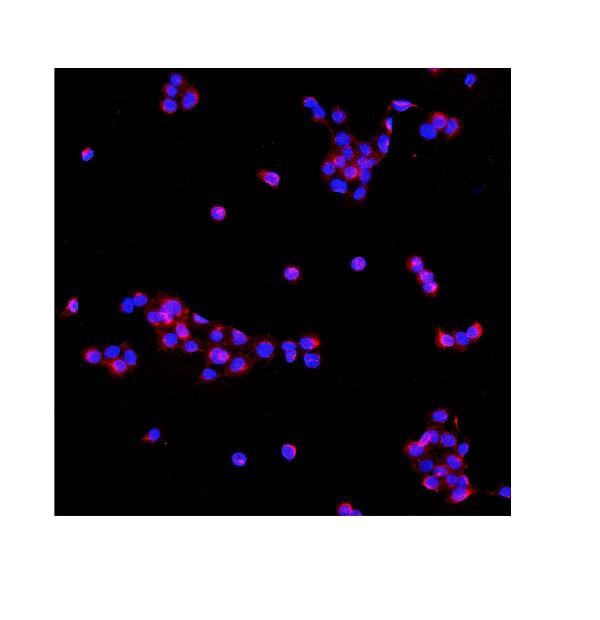
 IF (Immunofluorescence)
(Figure 9. IF analysis of P4HB using anti- P4HB antibody (AAA19258).P4HB was detected in immunocytochemical section of T-47D cells. Enzyme antigen retrieval was performed using IHC enzyme antigen retrieval reagent for 15 mins. The cells were blocked with 10% goat serum. And then incubated with 5μg/mL rabbit anti- P4HB Antibody (AAA19258) overnight at 4 degree C. DyLight®488 Conjugated Goat Anti-Rabbit IgG was used as secondary antibody at 1:100 dilution and incubated for 30 minutes at 37 degree C. The section was counterstained with DAPI. Visualize using a fluorescence microscope and filter sets appropriate for the label used.)
IF (Immunofluorescence)
(Figure 9. IF analysis of P4HB using anti- P4HB antibody (AAA19258).P4HB was detected in immunocytochemical section of T-47D cells. Enzyme antigen retrieval was performed using IHC enzyme antigen retrieval reagent for 15 mins. The cells were blocked with 10% goat serum. And then incubated with 5μg/mL rabbit anti- P4HB Antibody (AAA19258) overnight at 4 degree C. DyLight®488 Conjugated Goat Anti-Rabbit IgG was used as secondary antibody at 1:100 dilution and incubated for 30 minutes at 37 degree C. The section was counterstained with DAPI. Visualize using a fluorescence microscope and filter sets appropriate for the label used.)
P4HB, Polyclonal Antibody (Cat# AAA19258)
Full Name
Anti-P4HB Antibody
Gene Names
P4HB; DSI; GIT; PDI; PHDB; PDIA1; PO4DB; PO4HB; PROHB; ERBA2L; P4Hbeta
Reactivity
Human, Monkey, Mouse, Rat
Applications
WB, IHC-P, ICC, IF, FC/FACS/FCM, EIA
Purity
Immunogen affinity purified.
Pricing


 FCM (Flow Cytometry)
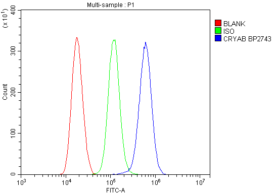
(Figure 13. Flow Cytometry analysis of THP-1 cells using anti-Alpha B Crystallin/CRYAB antibody (AAA19277).Overlay histogram showing THP-1 cells stained with AAA19277 (Blue line). The cells were blocked with 10% normal goat serum. And then incubated with rabbit anti-Alpha B Crystallin/CRYAB Antibody (AAA19277, 1μg/1x106 cells) for 30 min at 20 degree C. DyLight®488 conjugated goat anti-rabbit IgG (5-10μg/1x106 cells) was used as secondary antibody for 30 minutes at 20 degree C. Isotype control antibody (Green line) was rabbit IgG (1μg/1x106) used under the same conditions. Unlabelled sample (Red line) was also used as a control.)
FCM (Flow Cytometry)
(Figure 13. Flow Cytometry analysis of THP-1 cells using anti-Alpha B Crystallin/CRYAB antibody (AAA19277).Overlay histogram showing THP-1 cells stained with AAA19277 (Blue line). The cells were blocked with 10% normal goat serum. And then incubated with rabbit anti-Alpha B Crystallin/CRYAB Antibody (AAA19277, 1μg/1x106 cells) for 30 min at 20 degree C. DyLight®488 conjugated goat anti-rabbit IgG (5-10μg/1x106 cells) was used as secondary antibody for 30 minutes at 20 degree C. Isotype control antibody (Green line) was rabbit IgG (1μg/1x106) used under the same conditions. Unlabelled sample (Red line) was also used as a control.)
Alpha B Crystallin/CRYAB, Polyclonal Antibody (Cat# AAA19277)
Full Name
Anti-Alpha B Crystallin/CRYAB Antibody
Gene Names
CRYAB; CRYA2; CTPP2; HSPB5; CMD1II
Reactivity
Human, Mouse, Rat, Monkey
Applications
WB, IHC-P, ICC, IF, FC/FACS/FCM, EIA
Purity
Immunogen affinity purified.
Pricing


 FCM (Flow Cytometry)
(Figure 7. Flow Cytometry analysis of THP-1 cells using anti-PNPT1 antibody (AAA19306).Overlay histogram showing THP-1 cells stained with AAA19306 (Blue line). The cells were blocked with 10% normal goat serum. And then incubated with rabbit anti-PNPT1 Antibody (AAA19306, 1μg/1x106 cells) for 30 min at 20 degree C. DyLight®488 conjugated goat anti-rabbit IgG (5-10μg/1x106 cells) was used as secondary antibody for 30 minutes at 20 degree C. Isotype control antibody (Green line) was rabbit IgG (1μg/1x106) used under the same conditions. Unlabelled sample (Red line) was also used as a control.)
FCM (Flow Cytometry)
(Figure 7. Flow Cytometry analysis of THP-1 cells using anti-PNPT1 antibody (AAA19306).Overlay histogram showing THP-1 cells stained with AAA19306 (Blue line). The cells were blocked with 10% normal goat serum. And then incubated with rabbit anti-PNPT1 Antibody (AAA19306, 1μg/1x106 cells) for 30 min at 20 degree C. DyLight®488 conjugated goat anti-rabbit IgG (5-10μg/1x106 cells) was used as secondary antibody for 30 minutes at 20 degree C. Isotype control antibody (Green line) was rabbit IgG (1μg/1x106) used under the same conditions. Unlabelled sample (Red line) was also used as a control.)
PNPT1, Polyclonal Antibody (Cat# AAA19306)
Full Name
Anti-PNPT1 Antibody
Gene Names
PNPT1; OLD35; DFNB70; PNPASE; old-35; COXPD13
Reactivity
Human, Mouse, Rat
Applications
WB, IHC-P, ICC, IF, FC/FACS/FCM, EIA
Purity
Immunogen affinity purified.
Pricing


 WB (Western Blot)
(Figure 1. Western blot analysis of CISD2 using anti-CISD2 antibody (AAA19311).Electrophoresis was performed on a 5-20% SDS-PAGE gel at 70V (Stacking gel) / 90V (Resolving gel) for 2-3 hours. The sample well of each lane was loaded with 50ug of sample under reducing conditions.Lane 1: human HEK293 whole cell lysatesLane 2: human HELA whole cell lysatesLane 3: human MCF-7 whole cell lysatesLane 4: monkey kidney tissue lysatesLane 5: human SW620 whole cell lysatesLane 6: human Raji whole cell lysatesLane 7: rat kidney tissue lysatesLane 8: mouse kidney tissue lysates.After Electrophoresis, proteins were transferred to a Nitrocellulose membrane at 150mA for 50-90 minutes. Blocked the membrane with 5% Non-fat Milk/ TBS for 1. 5 hour at RT. The membrane was incubated with rabbit anti-CISD2 antigen affinity purified polyclonal antibody (Catalog # AAA19311) at 0. 5 μg/mL overnight at 4 degree C, then washed with TBS-0. 1%Tween 3 times with 5 minutes each and probed with a goat anti-rabbit IgG-HRP secondary antibody at a dilution of 1:10000 for 1. 5 hour at RT. The signal is developed using an Enhanced Chemiluminescent detection (ECL) kit (Catalog # with Tanon 5200 system. A specific band was detected for CISD2 at approximately 15KD. The expected band size for CISD2 is at 15KD.)
WB (Western Blot)
(Figure 1. Western blot analysis of CISD2 using anti-CISD2 antibody (AAA19311).Electrophoresis was performed on a 5-20% SDS-PAGE gel at 70V (Stacking gel) / 90V (Resolving gel) for 2-3 hours. The sample well of each lane was loaded with 50ug of sample under reducing conditions.Lane 1: human HEK293 whole cell lysatesLane 2: human HELA whole cell lysatesLane 3: human MCF-7 whole cell lysatesLane 4: monkey kidney tissue lysatesLane 5: human SW620 whole cell lysatesLane 6: human Raji whole cell lysatesLane 7: rat kidney tissue lysatesLane 8: mouse kidney tissue lysates.After Electrophoresis, proteins were transferred to a Nitrocellulose membrane at 150mA for 50-90 minutes. Blocked the membrane with 5% Non-fat Milk/ TBS for 1. 5 hour at RT. The membrane was incubated with rabbit anti-CISD2 antigen affinity purified polyclonal antibody (Catalog # AAA19311) at 0. 5 μg/mL overnight at 4 degree C, then washed with TBS-0. 1%Tween 3 times with 5 minutes each and probed with a goat anti-rabbit IgG-HRP secondary antibody at a dilution of 1:10000 for 1. 5 hour at RT. The signal is developed using an Enhanced Chemiluminescent detection (ECL) kit (Catalog # with Tanon 5200 system. A specific band was detected for CISD2 at approximately 15KD. The expected band size for CISD2 is at 15KD.)
CISD2, Polyclonal Antibody (Cat# AAA19311)
Full Name
Anti-CISD2 Antibody
Gene Names
CISD2; ERIS; WFS2; ZCD2; NAF-1; Miner1
Reactivity
Human, Mouse, Rat, Monkey
Applications
WB, IHC-P, ICC, IF, FC/FACS/FCM, EIA
Purity
Immunogen affinity purified.
Pricing


 FCM (Flow Cytometry)
(Figure 8. Flow Cytometry analysis of SiHa cells using anti-Astrin/Deepest/SPAG5 antibody (AAA19313).Overlay histogram showing SiHa cells stained with AAA19313 (Blue line). The cells were blocked with 10% normal goat serum. And then incubated with rabbit anti-Astrin/Deepest/SPAG5 Antibody (AAA19313, 1μg/1x106 cells) for 30 min at 20 degree C. DyLight®488 conjugated goat anti-rabbit IgG (5-10μg/1x106 cells) was used as secondary antibody for 30 minutes at 20 degree C. Isotype control antibody (Green line) was rabbit IgG (1μg/1x106) used under the same conditions. Unlabelled sample (Red line) was also used as a control.)
FCM (Flow Cytometry)
(Figure 8. Flow Cytometry analysis of SiHa cells using anti-Astrin/Deepest/SPAG5 antibody (AAA19313).Overlay histogram showing SiHa cells stained with AAA19313 (Blue line). The cells were blocked with 10% normal goat serum. And then incubated with rabbit anti-Astrin/Deepest/SPAG5 Antibody (AAA19313, 1μg/1x106 cells) for 30 min at 20 degree C. DyLight®488 conjugated goat anti-rabbit IgG (5-10μg/1x106 cells) was used as secondary antibody for 30 minutes at 20 degree C. Isotype control antibody (Green line) was rabbit IgG (1μg/1x106) used under the same conditions. Unlabelled sample (Red line) was also used as a control.)
Astrin/Deepest/SPAG5, Polyclonal Antibody (Cat# AAA19313)
Full Name
Anti-Astrin/Deepest/SPAG5 Antibody
Gene Names
SPAG5; MAP126; DEEPEST; hMAP126
Reactivity
Human
Applications
WB, IHC-P, ICC, IF, FC/FACS/FCM, EIA
Purity
Immunogen affinity purified.
Pricing


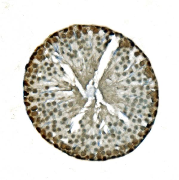
 IHC (Immunohistochemistry)
(Figure 7. IHC analysis of GCHFR using anti-GCHFR antibody (AAA19322).GCHFR was detected in paraffin-embedded section of mouse liver tissue. Heat mediated antigen retrieval was performed in EDTA buffer (pH8. 0, epitope retrieval solution). The tissue section was blocked with 10% goat serum. The tissue section was then incubated with 2μg/ml rabbit anti-GCHFR Antibody (AAA19322) overnight at 4 degree C. Biotinylated goat anti-rabbit IgG was used as secondary antibody and incubated for 30 minutes at 37 degree C. The tissue section was developed using Strepavidin-Biotin-Complex (SABC) (Catalog # with DAB as the chromogen.)
IHC (Immunohistochemistry)
(Figure 7. IHC analysis of GCHFR using anti-GCHFR antibody (AAA19322).GCHFR was detected in paraffin-embedded section of mouse liver tissue. Heat mediated antigen retrieval was performed in EDTA buffer (pH8. 0, epitope retrieval solution). The tissue section was blocked with 10% goat serum. The tissue section was then incubated with 2μg/ml rabbit anti-GCHFR Antibody (AAA19322) overnight at 4 degree C. Biotinylated goat anti-rabbit IgG was used as secondary antibody and incubated for 30 minutes at 37 degree C. The tissue section was developed using Strepavidin-Biotin-Complex (SABC) (Catalog # with DAB as the chromogen.)
GCHFR, Polyclonal Antibody (Cat# AAA19322)
Full Name
Anti-GCHFR Antibody
Gene Names
GCHFR; P35; GFRP; HsT16933
Reactivity
Human, Mouse, Rat
Applications
WB, IHC-P, ICC, IF, FC/FACS/FCM, EIA
Purity
Immunogen affinity purified.
Pricing


 FCM (Flow Cytometry)
(Figure 8. Flow Cytometry analysis of THP-1 cells using anti-NOX2/gp91phox/CYBB antibody (AAA19218).Overlay histogram showing THP-1 cells stained with AAA19218 (Blue line). The cells were blocked with 10% normal goat serum. And then incubated with rabbit anti-NOX2/gp91phox/CYBB Antibody (AAA19218, 1μg/1x106 cells) for 30 min at 20 degree C. DyLight®488 conjugated goat anti-rabbit IgG (5-10μg/1x106 cells) was used as secondary antibody for 30 minutes at 20 degree C. Isotype control antibody (Green line) was rabbit IgG (1μg/1x106) used under the same conditions. Unlabelled sample (Red line) was also used as a control.)
FCM (Flow Cytometry)
(Figure 8. Flow Cytometry analysis of THP-1 cells using anti-NOX2/gp91phox/CYBB antibody (AAA19218).Overlay histogram showing THP-1 cells stained with AAA19218 (Blue line). The cells were blocked with 10% normal goat serum. And then incubated with rabbit anti-NOX2/gp91phox/CYBB Antibody (AAA19218, 1μg/1x106 cells) for 30 min at 20 degree C. DyLight®488 conjugated goat anti-rabbit IgG (5-10μg/1x106 cells) was used as secondary antibody for 30 minutes at 20 degree C. Isotype control antibody (Green line) was rabbit IgG (1μg/1x106) used under the same conditions. Unlabelled sample (Red line) was also used as a control.)
NOX2/gp91phox/CYBB, Polyclonal Antibody (Cat# AAA19218)
Full Name
Anti-NOX2/gp91phox/CYBB Antibody
Gene Names
CYBB; CGD; NOX2; AMCBX2; GP91-1; GP91PHOX; p91-PHOX; GP91-PHOX
Reactivity
Human
Applications
WB, IHC-P, ICC, IF, FC/FACS/FCM, EIA
Purity
Immunogen affinity purified.
Pricing


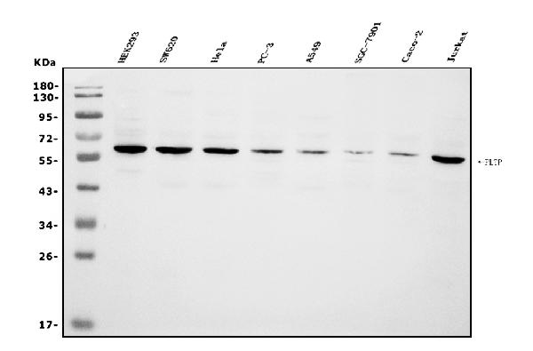
 FCM (Flow Cytometry)
(Figure 6. Flow Cytometry analysis of CACO-2 cells using anti-PLTP antibody (AAA19255).Overlay histogram showing CACO-2 cells stained with AAA19255 (Blue line). The cells were blocked with 10% normal goat serum. And then incubated with rabbit anti-PLTP Antibody (AAA19255, 1μg/1x106 cells) for 30 min at 20 degree C. DyLight®488 conjugated goat anti-rabbit IgG (5-10μg/1x106 cells) was used as secondary antibody for 30 minutes at 20 degree C. Isotype control antibody (Green line) was rabbit IgG (1μg/1x106) used under the same conditions. Unlabelled sample (Red line) was also used as a control.)
FCM (Flow Cytometry)
(Figure 6. Flow Cytometry analysis of CACO-2 cells using anti-PLTP antibody (AAA19255).Overlay histogram showing CACO-2 cells stained with AAA19255 (Blue line). The cells were blocked with 10% normal goat serum. And then incubated with rabbit anti-PLTP Antibody (AAA19255, 1μg/1x106 cells) for 30 min at 20 degree C. DyLight®488 conjugated goat anti-rabbit IgG (5-10μg/1x106 cells) was used as secondary antibody for 30 minutes at 20 degree C. Isotype control antibody (Green line) was rabbit IgG (1μg/1x106) used under the same conditions. Unlabelled sample (Red line) was also used as a control.)
PLTP, Polyclonal Antibody (Cat# AAA19255)
Full Name
Anti-PLTP Antibody
Gene Names
PLTP; BPIFE; HDLCQ9
Reactivity
Human
Applications
WB, IHC-P, ICC, IF, FC/FACS/FCM, EIA
Purity
Immunogen affinity purified.
Pricing


 FCM (Flow Cytometry)
(Figure 7. Flow Cytometry analysis of U87 cells using anti-PDE6 beta/PDE6B antibody (AAA19261).Overlay histogram showing U87 cells stained with AAA19261 (Blue line). The cells were blocked with 10% normal goat serum. And then incubated with rabbit anti-PDE6 beta/PDE6B Antibody (AAA19261,1μg/1x106 cells) for 30 min at 20 degree C. DyLight®488 conjugated goat anti-rabbit IgG (5-10μg/1x106 cells) was used as secondary antibody for 30 minutes at 20 degree C. Isotype control antibody (Green line) was rabbit IgG (1μg/1x106) used under the same conditions. Unlabelled sample (Red line) was also used as a control.)
FCM (Flow Cytometry)
(Figure 7. Flow Cytometry analysis of U87 cells using anti-PDE6 beta/PDE6B antibody (AAA19261).Overlay histogram showing U87 cells stained with AAA19261 (Blue line). The cells were blocked with 10% normal goat serum. And then incubated with rabbit anti-PDE6 beta/PDE6B Antibody (AAA19261,1μg/1x106 cells) for 30 min at 20 degree C. DyLight®488 conjugated goat anti-rabbit IgG (5-10μg/1x106 cells) was used as secondary antibody for 30 minutes at 20 degree C. Isotype control antibody (Green line) was rabbit IgG (1μg/1x106) used under the same conditions. Unlabelled sample (Red line) was also used as a control.)
PDE6 beta/PDE6B, Polyclonal Antibody (Cat# AAA19261)
Full Name
Anti-PDE6 beta/PDE6B Antibody
Reactivity
Human, Mouse, Rat
Applications
WB, IHC-P, ICC, IF, FC/FACS/FCM, EIA
Purity
Immunogen affinity purified.
Pricing


 FCM (Flow Cytometry)
(Figure 7. Flow Cytometry analysis of SiHa cells using anti-Aconitase 1/ACO1 antibody (AAA19267).Overlay histogram showing SiHa cells stained with AAA19267 (Blue line). The cells were blocked with 10% normal goat serum. And then incubated with rabbit anti-Aconitase 1/ACO1 Antibody (AAA19267,1μg/1x106 cells) for 30 min at 20 degree C. DyLight®488 conjugated goat anti-rabbit IgG (5-10μg/1x106 cells) was used as secondary antibody for 30 minutes at 20 degree C. Isotype control antibody (Green line) was rabbit IgG (1μg/1x106) used under the same conditions. Unlabelled sample (Red line) was also used as a control.)
FCM (Flow Cytometry)
(Figure 7. Flow Cytometry analysis of SiHa cells using anti-Aconitase 1/ACO1 antibody (AAA19267).Overlay histogram showing SiHa cells stained with AAA19267 (Blue line). The cells were blocked with 10% normal goat serum. And then incubated with rabbit anti-Aconitase 1/ACO1 Antibody (AAA19267,1μg/1x106 cells) for 30 min at 20 degree C. DyLight®488 conjugated goat anti-rabbit IgG (5-10μg/1x106 cells) was used as secondary antibody for 30 minutes at 20 degree C. Isotype control antibody (Green line) was rabbit IgG (1μg/1x106) used under the same conditions. Unlabelled sample (Red line) was also used as a control.)
Aconitase 1/ACO1, Polyclonal Antibody (Cat# AAA19267)
Full Name
Anti-Aconitase 1/ACO1 Antibody
Gene Names
ACO1; IRP1; ACONS; IREB1; IREBP; IREBP1
Reactivity
Human, Mouse, Monkey, Rat
Applications
WB, IHC-P, ICC, IF, FC/FACS/FCM, EIA
Purity
Immunogen affinity purified.
Pricing


 IF (Immunofluorescence)
(Figure 6. IF analysis of UNG using anti-UNG antibody (AAA19245).UNG was detected in immunocytochemical section of MCF-7 cells. Enzyme antigen retrieval was performed using IHC enzyme antigen retrieval reagent for 15 mins. The cells were blocked with 10% goat serum. And then incubated with 5μg/mL rabbit anti- UNG Antibody (AAA19245) overnight at 4 degree C. DyLight®594 Conjugated Goat Anti-Rabbit IgG (BA1142) was used as secondary antibody at 1:100 dilution and incubated for 30 minutes at 37 degree C. The section was counterstained with DAPI. Visualize using a fluorescence microscope and filter sets appropriate for the label used.)
IF (Immunofluorescence)
(Figure 6. IF analysis of UNG using anti-UNG antibody (AAA19245).UNG was detected in immunocytochemical section of MCF-7 cells. Enzyme antigen retrieval was performed using IHC enzyme antigen retrieval reagent for 15 mins. The cells were blocked with 10% goat serum. And then incubated with 5μg/mL rabbit anti- UNG Antibody (AAA19245) overnight at 4 degree C. DyLight®594 Conjugated Goat Anti-Rabbit IgG (BA1142) was used as secondary antibody at 1:100 dilution and incubated for 30 minutes at 37 degree C. The section was counterstained with DAPI. Visualize using a fluorescence microscope and filter sets appropriate for the label used.)
UNG, Polyclonal Antibody (Cat# AAA19245)
Full Name
Anti-UNG Antibody
Gene Names
UNG; DGU; UDG; UNG1; UNG2; HIGM4; HIGM5; UNG15
Reactivity
Human, Mouse, Rat, Monkey
Applications
WB, IHC-P, ICC, IF, FC/FACS/FCM, EIA
Purity
Immunogen affinity purified.
Pricing


 FCM (Flow Cytometry)
(Figure 7. Flow Cytometry analysis of U251 cells using anti-CGKI/PRKG1 antibody (AAA19246).Overlay histogram showing U251 cells stained with AAA19246 (Blue line). The cells were blocked with 10% normal goat serum. And then incubated with rabbit anti-CGKI/PRKG1 Antibody (AAA19246, 1μg/1x106 cells) for 30 min at 20 degree C. DyLight®488 conjugated goat anti-rabbit IgG (5-10μg/1x106 cells) was used as secondary antibody for 30 minutes at 20 degree C. Isotype control antibody (Green line) was rabbit IgG (1μg/1x106) used under the same conditions. Unlabelled sample (Red line) was also used as a control.)
FCM (Flow Cytometry)
(Figure 7. Flow Cytometry analysis of U251 cells using anti-CGKI/PRKG1 antibody (AAA19246).Overlay histogram showing U251 cells stained with AAA19246 (Blue line). The cells were blocked with 10% normal goat serum. And then incubated with rabbit anti-CGKI/PRKG1 Antibody (AAA19246, 1μg/1x106 cells) for 30 min at 20 degree C. DyLight®488 conjugated goat anti-rabbit IgG (5-10μg/1x106 cells) was used as secondary antibody for 30 minutes at 20 degree C. Isotype control antibody (Green line) was rabbit IgG (1μg/1x106) used under the same conditions. Unlabelled sample (Red line) was also used as a control.)
cGKI/PRKG1, Polyclonal Antibody (Cat# AAA19246)
Full Name
Anti-cGKI/PRKG1 Antibody
Gene Names
PRKG1; 1; PKG; cGK; AAT8; cGK1; cGKI; cGK 1; PRKG1B; PRKGR1B; cGKI-BETA; cGKI-alpha
Reactivity
Human, Mouse, Rat
Applications
WB, IHC-P, ICC, IF, FC/FACS/FCM, EIA
Purity
Immunogen affinity purified.
Pricing


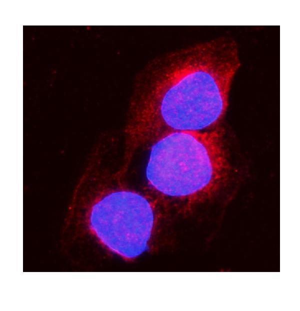
 IF (Immunofluorescence)
(Figure 11. IF analysis of H Cadherin/CDH13 using anti- H Cadherin/CDH13 antibody (AAA19252).H Cadherin/CDH13 was detected in immunocytochemical section of SIHA cells. Enzyme antigen retrieval was performed using IHC enzyme antigen retrieval reagent for 15 mins. The cells were blocked with 10% goat serum. And then incubated with 5μg/mL rabbit anti- H Cadherin/CDH13 Antibody (AAA19252) overnight at 4 degree C. DyLight®488 Conjugated Goat Anti-Rabbit IgG was used as secondary antibody at 1:100 dilution and incubated for 30 minutes at 37 degree C. The section was counterstained with DAPI. Visualize using a fluorescence microscope and filter sets appropriate for the label used.)
IF (Immunofluorescence)
(Figure 11. IF analysis of H Cadherin/CDH13 using anti- H Cadherin/CDH13 antibody (AAA19252).H Cadherin/CDH13 was detected in immunocytochemical section of SIHA cells. Enzyme antigen retrieval was performed using IHC enzyme antigen retrieval reagent for 15 mins. The cells were blocked with 10% goat serum. And then incubated with 5μg/mL rabbit anti- H Cadherin/CDH13 Antibody (AAA19252) overnight at 4 degree C. DyLight®488 Conjugated Goat Anti-Rabbit IgG was used as secondary antibody at 1:100 dilution and incubated for 30 minutes at 37 degree C. The section was counterstained with DAPI. Visualize using a fluorescence microscope and filter sets appropriate for the label used.)
H Cadherin/CDH13, Polyclonal Antibody (Cat# AAA19252)
Full Name
Anti-H Cadherin/CDH13 Antibody
Gene Names
CDH13; CDHH; P105
Reactivity
Human, Rat
Applications
WB, IHC-P, ICC, IF, FC/FACS/FCM, EIA
Purity
Immunogen affinity purified.
Pricing


 FCM (Flow Cytometry)
(Figure 6. Flow Cytometry analysis of U937 cells using anti-BDH1 antibody (AAA19328).Overlay histogram showing U937 cells stained with AAA19328 (Blue line). The cells were blocked with 10% normal goat serum. And then incubated with rabbit anti-BDH1 Antibody (AAA19328,1μg/1x106 cells) for 30 min at 20 degree C. DyLight®488 conjugated goat anti-rabbit IgG (5-10μg/1x106 cells) was used as secondary antibody for 30 minutes at 20 degree C. Isotype control antibody (Green line) was rabbit IgG (1μg/1x106) used under the same conditions. Unlabelled sample (Red line) was also used as a control.)
FCM (Flow Cytometry)
(Figure 6. Flow Cytometry analysis of U937 cells using anti-BDH1 antibody (AAA19328).Overlay histogram showing U937 cells stained with AAA19328 (Blue line). The cells were blocked with 10% normal goat serum. And then incubated with rabbit anti-BDH1 Antibody (AAA19328,1μg/1x106 cells) for 30 min at 20 degree C. DyLight®488 conjugated goat anti-rabbit IgG (5-10μg/1x106 cells) was used as secondary antibody for 30 minutes at 20 degree C. Isotype control antibody (Green line) was rabbit IgG (1μg/1x106) used under the same conditions. Unlabelled sample (Red line) was also used as a control.)
BDH1, Polyclonal Antibody (Cat# AAA19328)
Full Name
Anti-BDH1 Antibody
Gene Names
BDH1; BDH; SDR9C1
Reactivity
Human, Rat
Applications
WB, IHC-P, ICC, IF, FC/FACS/FCM, EIA
Purity
Immunogen affinity purified.
Pricing


 FCM (Flow Cytometry)
(Figure 6. Flow Cytometry analysis of A431 cells using anti-NDUFB5 antibody (AAA19333).Overlay histogram showing A431 cells stained with AAA19333 (Blue line). The cells were blocked with 10% normal goat serum. And then incubated with rabbit anti-NDUFB5 Antibody (AAA19333, 1μg/1x106 cells) for 30 min at 20 degree C. DyLight®488 conjugated goat anti-rabbit IgG (5-10μg/1x106 cells) was used as secondary antibody for 30 minutes at 20 degree C. Isotype control antibody (Green line) was rabbit IgG (1μg/1x106) used under the same conditions. Unlabelled sample (Red line) was also used as a control.)
FCM (Flow Cytometry)
(Figure 6. Flow Cytometry analysis of A431 cells using anti-NDUFB5 antibody (AAA19333).Overlay histogram showing A431 cells stained with AAA19333 (Blue line). The cells were blocked with 10% normal goat serum. And then incubated with rabbit anti-NDUFB5 Antibody (AAA19333, 1μg/1x106 cells) for 30 min at 20 degree C. DyLight®488 conjugated goat anti-rabbit IgG (5-10μg/1x106 cells) was used as secondary antibody for 30 minutes at 20 degree C. Isotype control antibody (Green line) was rabbit IgG (1μg/1x106) used under the same conditions. Unlabelled sample (Red line) was also used as a control.)
NDUFB5, Polyclonal Antibody (Cat# AAA19333)
Full Name
Anti-NDUFB5 Antibody
Gene Names
NDUFB5; SGDH; CISGDH
Reactivity
Human, Mouse, Rat
Applications
WB, IHC-P, ICC, IF, FC/FACS/FCM, EIA
Purity
Immunogen affinity purified.
Pricing


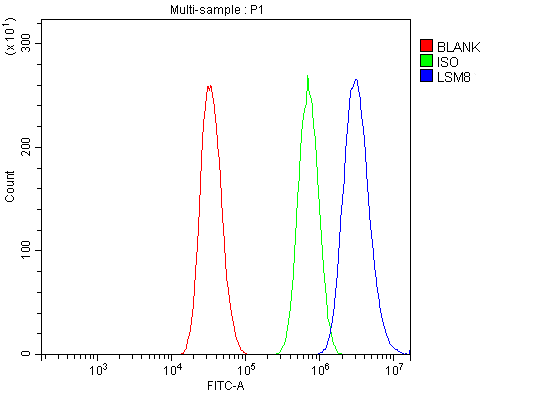 FCM (Flow Cytometry)
(Figure 13. Flow Cytometry analysis of A431 cells using anti-LSM8 antibody (AAA19337).Overlay histogram showing A431 cells stained with AAA19337 (Blue line). The cells were blocked with 10% normal goat serum. And then incubated with rabbit anti-LSM8 Antibody (AAA19337, 1μg/1x106 cells) for 30 min at 20 degree C. DyLight®488 conjugated goat anti-rabbit IgG (5-10μg/1x106 cells) was used as secondary antibody for 30 minutes at 20 degree C. Isotype control antibody (Green line) was rabbit IgG (1μg/1x106) used under the same conditions. Unlabelled sample (Red line) was also used as a control.)
FCM (Flow Cytometry)
(Figure 13. Flow Cytometry analysis of A431 cells using anti-LSM8 antibody (AAA19337).Overlay histogram showing A431 cells stained with AAA19337 (Blue line). The cells were blocked with 10% normal goat serum. And then incubated with rabbit anti-LSM8 Antibody (AAA19337, 1μg/1x106 cells) for 30 min at 20 degree C. DyLight®488 conjugated goat anti-rabbit IgG (5-10μg/1x106 cells) was used as secondary antibody for 30 minutes at 20 degree C. Isotype control antibody (Green line) was rabbit IgG (1μg/1x106) used under the same conditions. Unlabelled sample (Red line) was also used as a control.)
LSM8, Polyclonal Antibody (Cat# AAA19337)
Full Name
Anti-LSM8 Antibody
Reactivity
Human, Mouse, Rat
Applications
WB, IHC-P, ICC, IF, FC/FACS/FCM, EIA
Purity
Immunogen affinity purified.
Pricing


 FCM (Flow Cytometry)
(Figure 9. Flow Cytometry analysis of HL-60 cells using anti-NDUFB10 antibody (AAA19339).Overlay histogram showing HL-60 cells stained with AAA19339 (Blue line). The cells were blocked with 10% normal goat serum. And then incubated with rabbit anti-NDUFB10 Antibody (AAA19339, 1μg/1x106 cells) for 30 min at 20 degree C. DyLight®488 conjugated goat anti-rabbit IgG (5-10μg/1x106 cells) was used as secondary antibody for 30 minutes at 20 degree C. Isotype control antibody (Green line) was rabbit IgG (1μg/1x106) used under the same conditions. Unlabelled sample (Red line) was also used as a control.)
FCM (Flow Cytometry)
(Figure 9. Flow Cytometry analysis of HL-60 cells using anti-NDUFB10 antibody (AAA19339).Overlay histogram showing HL-60 cells stained with AAA19339 (Blue line). The cells were blocked with 10% normal goat serum. And then incubated with rabbit anti-NDUFB10 Antibody (AAA19339, 1μg/1x106 cells) for 30 min at 20 degree C. DyLight®488 conjugated goat anti-rabbit IgG (5-10μg/1x106 cells) was used as secondary antibody for 30 minutes at 20 degree C. Isotype control antibody (Green line) was rabbit IgG (1μg/1x106) used under the same conditions. Unlabelled sample (Red line) was also used as a control.)
NDUFB10, Polyclonal Antibody (Cat# AAA19339)
Full Name
Anti-NDUFB10 Antibody
Gene Names
NDUFB10; PDSW
Reactivity
Human, Mouse, Rat
Applications
WB, IHC-P, ICC, IF, FC/FACS/FCM, EIA
Purity
Immunogen affinity purified.
Pricing
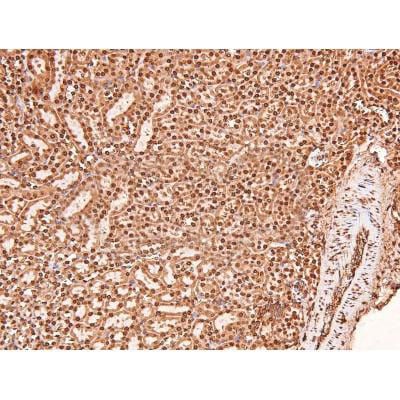
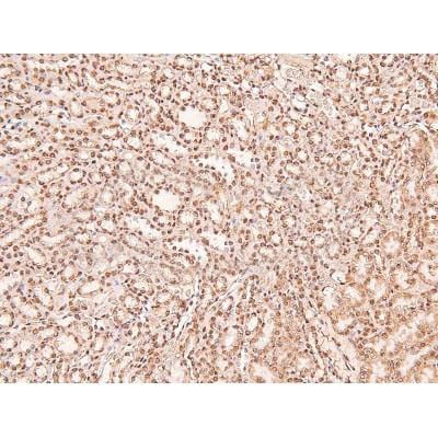


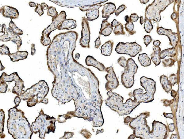
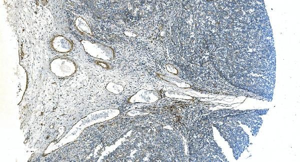
 IHC (Immunohistochemistry)
(At 1/200 staining Human lung cancer tissue sections by IHC-P. The tissue was formaldehyde fixed and a heat mediated antigen retrieval step in citrate buffer was performed. The tissue was then blocked and incubated with the antibody for 1.5 hours at 22 degree C. An HRP conjugated goat anti-rabbit antibody was used as the secondary antibody.)
IHC (Immunohistochemistry)
(At 1/200 staining Human lung cancer tissue sections by IHC-P. The tissue was formaldehyde fixed and a heat mediated antigen retrieval step in citrate buffer was performed. The tissue was then blocked and incubated with the antibody for 1.5 hours at 22 degree C. An HRP conjugated goat anti-rabbit antibody was used as the secondary antibody.)
ERK1/2, Polyclonal Antibody (Cat# AAA31416)
Full Name
Phospho-ERK1/2 (Thr202+Tyr204/Thr185+Tyr187) Antibody
Gene Names
MAPK3; ERK1; ERT2; ERK-1; PRKM3; P44ERK1; P44MAPK; HS44KDAP; HUMKER1A; p44-ERK1; p44-MAPK
Reactivity
Human, Mouse, Rat, Monkey
Predicted Reactivity: Pig (100%), Zebrafish (100%), Bovine (100%), Horse (100%), Sheep (100%), Rabbit (100%)
Predicted Reactivity: Pig (100%), Zebrafish (100%), Bovine (100%), Horse (100%), Sheep (100%), Rabbit (100%)
Applications
WB, IHC, EIA
Purity
The antibody is from purified rabbit serum by affinity purification via sequential chromatography on phospho-peptide and non-phospho-peptide affinity columns.
Pricing


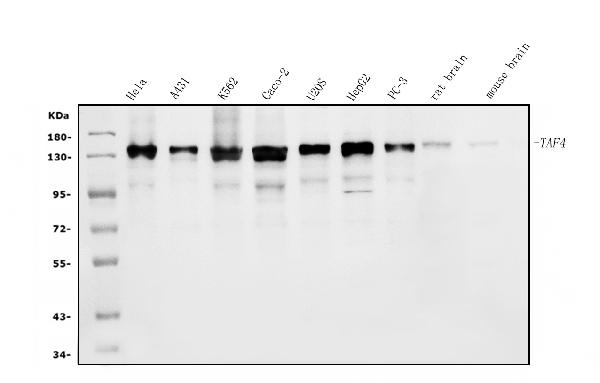
 FCM (Flow Cytometry)
(Figure 13. Flow Cytometry analysis of Hela cells using anti-TAF4 antibody (AAA19307).Overlay histogram showing Hela cells stained with AAA19307 (Blue line). The cells were blocked with 10% normal goat serum. And then incubated with rabbit anti-TAF4 Antibody (AAA19307, 1μg/1x106 cells) for 30 min at 20 degree C. DyLight®488 conjugated goat anti-rabbit IgG (5-10μg/1x106 cells) was used as secondary antibody for 30 minutes at 20 degree C. Isotype control antibody (Green line) was rabbit IgG (1μg/1x106) used under the same conditions. Unlabelled sample (Red line) was also used as a control.)
FCM (Flow Cytometry)
(Figure 13. Flow Cytometry analysis of Hela cells using anti-TAF4 antibody (AAA19307).Overlay histogram showing Hela cells stained with AAA19307 (Blue line). The cells were blocked with 10% normal goat serum. And then incubated with rabbit anti-TAF4 Antibody (AAA19307, 1μg/1x106 cells) for 30 min at 20 degree C. DyLight®488 conjugated goat anti-rabbit IgG (5-10μg/1x106 cells) was used as secondary antibody for 30 minutes at 20 degree C. Isotype control antibody (Green line) was rabbit IgG (1μg/1x106) used under the same conditions. Unlabelled sample (Red line) was also used as a control.)
TAF4, Polyclonal Antibody (Cat# AAA19307)
Full Name
Anti-TAF4 Antibody
Gene Names
TAF4; TAF2C; TAF4A; TAF2C1; TAFII130; TAFII135
Reactivity
Human, Mouse, Rat
Applications
WB, IHC-P, ICC, IF, EIA
Purity
Immunogen affinity purified.
Pricing


 FCM (Flow Cytometry)
(Figure 8. Flow Cytometry analysis of A431 cells using anti-EGFR antibody (AAA19214).Overlay histogram showing A431 cells stained with AAA19214 (Blue line). The cells were blocked with 10% normal goat serum. And then incubated with rabbit anti-EGFR Antibody (AAA19214,1μg/1x106 cells) for 30 min at 20 degree C. DyLight®488 conjugated goat anti-rabbit IgG (5-10μg/1x106 cells) was used as secondary antibody for 30 minutes at 20 degree C. Isotype control antibody (Green line) was rabbit IgG (1μg/1x106) used under the same conditions. Unlabelled sample (Red line) was also used as a control.)
FCM (Flow Cytometry)
(Figure 8. Flow Cytometry analysis of A431 cells using anti-EGFR antibody (AAA19214).Overlay histogram showing A431 cells stained with AAA19214 (Blue line). The cells were blocked with 10% normal goat serum. And then incubated with rabbit anti-EGFR Antibody (AAA19214,1μg/1x106 cells) for 30 min at 20 degree C. DyLight®488 conjugated goat anti-rabbit IgG (5-10μg/1x106 cells) was used as secondary antibody for 30 minutes at 20 degree C. Isotype control antibody (Green line) was rabbit IgG (1μg/1x106) used under the same conditions. Unlabelled sample (Red line) was also used as a control.)
EGFR, Polyclonal Antibody (Cat# AAA19214)
Full Name
Anti-EGFR Antibody
Gene Names
EGFR; ERBB; HER1; mENA; ERBB1; PIG61
Reactivity
Human, Mouse, Rat
Applications
WB, IHC-P, ICC, IF, FC/FACS/FCM, EIA
Purity
Immunogen affinity purified.
Pricing


 FCM (Flow Cytometry)
(Figure 18. Flow Cytometry analysis of A431 cells using anti-LSM7 antibody (AAA19327).Overlay histogram showing A431 cells stained with AAA19327 (Blue line). The cells were blocked with 10% normal goat serum. And then incubated with rabbit anti-LSM7 Antibody (AAA19327, 1μg/1x106 cells) for 30 min at 20 degree C. DyLight®488 conjugated goat anti-rabbit IgG (5-10μg/1x106 cells) was used as secondary antibody for 30 minutes at 20 degree C. Isotype control antibody (Green line) was rabbit IgG (1μg/1x106) used under the same conditions. Unlabelled sample (Red line) was also used as a control.)
FCM (Flow Cytometry)
(Figure 18. Flow Cytometry analysis of A431 cells using anti-LSM7 antibody (AAA19327).Overlay histogram showing A431 cells stained with AAA19327 (Blue line). The cells were blocked with 10% normal goat serum. And then incubated with rabbit anti-LSM7 Antibody (AAA19327, 1μg/1x106 cells) for 30 min at 20 degree C. DyLight®488 conjugated goat anti-rabbit IgG (5-10μg/1x106 cells) was used as secondary antibody for 30 minutes at 20 degree C. Isotype control antibody (Green line) was rabbit IgG (1μg/1x106) used under the same conditions. Unlabelled sample (Red line) was also used as a control.)
LSM7, Polyclonal Antibody (Cat# AAA19327)
Full Name
Anti-LSM7 Antibody
Gene Names
LSM7; YNL147W
Reactivity
Human, Mouse, Rat
Applications
WB, IHC-P, ICC, IF, FC/FACS/FCM, EIA
Purity
Immunogen affinity purified.
Pricing


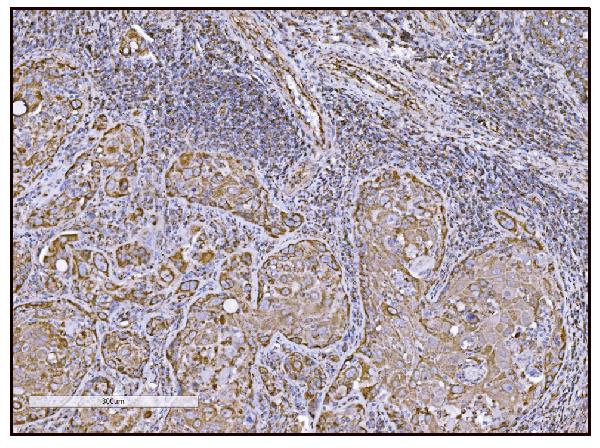


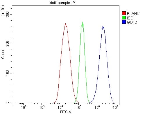


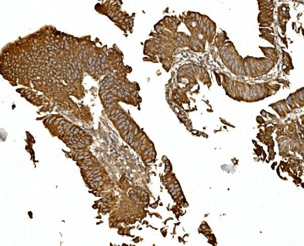
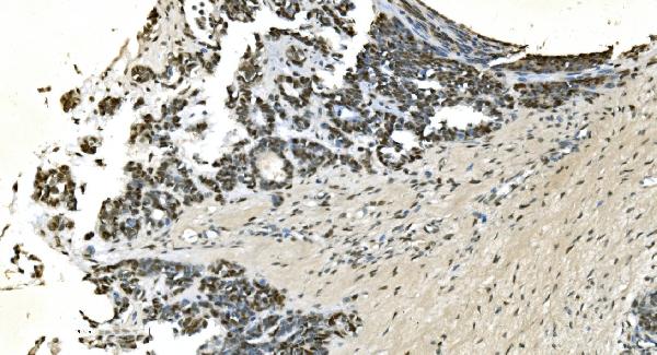
 IHC (Immunohistochemistry)
(Figure 14. IHC analysis of FABP-1/GOT2 using anti-FABP-1/GOT2 antibody (AAA19309).FABP-1/GOT2 was detected in paraffin-embedded section of rat lymph node tissue. Heat mediated antigen retrieval was performed in EDTA buffer (pH8. 0, epitope retrieval solution). The tissue section was blocked with 10% goat serum. The tissue section was then incubated with 2μg/ml rabbit anti-FABP-1/GOT2 Antibody (AAA19309) overnight at 4 degree C. Biotinylated goat anti-rabbit IgG was used as secondary antibody and incubated for 30 minutes at 37 degree C. The tissue section was developed using Strepavidin-Biotin-Complex (SABC) (Catalog # with DAB as the chromogen.)
IHC (Immunohistochemistry)
(Figure 14. IHC analysis of FABP-1/GOT2 using anti-FABP-1/GOT2 antibody (AAA19309).FABP-1/GOT2 was detected in paraffin-embedded section of rat lymph node tissue. Heat mediated antigen retrieval was performed in EDTA buffer (pH8. 0, epitope retrieval solution). The tissue section was blocked with 10% goat serum. The tissue section was then incubated with 2μg/ml rabbit anti-FABP-1/GOT2 Antibody (AAA19309) overnight at 4 degree C. Biotinylated goat anti-rabbit IgG was used as secondary antibody and incubated for 30 minutes at 37 degree C. The tissue section was developed using Strepavidin-Biotin-Complex (SABC) (Catalog # with DAB as the chromogen.)
FABP-1/GOT2, Polyclonal Antibody (Cat# AAA19309)
Full Name
Anti-FABP-1/GOT2 Antibody
Gene Names
GOT2; KAT4; KATIV; mitAAT
Reactivity
Human, Mouse, Rat
Applications
WB, IHC-P, ICC, IF, FC/FACS/FCM, EIA
Purity
Immunogen affinity purified.
Pricing


 FCM (Flow Cytometry)
(Figure 11. Flow Cytometry analysis of THP-1 cells using anti-EPRS1/PARS antibody (AAA19268).Overlay histogram showing THP-1 cells stained with AAA19268 (Blue line). The cells were blocked with 10% normal goat serum. And then incubated with rabbit anti-EPRS1/PARS Antibody (AAA19268, 1μg/1x106 cells) for 30 min at 20 degree C. DyLight®488 conjugated goat anti-rabbit IgG (5-10μg/1x106 cells) was used as secondary antibody for 30 minutes at 20 degree C. Isotype control antibody (Green line) was rabbit IgG (1μg/1x106) used under the same conditions. Unlabelled sample (Red line) was also used as a control.)
FCM (Flow Cytometry)
(Figure 11. Flow Cytometry analysis of THP-1 cells using anti-EPRS1/PARS antibody (AAA19268).Overlay histogram showing THP-1 cells stained with AAA19268 (Blue line). The cells were blocked with 10% normal goat serum. And then incubated with rabbit anti-EPRS1/PARS Antibody (AAA19268, 1μg/1x106 cells) for 30 min at 20 degree C. DyLight®488 conjugated goat anti-rabbit IgG (5-10μg/1x106 cells) was used as secondary antibody for 30 minutes at 20 degree C. Isotype control antibody (Green line) was rabbit IgG (1μg/1x106) used under the same conditions. Unlabelled sample (Red line) was also used as a control.)
EPRS1/PARS, Polyclonal Antibody (Cat# AAA19268)
Full Name
Anti-EPRS1/PARS Antibody
Gene Names
EPRS; EARS; PARS; QARS; QPRS; PIG32; GLUPRORS
Reactivity
Human, Mouse, Rat
Applications
WB, IHC-P, ICC, IF, FC/FACS/FCM, EIA
Purity
Immunogen affinity purified.
Pricing


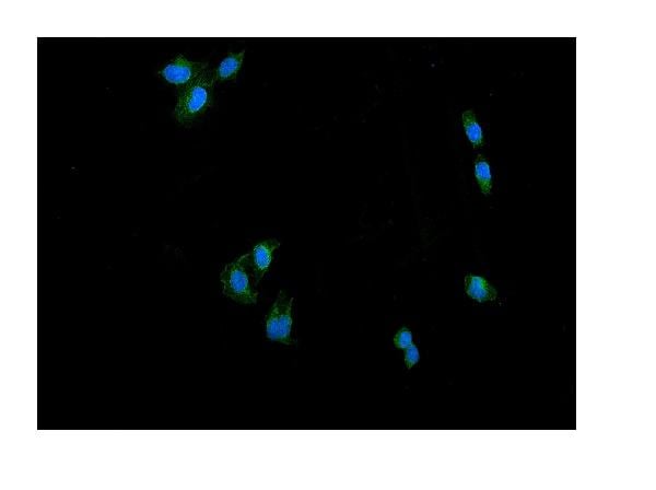
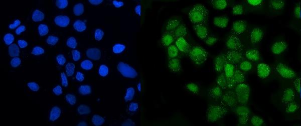
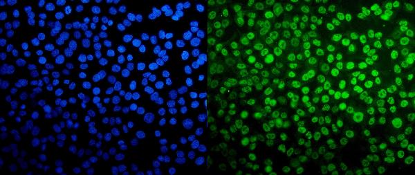
 Application Data
(At 25 degree C. Samples were then incubated with primary Ab(At 37 degree C. An AlexaFluor594 conjugated goat anti-rabbit IgG(H+L) Ab(Red) and an AlexaFluor488 conjugated goat anti-mouse IgG(H+L) Ab(Green) were used as the secondary antibody.The nuclear counter stain is DAPI (blue).)
Application Data
(At 25 degree C. Samples were then incubated with primary Ab(At 37 degree C. An AlexaFluor594 conjugated goat anti-rabbit IgG(H+L) Ab(Red) and an AlexaFluor488 conjugated goat anti-mouse IgG(H+L) Ab(Green) were used as the secondary antibody.The nuclear counter stain is DAPI (blue).)
ERK1/2, Polyclonal Antibody (Cat# AAA31300)
Full Name
Phospho-ERK1/2 (Thr177/Thr160) Antibody
Gene Names
MAPK3; ERK1; ERT2; ERK-1; PRKM3; P44ERK1; P44MAPK; HS44KDAP; HUMKER1A; p44-ERK1; p44-MAPK
Reactivity
Human, Mouse, Rat
Applications
IHC, IF, ICC, EIA
Purity
The antibody is from purified rabbit serum by affinity purification via sequential chromatography on phospho-peptide and non-phospho-peptide affinity columns.
Pricing