Filters
Clonality
Type
Reactivity
Gene Name
Isotype
Host
Application
Clone
6878 results for "ELISA Kit" - showing 6750-6800


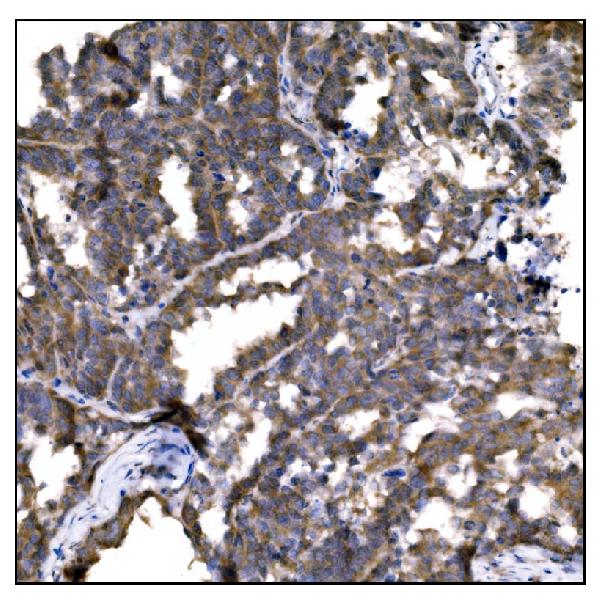
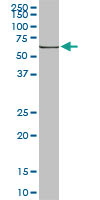 WB (Western Blot)
(HSPA1B monoclonal antibody Western Blot analysis of HSPA1B expression in Raw 264.7.)
WB (Western Blot)
(HSPA1B monoclonal antibody Western Blot analysis of HSPA1B expression in Raw 264.7.)
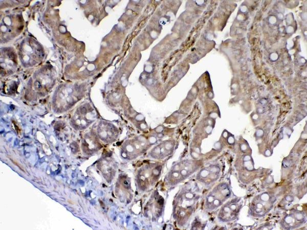
HSPA1A, Monoclonal Antibody (Cat# AAA24241)
Full Name
HSPA1A (Heat Shock 70kD Protein 1A/1B, Heat Shock 70kD Protein 1/2, HSP70.1/HSP70.2, HSP70-1/HSP70-2, HSPA1, HSPA1B) (AP)
Gene Names
HSPA1A; HSP72; HSPA1; HSP70I; HSP70-1; HSP70.1; HSP70-1A; HEL-S-103
Reactivity
Human, Mouse, Rat
Applications
EIA, IHC, WB
Purity
Purified by Protein A Affinity Chromatography.
Pricing


 IHC (Immunohistchemistry)
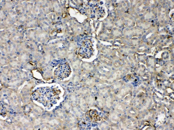
(Figure 6. IHC analysis of GALE using anti-GALE antibody (AAA19135).GALE was detected in paraffin-embedded section of rat kidney tissue. Heat mediated antigen retrieval was performed in citrate buffer (pH6, epitope retrieval solution) for 20 mins. The tissue section was blocked with 10% goat serum. The tissue section was then incubated with 1ug/ml rabbit anti-GALE Antibody (AAA19135) overnight at 4 degree C. Biotinylated goat anti-rabbit IgG was used as secondary antibody and incubated for 30 minutes at 37 degree C. The tissue section was developed using Strepavidin-Biotin-Complex (SABC) with DAB as the chromogen.)
IHC (Immunohistchemistry)
(Figure 6. IHC analysis of GALE using anti-GALE antibody (AAA19135).GALE was detected in paraffin-embedded section of rat kidney tissue. Heat mediated antigen retrieval was performed in citrate buffer (pH6, epitope retrieval solution) for 20 mins. The tissue section was blocked with 10% goat serum. The tissue section was then incubated with 1ug/ml rabbit anti-GALE Antibody (AAA19135) overnight at 4 degree C. Biotinylated goat anti-rabbit IgG was used as secondary antibody and incubated for 30 minutes at 37 degree C. The tissue section was developed using Strepavidin-Biotin-Complex (SABC) with DAB as the chromogen.)
GALE, Polyclonal Antibody (Cat# AAA19135)
Full Name
Anti-GALE Picoband antibody
Gene Names
GALE; SDR1E1
Reactivity
Human, Mouse, Rat
No cross reactivity with other proteins.
No cross reactivity with other proteins.
Applications
EIA, IHC, WB
Pricing


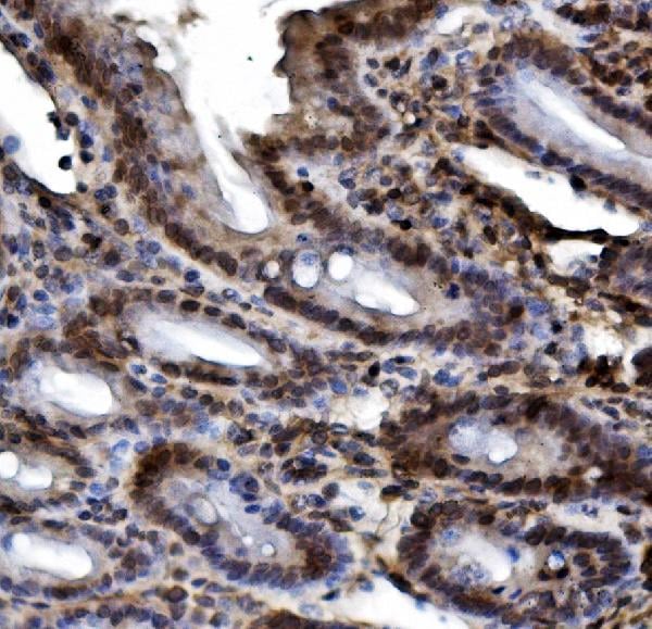
 IHC (Immunohistchemistry)
(Figure 6. IHC analysis of Integrin alpha 5 using anti-Integrin alpha 5 antibody (AAA19156).Integrin alpha 5 was detected in paraffin-embedded section of human placenta tissue. Heat mediated antigen retrieval was performed in citrate buffer (pH6, epitope retrieval solution) for 20 mins. The tissue section was blocked with 10% goat serum. The tissue section was then incubated with 1ug/ml rabbit anti-Integrin alpha 5 Antibody (AAA19156) overnight at 4 degree C. Biotinylated goat anti-rabbit IgG was used as secondary antibody and incubated for 30 minutes at 37 degree C. The tissue section was developed using Strepavidin-Biotin-Complex (SABC) with DAB as the chromogen.)
IHC (Immunohistchemistry)
(Figure 6. IHC analysis of Integrin alpha 5 using anti-Integrin alpha 5 antibody (AAA19156).Integrin alpha 5 was detected in paraffin-embedded section of human placenta tissue. Heat mediated antigen retrieval was performed in citrate buffer (pH6, epitope retrieval solution) for 20 mins. The tissue section was blocked with 10% goat serum. The tissue section was then incubated with 1ug/ml rabbit anti-Integrin alpha 5 Antibody (AAA19156) overnight at 4 degree C. Biotinylated goat anti-rabbit IgG was used as secondary antibody and incubated for 30 minutes at 37 degree C. The tissue section was developed using Strepavidin-Biotin-Complex (SABC) with DAB as the chromogen.)
Integrin alpha 5, Polyclonal Antibody (Cat# AAA19156)
Full Name
Anti-Integrin alpha 5 Picoband antibody
Gene Names
ITGA5; FNRA; CD49e; VLA-5; VLA5A
Reactivity
Human, Mouse, Rat
No cross reactivity with other proteins.
No cross reactivity with other proteins.
Applications
EIA, IHC, WB
Pricing

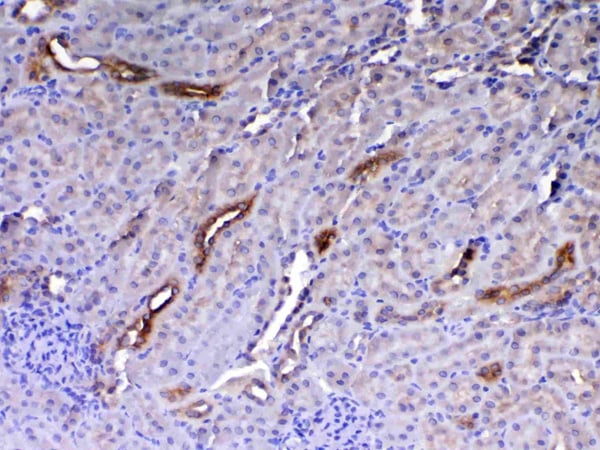


 IHC (Immunohistchemistry)
(Figure 6. IHC analysis of HE4 using anti-HE4 antibody (AAA19161).HE4 was detected in paraffin-embedded section of rat small intestine tissue. Heat mediated antigen retrieval was performed in citrate buffer (pH6, epitope retrieval solution) for 20 mins. The tissue section was blocked with 10% goat serum. The tissue section was then incubated with 1ug/ml rabbit anti-HE4 Antibody (AAA19161) overnight at 4 degree C. Biotinylated goat anti-rabbit IgG was used as secondary antibody and incubated for 30 minutes at 37 degree C. The tissue section was developed using Strepavidin-Biotin-Complex (SABC) with DAB as the chromogen.)
IHC (Immunohistchemistry)
(Figure 6. IHC analysis of HE4 using anti-HE4 antibody (AAA19161).HE4 was detected in paraffin-embedded section of rat small intestine tissue. Heat mediated antigen retrieval was performed in citrate buffer (pH6, epitope retrieval solution) for 20 mins. The tissue section was blocked with 10% goat serum. The tissue section was then incubated with 1ug/ml rabbit anti-HE4 Antibody (AAA19161) overnight at 4 degree C. Biotinylated goat anti-rabbit IgG was used as secondary antibody and incubated for 30 minutes at 37 degree C. The tissue section was developed using Strepavidin-Biotin-Complex (SABC) with DAB as the chromogen.)
HE4, Polyclonal Antibody (Cat# AAA19161)
Full Name
Anti-HE4 Picoband Antibody
Gene Names
Wfdc2; re4
Reactivity
Mouse, Rat
No cross reactivity with other proteins.
No cross reactivity with other proteins.
Applications
IHC, WB
Purity
Immunogen affinity purified
Pricing


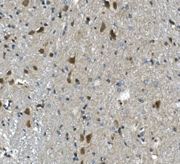
 IHC (Immunohistochemistry)
(Figure 8. IHC analysis of GSTM3 using anti-GSTM3 antibody (AAA19168).GSTM3 was detected in paraffin-embedded section of rat testis tissue. Heat mediated antigen retrieval was performed in citrate buffer (pH6, epitope retrieval solution) for 20 mins. The tissue section was blocked with 10% goat serum. The tissue section was then incubated with 1ug/ml rabbit anti-GSTM3 Antibody (AAA19168) overnight at 4 degree C. Biotinylated goat anti-rabbit IgG was used as secondary antibody and incubated for 30 minutes at 37 degree C. The tissue section was developed using Strepavidin-Biotin-Complex (SABC) with DAB as the chromogen.)
IHC (Immunohistochemistry)
(Figure 8. IHC analysis of GSTM3 using anti-GSTM3 antibody (AAA19168).GSTM3 was detected in paraffin-embedded section of rat testis tissue. Heat mediated antigen retrieval was performed in citrate buffer (pH6, epitope retrieval solution) for 20 mins. The tissue section was blocked with 10% goat serum. The tissue section was then incubated with 1ug/ml rabbit anti-GSTM3 Antibody (AAA19168) overnight at 4 degree C. Biotinylated goat anti-rabbit IgG was used as secondary antibody and incubated for 30 minutes at 37 degree C. The tissue section was developed using Strepavidin-Biotin-Complex (SABC) with DAB as the chromogen.)
GSTM3, Polyclonal Antibody (Cat# AAA19168)
Full Name
Anti-GSTM3 Picoband antibody
Gene Names
GSTM3; GST5; GSTB; GTM3; GSTM3-3
Reactivity
Human, Mouse, Rat
No cross reactivity with other proteins.
No cross reactivity with other proteins.
Applications
EIA, IHC, WB
Pricing


 FCM (Flow Cytometry)
(Figure 6. Flow Cytometry analysis of THP-1 cells using anti-TLR1 antibody (AAA19134).Overlay histogram showing THP-1 cells stained with AAA19134 (Blue line).The cells were blocked with 10% normal goat serum. And then incubated with rabbit anti-TLR1 Antibody (AAA19134,1ug/1x10^6 cells) for 30 min at 20 degree C. DyLight®488 conjugated goat anti-rabbit IgG (5-10ug/1x10^6 cells) was used as secondary antibody for 30 minutes at 20 degree C. Isotype control antibody (Green line) was rabbit IgG (1ug/1x106) used under the same conditions. Unlabelled sample (Red line) was also used as a control.)
FCM (Flow Cytometry)
(Figure 6. Flow Cytometry analysis of THP-1 cells using anti-TLR1 antibody (AAA19134).Overlay histogram showing THP-1 cells stained with AAA19134 (Blue line).The cells were blocked with 10% normal goat serum. And then incubated with rabbit anti-TLR1 Antibody (AAA19134,1ug/1x10^6 cells) for 30 min at 20 degree C. DyLight®488 conjugated goat anti-rabbit IgG (5-10ug/1x10^6 cells) was used as secondary antibody for 30 minutes at 20 degree C. Isotype control antibody (Green line) was rabbit IgG (1ug/1x106) used under the same conditions. Unlabelled sample (Red line) was also used as a control.)
TLR1, Polyclonal Antibody (Cat# AAA19134)
Full Name
Anti-TLR1 Picoband antibody
Gene Names
TLR1; TIL; CD281; rsc786; TIL. LPRS5
Reactivity
Human, Mouse, Rat
No cross reactivity with other proteins.
No cross reactivity with other proteins.
Applications
EIA, FC/FACS, IHC, ICC, WB
Pricing


 IHC (Immunohistchemistry)
(Figure 6. IHC analysis of Flt3 / CD135 using anti-Flt3 / CD135 antibody (AAA19130).Flt3 / CD135 was detected in paraffin-embedded section of mouse kidney tissue. Heat mediated antigen retrieval was performed in citrate buffer (pH6, epitope retrieval solution) for 20 mins. The tissue section was blocked with 10% goat serum. The tissue section was then incubated with 1ug/ml rabbit anti-Flt3 / CD135 Antibody (AAA19130) overnight at 4 degree C. Biotinylated goat anti-rabbit IgG was used as secondary antibody and incubated for 30 minutes at 37 degree C. The tissue section was developed using Strepavidin-Biotin-Complex (SABC) with DAB as the chromogen.)
IHC (Immunohistchemistry)
(Figure 6. IHC analysis of Flt3 / CD135 using anti-Flt3 / CD135 antibody (AAA19130).Flt3 / CD135 was detected in paraffin-embedded section of mouse kidney tissue. Heat mediated antigen retrieval was performed in citrate buffer (pH6, epitope retrieval solution) for 20 mins. The tissue section was blocked with 10% goat serum. The tissue section was then incubated with 1ug/ml rabbit anti-Flt3 / CD135 Antibody (AAA19130) overnight at 4 degree C. Biotinylated goat anti-rabbit IgG was used as secondary antibody and incubated for 30 minutes at 37 degree C. The tissue section was developed using Strepavidin-Biotin-Complex (SABC) with DAB as the chromogen.)
Flt3/CD135, Polyclonal Antibody (Cat# AAA19130)
Full Name
Anti-Flt3/CD135 Picoband Antibody
Reactivity
Mouse, Rat
No cross reactivity with other proteins.
No cross reactivity with other proteins.
Applications
EIA, IHC, WB
Purity
Immunogen affinity purified
Pricing


 IHC (Immunohistochemistry)
(Figure 8. IHC analysis of beta Catenin using anti-beta Catenin antibody (AAA19129).beta Catenin was detected in paraffin-embedded section of rat small intestine tissue. Heat mediated antigen retrieval was performed in citrate buffer (pH6, epitope retrieval solution) for 20 mins. The tissue section was blocked with 10% goat serum. The tissue section was then incubated with 1ug/ml rabbit anti-beta Catenin Antibody (AAA19129) overnight at 4 degree C. Biotinylated goat anti-rabbit IgG was used as secondary antibody and incubated for 30 minutes at 37 degree C. The tissue section was developed using Strepavidin-Biotin-Complex (SABC) with DAB as the chromogen.)
IHC (Immunohistochemistry)
(Figure 8. IHC analysis of beta Catenin using anti-beta Catenin antibody (AAA19129).beta Catenin was detected in paraffin-embedded section of rat small intestine tissue. Heat mediated antigen retrieval was performed in citrate buffer (pH6, epitope retrieval solution) for 20 mins. The tissue section was blocked with 10% goat serum. The tissue section was then incubated with 1ug/ml rabbit anti-beta Catenin Antibody (AAA19129) overnight at 4 degree C. Biotinylated goat anti-rabbit IgG was used as secondary antibody and incubated for 30 minutes at 37 degree C. The tissue section was developed using Strepavidin-Biotin-Complex (SABC) with DAB as the chromogen.)
beta Catenin, Polyclonal Antibody (Cat# AAA19129)
Full Name
Anti-beta Catenin Picoband Antibody
Gene Names
CTNNB1; EVR7; CTNNB; MRD19; armadillo
Reactivity
Human, Mouse, Rat
No cross reactivity with other proteins.
No cross reactivity with other proteins.
Applications
EIA, IHC, WB
Purity
Immunogen affinity purified
Pricing


 IHC (Immunohistchemistry)
(Figure 6. IHC analysis of HP1 gamma using anti-HP1 gamma antibody (AAA19145).HP1 gamma was detected in paraffin-embedded section of rat small intestine tissue. Heat mediated antigen retrieval was performed in citrate buffer (pH6, epitope retrieval solution) for 20 mins. The tissue section was blocked with 10% goat serum. The tissue section was then incubated with 1ug/ml rabbit anti-HP1 gamma Antibody (AAA19145) overnight at 4 degree C. Biotinylated goat anti-rabbit IgG was used as secondary antibody and incubated for 30 minutes at 37 degree C. The tissue section was developed using Strepavidin-Biotin-Complex (SABC) with DAB as the chromogen.)
IHC (Immunohistchemistry)
(Figure 6. IHC analysis of HP1 gamma using anti-HP1 gamma antibody (AAA19145).HP1 gamma was detected in paraffin-embedded section of rat small intestine tissue. Heat mediated antigen retrieval was performed in citrate buffer (pH6, epitope retrieval solution) for 20 mins. The tissue section was blocked with 10% goat serum. The tissue section was then incubated with 1ug/ml rabbit anti-HP1 gamma Antibody (AAA19145) overnight at 4 degree C. Biotinylated goat anti-rabbit IgG was used as secondary antibody and incubated for 30 minutes at 37 degree C. The tissue section was developed using Strepavidin-Biotin-Complex (SABC) with DAB as the chromogen.)
HP1 gamma, Polyclonal Antibody (Cat# AAA19145)
Full Name
Anti-HP1 gamma Picoband antibody
Gene Names
CBX3; HECH; HP1-GAMMA; HP1Hs-gamma
Reactivity
Human, Mouse, Rat
No cross reactivity with other proteins.
No cross reactivity with other proteins.
Applications
EIA, IHC, WB
Pricing


 IHC (Immunohistchemistry)
(Figure 6. IHC analysis of Cyclophilin B using anti-Cyclophilin B antibody (AAA19167).Cyclophilin B was detected in paraffin-embedded section of rat thyroid gland tissue. Heat mediated antigen retrieval was performed in citrate buffer (pH6, epitope retrieval solution) for 20 mins. The tissue section was blocked with 10% goat serum. The tissue section was then incubated with 1ug/ml rabbit anti-Cyclophilin B Antibody (AAA19167) overnight at 4 degree C. Biotinylated goat anti-rabbit IgG was used as secondary antibody and incubated for 30 minutes at 37 degree C. The tissue section was developed using Strepavidin-Biotin-Complex (SABC) with DAB as the chromogen.)
IHC (Immunohistchemistry)
(Figure 6. IHC analysis of Cyclophilin B using anti-Cyclophilin B antibody (AAA19167).Cyclophilin B was detected in paraffin-embedded section of rat thyroid gland tissue. Heat mediated antigen retrieval was performed in citrate buffer (pH6, epitope retrieval solution) for 20 mins. The tissue section was blocked with 10% goat serum. The tissue section was then incubated with 1ug/ml rabbit anti-Cyclophilin B Antibody (AAA19167) overnight at 4 degree C. Biotinylated goat anti-rabbit IgG was used as secondary antibody and incubated for 30 minutes at 37 degree C. The tissue section was developed using Strepavidin-Biotin-Complex (SABC) with DAB as the chromogen.)
Cyclophilin B, Polyclonal Antibody (Cat# AAA19167)
Full Name
Anti-Cyclophilin B Picoband Antibody
Gene Names
PPIB; OI9; CYPB; SCYLP; CYP-S1; HEL-S-39
Reactivity
Human, Mouse, Rat
No cross reactivity with other proteins.
No cross reactivity with other proteins.
Applications
EIA, IHC, WB
Purity
Immunogen affinity purified
Pricing


 IHC (Immunohistchemistry)
(Figure 6. IHC analysis of ADA using anti-ADA antibody (AAA19140).ADA was detected in paraffin-embedded section of rat spleen tissue. Heat mediated antigen retrieval was performed in citrate buffer (pH6, epitope retrieval solution) for 20 mins. The tissue section was blocked with 10% goat serum. The tissue section was then incubated with 1ug/ml rabbit anti-ADA Antibody (AAA19140) overnight at 4 degree C. Biotinylated goat anti-rabbit IgG was used as secondary antibody and incubated for 30 minutes at 37 degree C. The tissue section was developed using Strepavidin-Biotin-Complex (SABC) with DAB as the chromogen.)
IHC (Immunohistchemistry)
(Figure 6. IHC analysis of ADA using anti-ADA antibody (AAA19140).ADA was detected in paraffin-embedded section of rat spleen tissue. Heat mediated antigen retrieval was performed in citrate buffer (pH6, epitope retrieval solution) for 20 mins. The tissue section was blocked with 10% goat serum. The tissue section was then incubated with 1ug/ml rabbit anti-ADA Antibody (AAA19140) overnight at 4 degree C. Biotinylated goat anti-rabbit IgG was used as secondary antibody and incubated for 30 minutes at 37 degree C. The tissue section was developed using Strepavidin-Biotin-Complex (SABC) with DAB as the chromogen.)
ADA/Adenosine Deaminase, Polyclonal Antibody (Cat# AAA19140)
Full Name
Anti-ADA/Adenosine Deaminase Picoband antibody
Reactivity
Mouse, Rat
No cross reactivity with other proteins.
No cross reactivity with other proteins.
Applications
EIA, IHC, WB
Pricing


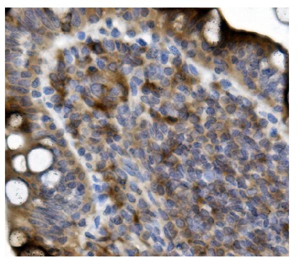
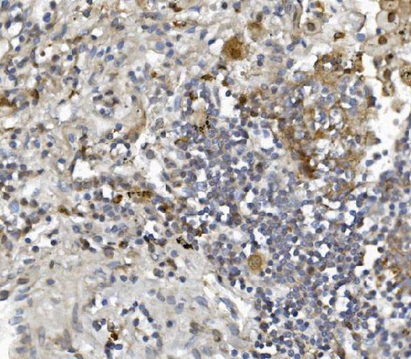
 IHC (Immunohistochemistry)
(Figure 8. IHC analysis of PRDM1/Blimp1 using anti-PRDM1/Blimp1 antibody (AAA19133).PRDM1/Blimp1 was detected in paraffin-embedded section of rat small intestine tissue. Heat mediated antigen retrieval was performed in citrate buffer (pH6, epitope retrieval solution) for 20 mins. The tissue section was blocked with 10% goat serum. The tissue section was then incubated with 1ug/ml rabbit anti-PRDM1/Blimp1 Antibody (AAA19133) overnight at 4 degree C. Biotinylated goat anti-rabbit IgG was used as secondary antibody and incubated for 30 minutes at 37 degree C. The tissue section was developed using Strepavidin-Biotin-Complex (SABC) with DAB as the chromogen.)
IHC (Immunohistochemistry)
(Figure 8. IHC analysis of PRDM1/Blimp1 using anti-PRDM1/Blimp1 antibody (AAA19133).PRDM1/Blimp1 was detected in paraffin-embedded section of rat small intestine tissue. Heat mediated antigen retrieval was performed in citrate buffer (pH6, epitope retrieval solution) for 20 mins. The tissue section was blocked with 10% goat serum. The tissue section was then incubated with 1ug/ml rabbit anti-PRDM1/Blimp1 Antibody (AAA19133) overnight at 4 degree C. Biotinylated goat anti-rabbit IgG was used as secondary antibody and incubated for 30 minutes at 37 degree C. The tissue section was developed using Strepavidin-Biotin-Complex (SABC) with DAB as the chromogen.)
PRDM1/Blimp1, Polyclonal Antibody (Cat# AAA19133)
Full Name
Anti-PRDM1/Blimp1 Picoband antibody
Gene Names
PRDM1; BLIMP1; PRDI-BF1
Reactivity
Human, Mouse, Rat
No cross reactivity with other proteins.
No cross reactivity with other proteins.
Applications
EIA, IHC, WB
Pricing


 IHC (Immunohistochemistry)
(Figure 7. IHC analysis of splicing factor 1 using anti-splicing factor 1 antibody (AAA19141).splicing factor 1 was detected in paraffin-embedded section of rat small intestine tissue. Heat mediated antigen retrieval was performed in citrate buffer (pH6, epitope retrieval solution) for 20 mins. The tissue section was blocked with 10% goat serum. The tissue section was then incubated with 1ug/ml rabbit anti-splicing factor 1 Antibody (AAA19141) overnight at 4 degree C. Biotinylated goat anti-rabbit IgG was used as secondary antibody and incubated for 30 minutes at 37 degree C. The tissue section was developed using Strepavidin-Biotin-Complex (SABC) with DAB as the chromogen.)
IHC (Immunohistochemistry)
(Figure 7. IHC analysis of splicing factor 1 using anti-splicing factor 1 antibody (AAA19141).splicing factor 1 was detected in paraffin-embedded section of rat small intestine tissue. Heat mediated antigen retrieval was performed in citrate buffer (pH6, epitope retrieval solution) for 20 mins. The tissue section was blocked with 10% goat serum. The tissue section was then incubated with 1ug/ml rabbit anti-splicing factor 1 Antibody (AAA19141) overnight at 4 degree C. Biotinylated goat anti-rabbit IgG was used as secondary antibody and incubated for 30 minutes at 37 degree C. The tissue section was developed using Strepavidin-Biotin-Complex (SABC) with DAB as the chromogen.)
splicing factor 1, Polyclonal Antibody (Cat# AAA19141)
Full Name
Anti-splicing factor 1 Picoband antibody
Gene Names
SF1; BBP; MBBP; ZFM1; ZNF162; D11S636; ZCCHC25
Reactivity
Human, Mouse, Rat
No cross reactivity with other proteins.
No cross reactivity with other proteins.
Applications
EIA, IHC, WB
Pricing


 IHC (Immunohistchemistry)
(Figure 6. IHC analysis of NSF using anti-NSF antibody (AAA19136).NSF was detected in paraffin-embedded section of human mammary cancer tissue. Heat mediated antigen retrieval was performed in citrate buffer (pH6, epitope retrieval solution) for 20 mins. The tissue section was blocked with 10% goat serum. The tissue section was then incubated with 1ug/ml rabbit anti-NSF Antibody (AAA19136) overnight at 4 degree C. Biotinylated goat anti-rabbit IgG was used as secondary antibody and incubated for 30 minutes at 37 degree C. The tissue section was developed using Strepavidin-Biotin-Complex (SABC) with DAB as the chromogen.)
IHC (Immunohistchemistry)
(Figure 6. IHC analysis of NSF using anti-NSF antibody (AAA19136).NSF was detected in paraffin-embedded section of human mammary cancer tissue. Heat mediated antigen retrieval was performed in citrate buffer (pH6, epitope retrieval solution) for 20 mins. The tissue section was blocked with 10% goat serum. The tissue section was then incubated with 1ug/ml rabbit anti-NSF Antibody (AAA19136) overnight at 4 degree C. Biotinylated goat anti-rabbit IgG was used as secondary antibody and incubated for 30 minutes at 37 degree C. The tissue section was developed using Strepavidin-Biotin-Complex (SABC) with DAB as the chromogen.)
NSF, Polyclonal Antibody (Cat# AAA19136)
Full Name
Anti-NSF Picoband antibody
Gene Names
NSF; SKD2; SEC18
Reactivity
Human, Mouse, Rat
No cross reactivity with other proteins.
No cross reactivity with other proteins.
Applications
EIA, IHC, WB
Pricing


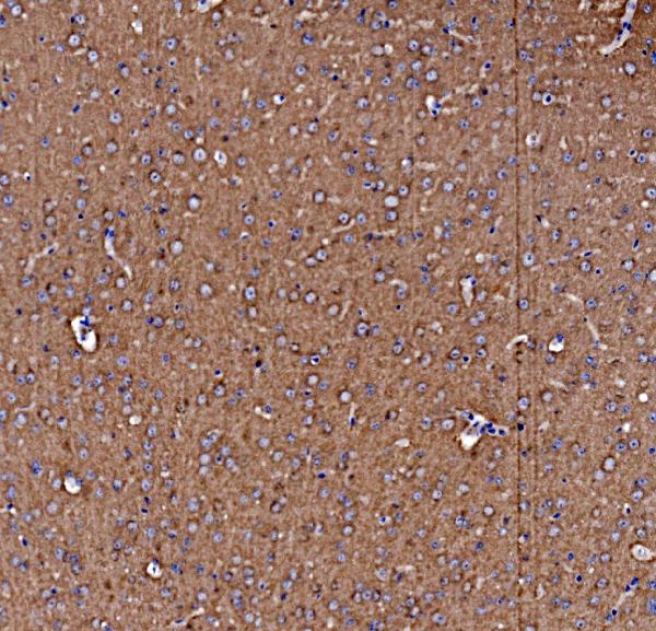
 IHC (Immunohistchemistry)
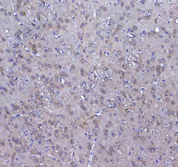
(Figure 6. IHC analysis of MAP1LC3A using anti-MAP1LC3A antibody (AAA19151).MAP1LC3A was detected in paraffin-embedded section of rat brain tissue. Heat mediated antigen retrieval was performed in citrate buffer (pH6, epitope retrieval solution) for 20 mins. The tissue section was blocked with 10% goat serum. The tissue section was then incubated with 1ug/ml rabbit anti-MAP1LC3A Antibody (AAA19151) overnight at 4 degree C. Biotinylated goat anti-rabbit IgG was used as secondary antibody and incubated for 30 minutes at 37 degree C. The tissue section was developed using Strepavidin-Biotin-Complex (SABC) with DAB as the chromogen.)
IHC (Immunohistchemistry)
(Figure 6. IHC analysis of MAP1LC3A using anti-MAP1LC3A antibody (AAA19151).MAP1LC3A was detected in paraffin-embedded section of rat brain tissue. Heat mediated antigen retrieval was performed in citrate buffer (pH6, epitope retrieval solution) for 20 mins. The tissue section was blocked with 10% goat serum. The tissue section was then incubated with 1ug/ml rabbit anti-MAP1LC3A Antibody (AAA19151) overnight at 4 degree C. Biotinylated goat anti-rabbit IgG was used as secondary antibody and incubated for 30 minutes at 37 degree C. The tissue section was developed using Strepavidin-Biotin-Complex (SABC) with DAB as the chromogen.)
MAP1LC3A, Polyclonal Antibody (Cat# AAA19151)
Full Name
Anti-MAP1LC3A Picoband antibody
Gene Names
MAP1LC3A; LC3; LC3A; ATG8E; MAP1ALC3; MAP1BLC3
Reactivity
Human, Mouse, Rat
No cross reactivity with other proteins.
No cross reactivity with other proteins.
Applications
EIA, IHC, WB
Pricing


 FCM (Flow Cytometry)
(Figure 6. Flow Cytometry analysis of Hela cells using anti-PDE4D antibody (AAA19143).Overlay histogram showing Hela cells stained with AAA19143 (Blue line).The cells were blocked with 10% normal goat serum. And then incubated with rabbit anti-PDE4D Antibody (AAA19143,1ug/1x10^6 cells) for 30 min at 20 degree C. DyLight®488 conjugated goat anti-rabbit IgG (5-10ug/1x10^6 cells) was used as secondary antibody for 30 minutes at 20 degree C. Isotype control antibody (Green line) was rabbit IgG (1ug/1x106) used under the same conditions. Unlabelled sample (Red line) was also used as a control.)
FCM (Flow Cytometry)
(Figure 6. Flow Cytometry analysis of Hela cells using anti-PDE4D antibody (AAA19143).Overlay histogram showing Hela cells stained with AAA19143 (Blue line).The cells were blocked with 10% normal goat serum. And then incubated with rabbit anti-PDE4D Antibody (AAA19143,1ug/1x10^6 cells) for 30 min at 20 degree C. DyLight®488 conjugated goat anti-rabbit IgG (5-10ug/1x10^6 cells) was used as secondary antibody for 30 minutes at 20 degree C. Isotype control antibody (Green line) was rabbit IgG (1ug/1x106) used under the same conditions. Unlabelled sample (Red line) was also used as a control.)
PDE4D, Polyclonal Antibody (Cat# AAA19143)
Full Name
Anti-PDE4D Picoband antibody
Gene Names
PDE4D; DPDE3; PDE43; STRK1; ACRDYS2; HSPDE4D; PDE4DN2
Reactivity
Human, Mouse, Rat
No cross reactivity with other proteins.
No cross reactivity with other proteins.
Applications
EIA, FC/FACS, IHC, ICC, WB
Pricing


 IHC (Immunohistochemistry)
(Figure 7. IHC analysis of MYLK using anti-MYLK antibody (AAA19154).MYLK was detected in paraffin-embedded section of rat lung tissue. Heat mediated antigen retrieval was performed in citrate buffer (pH6, epitope retrieval solution) for 20 mins. The tissue section was blocked with 10% goat serum. The tissue section was then incubated with 1ug/ml rabbit anti-MYLK Antibody (AAA19154) overnight at 4 degree C. Biotinylated goat anti-rabbit IgG was used as secondary antibody and incubated for 30 minutes at 37 degree C. The tissue section was developed using Strepavidin-Biotin-Complex (SABC) with DAB as the chromogen. )
IHC (Immunohistochemistry)
(Figure 7. IHC analysis of MYLK using anti-MYLK antibody (AAA19154).MYLK was detected in paraffin-embedded section of rat lung tissue. Heat mediated antigen retrieval was performed in citrate buffer (pH6, epitope retrieval solution) for 20 mins. The tissue section was blocked with 10% goat serum. The tissue section was then incubated with 1ug/ml rabbit anti-MYLK Antibody (AAA19154) overnight at 4 degree C. Biotinylated goat anti-rabbit IgG was used as secondary antibody and incubated for 30 minutes at 37 degree C. The tissue section was developed using Strepavidin-Biotin-Complex (SABC) with DAB as the chromogen. )
MYLK, Polyclonal Antibody (Cat# AAA19154)
Full Name
Anti-MYLK Picoband antibody
Gene Names
MYLK; KRP; AAT7; MLCK; MLCK1; MYLK1; smMLCK; MLCK108; MLCK210; MSTP083
Reactivity
Human, Mouse, Rat
No cross reactivity with other proteins.
No cross reactivity with other proteins.
Applications
EIA, IHC, WB
Pricing


 FCM (Flow Cytometry)
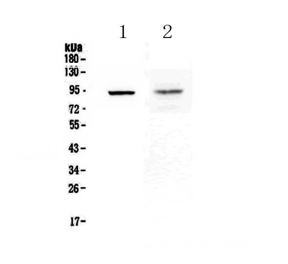
(Figure 11. Flow Cytometry analysis of U937 cells using anti-Coronin 1a/TACO/CORO1A antibody (AAA19291).Overlay histogram showing U937 cells stained with AAA19291 (Blue line). The cells were blocked with 10% normal goat serum. And then incubated with rabbit anti-Coronin 1a/TACO/CORO1A Antibody (AAA19291, 1μg/1x106 cells) for 30 min at 20 degree C. DyLight®488 conjugated goat anti-rabbit IgG (5-10μg/1x106 cells) was used as secondary antibody for 30 minutes at 20 degree C. Isotype control antibody (Green line) was rabbit IgG (1μg/1x106) used under the same conditions. Unlabelled sample (Red line) was also used as a control.)
FCM (Flow Cytometry)
(Figure 11. Flow Cytometry analysis of U937 cells using anti-Coronin 1a/TACO/CORO1A antibody (AAA19291).Overlay histogram showing U937 cells stained with AAA19291 (Blue line). The cells were blocked with 10% normal goat serum. And then incubated with rabbit anti-Coronin 1a/TACO/CORO1A Antibody (AAA19291, 1μg/1x106 cells) for 30 min at 20 degree C. DyLight®488 conjugated goat anti-rabbit IgG (5-10μg/1x106 cells) was used as secondary antibody for 30 minutes at 20 degree C. Isotype control antibody (Green line) was rabbit IgG (1μg/1x106) used under the same conditions. Unlabelled sample (Red line) was also used as a control.)
Coronin 1a/TACO/CORO1A, Polyclonal Antibody (Cat# AAA19291)
Full Name
Anti-Coronin 1a/TACO/CORO1A Antibody
Gene Names
CORO1A; p57; TACO; CLABP; HCORO1; CLIPINA
Reactivity
Human, Mouse, Rat
Applications
WB, IHC-P, FC/FACS/FCM, EIA
Purity
Immunogen affinity purified.
Pricing


 IF (Immunofluorescence)
(Figure 6. IF analysis of SCG10/STMN2 using anti- SCG10/STMN2 antibody (AAA19299).SCG10/STMN2 was detected in immunocytochemical section of U20S cells. Enzyme antigen retrieval was performed using IHC enzyme antigen retrieval reagent for 15 mins. The cells were blocked with 10% goat serum. And then incubated with 5μg/mL rabbit anti- SCG10/STMN2 Antibody (AAA19299) overnight at 4 degree C. DyLight®488 Conjugated Goat Anti-Rabbit IgG was used as secondary antibody at 1:100 dilution and incubated for 30 minutes at 37 degree C. The section was counterstained with DAPI. Visualize using a fluorescence microscope and filter sets appropriate for the label used.)
IF (Immunofluorescence)
(Figure 6. IF analysis of SCG10/STMN2 using anti- SCG10/STMN2 antibody (AAA19299).SCG10/STMN2 was detected in immunocytochemical section of U20S cells. Enzyme antigen retrieval was performed using IHC enzyme antigen retrieval reagent for 15 mins. The cells were blocked with 10% goat serum. And then incubated with 5μg/mL rabbit anti- SCG10/STMN2 Antibody (AAA19299) overnight at 4 degree C. DyLight®488 Conjugated Goat Anti-Rabbit IgG was used as secondary antibody at 1:100 dilution and incubated for 30 minutes at 37 degree C. The section was counterstained with DAPI. Visualize using a fluorescence microscope and filter sets appropriate for the label used.)
SCG10/STMN2, Polyclonal Antibody (Cat# AAA19299)
Full Name
Anti-SCG10/STMN2 Antibody
Gene Names
STMN2; SCG10; SCGN10
Reactivity
Human, Mouse, Rat
Applications
WB, IHC-P, ICC, IF, FC/FACS/FCM, EIA
Purity
Immunogen affinity purified.
Pricing


 FCM (Flow Cytometry)
(Figure 7. Flow Cytometry analysis of CACO-2 cells using anti-Glycine decarboxylase/GLDC antibody (AAA19300).Overlay histogram showing CACO-2 cells stained with AAA19300 (Blue line). The cells were blocked with 10% normal goat serum. And then incubated with rabbit anti-Glycine decarboxylase/GLDC Antibody (AAA19300, 1μg/1x106 cells) for 30 min at 20 degree C. DyLight®488 conjugated goat anti-rabbit IgG (5-10μg/1x106 cells) was used as secondary antibody for 30 minutes at 20 degree C. Isotype control antibody (Green line) was rabbit IgG (1μg/1x106) used under the same conditions. Unlabelled sample (Red line) was also used as a control.)
FCM (Flow Cytometry)
(Figure 7. Flow Cytometry analysis of CACO-2 cells using anti-Glycine decarboxylase/GLDC antibody (AAA19300).Overlay histogram showing CACO-2 cells stained with AAA19300 (Blue line). The cells were blocked with 10% normal goat serum. And then incubated with rabbit anti-Glycine decarboxylase/GLDC Antibody (AAA19300, 1μg/1x106 cells) for 30 min at 20 degree C. DyLight®488 conjugated goat anti-rabbit IgG (5-10μg/1x106 cells) was used as secondary antibody for 30 minutes at 20 degree C. Isotype control antibody (Green line) was rabbit IgG (1μg/1x106) used under the same conditions. Unlabelled sample (Red line) was also used as a control.)
Glycine decarboxylase/GLDC, Polyclonal Antibody (Cat# AAA19300)
Full Name
Anti-Glycine decarboxylase/GLDC Antibody
Gene Names
GLDC; GCE; GCSP; HYGN1
Reactivity
Human, Mouse, Rat
Applications
WB, IHC-P, FC/FACS/FCM, EIA
Purity
Immunogen affinity purified.
Pricing


 FCM (Flow Cytometry)
(Figure 7. Flow Cytometry analysis of U20S cells using anti-VDAC3 antibody (AAA19301).Overlay histogram showing U20S cells stained with AAA19301 (Blue line). The cells were blocked with 10% normal goat serum. And then incubated with rabbit anti-VDAC3 Antibody (AAA19301, 1μg/1x106 cells) for 30 min at 20 degree C. DyLight®488 conjugated goat anti-rabbit IgG (5-10μg/1x106 cells) was used as secondary antibody for 30 minutes at 20 degree C. Isotype control antibody (Green line) was rabbit IgG (1μg/1x106) used under the same conditions. Unlabelled sample (Red line) was also used as a control.)
FCM (Flow Cytometry)
(Figure 7. Flow Cytometry analysis of U20S cells using anti-VDAC3 antibody (AAA19301).Overlay histogram showing U20S cells stained with AAA19301 (Blue line). The cells were blocked with 10% normal goat serum. And then incubated with rabbit anti-VDAC3 Antibody (AAA19301, 1μg/1x106 cells) for 30 min at 20 degree C. DyLight®488 conjugated goat anti-rabbit IgG (5-10μg/1x106 cells) was used as secondary antibody for 30 minutes at 20 degree C. Isotype control antibody (Green line) was rabbit IgG (1μg/1x106) used under the same conditions. Unlabelled sample (Red line) was also used as a control.)
VDAC3, Polyclonal Antibody (Cat# AAA19301)
Full Name
Anti-VDAC3 Antibody
Gene Names
VDAC3; VDAC-3; HD-VDAC3
Reactivity
Human, Mouse, Rat
Applications
WB, IHC-P, FC/FACS/FCM, EIA
Purity
Immunogen affinity purified.
Pricing


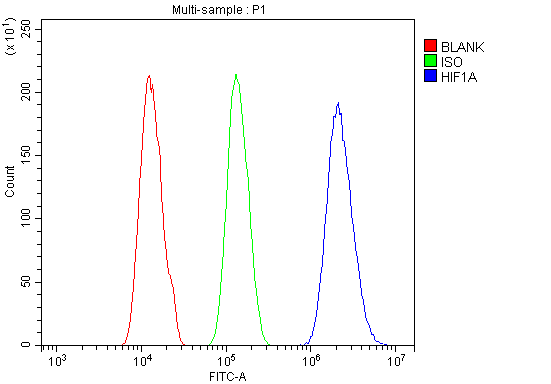 FCM (Flow Cytometry)
(Figure 6. Flow Cytometry analysis of A431 cells using anti-HIF-1 alpha/HIF1A antibody (AAA19213).Overlay histogram showing A431 cells stained with AAA19213 (Blue line). The cells were blocked with 10% normal goat serum. And then incubated with rabbit anti-HIF-1 alpha/HIF1A Antibody (AAA19213, 1μg/1x106 cells) for 30 min at 20 degree C. DyLight®488 conjugated goat anti-rabbit IgG (5-10μg/1x106 cells) was used as secondary antibody for 30 minutes at 20 degree C. Isotype control antibody (Green line) was rabbit IgG (1μg/1x106) used under the same conditions. Unlabelled sample (Red line) was also used as a control.)
FCM (Flow Cytometry)
(Figure 6. Flow Cytometry analysis of A431 cells using anti-HIF-1 alpha/HIF1A antibody (AAA19213).Overlay histogram showing A431 cells stained with AAA19213 (Blue line). The cells were blocked with 10% normal goat serum. And then incubated with rabbit anti-HIF-1 alpha/HIF1A Antibody (AAA19213, 1μg/1x106 cells) for 30 min at 20 degree C. DyLight®488 conjugated goat anti-rabbit IgG (5-10μg/1x106 cells) was used as secondary antibody for 30 minutes at 20 degree C. Isotype control antibody (Green line) was rabbit IgG (1μg/1x106) used under the same conditions. Unlabelled sample (Red line) was also used as a control.)
HIF-1 alpha/HIF1A, Polyclonal Antibody (Cat# AAA19213)
Full Name
Anti-HIF-1 alpha/HIF1A Antibody
Gene Names
HIF1A; HIF1; MOP1; PASD8; HIF-1A; bHLHe78; HIF-1alpha; HIF1-ALPHA
Reactivity
Human, Mouse, Rat
Applications
WB, IHC-P, FC/FACS/FCM, EIA
Purity
Immunogen affinity purified.
Pricing


 FCM (Flow Cytometry)
(Figure 8. Flow Cytometry analysis of HepG2 cells using anti-MERTK antibody (AAA19219).Overlay histogram showing HepG2 cells stained with AAA19219 (Blue line). The cells were blocked with 10% normal goat serum. And then incubated with rabbit anti-MERTK Antibody (AAA19219, 1ug/1x106 cells) for 30 min at 20 degree C. DyLight488 conjugated goat anti-rabbit IgG (5-10ug/1x106 cells) was used as secondary antibody for 30 minutes at 20 degree C. Isotype control antibody (Green line) was rabbit IgG (1ug/1x106) used under the same conditions. Unlabelled sample (Red line) was also used as a control.)
FCM (Flow Cytometry)
(Figure 8. Flow Cytometry analysis of HepG2 cells using anti-MERTK antibody (AAA19219).Overlay histogram showing HepG2 cells stained with AAA19219 (Blue line). The cells were blocked with 10% normal goat serum. And then incubated with rabbit anti-MERTK Antibody (AAA19219, 1ug/1x106 cells) for 30 min at 20 degree C. DyLight488 conjugated goat anti-rabbit IgG (5-10ug/1x106 cells) was used as secondary antibody for 30 minutes at 20 degree C. Isotype control antibody (Green line) was rabbit IgG (1ug/1x106) used under the same conditions. Unlabelled sample (Red line) was also used as a control.)
MERTK, Polyclonal Antibody (Cat# AAA19219)
Full Name
Anti-MERTK Antibody
Gene Names
MERTK; MER; RP38; c-mer
Reactivity
Human, Mouse, Rat
Applications
WB, IHC-P, FC/FACS/FCM, EIA
Purity
Immunogen affinity purified.
Pricing


 IF (Immunofluorescence)
(Figure 6. IF analysis of CD72 using anti- CD72 antibody (AAA19329).CD72 was detected in immunocytochemical section of HEPA1-6 cells. Enzyme antigen retrieval was performed using IHC enzyme antigen retrieval reagent for 15 mins. The cells were blocked with 10% goat serum. And then incubated with 5μg/mL rabbit anti- CD72 Antibody (AAA19329) overnight at 4 degree C. DyLight®488 Conjugated Goat Anti-Rabbit IgG was used as secondary antibody at 1:100 dilution and incubated for 30 minutes at 37 degree C. The section was counterstained with DAPI. Visualize using a fluorescence microscope and filter sets appropriate for the label used.)
IF (Immunofluorescence)
(Figure 6. IF analysis of CD72 using anti- CD72 antibody (AAA19329).CD72 was detected in immunocytochemical section of HEPA1-6 cells. Enzyme antigen retrieval was performed using IHC enzyme antigen retrieval reagent for 15 mins. The cells were blocked with 10% goat serum. And then incubated with 5μg/mL rabbit anti- CD72 Antibody (AAA19329) overnight at 4 degree C. DyLight®488 Conjugated Goat Anti-Rabbit IgG was used as secondary antibody at 1:100 dilution and incubated for 30 minutes at 37 degree C. The section was counterstained with DAPI. Visualize using a fluorescence microscope and filter sets appropriate for the label used.)
CD72, Polyclonal Antibody (Cat# AAA19329)
Full Name
Anti-CD72 Antibody
Gene Names
Cd72; CD72c; Ly-19; Ly-32; Lyb-2; Ly-m19
Reactivity
Mouse, Rat
Applications
WB, IHC-P, ICC, IF, FC/FACS/FCM, EIA
Purity
Immunogen affinity purified.
Pricing


 IF (Immunofluorescence)
(Figure 7. IF analysis of METTL1 using anti- METTL1 antibody (AAA19336).METTL1 was detected in immunocytochemical section of A431 cells. Enzyme antigen retrieval was performed using IHC enzyme antigen retrieval reagent for 15 mins. The cells were blocked with 10% goat serum. And then incubated with 5μg/mL rabbit anti-METTL1 Antibody (AAA19336) overnight at 4 degree C. DyLight®488 Conjugated Goat Anti-Rabbit IgG was used as secondary antibody at 1:100 dilution and incubated for 30 minutes at 37 degree C. The section was counterstained with DAPI. Visualize using a fluorescence microscope and filter sets appropriate for the label used.)
IF (Immunofluorescence)
(Figure 7. IF analysis of METTL1 using anti- METTL1 antibody (AAA19336).METTL1 was detected in immunocytochemical section of A431 cells. Enzyme antigen retrieval was performed using IHC enzyme antigen retrieval reagent for 15 mins. The cells were blocked with 10% goat serum. And then incubated with 5μg/mL rabbit anti-METTL1 Antibody (AAA19336) overnight at 4 degree C. DyLight®488 Conjugated Goat Anti-Rabbit IgG was used as secondary antibody at 1:100 dilution and incubated for 30 minutes at 37 degree C. The section was counterstained with DAPI. Visualize using a fluorescence microscope and filter sets appropriate for the label used.)
METTL1, Polyclonal Antibody (Cat# AAA19336)
Full Name
Anti-METTL1 Antibody
Gene Names
METTL1; TRM8; TRMT8; C12orf1; YDL201w
Reactivity
Human, Rat
Applications
WB, IHC-P, ICC, IF, EIA
Purity
Immunogen affinity purified.
Pricing


 FCM (Flow Cytometry)
(Figure 7. Flow Cytometry analysis of HEPA1-6 cells using anti-Rad9b antibody (AAA19340).Overlay histogram showing HEPA1-6 cells stained with AAA19340 (Blue line). The cells were blocked with 10% normal goat serum. And then incubated with rabbit anti-Rad9b Antibody (AAA19340, 1μg/1x106 cells) for 30 min at 20 degree C. DyLight®488 conjugated goat anti-rabbit IgG (5-10μg/1x106 cells) was used as secondary antibody for 30 minutes at 20 degree C. Isotype control antibody (Green line) was rabbit IgG (1μg/1x106) used under the same conditions. Unlabelled sample (Red line) was also used as a control.)
FCM (Flow Cytometry)
(Figure 7. Flow Cytometry analysis of HEPA1-6 cells using anti-Rad9b antibody (AAA19340).Overlay histogram showing HEPA1-6 cells stained with AAA19340 (Blue line). The cells were blocked with 10% normal goat serum. And then incubated with rabbit anti-Rad9b Antibody (AAA19340, 1μg/1x106 cells) for 30 min at 20 degree C. DyLight®488 conjugated goat anti-rabbit IgG (5-10μg/1x106 cells) was used as secondary antibody for 30 minutes at 20 degree C. Isotype control antibody (Green line) was rabbit IgG (1μg/1x106) used under the same conditions. Unlabelled sample (Red line) was also used as a control.)
Rad9b, Polyclonal Antibody (Cat# AAA19340)
Full Name
Anti-Rad9b Antibody
Gene Names
Rad9b; BC021784; A630082N15Rik
Reactivity
Mouse, Rat
Applications
WB, IHC-P, FC/FACS/FCM, EIA
Purity
Immunogen affinity purified.
Pricing


 FCM (Flow Cytometry)
(Figure 6. Flow Cytometry analysis of U87 cells using anti-GRB10 antibody (AAA19244).Overlay histogram showing U87 cells stained with AAA19244 (Blue line). The cells were blocked with 10% normal goat serum. And then incubated with rabbit anti-GRB10 Antibody (AAA19244,1μg/1x106 cells) for 30 min at 20 degree C. DyLight®488 conjugated goat anti-rabbit IgG (5-10μg/1x106 cells) was used as secondary antibody for 30 minutes at 20 degree C. Isotype control antibody (Green line) was rabbit IgG (1μg/1x106) used under the same conditions. Unlabelled sample (Red line) was also used as a control.)
FCM (Flow Cytometry)
(Figure 6. Flow Cytometry analysis of U87 cells using anti-GRB10 antibody (AAA19244).Overlay histogram showing U87 cells stained with AAA19244 (Blue line). The cells were blocked with 10% normal goat serum. And then incubated with rabbit anti-GRB10 Antibody (AAA19244,1μg/1x106 cells) for 30 min at 20 degree C. DyLight®488 conjugated goat anti-rabbit IgG (5-10μg/1x106 cells) was used as secondary antibody for 30 minutes at 20 degree C. Isotype control antibody (Green line) was rabbit IgG (1μg/1x106) used under the same conditions. Unlabelled sample (Red line) was also used as a control.)
GRB10, Polyclonal Antibody (Cat# AAA19244)
Full Name
Anti-GRB10 Antibody
Gene Names
GRB10; RSS; IRBP; MEG1; GRB-IR; Grb-10
Reactivity
Human, Mouse, Rat
Applications
WB, IHC-P, FC/FACS/FCM, EIA
Purity
Immunogen affinity purified.
Pricing


 FCM (Flow Cytometry)
(Figure 7. Flow Cytometry analysis of 293T cells using anti-TBP-1/PSMC3 antibody (AAA19314).Overlay histogram showing 293T cells stained with AAA19314 (Blue line). The cells were blocked with 10% normal goat serum. And then incubated with rabbit anti-TBP-1/PSMC3 Antibody (AAA19314, 1μg/1x106 cells) for 30 min at 20 degree C. DyLight®488 conjugated goat anti-rabbit IgG (5-10μg/1x106 cells) was used as secondary antibody for 30 minutes at 20 degree C. Isotype control antibody (Green line) was rabbit IgG (1μg/1x106) used under the same conditions. Unlabelled sample (Red line) was also used as a control.)
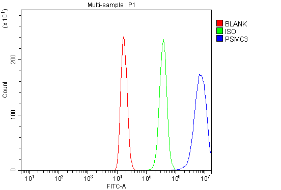
FCM (Flow Cytometry)
(Figure 7. Flow Cytometry analysis of 293T cells using anti-TBP-1/PSMC3 antibody (AAA19314).Overlay histogram showing 293T cells stained with AAA19314 (Blue line). The cells were blocked with 10% normal goat serum. And then incubated with rabbit anti-TBP-1/PSMC3 Antibody (AAA19314, 1μg/1x106 cells) for 30 min at 20 degree C. DyLight®488 conjugated goat anti-rabbit IgG (5-10μg/1x106 cells) was used as secondary antibody for 30 minutes at 20 degree C. Isotype control antibody (Green line) was rabbit IgG (1μg/1x106) used under the same conditions. Unlabelled sample (Red line) was also used as a control.)
TBP-1/PSMC3, Polyclonal Antibody (Cat# AAA19314)
Full Name
Anti-TBP-1/PSMC3 Antibody
Gene Names
PSMC3; TBP1
Reactivity
Human, Mouse, Rat
Applications
WB, IHC-P, FC/FACS/FCM, EIA
Purity
Immunogen affinity purified.
Pricing


 IF (Immunofluorescence)
(Figure 6. IF analysis of GRSF1 using anti- GRSF1 antibody (AAA19315).GRSF1 was detected in immunocytochemical section of MCF-7 cells. Enzyme antigen retrieval was performed using IHC enzyme antigen retrieval reagent for 15 mins. The cells were blocked with 10% goat serum. And then incubated with 5μg/mL rabbit anti- GRSF1 Antibody (AAA19315) overnight at 4 degree C. DyLight®488 Conjugated Goat Anti-Rabbit IgG was used as secondary antibody at 1:100 dilution and incubated for 30 minutes at 37 degree C. The section was counterstained with DAPI. Visualize using a fluorescence microscope and filter sets appropriate for the label used.)
IF (Immunofluorescence)
(Figure 6. IF analysis of GRSF1 using anti- GRSF1 antibody (AAA19315).GRSF1 was detected in immunocytochemical section of MCF-7 cells. Enzyme antigen retrieval was performed using IHC enzyme antigen retrieval reagent for 15 mins. The cells were blocked with 10% goat serum. And then incubated with 5μg/mL rabbit anti- GRSF1 Antibody (AAA19315) overnight at 4 degree C. DyLight®488 Conjugated Goat Anti-Rabbit IgG was used as secondary antibody at 1:100 dilution and incubated for 30 minutes at 37 degree C. The section was counterstained with DAPI. Visualize using a fluorescence microscope and filter sets appropriate for the label used.)
GRSF1, Polyclonal Antibody (Cat# AAA19315)
Full Name
Anti-GRSF1 Antibody
Reactivity
Human
Applications
WB, IHC-P, ICC, IF, FC/FACS/FCM, EIA
Purity
Immunogen affinity purified.
Pricing


 FCM (Flow Cytometry)
(Figure 6. Flow Cytometry analysis of THP-1 cells using anti-PRMT8 antibody (AAA19321).Overlay histogram showing THP-1 cells stained with AAA19321 (Blue line). The cells were blocked with 10% normal goat serum. And then incubated with rabbit anti-PRMT8 Antibody (AAA19321,1μg/1x106 cells) for 30 min at 20 degree C. DyLight®488 conjugated goat anti-rabbit IgG (5-10μg/1x106 cells) was used as secondary antibody for 30 minutes at 20 degree C. Isotype control antibody (Green line) was rabbit IgG (1μg/1x106) used under the same conditions. Unlabelled sample (Red line) was also used as a control.)
FCM (Flow Cytometry)
(Figure 6. Flow Cytometry analysis of THP-1 cells using anti-PRMT8 antibody (AAA19321).Overlay histogram showing THP-1 cells stained with AAA19321 (Blue line). The cells were blocked with 10% normal goat serum. And then incubated with rabbit anti-PRMT8 Antibody (AAA19321,1μg/1x106 cells) for 30 min at 20 degree C. DyLight®488 conjugated goat anti-rabbit IgG (5-10μg/1x106 cells) was used as secondary antibody for 30 minutes at 20 degree C. Isotype control antibody (Green line) was rabbit IgG (1μg/1x106) used under the same conditions. Unlabelled sample (Red line) was also used as a control.)
PRMT8, Polyclonal Antibody (Cat# AAA19321)
Full Name
Anti-PRMT8 Antibody
Gene Names
PRMT8; HRMT1L3; HRMT1L4
Reactivity
Human, Rat
Applications
WB, IHC-P, FC/FACS/FCM, EIA
Purity
Immunogen affinity purified.
Pricing


 IHC (Immunohistchemistry)
(Figure 6. IHC analysis of DYNLT1 using anti-DYNLT1 antibody (AAA19175).DYNLT1 was detected in paraffin-embedded section of rat lung tissue. Heat mediated antigen retrieval was performed in citrate buffer (pH6, epitope retrieval solution) for 20 mins. The tissue section was blocked with 10% goat serum. The tissue section was then incubated with 1ug/ml rabbit anti-DYNLT1 Antibody (AAA19175) overnight at 4 degree C. Biotinylated goat anti-rabbit IgG was used as secondary antibody and incubated for 30 minutes at 37 degree C. The tissue section was developed using Strepavidin-Biotin-Complex (SABC) with DAB as the chromogen.)
IHC (Immunohistchemistry)
(Figure 6. IHC analysis of DYNLT1 using anti-DYNLT1 antibody (AAA19175).DYNLT1 was detected in paraffin-embedded section of rat lung tissue. Heat mediated antigen retrieval was performed in citrate buffer (pH6, epitope retrieval solution) for 20 mins. The tissue section was blocked with 10% goat serum. The tissue section was then incubated with 1ug/ml rabbit anti-DYNLT1 Antibody (AAA19175) overnight at 4 degree C. Biotinylated goat anti-rabbit IgG was used as secondary antibody and incubated for 30 minutes at 37 degree C. The tissue section was developed using Strepavidin-Biotin-Complex (SABC) with DAB as the chromogen.)
DYNLT1, Polyclonal Antibody (Cat# AAA19175)
Full Name
Anti-DYNLT1 Picoband antibody
Gene Names
DYNLT1; CW-1; TCTEL1; tctex-1
Reactivity
Human, Mouse, Rat
No cross reactivity with other proteins.
No cross reactivity with other proteins.
Applications
EIA, IHC, WB
Pricing


 IHC (Immunohistchemistry)
(Figure 6. IHC analysis of SAE2/UBA2 using anti-SAE2/UBA2 antibody (AAA19171).SAE2/UBA2 was detected in paraffin-embedded section of mouse testis tissue. Heat mediated antigen retrieval was performed in citrate buffer (pH6, epitope retrieval solution) for 20 mins. The tissue section was blocked with 10% goat serum. The tissue section was then incubated with 1ug/ml rabbit anti-SAE2/UBA2 Antibody (AAA19171) overnight at 4 degree C. Biotinylated goat anti-rabbit IgG was used as secondary antibody and incubated for 30 minutes at 37 degree C. The tissue section was developed using Strepavidin-Biotin-Complex (SABC) with DAB as the chromogen.)
IHC (Immunohistchemistry)
(Figure 6. IHC analysis of SAE2/UBA2 using anti-SAE2/UBA2 antibody (AAA19171).SAE2/UBA2 was detected in paraffin-embedded section of mouse testis tissue. Heat mediated antigen retrieval was performed in citrate buffer (pH6, epitope retrieval solution) for 20 mins. The tissue section was blocked with 10% goat serum. The tissue section was then incubated with 1ug/ml rabbit anti-SAE2/UBA2 Antibody (AAA19171) overnight at 4 degree C. Biotinylated goat anti-rabbit IgG was used as secondary antibody and incubated for 30 minutes at 37 degree C. The tissue section was developed using Strepavidin-Biotin-Complex (SABC) with DAB as the chromogen.)
SAE2/UBA2, Polyclonal Antibody (Cat# AAA19171)
Full Name
Anti-SAE2/UBA2 Picoband antibody
Gene Names
UBA2; ARX; SAE2; HRIHFB2115
Reactivity
Human, Mouse, Rat
No cross reactivity with other proteins.
No cross reactivity with other proteins.
Applications
EIA, IHC, WB
Pricing


 FCM (Flow Cytometry)
(Figure 6. Flow Cytometry analysis of K562 cells using anti-IRF3 antibody (AAA19216).Overlay histogram showing K562 cells stained with AAA19216 (Blue line). The cells were blocked with 10% normal goat serum. And then incubated with rabbit anti-IRF3 Antibody (AAA19216, 1μg/1x106 cells) for 30 min at 20 degree C. DyLight®488 conjugated goat anti-rabbit IgG (5-10μg/1x106 cells) was used as secondary antibody for 30 minutes at 20 degree C. Isotype control antibody (Green line) was rabbit IgG (1μg/1x106) used under the same conditions. Unlabelled sample (Red line) was also used as a control.)
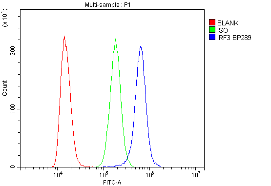
FCM (Flow Cytometry)
(Figure 6. Flow Cytometry analysis of K562 cells using anti-IRF3 antibody (AAA19216).Overlay histogram showing K562 cells stained with AAA19216 (Blue line). The cells were blocked with 10% normal goat serum. And then incubated with rabbit anti-IRF3 Antibody (AAA19216, 1μg/1x106 cells) for 30 min at 20 degree C. DyLight®488 conjugated goat anti-rabbit IgG (5-10μg/1x106 cells) was used as secondary antibody for 30 minutes at 20 degree C. Isotype control antibody (Green line) was rabbit IgG (1μg/1x106) used under the same conditions. Unlabelled sample (Red line) was also used as a control.)
IRF3, Polyclonal Antibody (Cat# AAA19216)
Full Name
Anti-IRF3 Antibody
Reactivity
Human
Applications
WB, IHC-P, FC/FACS/FCM, EIA
Purity
Immunogen affinity purified.
Pricing


 IHC (Immunohistchemistry)
(Figure 5. IHC analysis of GNG4 using anti-GNG4 antibody (AAA19341).GNG4 was detected in paraffin-embedded section of human placenta tissue. Heat mediated antigen retrieval was performed in EDTA buffer (pH8. 0, epitope retrieval solution). The tissue section was blocked with 10% goat serum. The tissue section was then incubated with 2μg/ml rabbit anti-GNG4 Antibody (AAA19341) overnight at 4 degree C. Biotinylated goat anti-rabbit IgG was used as secondary antibody and incubated for 30 minutes at 37 degree C. The tissue section was developed using Strepavidin-Biotin-Complex (SABC) (Catalog # with DAB as the chromogen.)
IHC (Immunohistchemistry)
(Figure 5. IHC analysis of GNG4 using anti-GNG4 antibody (AAA19341).GNG4 was detected in paraffin-embedded section of human placenta tissue. Heat mediated antigen retrieval was performed in EDTA buffer (pH8. 0, epitope retrieval solution). The tissue section was blocked with 10% goat serum. The tissue section was then incubated with 2μg/ml rabbit anti-GNG4 Antibody (AAA19341) overnight at 4 degree C. Biotinylated goat anti-rabbit IgG was used as secondary antibody and incubated for 30 minutes at 37 degree C. The tissue section was developed using Strepavidin-Biotin-Complex (SABC) (Catalog # with DAB as the chromogen.)
GNG4, Polyclonal Antibody (Cat# AAA19341)
Full Name
Anti-GNG4 Antibody
Reactivity
Human, Mouse, Rat
Applications
WB, IHC-P, FC/FACS/FCM, EIA
Purity
Immunogen affinity purified.
Pricing


 IHC (Immunohistochemistry)
(Figure 7. IHC analysis of GALNS using anti-GALNS antibody (AAA19164).GALNS was detected in paraffin-embedded section of rat small intestine tissue. Heat mediated antigen retrieval was performed in citrate buffer (pH6, epitope retrieval solution) for 20 mins. The tissue section was blocked with 10% goat serum. The tissue section was then incubated with 1ug/ml rabbit anti-GALNS Antibody (AAA19164) overnight at 4 degree C. Biotinylated goat anti-rabbit IgG was used as secondary antibody and incubated for 30 minutes at 37 degree C. The tissue section was developed using Strepavidin-Biotin-Complex (SABC) with DAB as the chromogen.)
IHC (Immunohistochemistry)
(Figure 7. IHC analysis of GALNS using anti-GALNS antibody (AAA19164).GALNS was detected in paraffin-embedded section of rat small intestine tissue. Heat mediated antigen retrieval was performed in citrate buffer (pH6, epitope retrieval solution) for 20 mins. The tissue section was blocked with 10% goat serum. The tissue section was then incubated with 1ug/ml rabbit anti-GALNS Antibody (AAA19164) overnight at 4 degree C. Biotinylated goat anti-rabbit IgG was used as secondary antibody and incubated for 30 minutes at 37 degree C. The tissue section was developed using Strepavidin-Biotin-Complex (SABC) with DAB as the chromogen.)
GALNS, Antibody (Cat# AAA19164)
Full Name
Anti-GALNS Picoband antibody
Gene Names
GALNS; GAS; MPS4A; GalN6S; GALNAC6S
Reactivity
Reacts with: Human, Mouse, Rat
Applications
WB, EIA
Purity
Immunogen affinity purified
Pricing


 IHC (Immunohistchemistry)
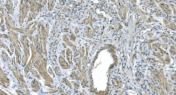
(Figure 6. IHC analysis of Thrombopoietin using anti- Thrombopoietin antibody (AAA19166).Thrombopoietin was detected in paraffin-embedded section of rat kidney tissues. Heat mediated antigen retrieval was performed in citrate buffer (pH6, epitope retrieval solution) for 20 mins. The tissue section was blocked with 10% goat serum. The tissue section was then incubated with 1ug/ml rabbit anti- Thrombopoietin Antibody (AAA19166) overnight at 4 degree C. Biotinylated goat anti-rabbit IgG was used as secondary antibody and incubated for 30 minutes at 37 degree C. The tissue section was developed using Strepavidin-Biotin-Complex (SABC) with DAB as the chromogen.)
IHC (Immunohistchemistry)
(Figure 6. IHC analysis of Thrombopoietin using anti- Thrombopoietin antibody (AAA19166).Thrombopoietin was detected in paraffin-embedded section of rat kidney tissues. Heat mediated antigen retrieval was performed in citrate buffer (pH6, epitope retrieval solution) for 20 mins. The tissue section was blocked with 10% goat serum. The tissue section was then incubated with 1ug/ml rabbit anti- Thrombopoietin Antibody (AAA19166) overnight at 4 degree C. Biotinylated goat anti-rabbit IgG was used as secondary antibody and incubated for 30 minutes at 37 degree C. The tissue section was developed using Strepavidin-Biotin-Complex (SABC) with DAB as the chromogen.)
Thrombopoietin, Polyclonal Antibody (Cat# AAA19166)
Full Name
Anti-Thrombopoietin Picoband Antibody
Gene Names
Thpo; Ml; Tpo; Mgdf; Mpllg
Reactivity
Mouse, Rat
No cross reactivity with other proteins
No cross reactivity with other proteins
Applications
EIA, IHC, WB
Purity
Immunogen affinity purified
Pricing


 FCM (Flow Cytometry)
(Figure 8. Flow Cytometry analysis of U20S cells using anti-Dynamin 1 antibody (AAA19160).Overlay histogram showing U20S cells stained with AAA19160 (Blue line).The cells were blocked with 10% normal goat serum. And then incubated with rabbit anti-DNM1 Antibody (AAA19160,1ug/1x10^6 cells) for 30 min at 20 degree C. DyLight®488 conjugated goat anti-rabbit IgG (5-10ug/1x10^6 cells) was used as secondary antibody for 30 minutes at 20 degree C. Isotype control antibody (Green line) was rabbit IgG (1ug/1x106) used under the same conditions. Unlabelled sample (Red line) was also used as a control.)
FCM (Flow Cytometry)
(Figure 8. Flow Cytometry analysis of U20S cells using anti-Dynamin 1 antibody (AAA19160).Overlay histogram showing U20S cells stained with AAA19160 (Blue line).The cells were blocked with 10% normal goat serum. And then incubated with rabbit anti-DNM1 Antibody (AAA19160,1ug/1x10^6 cells) for 30 min at 20 degree C. DyLight®488 conjugated goat anti-rabbit IgG (5-10ug/1x10^6 cells) was used as secondary antibody for 30 minutes at 20 degree C. Isotype control antibody (Green line) was rabbit IgG (1ug/1x106) used under the same conditions. Unlabelled sample (Red line) was also used as a control.)
Dynamin 1, Polyclonal Antibody (Cat# AAA19160)
Full Name
Anti-Dynamin 1 Picoband antibody
Gene Names
DNM1; DNM; EIEE31
Reactivity
Human, Mouse, Rat
No cross reactivity with other proteins.
No cross reactivity with other proteins.
Applications
EIA, FC/FACS, IHC, ICC, WB
Pricing


 FCM (Flow Cytometry)
(Figure 11. Flow Cytometry analysis of U937 cells using anti-NSF antibody (AAA19222).Overlay histogram showing U937 cells stained with AAA19222 (Blue line). The cells were blocked with 10% normal goat serum. And then incubated with rabbit anti-NSF Antibody (AAA19222, 1μg/1x106 cells) for 30 min at 20 degree C. DyLight®488 conjugated goat anti-rabbit IgG (5-10μg/1x106 cells) was used as secondary antibody for 30 minutes at 20 degree C. Isotype control antibody (Green line) was rabbit IgG (1μg/1x106) used under the same conditions. Unlabelled sample (Red line) was also used as a control.)
FCM (Flow Cytometry)
(Figure 11. Flow Cytometry analysis of U937 cells using anti-NSF antibody (AAA19222).Overlay histogram showing U937 cells stained with AAA19222 (Blue line). The cells were blocked with 10% normal goat serum. And then incubated with rabbit anti-NSF Antibody (AAA19222, 1μg/1x106 cells) for 30 min at 20 degree C. DyLight®488 conjugated goat anti-rabbit IgG (5-10μg/1x106 cells) was used as secondary antibody for 30 minutes at 20 degree C. Isotype control antibody (Green line) was rabbit IgG (1μg/1x106) used under the same conditions. Unlabelled sample (Red line) was also used as a control.)
NSF, Polyclonal Antibody (Cat# AAA19222)
Full Name
Anti-NSF Antibody
Gene Names
NSF; SKD2
Reactivity
Human, Mouse, Rat
Applications
WB, IHC-P, ICC, IF, FC/FACS/FCM, EIA
Purity
Immunogen affinity purified.
Pricing


 IF (Immunofluorescence)
(Figure 6. IF analysis of EEF2 using anti- EEF2 antibody (AAA19228).EEF2 was detected in immunocytochemical section of MCF-7 cells. Enzyme antigen retrieval was performed using IHC enzyme antigen retrieval reagent for 15 mins. The cells were blocked with 10% goat serum. And then incubated with 5μg/mL rabbit anti-EEF2 Antibody (AAA19228) overnight at 4 degree C. DyLight®488 Conjugated Goat Anti-Rabbit IgG was used as secondary antibody at 1:100 dilution and incubated for 30 minutes at 37 degree C. The section was counterstained with DAPI. Visualize using a fluorescence microscope and filter sets appropriate for the label used.)
IF (Immunofluorescence)
(Figure 6. IF analysis of EEF2 using anti- EEF2 antibody (AAA19228).EEF2 was detected in immunocytochemical section of MCF-7 cells. Enzyme antigen retrieval was performed using IHC enzyme antigen retrieval reagent for 15 mins. The cells were blocked with 10% goat serum. And then incubated with 5μg/mL rabbit anti-EEF2 Antibody (AAA19228) overnight at 4 degree C. DyLight®488 Conjugated Goat Anti-Rabbit IgG was used as secondary antibody at 1:100 dilution and incubated for 30 minutes at 37 degree C. The section was counterstained with DAPI. Visualize using a fluorescence microscope and filter sets appropriate for the label used.)
EEF2, Polyclonal Antibody (Cat# AAA19228)
Full Name
Anti-EEF2 Antibody
Gene Names
EEF2; EF2; EF-2; EEF-2
Reactivity
Human
Applications
WB, IHC-P, ICC, IF, EIA
Purity
Immunogen affinity purified.
Pricing


 FCM (Flow Cytometry)
(Figure 6. Flow Cytometry analysis of U20S cells using anti-PPID antibody (AAA19159).Overlay histogram showing U20S cells stained with AAA19159 (Blue line).The cells were blocked with 10% normal goat serum. And then incubated with rabbit anti-PPID Antibody (AAA19159,1ug/1x10^6 cells) for 30 min at 20 degree C. DyLight®488 conjugated goat anti-rabbit IgG (5-10ug/1x10^6 cells) was used as secondary antibody for 30 minutes at 20 degree C. Isotype control antibody (Green line) was rabbit IgG (1ug/1x106) used under the same conditions. Unlabelled sample (Red line) was also used as a control.)
FCM (Flow Cytometry)
(Figure 6. Flow Cytometry analysis of U20S cells using anti-PPID antibody (AAA19159).Overlay histogram showing U20S cells stained with AAA19159 (Blue line).The cells were blocked with 10% normal goat serum. And then incubated with rabbit anti-PPID Antibody (AAA19159,1ug/1x10^6 cells) for 30 min at 20 degree C. DyLight®488 conjugated goat anti-rabbit IgG (5-10ug/1x10^6 cells) was used as secondary antibody for 30 minutes at 20 degree C. Isotype control antibody (Green line) was rabbit IgG (1ug/1x106) used under the same conditions. Unlabelled sample (Red line) was also used as a control.)
PPID/Cyclophilin 40, Polyclonal Antibody (Cat# AAA19159)
Full Name
Anti-PPID/Cyclophilin 40 Picoband antibody
Gene Names
PPID; CYPD; CYP-40
Reactivity
Human, Mouse, Rat
No cross reactivity with other proteins.
No cross reactivity with other proteins.
Applications
EIA, FC/FACS, IHC, ICC, WB
Pricing


 IHC (Immunohistchemistry)
(Figure 6. IHC analysis of MED15 using anti-MED15 antibody (AAA19169).MED15 was detected in paraffin-embedded section of rat brain tissue. Heat mediated antigen retrieval was performed in citrate buffer (pH6, epitope retrieval solution) for 20 mins. The tissue section was blocked with 10% goat serum. The tissue section was then incubated with 1ug/ml rabbit anti-MED15 Antibody (AAA19169) overnight at 4 degree C. Biotinylated goat anti-rabbit IgG was used as secondary antibody and incubated for 30 minutes at 37 degree C. The tissue section was developed using Strepavidin-Biotin-Complex (SABC) with DAB as the chromogen.)
IHC (Immunohistchemistry)
(Figure 6. IHC analysis of MED15 using anti-MED15 antibody (AAA19169).MED15 was detected in paraffin-embedded section of rat brain tissue. Heat mediated antigen retrieval was performed in citrate buffer (pH6, epitope retrieval solution) for 20 mins. The tissue section was blocked with 10% goat serum. The tissue section was then incubated with 1ug/ml rabbit anti-MED15 Antibody (AAA19169) overnight at 4 degree C. Biotinylated goat anti-rabbit IgG was used as secondary antibody and incubated for 30 minutes at 37 degree C. The tissue section was developed using Strepavidin-Biotin-Complex (SABC) with DAB as the chromogen.)
MED15/Pcqap, Polyclonal Antibody (Cat# AAA19169)
Full Name
Anti-MED15/Pcqap Picoband Antibody
Gene Names
MED15; TIG1; CAG7A; CTG7A; PCQAP; TIG-1; TNRC7; ARC105
Reactivity
Human, Mouse, Rat
No cross reactivity with other proteins.
No cross reactivity with other proteins.
Applications
EIA, IHC, WB
Purity
Immunogen affinity purified
Pricing


 IHC (Immunohistchemistry)
(Figure 6. IHC analysis of Integrin beta 4/ITGB4 using anti-Integrin beta 4/ITGB4 antibody (AAA19269).Integrin beta 4/ITGB4 was detected in paraffin-embedded section of rat brain tissue. Heat mediated antigen retrieval was performed in EDTA buffer (pH8. 0, epitope retrieval solution). The tissue section was blocked with 10% goat serum. The tissue section was then incubated with 1μg/ml rabbit anti-Integrin beta 4/ITGB4 Antibody (AAA19269) overnight at 4 degree C. Biotinylated goat anti-rabbit IgG was used as secondary antibody and incubated for 30 minutes at 37 degree C. The tissue section was developed using Strepavidin-Biotin-Complex (SABC) (Catalog # with DAB as the chromogen.)
IHC (Immunohistchemistry)
(Figure 6. IHC analysis of Integrin beta 4/ITGB4 using anti-Integrin beta 4/ITGB4 antibody (AAA19269).Integrin beta 4/ITGB4 was detected in paraffin-embedded section of rat brain tissue. Heat mediated antigen retrieval was performed in EDTA buffer (pH8. 0, epitope retrieval solution). The tissue section was blocked with 10% goat serum. The tissue section was then incubated with 1μg/ml rabbit anti-Integrin beta 4/ITGB4 Antibody (AAA19269) overnight at 4 degree C. Biotinylated goat anti-rabbit IgG was used as secondary antibody and incubated for 30 minutes at 37 degree C. The tissue section was developed using Strepavidin-Biotin-Complex (SABC) (Catalog # with DAB as the chromogen.)
mGluR1/GRM1, Polyclonal Antibody (Cat# AAA19269)
Full Name
Anti-mGluR1/GRM1 Antibody
Gene Names
GRM1; MGLU1; GPRC1A; MGLUR1; SCAR13; PPP1R85
Reactivity
Human, Mouse, Rat
Applications
WB, IHC-P, FC/FACS/FCM, EIA
Purity
Immunogen affinity purified.
Pricing


 FCM (Flow Cytometry)
(Figure 6. Flow Cytometry analysis of HEPA1-6 cells using anti-PAR4/Pawr antibody (AAA19278).Overlay histogram showing HEPA1-6 cells stained with AAA19278 (Blue line). The cells were blocked with 10% normal goat serum. And then incubated with rabbit anti-PAR4/Pawr Antibody (AAA19278, 1μg/1x106 cells) for 30 min at 20 degree C. DyLight®488 conjugated goat anti-rabbit IgG (5-10μg/1x106 cells) was used as secondary antibody for 30 minutes at 20 degree C. Isotype control antibody (Green line) was rabbit IgG (1μg/1x106) used under the same conditions. Unlabelled sample (Red line) was also used as a control.)
FCM (Flow Cytometry)
(Figure 6. Flow Cytometry analysis of HEPA1-6 cells using anti-PAR4/Pawr antibody (AAA19278).Overlay histogram showing HEPA1-6 cells stained with AAA19278 (Blue line). The cells were blocked with 10% normal goat serum. And then incubated with rabbit anti-PAR4/Pawr Antibody (AAA19278, 1μg/1x106 cells) for 30 min at 20 degree C. DyLight®488 conjugated goat anti-rabbit IgG (5-10μg/1x106 cells) was used as secondary antibody for 30 minutes at 20 degree C. Isotype control antibody (Green line) was rabbit IgG (1μg/1x106) used under the same conditions. Unlabelled sample (Red line) was also used as a control.)
PAR4/Pawr, Polyclonal Antibody (Cat# AAA19278)
Full Name
Anti-PAR4/Pawr Antibody
Gene Names
Pawr; PAR4; Par-4; 2310001G03Rik
Reactivity
Mouse, Rat
Applications
WB, IHC-P, ICC, IF, FC/FACS/FCM, EIA
Purity
Immunogen affinity purified.
Pricing


 FCM (Flow Cytometry)
(Figure 6. Flow Cytometry analysis of CACO-2 cells using anti-Galectin 2/LGALS2 antibody (AAA19287).Overlay histogram showing CACO-2 cells stained with AAA19287 (Blue line). The cells were blocked with 10% normal goat serum. And then incubated with rabbit anti-Galectin 2/LGALS2 Antibody (AAA19287, 1μg/1x106 cells) for 30 min at 20 degree C. DyLight®488 conjugated goat anti-rabbit IgG (5-10μg/1x106 cells) was used as secondary antibody for 30 minutes at 20 degree C. Isotype control antibody (Green line) was rabbit IgG (1μg/1x106) used under the same conditions. Unlabelled sample (Red line) was also used as a control.)
FCM (Flow Cytometry)
(Figure 6. Flow Cytometry analysis of CACO-2 cells using anti-Galectin 2/LGALS2 antibody (AAA19287).Overlay histogram showing CACO-2 cells stained with AAA19287 (Blue line). The cells were blocked with 10% normal goat serum. And then incubated with rabbit anti-Galectin 2/LGALS2 Antibody (AAA19287, 1μg/1x106 cells) for 30 min at 20 degree C. DyLight®488 conjugated goat anti-rabbit IgG (5-10μg/1x106 cells) was used as secondary antibody for 30 minutes at 20 degree C. Isotype control antibody (Green line) was rabbit IgG (1μg/1x106) used under the same conditions. Unlabelled sample (Red line) was also used as a control.)
Galectin 2/LGALS2, Polyclonal Antibody (Cat# AAA19287)
Full Name
Anti-Galectin 2/LGALS2 Antibody
Gene Names
LGALS2; HL14
Reactivity
Human
Applications
WB, IHC-P, ICC, IF, FC/FACS/FCM, EIA
Purity
Immunogen affinity purified.
Pricing


 FCM (Flow Cytometry)
(Figure 6. Flow Cytometry analysis of HEPA1-6 cells using anti-Rad9/Rad9a antibody (AAA19288).Overlay histogram showing HEPA1-6 cells stained with AAA19288 (Blue line). The cells were blocked with 10% normal goat serum. And then incubated with rabbit anti-Rad9/Rad9a Antibody (AAA19288,1μg/1x106 cells) for 30 min at 20 degree C. DyLight®488 conjugated goat anti-rabbit IgG (5-10μg/1x106 cells) was used as secondary antibody for 30 minutes at 20 degree C. Isotype control antibody (Green line) was rabbit IgG (1μg/1x106) used under the same conditions. Unlabelled sample (Red line) was also used as a control.)
FCM (Flow Cytometry)
(Figure 6. Flow Cytometry analysis of HEPA1-6 cells using anti-Rad9/Rad9a antibody (AAA19288).Overlay histogram showing HEPA1-6 cells stained with AAA19288 (Blue line). The cells were blocked with 10% normal goat serum. And then incubated with rabbit anti-Rad9/Rad9a Antibody (AAA19288,1μg/1x106 cells) for 30 min at 20 degree C. DyLight®488 conjugated goat anti-rabbit IgG (5-10μg/1x106 cells) was used as secondary antibody for 30 minutes at 20 degree C. Isotype control antibody (Green line) was rabbit IgG (1μg/1x106) used under the same conditions. Unlabelled sample (Red line) was also used as a control.)
Rad9/Rad9a, Polyclonal Antibody (Cat# AAA19288)
Full Name
Anti-Rad9/Rad9a Antibody
Gene Names
Rad9a; Rad9
Reactivity
Mouse, Rat
Applications
WB, IHC-P, FC/FACS/FCM, EIA
Purity
Immunogen affinity purified.
Pricing


 FCM (Flow Cytometry)
(Figure 7. Flow Cytometry analysis of A549 cells using anti-AGO3 antibody (AAA19290).Overlay histogram showing A549 cells stained with AAA19290 (Blue line). The cells were blocked with 10% normal goat serum. And then incubated with rabbit anti-AGO3 Antibody (AAA19290, 1μg/1x106 cells) for 30 min at 20 degree C. DyLight®488 conjugated goat anti-rabbit IgG (5-10μg/1x106 cells) was used as secondary antibody for 30 minutes at 20 degree C. Isotype control antibody (Green line) was rabbit IgG (1μg/1x106) used under the same conditions. Unlabelled sample (Red line) was also used as a control.)
FCM (Flow Cytometry)
(Figure 7. Flow Cytometry analysis of A549 cells using anti-AGO3 antibody (AAA19290).Overlay histogram showing A549 cells stained with AAA19290 (Blue line). The cells were blocked with 10% normal goat serum. And then incubated with rabbit anti-AGO3 Antibody (AAA19290, 1μg/1x106 cells) for 30 min at 20 degree C. DyLight®488 conjugated goat anti-rabbit IgG (5-10μg/1x106 cells) was used as secondary antibody for 30 minutes at 20 degree C. Isotype control antibody (Green line) was rabbit IgG (1μg/1x106) used under the same conditions. Unlabelled sample (Red line) was also used as a control.)
AGO3, Polyclonal Antibody (Cat# AAA19290)
Full Name
Anti-AGO3 Antibody
Gene Names
AGO3; EIF2C3
Reactivity
Human
Applications
WB, IHC-P, ICC, IF, FC/FACS/FCM, EIA
Purity
Immunogen affinity purified.
Pricing


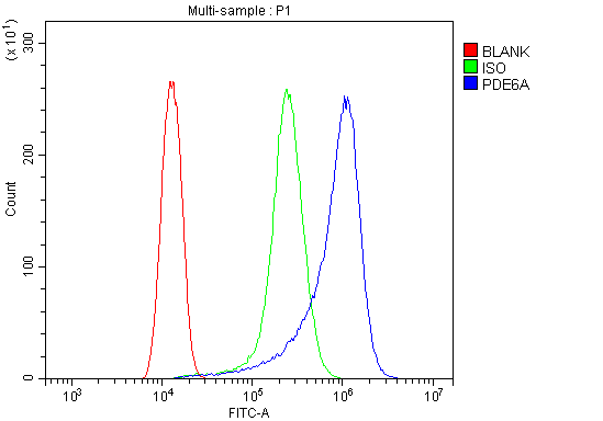 FCM (Flow Cytometry)
(Figure 6. Flow Cytometry analysis of HELA cells using anti-PDE6 alpha/PDE6A antibody (AAA19318).Overlay histogram showing HELA cells stained with AAA19318 (Blue line). The cells were blocked with 10% normal goat serum. And then incubated with rabbit anti-PDE6 alpha/PDE6A Antibody (AAA19318, 1μg/1x106 cells) for 30 min at 20 degree C. DyLight®488 conjugated goat anti-rabbit IgG (5-10μg/1x106 cells) was used as secondary antibody for 30 minutes at 20 degree C. Isotype control antibody (Green line) was rabbit IgG (1μg/1x106) used under the same conditions. Unlabelled sample (Red line) was also used as a control.)
FCM (Flow Cytometry)
(Figure 6. Flow Cytometry analysis of HELA cells using anti-PDE6 alpha/PDE6A antibody (AAA19318).Overlay histogram showing HELA cells stained with AAA19318 (Blue line). The cells were blocked with 10% normal goat serum. And then incubated with rabbit anti-PDE6 alpha/PDE6A Antibody (AAA19318, 1μg/1x106 cells) for 30 min at 20 degree C. DyLight®488 conjugated goat anti-rabbit IgG (5-10μg/1x106 cells) was used as secondary antibody for 30 minutes at 20 degree C. Isotype control antibody (Green line) was rabbit IgG (1μg/1x106) used under the same conditions. Unlabelled sample (Red line) was also used as a control.)
PDE6 alpha/PDE6A, Polyclonal Antibody (Cat# AAA19318)
Full Name
Anti-PDE6 alpha/PDE6A Antibody
Gene Names
PDE6A; PDEA; RP43; CGPR-A; PDE6A
Reactivity
Human, Mouse, Rat
Applications
WB, IHC-P, ICC, IF, FC/FACS/FCM, EIA
Purity
Immunogen affinity purified.
Pricing


 FCM (Flow Cytometry)
(Figure 6. Flow Cytometry analysis of Hela cells using anti-Caspase-9/CASP9 antibody (AAA19215).Overlay histogram showing Hela cells stained with AAA19215 (Blue line). The cells were blocked with 10% normal goat serum. And then incubated with rabbit anti-Caspase-9/CASP9 Antibody (AAA19215, 1 μg/1x106 cells) for 30 min at 20 degree C. DyLight®488 conjugated goat anti-rabbit IgG (5-10 μg/1x106 cells) was used as secondary antibody for 30 minutes at 20 degree C. Isotype control antibody (Green line) was rabbit IgG (1 μg/1x106) used under the same conditions. Unlabelled sample (Red line) was also used as a control.)
FCM (Flow Cytometry)
(Figure 6. Flow Cytometry analysis of Hela cells using anti-Caspase-9/CASP9 antibody (AAA19215).Overlay histogram showing Hela cells stained with AAA19215 (Blue line). The cells were blocked with 10% normal goat serum. And then incubated with rabbit anti-Caspase-9/CASP9 Antibody (AAA19215, 1 μg/1x106 cells) for 30 min at 20 degree C. DyLight®488 conjugated goat anti-rabbit IgG (5-10 μg/1x106 cells) was used as secondary antibody for 30 minutes at 20 degree C. Isotype control antibody (Green line) was rabbit IgG (1 μg/1x106) used under the same conditions. Unlabelled sample (Red line) was also used as a control.)
Caspase-9/CASP9, Polyclonal Antibody (Cat# AAA19215)
Full Name
Anti-Caspase-9/CASP9 Antibody
Gene Names
CASP9; MCH6; APAF3; APAF-3; PPP1R56; ICE-LAP6
Reactivity
Human
Applications
WB, IHC-P, FC/FACS/FCM, EIA
Purity
Immunogen affinity purified.
Pricing


 FCM (Flow Cytometry)
(Figure 7. Flow Cytometry analysis of U251 cells using anti-Serum Response Factor/SRF antibody (AAA19221).Overlay histogram showing U251 cells stained with AAA19221 (Blue line). The cells were blocked with 10% normal goat serum. And then incubated with rabbit anti-Serum Response Factor/SRF Antibody (AAA19221, 1μg/1x106 cells) for 30 min at 20 degree C. DyLight®488 conjugated goat anti-rabbit IgG (5-10μg/1x106 cells) was used as secondary antibody for 30 minutes at 20 degree C. Isotype control antibody (Green line) was rabbit IgG (1μg/1x106) used under the same conditions. Unlabelled sample (Red line) was also used as a control.)
FCM (Flow Cytometry)
(Figure 7. Flow Cytometry analysis of U251 cells using anti-Serum Response Factor/SRF antibody (AAA19221).Overlay histogram showing U251 cells stained with AAA19221 (Blue line). The cells were blocked with 10% normal goat serum. And then incubated with rabbit anti-Serum Response Factor/SRF Antibody (AAA19221, 1μg/1x106 cells) for 30 min at 20 degree C. DyLight®488 conjugated goat anti-rabbit IgG (5-10μg/1x106 cells) was used as secondary antibody for 30 minutes at 20 degree C. Isotype control antibody (Green line) was rabbit IgG (1μg/1x106) used under the same conditions. Unlabelled sample (Red line) was also used as a control.)
Serum Response Factor/SRF, Polyclonal Antibody (Cat# AAA19221)
Full Name
Anti-Serum Response Factor/SRF Antibody
Gene Names
SRF; MCM1
Reactivity
Human, Mouse, Rat
Applications
WB, IHC-P, ICC, IF, FC/FACS/FCM, EIA
Purity
Immunogen affinity purified.
Pricing


 FCM (Flow Cytometry)
(Figure 6. Flow Cytometry analysis of MCF-7 cells using anti-Caspase-7/CASP7 antibody (AAA19232).Overlay histogram showing MCF-7 cells stained with AAA19232 (Blue line). The cells were blocked with 10% normal goat serum. And then incubated with rabbit anti-Caspase-7/CASP7 Antibody (AAA19232, 1μg/1x106 cells) for 30 min at 20 degree C. DyLight®488 conjugated goat anti-rabbit IgG (5-10μg/1x106 cells) was used as secondary antibody for 30 minutes at 20 degree C. Isotype control antibody (Green line) was rabbit IgG (1μg/1x106) used under the same conditions. Unlabelled sample (Red line) was also used as a control.)
FCM (Flow Cytometry)
(Figure 6. Flow Cytometry analysis of MCF-7 cells using anti-Caspase-7/CASP7 antibody (AAA19232).Overlay histogram showing MCF-7 cells stained with AAA19232 (Blue line). The cells were blocked with 10% normal goat serum. And then incubated with rabbit anti-Caspase-7/CASP7 Antibody (AAA19232, 1μg/1x106 cells) for 30 min at 20 degree C. DyLight®488 conjugated goat anti-rabbit IgG (5-10μg/1x106 cells) was used as secondary antibody for 30 minutes at 20 degree C. Isotype control antibody (Green line) was rabbit IgG (1μg/1x106) used under the same conditions. Unlabelled sample (Red line) was also used as a control.)
Caspase-7/CASP7, Polyclonal Antibody (Cat# AAA19232)
Full Name
Anti-Caspase-7/CASP7 Antibody
Gene Names
CASP7; MCH3; CMH-1; LICE2; CASP-7; ICE-LAP3
Reactivity
Human, Rat
Applications
WB, IHC-P, ICC, IF, FC/FACS/FCM, EIA
Purity
Immunogen affinity purified.
Pricing