
Filters
Clonality
Type
Reactivity
Gene Name
Isotype
Host
Application
Clone
10000 results for "Recombinant Proteins" - showing 9750-9800
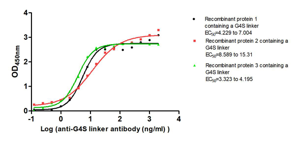
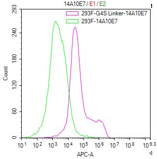 FCM/FACS (Flow Cytometry)
(Untransfected HEK-293F cells (green line) and transfected scFv-based Anti-CD19 CAR containing a G4S linker stable cells (red line) were stained with anti-G4S linker antibody (2ug/1*106cells), washed and then followed by APC-conjugated anti-Mouse IgG Fc antibody and analyzed with flow cytometry.)
FCM/FACS (Flow Cytometry)
(Untransfected HEK-293F cells (green line) and transfected scFv-based Anti-CD19 CAR containing a G4S linker stable cells (red line) were stained with anti-G4S linker antibody (2ug/1*106cells), washed and then followed by APC-conjugated anti-Mouse IgG Fc antibody and analyzed with flow cytometry.)
G4S linker, Monoclonal Antibody (Cat# AAA279298)
Full Name
G4S linker Monoclonal Antibody
Reactivity
Species Independent
Applications
Flow Cytometry
Purity
>95%, Protein G purified
Pricing
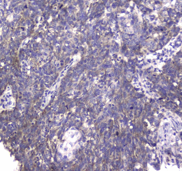
 IHC (Immunohistochemistry)
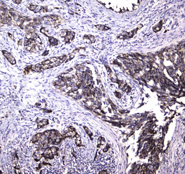
(Figure 4. IHC analysis of MRE11 using anti-MRE11 antibody (AAA124590).MRE11 was detected in paraffin-embedded section of human mammary cancer tissue. Heat mediated antigen retrieval was performed in citrate buffer (pH6, epitope retrieval solution) for 20 mins. The tissue section was blocked with 10% goat serum. The tissue section was then incubated with 1ug/ml rabbit anti-MRE11 Antibody (AAA124590) overnight at 4 degree C. Biotinylated goat anti-rabbit IgG was used as secondary antibody and incubated for 30 minutes at 37 degree C. The tissue section was developed using Strepavidin-Biotin-Complex (SABC) with DAB as the chromogen.)
IHC (Immunohistochemistry)
(Figure 4. IHC analysis of MRE11 using anti-MRE11 antibody (AAA124590).MRE11 was detected in paraffin-embedded section of human mammary cancer tissue. Heat mediated antigen retrieval was performed in citrate buffer (pH6, epitope retrieval solution) for 20 mins. The tissue section was blocked with 10% goat serum. The tissue section was then incubated with 1ug/ml rabbit anti-MRE11 Antibody (AAA124590) overnight at 4 degree C. Biotinylated goat anti-rabbit IgG was used as secondary antibody and incubated for 30 minutes at 37 degree C. The tissue section was developed using Strepavidin-Biotin-Complex (SABC) with DAB as the chromogen.)
MRE11, Polyclonal Antibody (Cat# AAA124590)
Full Name
Anti-MRE11 Picoband antibody
Gene Names
MRE11; ATLD; HNGS1; MRE11A; MRE11B
Reactivity
Human, Mouse, Rat
No cross reactivity with other proteins.
No cross reactivity with other proteins.
Applications
Western Blot, Immunohistochemistry
Pricing
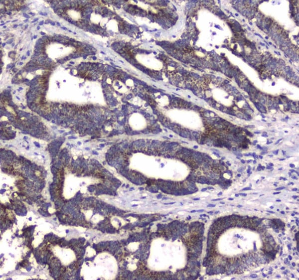
 IHC (Immunohiostchemistry)
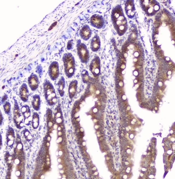
(Figure 3. IHC analysis of GOLPH3 using anti-GOLPH3 antibody.GOLPH3 was detected in paraffin-embedded section of human intestinal cancer tissue. Heat mediated antigen retrieval was performed in citrate buffer (pH6, epitope retrieval solution) for 20 mins. The tissue section was blocked with 10% goat serum. The tissue section was then incubated with 2ug/ml rabbit anti-GOLPH3 antibody.GOLPH3 was detected in paraffin-embedded section of human intestinal cancer tissue. Heat mediated antigen retrieval was performed in citrate buffer (pH6, epitope retrieval solution) for 20 mins. The tissue section was blocked with 10% goat serum. The tissue section was then incubated with 2ug/ml rabbit anti-GOLPH3 antibody.GOLPH3 was detected in paraffin-embedded section of human lung cancer tissue. Heat mediated antigen retrieval was performed in citrate buffer (pH6, epitope retrieval solution) for 20 mins. The tissue section was blocked with 10% goat serum. The tissue section was then incubated with 2ug/ml rabbit anti-GOLPH3 antibody.GOLPH3 was detected in paraffin-embedded section of rat brain tissue. Heat mediated antigen retrieval was performed in citrate buffer (pH6, epitope retrieval solution) for 20 mins. The tissue section was blocked with 10% goat serum. The tissue section was then incubated with 2ug/ml rabbit anti-GOLPH3 antibody.GOLPH3 was detected in paraffin-embedded section of rat small intestine tissue. Heat mediated antigen retrieval was performed in citrate buffer (pH6, epitope retrieval solution) for 20 mins. The tissue section was blocked with 10% goat serum. The tissue section was then incubated with 2ug/ml rabbit anti-GOLPH3 Antibody overnight at 4 degree C. Biotinylated goat anti-rabbit IgG was used as secondary antibody and incubated for 30 minutes at 37 degree C. The tissue section was developed using Strepavidin-Biotin-Complex (SABC) with DAB as the chromogen.)
IHC (Immunohiostchemistry)
(Figure 3. IHC analysis of GOLPH3 using anti-GOLPH3 antibody.GOLPH3 was detected in paraffin-embedded section of human intestinal cancer tissue. Heat mediated antigen retrieval was performed in citrate buffer (pH6, epitope retrieval solution) for 20 mins. The tissue section was blocked with 10% goat serum. The tissue section was then incubated with 2ug/ml rabbit anti-GOLPH3 antibody.GOLPH3 was detected in paraffin-embedded section of human intestinal cancer tissue. Heat mediated antigen retrieval was performed in citrate buffer (pH6, epitope retrieval solution) for 20 mins. The tissue section was blocked with 10% goat serum. The tissue section was then incubated with 2ug/ml rabbit anti-GOLPH3 antibody.GOLPH3 was detected in paraffin-embedded section of human lung cancer tissue. Heat mediated antigen retrieval was performed in citrate buffer (pH6, epitope retrieval solution) for 20 mins. The tissue section was blocked with 10% goat serum. The tissue section was then incubated with 2ug/ml rabbit anti-GOLPH3 antibody.GOLPH3 was detected in paraffin-embedded section of rat brain tissue. Heat mediated antigen retrieval was performed in citrate buffer (pH6, epitope retrieval solution) for 20 mins. The tissue section was blocked with 10% goat serum. The tissue section was then incubated with 2ug/ml rabbit anti-GOLPH3 antibody.GOLPH3 was detected in paraffin-embedded section of rat small intestine tissue. Heat mediated antigen retrieval was performed in citrate buffer (pH6, epitope retrieval solution) for 20 mins. The tissue section was blocked with 10% goat serum. The tissue section was then incubated with 2ug/ml rabbit anti-GOLPH3 Antibody overnight at 4 degree C. Biotinylated goat anti-rabbit IgG was used as secondary antibody and incubated for 30 minutes at 37 degree C. The tissue section was developed using Strepavidin-Biotin-Complex (SABC) with DAB as the chromogen.)
GOLPH3, Polyclonal Antibody (Cat# AAA124926)
Full Name
Anti-GOLPH3 Antibody
Gene Names
GOLPH3; GOPP1; GPP34; MIDAS; Vps74
Reactivity
Human, Mouse, Rat
Applications
Direct ELISA, Immunohistochemistry, Western Blot
Purity
Immunogen Affinity Purified
Pricing
 IHC (Immunohiostchemistry)
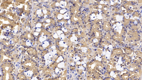
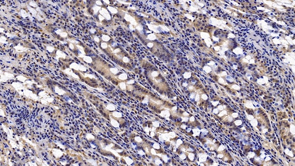
(DAB staining on IHCP;Sample: Human Small intestine Tissue; Primary Ab: 10ug/ml Mouse AntiHuman IFNb AntibodySecond Ab: 2ug/mL HRPLinked Caprine AntiMouse IgG Polyclonal Antibody(Catalog: SAA544Mu19))
IHC (Immunohiostchemistry)
(DAB staining on IHCP;Sample: Human Small intestine Tissue; Primary Ab: 10ug/ml Mouse AntiHuman IFNb AntibodySecond Ab: 2ug/mL HRPLinked Caprine AntiMouse IgG Polyclonal Antibody(Catalog: SAA544Mu19))
Interferon Beta (IFNb), Monoclonal Antibody (Cat# AAA151508)
Full Name
Monoclonal Antibody to Interferon Beta (IFNb)
Gene Names
IFNB1; IFB; IFF; IFNB
Reactivity
Human, Pig
Applications
Immunoprecipitation, Immunocytochemistry, Immunohistochemistry, Western Blot
Purity
Protein A + Protein G affinity chromatography
Pricing
 FCM/FACS (Flow Cytometry)
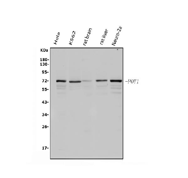
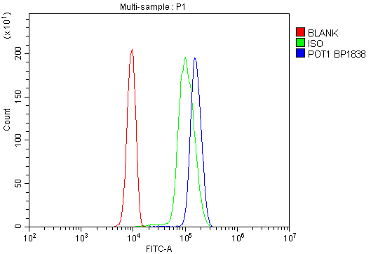
(Figure 2. Flow Cytometry analysis of U937 cells using anti-POT1 antibody (AAA125569).Overlay histogram showing U937 cells stained with AAA125569 (Blue line). The cells were blocked with 10% normal goat serum. And then incubated with rabbit anti-POT1 Antibody (AAA125569, 1μg/1x106 cells) for 30 min at 20 degree C. DyLight®488 conjugated goat anti-rabbit IgG (5-10μg/1x106 cells) was used as secondary antibody for 30 minutes at 20 degree C. Isotype control antibody (Green line) was rabbit IgG (1μg/1x106) used under the same conditions. Unlabelled sample (Red line) was also used as a control.)
FCM/FACS (Flow Cytometry)
(Figure 2. Flow Cytometry analysis of U937 cells using anti-POT1 antibody (AAA125569).Overlay histogram showing U937 cells stained with AAA125569 (Blue line). The cells were blocked with 10% normal goat serum. And then incubated with rabbit anti-POT1 Antibody (AAA125569, 1μg/1x106 cells) for 30 min at 20 degree C. DyLight®488 conjugated goat anti-rabbit IgG (5-10μg/1x106 cells) was used as secondary antibody for 30 minutes at 20 degree C. Isotype control antibody (Green line) was rabbit IgG (1μg/1x106) used under the same conditions. Unlabelled sample (Red line) was also used as a control.)
POT1, Polyclonal Antibody (Cat# AAA125569)
Full Name
Anti-POT1 Antibody
Reactivity
Human, Mouse, Rat
Applications
Direct ELISA, Flow Cytometry, Western Blot
Purity
Immunogen affinity purified.
Pricing
 IP (Immunoprecipitation)
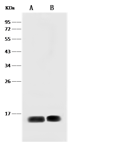
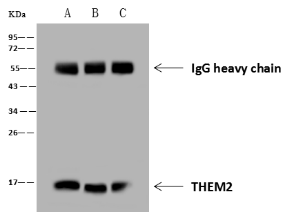
(THEM2 was immunoprecipitated using:Lane A:0.5 mg A431 Whole Cell LysateLane B:0.5 mg Jurkat Whole Cell LysateLane C:0.5 mg MCF-7 Whole Cell Lysate4 uL anti-THEM2 rabbit polyclonal antibody and 60 ug of Immunomagnetic beads Protein A/G.Primary antibody:Anti-THEM2 rabbit polyclonal antibody,at 1:100 dilutionSecondary antibody:Goat Anti-Rabbit IgG (H+L)/HRP at 1/10000 dilutionDeveloped using the ECL technique.Performed under reducing conditions.Predicted band size: 15 kDaObserved band size :15 kDa)
IP (Immunoprecipitation)
(THEM2 was immunoprecipitated using:Lane A:0.5 mg A431 Whole Cell LysateLane B:0.5 mg Jurkat Whole Cell LysateLane C:0.5 mg MCF-7 Whole Cell Lysate4 uL anti-THEM2 rabbit polyclonal antibody and 60 ug of Immunomagnetic beads Protein A/G.Primary antibody:Anti-THEM2 rabbit polyclonal antibody,at 1:100 dilutionSecondary antibody:Goat Anti-Rabbit IgG (H+L)/HRP at 1/10000 dilutionDeveloped using the ECL technique.Performed under reducing conditions.Predicted band size: 15 kDaObserved band size :15 kDa)
ACOT13/THEM2, Polyclonal Antibody (Cat# AAA258925)
Full Name
Anti-ACOT13/THEM2 Antibody, Rabbit Polyclonal
Reactivity
Human
Applications
Immunoprecipitation, Western Blot
Purity
Protein A & Antigen Affinity
Pricing
 IHC (Immunohiostchemistry)
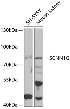
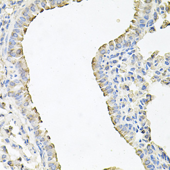
(Immunohistochemistry of paraffin-embedded mouse lung using SCNN1G at dilution of 1:100 (40x lens).)
IHC (Immunohiostchemistry)
(Immunohistochemistry of paraffin-embedded mouse lung using SCNN1G at dilution of 1:100 (40x lens).)
SCNN1G, Polyclonal Antibody (Cat# AAA313922)
Full Name
SCNN1G Rabbit Polyclonal Antibody
Gene Names
SCNN1G; PHA1; BESC3; ENaCg; SCNEG; ENaCgamma
Reactivity
Human, Mouse
Applications
Immunohistochemistry, Western Blot
Purity
Affinity purification
Pricing
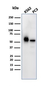 WB (Western Blot)
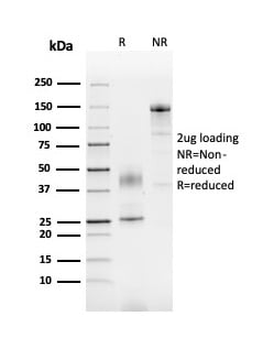
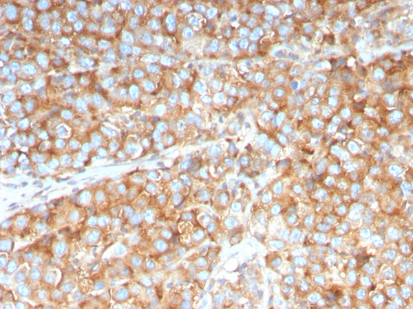
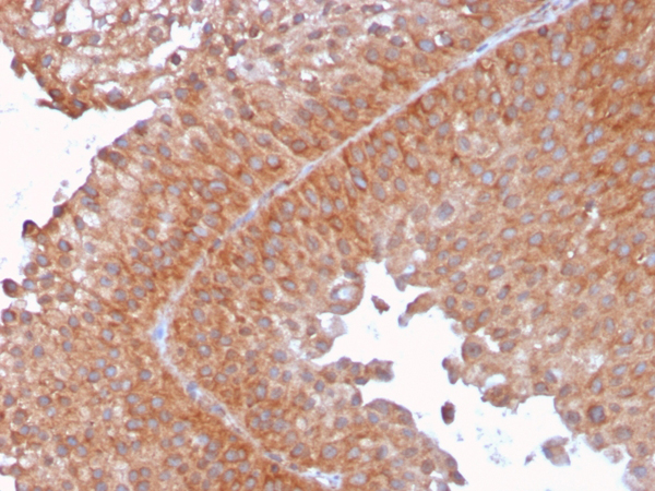
(Western Blot Analysis of K562 and PC3 cell lysate using SERBP1 Mouse Monoclonal Antibody (SERBP1/3492).)
WB (Western Blot)
(Western Blot Analysis of K562 and PC3 cell lysate using SERBP1 Mouse Monoclonal Antibody (SERBP1/3492).)
SERBP, Monoclonal Antibody (Cat# AAA215401)
Full Name
SERBP1/PAI-RBP1
Gene Names
SERBP1; CGI-55; CHD3IP; HABP4L; PAIRBP1; PAI-RBP1
Reactivity
Human
Applications
Western Blot, Immunohistochemistry
Purity
Purified Ab with BSA and Azide at 200ug/ml or Purified Ab with BSA and Azide at 200ug/ml or Purified Ab WITHOUT BSA and Azide at 1.0mg/ml
Pricing
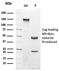
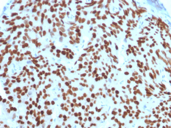
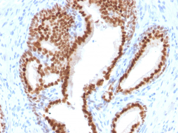
 Application Data
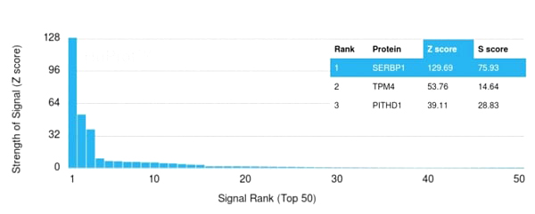
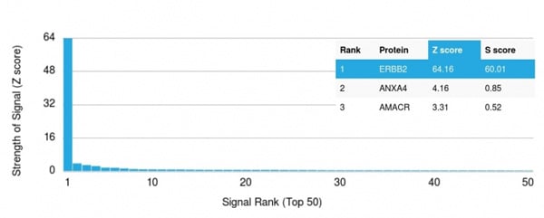
(Analysis of Protein Array containing more than 19,000 full-length human proteins using NKX3.1-Monospecific Mouse Monoclonal Antibody (NKX3.1/2576). Z- and S- Score: The Z-score represents the strength of a signal that a monoclonal antibody (MAb) (in combination with a fluorescently-tagged anti-IgG secondary antibody) produces when binding to a particular protein on the HuProtTM array. Z-scores are described in units of standard deviations (SD’s) above the mean value of all signals generated on that array. If targets on HuProtTM are arranged in descending order of the Z-score, the S-score is the difference (also in units of SD’s) between the Z-score. S-score therefore represents the relative target specificity of a MAb to its intended target. A MAb is considered to specific to its intended target, if the MAb has an S-score of at least 2.5. For example, if a MAb binds to protein X with a Z-score of 43 and to protein Y with a Z-score of 14, then the S-score for the binding of that MAb to protein X is equal to 29.)
Application Data
(Analysis of Protein Array containing more than 19,000 full-length human proteins using NKX3.1-Monospecific Mouse Monoclonal Antibody (NKX3.1/2576). Z- and S- Score: The Z-score represents the strength of a signal that a monoclonal antibody (MAb) (in combination with a fluorescently-tagged anti-IgG secondary antibody) produces when binding to a particular protein on the HuProtTM array. Z-scores are described in units of standard deviations (SD’s) above the mean value of all signals generated on that array. If targets on HuProtTM are arranged in descending order of the Z-score, the S-score is the difference (also in units of SD’s) between the Z-score. S-score therefore represents the relative target specificity of a MAb to its intended target. A MAb is considered to specific to its intended target, if the MAb has an S-score of at least 2.5. For example, if a MAb binds to protein X with a Z-score of 43 and to protein Y with a Z-score of 14, then the S-score for the binding of that MAb to protein X is equal to 29.)
NKX3.1, Monoclonal Antibody (Cat# AAA215103)
Full Name
NKX3.1 (Metastatic Prostate Adenocarcinoma Marker)
Gene Names
NKX3-1; NKX3; BAPX2; NKX3A; NKX3.1
Reactivity
Human
Applications
Immunohistochemistry
Purity
Purified Ab with BSA and Azide at 200ug/ml OR Purified Ab WITHOUT BSA and Azide at 1.0mg/ml
Pricing
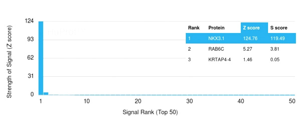 Application Data
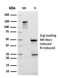
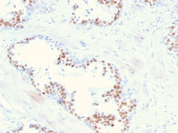
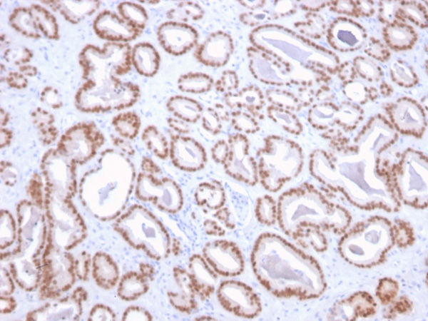
(Analysis of Protein Array containing more than 19,000 full-length human proteins using NKX3.1-Monospecific Mouse Monoclonal Antibody (NKX3.1/2836). Z- and S- Score: The Z-score represents the strength of a signal that a monoclonal antibody (MAb) (in combination with a fluorescently-tagged anti-IgG secondary antibody) produces when binding to a particular protein on the HuProtTM array. Z-scores are described in units of standard deviations (SD’s) above the mean value of all signals generated on that array. If targets on HuProtTM are arranged in descending order of the Z-score, the S-score is the difference (also in units of SD’s) between the Z-score. S-score therefore represents the relative target specificity of a MAb to its intended target. A MAb is considered to specific to its intended target, if the MAb has an S-score of at least 2.5. For example, if a MAb binds to protein X with a Z-score of 43 and to protein Y with a Z-score of 14, then the S-score for the binding of that MAb to protein X is equal to 29.)
Application Data
(Analysis of Protein Array containing more than 19,000 full-length human proteins using NKX3.1-Monospecific Mouse Monoclonal Antibody (NKX3.1/2836). Z- and S- Score: The Z-score represents the strength of a signal that a monoclonal antibody (MAb) (in combination with a fluorescently-tagged anti-IgG secondary antibody) produces when binding to a particular protein on the HuProtTM array. Z-scores are described in units of standard deviations (SD’s) above the mean value of all signals generated on that array. If targets on HuProtTM are arranged in descending order of the Z-score, the S-score is the difference (also in units of SD’s) between the Z-score. S-score therefore represents the relative target specificity of a MAb to its intended target. A MAb is considered to specific to its intended target, if the MAb has an S-score of at least 2.5. For example, if a MAb binds to protein X with a Z-score of 43 and to protein Y with a Z-score of 14, then the S-score for the binding of that MAb to protein X is equal to 29.)
NKX3.1, Monoclonal Antibody (Cat# AAA215104)
Full Name
NKX3.1 (Metastatic Prostate Adenocarcinoma Marker)
Gene Names
NKX3-1; NKX3; BAPX2; NKX3A; NKX3.1
Reactivity
Human
Applications
Immunohistochemistry
Purity
Purified Ab with BSA and Azide at 200ug/ml OR Purified Ab WITHOUT BSA and Azide at 1.0mg/ml
Pricing
 Application Data
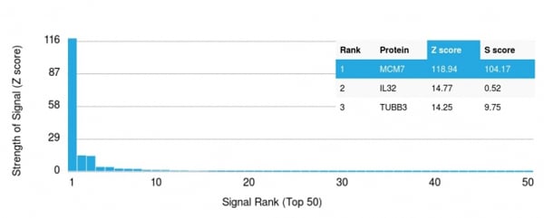
(Analysis of Protein Array containing more than 19, 000 full-length human proteins using MCM7 Mouse Monoclonal Antibody (MCM7/1469)Z- and S- Score: The Z-score represents the strength of a signal that a monoclonal antibody (MAb) (in combination with a fluorescently-tagged anti-IgG secondary antibody) produces when binding to a particular protein on the HuProtTM array. Z-scores are described in units of standard deviations (SD's) above the mean value of all signals generated on that array. If targets on HuProtTM are arranged in descending order of the Z-score, the S-score is the difference (also in units of SD's) between the Z-score. S-score therefore represents the relative target specificity of a MAb to its intended target. A MAb is considered to specific to its intended target, if the MAb has an S-score of at least 2.5. For example, if a MAb binds to protein X with a Z-score of 43 and to protein Y with a Z-score of 14, then the S-score for the binding of that MAb to protein X is equal to 29.)
Application Data
(Analysis of Protein Array containing more than 19, 000 full-length human proteins using MCM7 Mouse Monoclonal Antibody (MCM7/1469)Z- and S- Score: The Z-score represents the strength of a signal that a monoclonal antibody (MAb) (in combination with a fluorescently-tagged anti-IgG secondary antibody) produces when binding to a particular protein on the HuProtTM array. Z-scores are described in units of standard deviations (SD's) above the mean value of all signals generated on that array. If targets on HuProtTM are arranged in descending order of the Z-score, the S-score is the difference (also in units of SD's) between the Z-score. S-score therefore represents the relative target specificity of a MAb to its intended target. A MAb is considered to specific to its intended target, if the MAb has an S-score of at least 2.5. For example, if a MAb binds to protein X with a Z-score of 43 and to protein Y with a Z-score of 14, then the S-score for the binding of that MAb to protein X is equal to 29.)
MCM7, Monoclonal Antibody (Cat# AAA214445)
Full Name
MCM7 (Proliferation Marker)
Gene Names
MCM7; MCM2; CDC47; P85MCM; P1CDC47; PNAS146; PPP1R104; P1.1-MCM3
Reactivity
Human. Others not known.
Applications
Immunohistochemistry, Immunofluorescence, Flow Cytometry
Pricing
 FCM/FACS (Flow Cytometry)
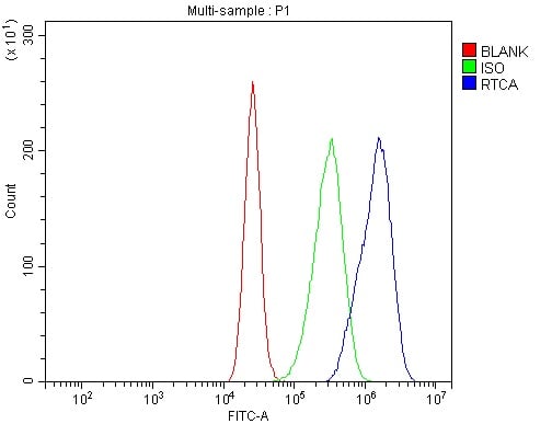
(Figure 3. Flow Cytometry analysis of CACO-2 cells using anti-RTCD1/RTCA antibody (AAA125792).Overlay histogram showing CACO-2 cells stained with AAA125792 (Blue line). The cells were blocked with 10% normal goat serum. And then incubated with rabbit anti-RTCD1/RTCA Antibody (AAA125792, 1μg/1x106 cells) for 30 min at 20 degree C. DyLight®488 conjugated goat anti-rabbit IgG (5-10μg/1x106 cells) was used as secondary antibody for 30 minutes at 20 degree C. Isotype control antibody (Green line) was rabbit IgG (1μg/1x106) used under the same conditions. Unlabelled sample (Red line) was also used as a control.)
FCM/FACS (Flow Cytometry)
(Figure 3. Flow Cytometry analysis of CACO-2 cells using anti-RTCD1/RTCA antibody (AAA125792).Overlay histogram showing CACO-2 cells stained with AAA125792 (Blue line). The cells were blocked with 10% normal goat serum. And then incubated with rabbit anti-RTCD1/RTCA Antibody (AAA125792, 1μg/1x106 cells) for 30 min at 20 degree C. DyLight®488 conjugated goat anti-rabbit IgG (5-10μg/1x106 cells) was used as secondary antibody for 30 minutes at 20 degree C. Isotype control antibody (Green line) was rabbit IgG (1μg/1x106) used under the same conditions. Unlabelled sample (Red line) was also used as a control.)
RTCD1/RTCA, Polyclonal Antibody (Cat# AAA125792)
Full Name
Anti-RTCD1/RTCA Antibody
Gene Names
RTCA; RPC; RTC1; RTCD1
Reactivity
Human
Applications
Direct ELISA, Flow Cytometry, Immunofluorescence, Immunocytochemistry, Western Blot
Purity
Immunogen affinity purified.
Pricing
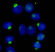
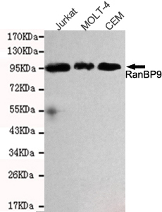 ICC (Immunocytochemistry)
(Immunocytochemistry stain of Hela using RanBP9 mouse mAb (1:300).)
ICC (Immunocytochemistry)
(Immunocytochemistry stain of Hela using RanBP9 mouse mAb (1:300).)
RanBP9, Monoclonal Antibody (Cat# AAA320342)
Full Name
RanBP9 Mouse mAb
Gene Names
RANBP9; BPM-L; BPM90; RANBPM; RanBP7
Reactivity
Human
Applications
Immunoprecipitation, Immunocytochemistry, Western Blot
Purity
The antibody was affinity-purified from mouse ascites by affinity-chromatography using epitope-specific immunogen.
Pricing
 FCM/FACS (Flow Cytometry)
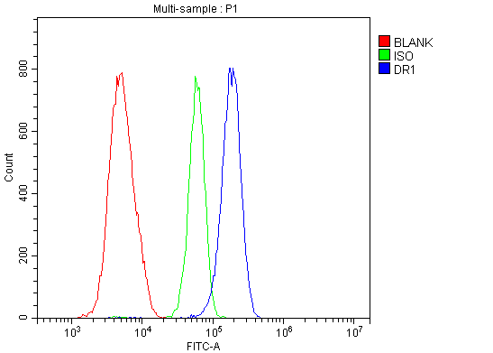
(Figure 5. Flow Cytometry analysis of HL-60 cells using anti-DR1 antibody (AAA125641).Overlay histogram showing HL-60 cells stained with AAA125641(Blue line). The cells were blocked with 10% normal goat serum. And then incubated with rabbit anti-DR1 Antibody (AAA125641, 1μg/1x106 cells) for 30 min at 20 degree C. DyLight®488 conjugated goat anti-rabbit IgG (5-10μg/1x106 cells) was used as secondary antibody for 30 minutes at 20 degree C. Isotype control antibody (Green line) was rabbit IgG (1μg/1x106) used under the same conditions. Unlabelled sample (Red line) was also used as a control.)
FCM/FACS (Flow Cytometry)
(Figure 5. Flow Cytometry analysis of HL-60 cells using anti-DR1 antibody (AAA125641).Overlay histogram showing HL-60 cells stained with AAA125641(Blue line). The cells were blocked with 10% normal goat serum. And then incubated with rabbit anti-DR1 Antibody (AAA125641, 1μg/1x106 cells) for 30 min at 20 degree C. DyLight®488 conjugated goat anti-rabbit IgG (5-10μg/1x106 cells) was used as secondary antibody for 30 minutes at 20 degree C. Isotype control antibody (Green line) was rabbit IgG (1μg/1x106) used under the same conditions. Unlabelled sample (Red line) was also used as a control.)
DR1, Polyclonal Antibody (Cat# AAA125641)
Full Name
Anti-DR1 Antibody
Gene Names
DR1; NC2; NC2-BETA
Reactivity
Human, Mouse, Rat
Applications
Direct ELISA, Flow Cytometry, Immunofluorescence, Immunocytochemistry, Immunohistochemistry, Western Blot
Purity
Immunogen affinity purified.
Pricing
 FCM/FACS (Flow Cytometry)
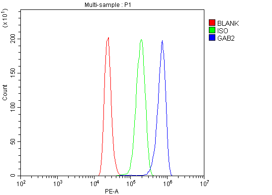
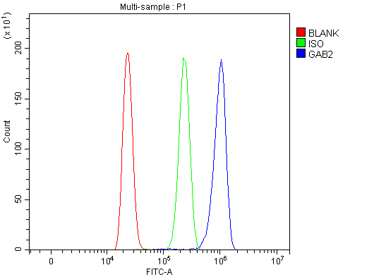
(Figure 4. Flow Cytometry analysis of THP-1 cells using anti-GAB2 antibody (AAA127111).Overlay histogram showing THP-1 cells stained with AAA127111 (Blue line). To facilitate intracellular staining, cells were fixed with 4% paraformaldehyde and permeabilized with permeabilization buffer. The cells were blocked with 10% normal goat serum. And then incubated with rabbit anti-GAB2 Antibody (AAA127111, 1ug/1x106 cells) for 30 min at 20 degree C. DyLight488 conjugated goat anti-rabbit IgG was used as secondary antibody for 30 minutes at 20 degree C. Isotype control antibody (Green line) was rabbit IgG (1ug/1x106) used under the same conditions. Unlabelled sample without incubation with primary antibody and secondary antibody (Red line) was used as a blank control.)
FCM/FACS (Flow Cytometry)
(Figure 4. Flow Cytometry analysis of THP-1 cells using anti-GAB2 antibody (AAA127111).Overlay histogram showing THP-1 cells stained with AAA127111 (Blue line). To facilitate intracellular staining, cells were fixed with 4% paraformaldehyde and permeabilized with permeabilization buffer. The cells were blocked with 10% normal goat serum. And then incubated with rabbit anti-GAB2 Antibody (AAA127111, 1ug/1x106 cells) for 30 min at 20 degree C. DyLight488 conjugated goat anti-rabbit IgG was used as secondary antibody for 30 minutes at 20 degree C. Isotype control antibody (Green line) was rabbit IgG (1ug/1x106) used under the same conditions. Unlabelled sample without incubation with primary antibody and secondary antibody (Red line) was used as a blank control.)
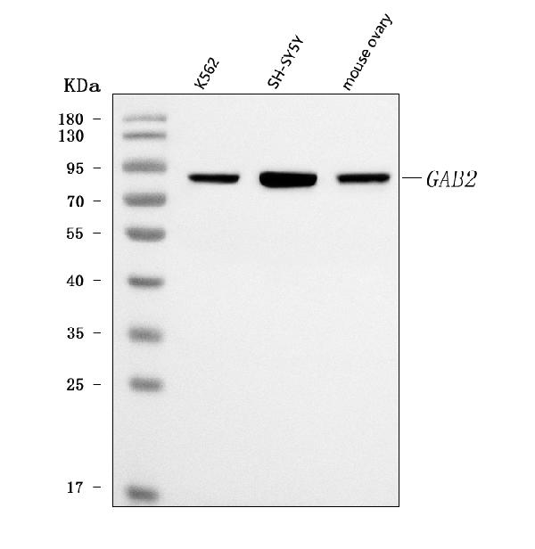
GAB2, Polyclonal Antibody (Cat# AAA127111)
Full Name
Anti-GAB2 Antibody Picoband
Reactivity
Human, Mouse
Applications
Flow Cytometry, Western Blot
Purity
Immunogen affinity purified.
Pricing
 FCM/FACS (Flow Cytometry)
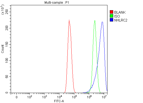
(Figure 2. Flow Cytometry analysis of U251 cells using anti-NHLRC2 antibody (AAA127907).Overlay histogram showing U251 cells stained with AAA127907 (Blue line). The cells were fixed with 4% paraformaldehyde and blocked with 10% normal goat serum. And then incubated with rabbit anti-NHLRC2 Antibody (AAA127907, 1ug/1x106 cells) for 30 min at 20 degree C. DyLight488 conjugated goat anti-rabbit IgG was used as secondary antibody for 30 minutes at 20 degree C. Isotype control antibody (Green line) was rabbit IgG (1ug/1x106) used under the same conditions. Unlabelled sample (Red line) was also used as a control.)
FCM/FACS (Flow Cytometry)
(Figure 2. Flow Cytometry analysis of U251 cells using anti-NHLRC2 antibody (AAA127907).Overlay histogram showing U251 cells stained with AAA127907 (Blue line). The cells were fixed with 4% paraformaldehyde and blocked with 10% normal goat serum. And then incubated with rabbit anti-NHLRC2 Antibody (AAA127907, 1ug/1x106 cells) for 30 min at 20 degree C. DyLight488 conjugated goat anti-rabbit IgG was used as secondary antibody for 30 minutes at 20 degree C. Isotype control antibody (Green line) was rabbit IgG (1ug/1x106) used under the same conditions. Unlabelled sample (Red line) was also used as a control.)
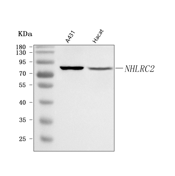
NHLRC2, Polyclonal Antibody (Cat# AAA127907)
Full Name
Anti-NHLRC2 Antibody Picoband
Reactivity
Human
Applications
Flow Cytometry, Western Blot
Purity
Immunogen affinity purified.
Pricing
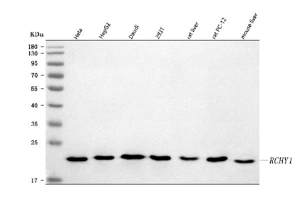
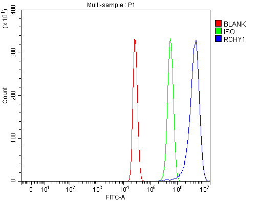 FCM/FACS (Flow Cytometry)
(Figure 2. Flow Cytometry analysis of HepG2 cells using anti-Pirh2/RCHY1 antibody (AAA127236).Overlay histogram showing HepG2 cells stained with AAA127236 (Blue line). To facilitate intracellular staining, cells were fixed with 4% paraformaldehyde and permeabilized with permeabilization buffer. The cells were blocked with 10% normal goat serum. And then incubated with rabbit anti-Pirh2/RCHY1 Antibody (AAA127236, 1ug/1x106 cells) for 30 min at 20 degree C. DyLight488 conjugated goat anti-rabbit IgG was used as secondary antibody for 30 minutes at 20 degree C. Isotype control antibody (Green line) was rabbit IgG (1ug/1x106) used under the same conditions. Unlabelled sample without incubation with primary antibody and secondary antibody (Red line) was used as a blank control.)
FCM/FACS (Flow Cytometry)
(Figure 2. Flow Cytometry analysis of HepG2 cells using anti-Pirh2/RCHY1 antibody (AAA127236).Overlay histogram showing HepG2 cells stained with AAA127236 (Blue line). To facilitate intracellular staining, cells were fixed with 4% paraformaldehyde and permeabilized with permeabilization buffer. The cells were blocked with 10% normal goat serum. And then incubated with rabbit anti-Pirh2/RCHY1 Antibody (AAA127236, 1ug/1x106 cells) for 30 min at 20 degree C. DyLight488 conjugated goat anti-rabbit IgG was used as secondary antibody for 30 minutes at 20 degree C. Isotype control antibody (Green line) was rabbit IgG (1ug/1x106) used under the same conditions. Unlabelled sample without incubation with primary antibody and secondary antibody (Red line) was used as a blank control.)
Pirh2/RCHY1, Polyclonal Antibody (Cat# AAA127236)
Full Name
Anti-Pirh2/RCHY1 Antibody Picoband
Gene Names
RCHY1; ZCHY; ARNIP; CHIMP; PIRH2; RNF199; ZNF363; PRO1996
Reactivity
Human, Mouse, Rat
Applications
Flow Cytometry, Western Blot
Purity
Immunogen affinity purified.
Pricing
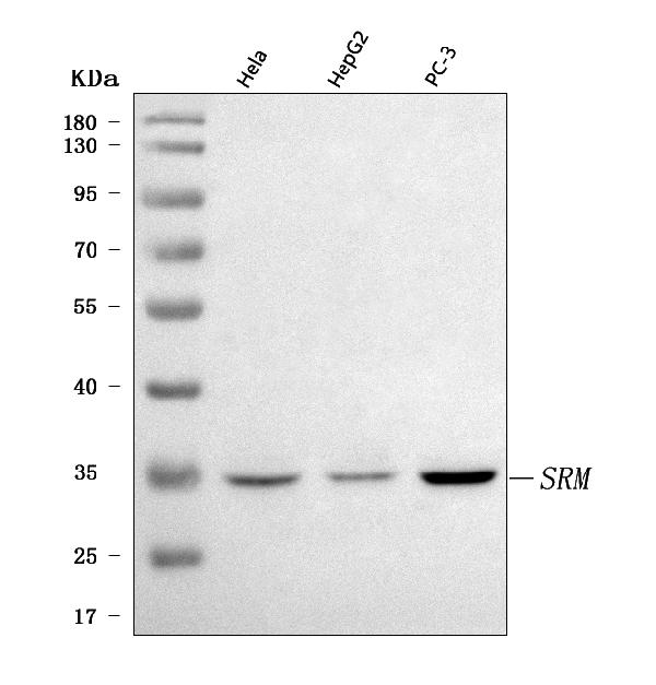
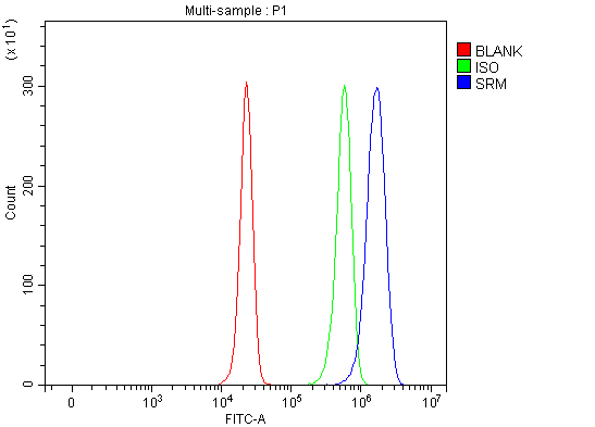 FCM/FACS (Flow Cytometry)
(Figure 2. Flow Cytometry analysis of HepG2 cells using anti-Spermidine synthase/SRM antibody (AAA127068).Overlay histogram showing HepG2 cells stained with AAA127068 (Blue line). To facilitate intracellular staining, cells were fixed with 4% paraformaldehyde and permeabilized with permeabilization buffer. The cells were blocked with 10% normal goat serum. And then incubated with rabbit anti-Spermidine synthase/SRM Antibody (AAA127068, 1ug/1x106 cells) for 30 min at 20 degree C. DyLight488 conjugated goat anti-rabbit IgG was used as secondary antibody for 30 minutes at 20 degree C. Isotype control antibody (Green line) was rabbit IgG (1ug/1x106) used under the same conditions. Unlabelled sample without incubation with primary antibody and secondary antibody (Red line) was used as a blank control.)
FCM/FACS (Flow Cytometry)
(Figure 2. Flow Cytometry analysis of HepG2 cells using anti-Spermidine synthase/SRM antibody (AAA127068).Overlay histogram showing HepG2 cells stained with AAA127068 (Blue line). To facilitate intracellular staining, cells were fixed with 4% paraformaldehyde and permeabilized with permeabilization buffer. The cells were blocked with 10% normal goat serum. And then incubated with rabbit anti-Spermidine synthase/SRM Antibody (AAA127068, 1ug/1x106 cells) for 30 min at 20 degree C. DyLight488 conjugated goat anti-rabbit IgG was used as secondary antibody for 30 minutes at 20 degree C. Isotype control antibody (Green line) was rabbit IgG (1ug/1x106) used under the same conditions. Unlabelled sample without incubation with primary antibody and secondary antibody (Red line) was used as a blank control.)
Spermidine synthase/SRM, Polyclonal Antibody (Cat# AAA127068)
Full Name
Anti-Spermidine synthase/SRM Antibody Picoband
Gene Names
SRM; PAPT; SPS1; SPDSY; SRML1
Reactivity
Human
Applications
Flow Cytometry, Western Blot
Purity
Immunogen affinity purified.
Pricing
 FCM/FACS (Flow Cytometry)
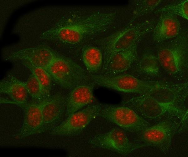
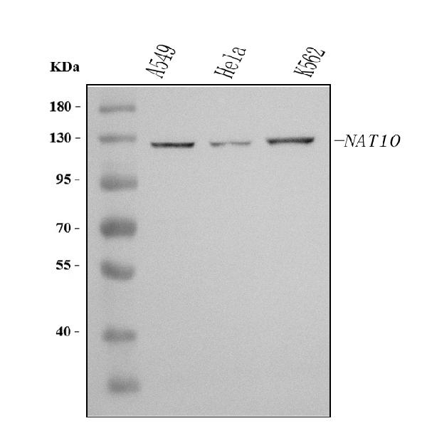
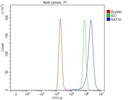
(Figure 3. Flow Cytometry analysis of Hela cells using anti-NAT10 antibody (AAA127352).Overlay histogram showing Hela cells stained with AAA127352 (Blue line). To facilitate intracellular staining, cells were fixed with 4% paraformaldehyde and permeabilized with permeabilization buffer. The cells were blocked with 10% normal goat serum. And then incubated with rabbit anti-NAT10 Antibody (AAA127352, 1ug/1x106 cells) for 30 min at 20 degree C. DyLight488 conjugated goat anti-rabbit IgG was used as secondary antibody for 30 minutes at 20 degree C. Isotype control antibody (Green line) was rabbit IgG (1ug/1x106) used under the same conditions. Unlabelled sample (Red line) was also used as a control.)
FCM/FACS (Flow Cytometry)
(Figure 3. Flow Cytometry analysis of Hela cells using anti-NAT10 antibody (AAA127352).Overlay histogram showing Hela cells stained with AAA127352 (Blue line). To facilitate intracellular staining, cells were fixed with 4% paraformaldehyde and permeabilized with permeabilization buffer. The cells were blocked with 10% normal goat serum. And then incubated with rabbit anti-NAT10 Antibody (AAA127352, 1ug/1x106 cells) for 30 min at 20 degree C. DyLight488 conjugated goat anti-rabbit IgG was used as secondary antibody for 30 minutes at 20 degree C. Isotype control antibody (Green line) was rabbit IgG (1ug/1x106) used under the same conditions. Unlabelled sample (Red line) was also used as a control.)
NAT10, Polyclonal Antibody (Cat# AAA127352)
Full Name
Anti-NAT10 Antibody Picoband
Gene Names
NAT10; ALP; NET43
Reactivity
Human
Applications
Flow Cytometry, Immunofluorescence, Immunocytochemistry, Western Blot
Purity
Immunogen affinity purified.
Pricing
 FCM/FACS (Flow Cytometry)
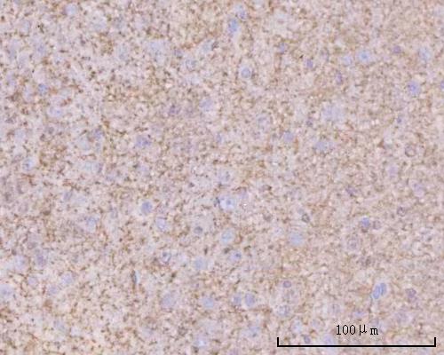
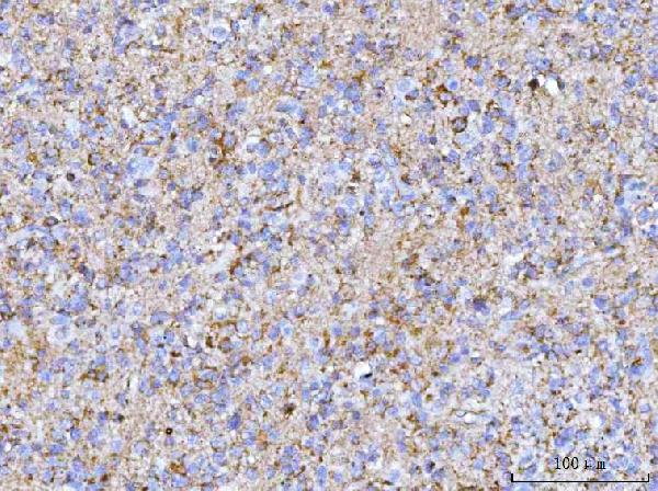
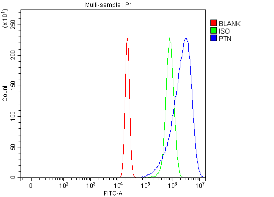
(Figure 5. Flow Cytometry analysis of U20S cells using anti-PTN antibody (AAA127055).Overlay histogram showing U20S cells stained with AAA127055 (Blue line). The cells were fixed with 4% paraformaldehyde and blocked with 10% normal goat serum. And then incubated with rabbit anti-PTN Antibody (AAA127055, 1ug/1x106 cells) for 30 min at 20 degree C. DyLight488 conjugated goat anti-rabbit IgG was used as secondary antibody for 30 minutes at 20 degree C. Isotype control antibody (Green line) was rabbit IgG (1ug/1x106) used under the same conditions. Unlabelled sample without incubation with primary antibody and secondary antibody (Red line) was used as a blank control.)
FCM/FACS (Flow Cytometry)
(Figure 5. Flow Cytometry analysis of U20S cells using anti-PTN antibody (AAA127055).Overlay histogram showing U20S cells stained with AAA127055 (Blue line). The cells were fixed with 4% paraformaldehyde and blocked with 10% normal goat serum. And then incubated with rabbit anti-PTN Antibody (AAA127055, 1ug/1x106 cells) for 30 min at 20 degree C. DyLight488 conjugated goat anti-rabbit IgG was used as secondary antibody for 30 minutes at 20 degree C. Isotype control antibody (Green line) was rabbit IgG (1ug/1x106) used under the same conditions. Unlabelled sample without incubation with primary antibody and secondary antibody (Red line) was used as a blank control.)
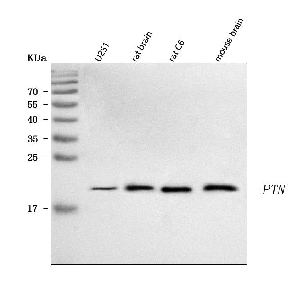
PTN, Polyclonal Antibody (Cat# AAA127055)
Full Name
Anti-PTN Antibody Picoband
Gene Names
PTN; HARP; HBNF; HBGF8; NEGF1
Reactivity
Human, Mouse, Rat
Applications
Flow Cytometry, Immunohistochemistry, Western Blot
Purity
Immunogen affinity purified.
Pricing
 FCM/FACS (Flow Cytometry)
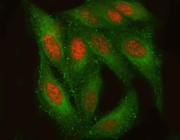
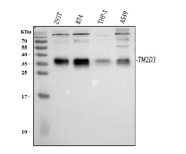
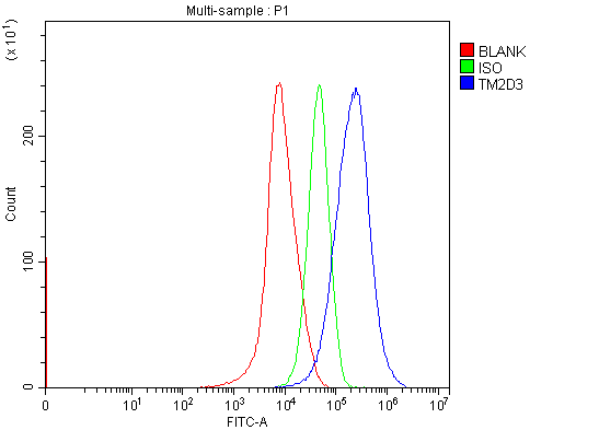
(Figure 3. Flow Cytometry analysis of HepG2 cells using anti-TM2D3 antibody (AAA127867).Overlay histogram showing HepG2 cells stained with AAA127867 (Blue line). To facilitate intracellular staining, cells were fixed with 4% paraformaldehyde and permeabilized with permeabilization buffer. The cells were blocked with 10% normal goat serum. And then incubated with rabbit anti-TM2D3 Antibody (AAA127867, 1ug/1x106 cells) for 30 min at 20 degree C. DyLight488 conjugated goat anti-rabbit IgG was used as secondary antibody for 30 minutes at 20 degree C. Isotype control antibody (Green line) was rabbit IgG (1ug/1x106) used under the same conditions. Unlabelled sample (Red line) was also used as a control.)
FCM/FACS (Flow Cytometry)
(Figure 3. Flow Cytometry analysis of HepG2 cells using anti-TM2D3 antibody (AAA127867).Overlay histogram showing HepG2 cells stained with AAA127867 (Blue line). To facilitate intracellular staining, cells were fixed with 4% paraformaldehyde and permeabilized with permeabilization buffer. The cells were blocked with 10% normal goat serum. And then incubated with rabbit anti-TM2D3 Antibody (AAA127867, 1ug/1x106 cells) for 30 min at 20 degree C. DyLight488 conjugated goat anti-rabbit IgG was used as secondary antibody for 30 minutes at 20 degree C. Isotype control antibody (Green line) was rabbit IgG (1ug/1x106) used under the same conditions. Unlabelled sample (Red line) was also used as a control.)
TM2D3, Polyclonal Antibody (Cat# AAA127867)
Full Name
Anti-TM2D3 Antibody Picoband
Gene Names
TM2D3; BLP2
Reactivity
Human
Applications
Flow Cytometry, Immunofluorescence, Immunocytochemistry, Western Blot
Purity
Immunogen affinity purified.
Pricing
 FCM/FACS (Flow Cytometry)
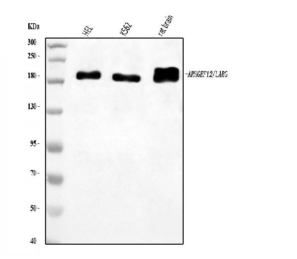
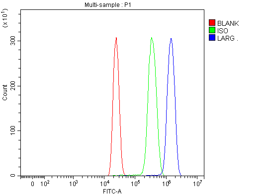
(Figure 2. Flow Cytometry analysis of HL-60 cells using anti-LARG/ARHGEF12 antibody (AAA126470).Overlay histogram showing HL-60 cells stained with AAA126470 (Blue line). The cells were blocked with 10% normal goat serum. And then incubated with rabbit anti-LARG/ARHGEF12 Antibody (AAA126470, 1 ug/1x10^6 cells) for 30 min at 20 degree C. DyLight488 conjugated goat anti-rabbit IgG was used as secondary antibody for 30 minutes at 20 degree C. Isotype control antibody (Green line) was rabbit IgG (1 ug/1x10^6) used under the same conditions. Unlabelled sample (Red line) was also used as a control.)
FCM/FACS (Flow Cytometry)
(Figure 2. Flow Cytometry analysis of HL-60 cells using anti-LARG/ARHGEF12 antibody (AAA126470).Overlay histogram showing HL-60 cells stained with AAA126470 (Blue line). The cells were blocked with 10% normal goat serum. And then incubated with rabbit anti-LARG/ARHGEF12 Antibody (AAA126470, 1 ug/1x10^6 cells) for 30 min at 20 degree C. DyLight488 conjugated goat anti-rabbit IgG was used as secondary antibody for 30 minutes at 20 degree C. Isotype control antibody (Green line) was rabbit IgG (1 ug/1x10^6) used under the same conditions. Unlabelled sample (Red line) was also used as a control.)
LARG/ARHGEF12, Polyclonal Antibody (Cat# AAA126470)
Full Name
Anti-LARG/ARHGEF12 Antibody Picoband
Gene Names
ARHGEF12; LARG; PRO2792
Reactivity
Human, Rat
Applications
Western Blot, Flow Cytometry
Purity
Immunogen affinity purified.
Pricing
 FCM/FACS (Flow Cytometry)
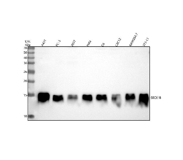
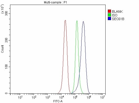
(Figure 2. Flow Cytometry analysis of PC-3 cells using anti-SEC61B antibody (AAA127438).Overlay histogram showing PC-3 cells stained with AAA127438 (Blue line). To facilitate intracellular staining, cells were fixed with 4% paraformaldehyde and permeabilized with permeabilization buffer. The cells were blocked with 10% normal goat serum. And then incubated with rabbit anti-SEC61B Antibody (AAA127438, 1ug/1x106 cells) for 30 min at 20 degree C. DyLight488 conjugated goat anti-rabbit IgG was used as secondary antibody for 30 minutes at 20 degree C. Isotype control antibody (Green line) was rabbit IgG (1ug/1x106) used under the same conditions. Unlabelled sample without incubation with primary antibody and secondary antibody (Red line) was used as a blank control.)
FCM/FACS (Flow Cytometry)
(Figure 2. Flow Cytometry analysis of PC-3 cells using anti-SEC61B antibody (AAA127438).Overlay histogram showing PC-3 cells stained with AAA127438 (Blue line). To facilitate intracellular staining, cells were fixed with 4% paraformaldehyde and permeabilized with permeabilization buffer. The cells were blocked with 10% normal goat serum. And then incubated with rabbit anti-SEC61B Antibody (AAA127438, 1ug/1x106 cells) for 30 min at 20 degree C. DyLight488 conjugated goat anti-rabbit IgG was used as secondary antibody for 30 minutes at 20 degree C. Isotype control antibody (Green line) was rabbit IgG (1ug/1x106) used under the same conditions. Unlabelled sample without incubation with primary antibody and secondary antibody (Red line) was used as a blank control.)
SEC61B, Polyclonal Antibody (Cat# AAA127438)
Full Name
Anti-SEC61B Antibody Picoband
Reactivity
Human, Mouse, Rat
Applications
Western Blot, Flow Cytometry
Purity
Immunogen affinity purified.
Pricing
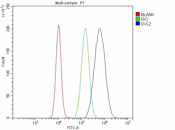 FCM/FACS (Flow Cytometry)
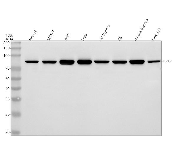
(Figure 2. Flow Cytometry analysis of A431 cells using anti-DVL2 antibody (AAA127114).Overlay histogram showing A431 cells stained with AAA127114 (Blue line). To facilitate intracellular staining, cells were fixed with 4% paraformaldehyde and permeabilized with permeabilization buffer. The cells were blocked with 10% normal goat serum. And then incubated with rabbit anti-DVL2 Antibody (AAA127114, 1ug/1x106 cells) for 30 min at 20 degree C. DyLight488 conjugated goat anti-rabbit IgG was used as secondary antibody for 30 minutes at 20 degree C. Isotype control antibody (Green line) was rabbit IgG (1ug/1x106) used under the same conditions. Unlabelled sample without incubation with primary antibody and secondary antibody (Red line) was used as a blank control.)
FCM/FACS (Flow Cytometry)
(Figure 2. Flow Cytometry analysis of A431 cells using anti-DVL2 antibody (AAA127114).Overlay histogram showing A431 cells stained with AAA127114 (Blue line). To facilitate intracellular staining, cells were fixed with 4% paraformaldehyde and permeabilized with permeabilization buffer. The cells were blocked with 10% normal goat serum. And then incubated with rabbit anti-DVL2 Antibody (AAA127114, 1ug/1x106 cells) for 30 min at 20 degree C. DyLight488 conjugated goat anti-rabbit IgG was used as secondary antibody for 30 minutes at 20 degree C. Isotype control antibody (Green line) was rabbit IgG (1ug/1x106) used under the same conditions. Unlabelled sample without incubation with primary antibody and secondary antibody (Red line) was used as a blank control.)
DVL2, Polyclonal Antibody (Cat# AAA127114)
Full Name
Anti-DVL2 Antibody Picoband
Reactivity
Human, Mouse, Rat
Applications
Western Blot
Purity
Immunogen affinity purified.
Pricing
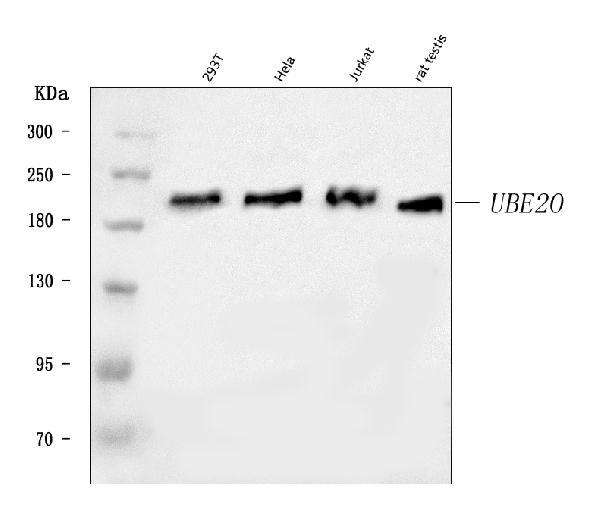
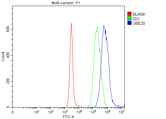 FCM/FACS (Flow Cytometry)
(Figure 2. Flow Cytometry analysis of Daudi cells using anti-UBE2O antibody (AAA126547).Overlay histogram showing Daudi cells stained with AAA126547 (Blue line). The cells were blocked with 10% normal goat serum. And then incubated with rabbit anti-UBE2O Antibody (AAA126547, 1 ug/1x10^6 cells) for 30 min at 20 degree C. DyLight488 conjugated goat anti-rabbit IgG was used as secondary antibody for 30 minutes at 20 degree C. Isotype control antibody (Green line) was rabbit IgG (1 ug/1x10^6) used under the same conditions. Unlabelled sample (Red line) was also used as a control.)
FCM/FACS (Flow Cytometry)
(Figure 2. Flow Cytometry analysis of Daudi cells using anti-UBE2O antibody (AAA126547).Overlay histogram showing Daudi cells stained with AAA126547 (Blue line). The cells were blocked with 10% normal goat serum. And then incubated with rabbit anti-UBE2O Antibody (AAA126547, 1 ug/1x10^6 cells) for 30 min at 20 degree C. DyLight488 conjugated goat anti-rabbit IgG was used as secondary antibody for 30 minutes at 20 degree C. Isotype control antibody (Green line) was rabbit IgG (1 ug/1x10^6) used under the same conditions. Unlabelled sample (Red line) was also used as a control.)
UBE2O, Polyclonal Antibody (Cat# AAA126547)
Full Name
Anti-UBE2O Antibody Picoband
Gene Names
UBE2O; E2-230K
Reactivity
Human, Rat
Applications
Flow Cytometry, Western Blot
Purity
Immunogen affinity purified.
Pricing
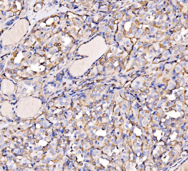
 IHC (Immunohistochemisry)
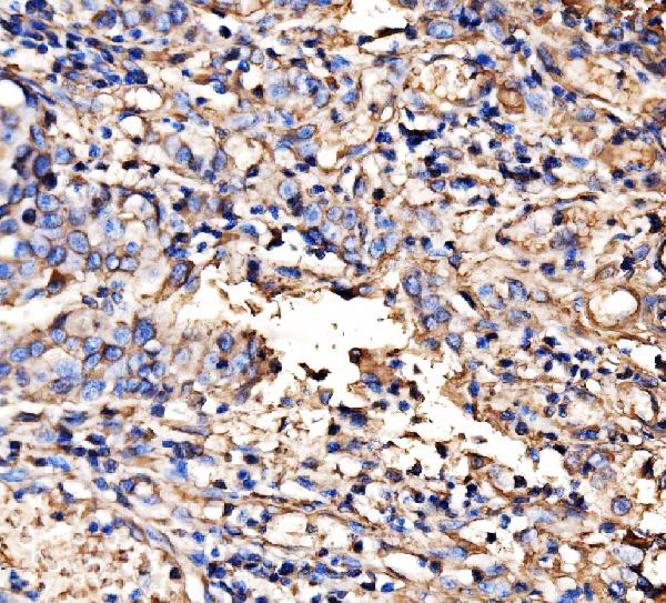
(Figure 3. IHC analysis of HECTD1 using anti-HECTD1 antibody (AAA126520).HECTD1 was detected in a paraffin-embedded section of human lung cancer tissue. Heat mediated antigen retrieval was performed in EDTA buffer (pH 8.0, epitope retrieval solution). The tissue section was blocked with 10% goat serum. The tissue section was then incubated with 2 ug/ml rabbit anti-HECTD1 Antibody (AAA126520) overnight at 4 degree C. Biotinylated goat anti-rabbit IgG was used as secondary antibody and incubated for 30 minutes at 37 degree C. The tissue section was developed using Strepavidin-Biotin-Complex (SABC) with DAB as the chromogen.)
IHC (Immunohistochemisry)
(Figure 3. IHC analysis of HECTD1 using anti-HECTD1 antibody (AAA126520).HECTD1 was detected in a paraffin-embedded section of human lung cancer tissue. Heat mediated antigen retrieval was performed in EDTA buffer (pH 8.0, epitope retrieval solution). The tissue section was blocked with 10% goat serum. The tissue section was then incubated with 2 ug/ml rabbit anti-HECTD1 Antibody (AAA126520) overnight at 4 degree C. Biotinylated goat anti-rabbit IgG was used as secondary antibody and incubated for 30 minutes at 37 degree C. The tissue section was developed using Strepavidin-Biotin-Complex (SABC) with DAB as the chromogen.)
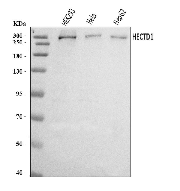
HECTD1, Polyclonal Antibody (Cat# AAA126520)
Full Name
Anti-HECTD1 Antibody Picoband
Gene Names
HECTD1; EULIR
Reactivity
Human
Applications
Immunohistochemistry, Western Blot
Purity
Immunogen affinity purified.
Pricing
 FCM/FACS (Flow Cytometry)
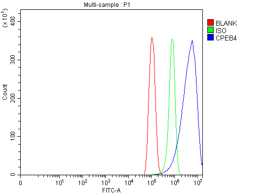
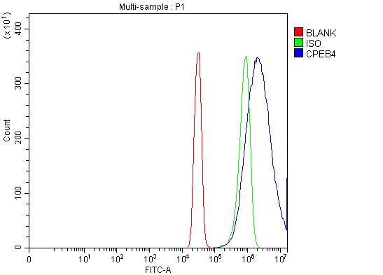
(Figure 3. Flow Cytometry analysis of SiHa cells using anti-CPEB4 antibody (AAA126451).Overlay histogram showing SiHa cells stained with AAA126451 (Blue line). The cells were blocked with 10% normal goat serum. And then incubated with rabbit anti-CPEB4 Antibody (AAA126451, 1 ug/1x10^6 cells) for 30 min at 20 degree C. DyLight488 conjugated goat anti-rabbit IgG was used as secondary antibody for 30 minutes at 20 degree C. Isotype control antibody (Green line) was rabbit IgG (1 ug/1x10^6) used under the same conditions. Unlabelled sample (Red line) was also used as a control.)
FCM/FACS (Flow Cytometry)
(Figure 3. Flow Cytometry analysis of SiHa cells using anti-CPEB4 antibody (AAA126451).Overlay histogram showing SiHa cells stained with AAA126451 (Blue line). The cells were blocked with 10% normal goat serum. And then incubated with rabbit anti-CPEB4 Antibody (AAA126451, 1 ug/1x10^6 cells) for 30 min at 20 degree C. DyLight488 conjugated goat anti-rabbit IgG was used as secondary antibody for 30 minutes at 20 degree C. Isotype control antibody (Green line) was rabbit IgG (1 ug/1x10^6) used under the same conditions. Unlabelled sample (Red line) was also used as a control.)
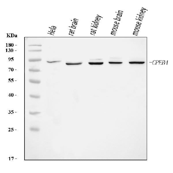
CPEB4, Polyclonal Antibody (Cat# AAA126451)
Full Name
Anti-CPEB4 Antibody Picoband
Reactivity
Human, Mouse, Rat
Applications
Flow Cytometry, Western Blot
Purity
Immunogen affinity purified.
Pricing
 FCM/FACS (Flow Cytometry)
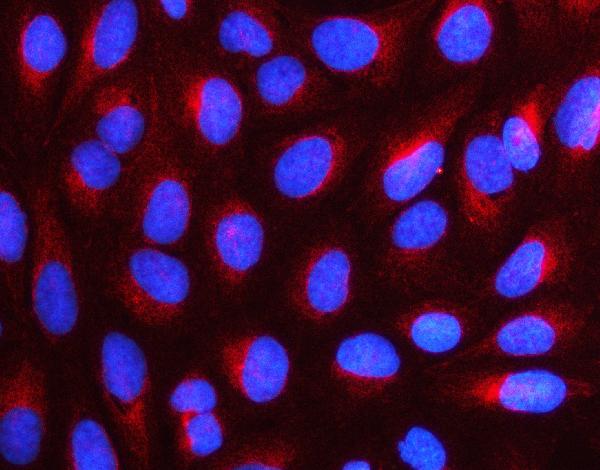
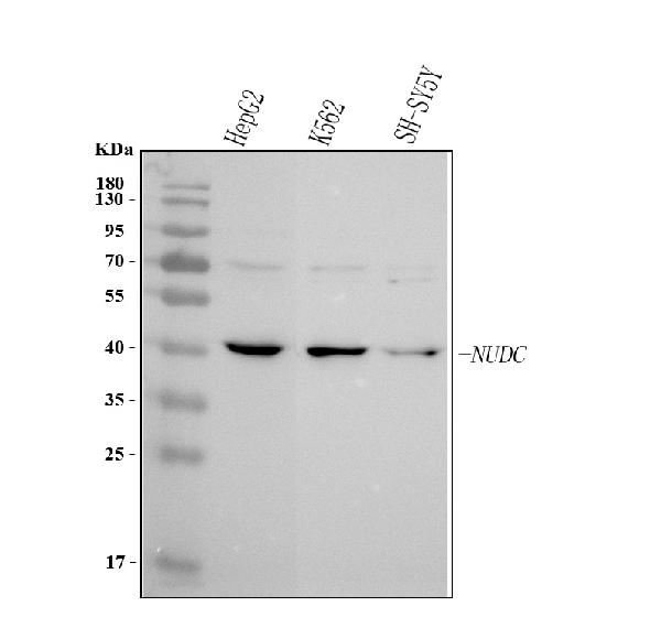
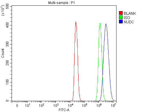
(Figure 3. Flow Cytometry analysis of 293T cells using anti-NUDC antibody (AAA127026).Overlay histogram showing 293T cells stained with AAA127026 (Blue line). To facilitate intracellular staining, cells were fixed with 4% paraformaldehyde and permeabilized with permeabilization buffer. The cells were blocked with 10% normal goat serum. And then incubated with rabbit anti-NUDC Antibody (AAA127026, 1ug/1x106 cells) for 30 min at 20 degree C. DyLight488 conjugated goat anti-rabbit IgG was used as secondary antibody for 30 minutes at 20 degree C. Isotype control antibody (Green line) was rabbit IgG (1ug/1x106) used under the same conditions. Unlabelled sample (Red line) was also used as a control.)
FCM/FACS (Flow Cytometry)
(Figure 3. Flow Cytometry analysis of 293T cells using anti-NUDC antibody (AAA127026).Overlay histogram showing 293T cells stained with AAA127026 (Blue line). To facilitate intracellular staining, cells were fixed with 4% paraformaldehyde and permeabilized with permeabilization buffer. The cells were blocked with 10% normal goat serum. And then incubated with rabbit anti-NUDC Antibody (AAA127026, 1ug/1x106 cells) for 30 min at 20 degree C. DyLight488 conjugated goat anti-rabbit IgG was used as secondary antibody for 30 minutes at 20 degree C. Isotype control antibody (Green line) was rabbit IgG (1ug/1x106) used under the same conditions. Unlabelled sample (Red line) was also used as a control.)
NUDC, Polyclonal Antibody (Cat# AAA127026)
Full Name
Anti-NUDC Antibody Picoband
Gene Names
NUDC; HNUDC; MNUDC; NPD011
Reactivity
Human
Applications
Flow Cytometry, Immunofluorescence, Immunocytochemistry, Western Blot
Purity
Immunogen affinity purified.
Pricing
 FCM/FACS (Flow Cytometry)
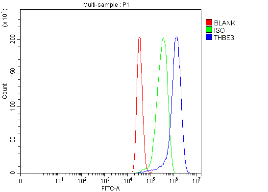
(Figure 4. Flow Cytometry analysis of MCF-7 cells using anti-THBS3 antibody (AAA126599).Overlay histogram showing MCF-7 cells stained with AAA126599 (Blue line). The cells were blocked with 10% normal goat serum. And then incubated with rabbit anti-THBS3 Antibody (AAA126599, 1 ug/1x10^6 cells) for 30 min at 20 degree C. DyLight488 conjugated goat anti-rabbit IgG was used as secondary antibody for 30 minutes at 20 degree C. Isotype control antibody (Green line) was rabbit IgG (1 ug/1x10^6) used under the same conditions. Unlabelled sample (Red line) was also used as a control.)
FCM/FACS (Flow Cytometry)
(Figure 4. Flow Cytometry analysis of MCF-7 cells using anti-THBS3 antibody (AAA126599).Overlay histogram showing MCF-7 cells stained with AAA126599 (Blue line). The cells were blocked with 10% normal goat serum. And then incubated with rabbit anti-THBS3 Antibody (AAA126599, 1 ug/1x10^6 cells) for 30 min at 20 degree C. DyLight488 conjugated goat anti-rabbit IgG was used as secondary antibody for 30 minutes at 20 degree C. Isotype control antibody (Green line) was rabbit IgG (1 ug/1x10^6) used under the same conditions. Unlabelled sample (Red line) was also used as a control.)
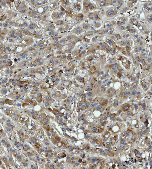
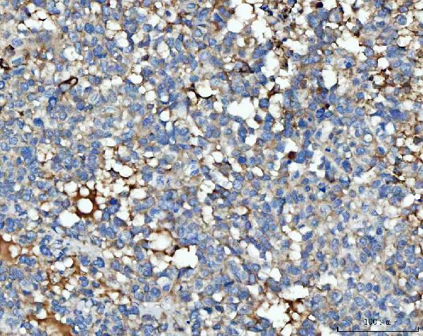
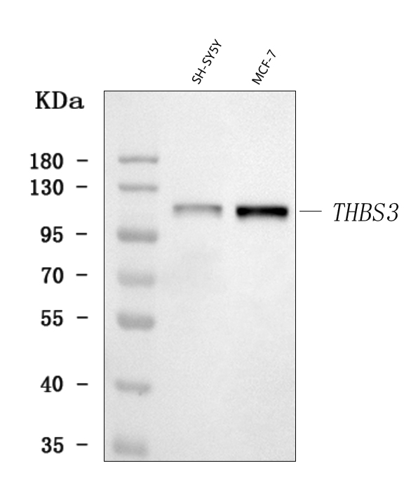
THBS3, Polyclonal Antibody (Cat# AAA126599)
Full Name
Anti-THBS3 Antibody Picoband
Gene Names
THBS3; TSP3
Reactivity
Human
Applications
Flow Cytometry, Immunohistochemistry, Western Blot
Purity
Immunogen affinity purified.
Pricing
 IF (Immunofluorescence)
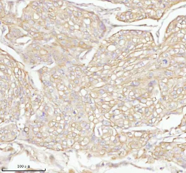
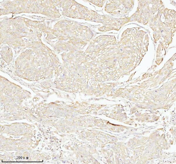
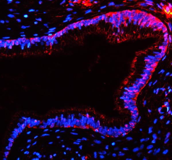
(Figure 5. IF analysis of TAS2R10 using anti-TAS2R10 antibody (AAA127686).TAS2R10 was detected in a paraffin-embedded section of human cervix cancer tissue. Heat mediated antigen retrieval was performed in EDTA buffer (pH 8.0, epitope retrieval solution). The tissue section was blocked with 10% goat serum. The tissue section was then incubated with 5ug/mL rabbit anti-TAS2R10 Antibody (AAA127686) overnight at 4 degree C. DyLight594 Conjugated Goat Anti-Rabbit IgG (BA1142) was used as secondary antibody at 1:500 dilution and incubated for 30 minutes at 37 degree C. The section was counterstained with DAPI. Visualize using a fluorescence microscope and filter sets appropriate for the label used.)
IF (Immunofluorescence)
(Figure 5. IF analysis of TAS2R10 using anti-TAS2R10 antibody (AAA127686).TAS2R10 was detected in a paraffin-embedded section of human cervix cancer tissue. Heat mediated antigen retrieval was performed in EDTA buffer (pH 8.0, epitope retrieval solution). The tissue section was blocked with 10% goat serum. The tissue section was then incubated with 5ug/mL rabbit anti-TAS2R10 Antibody (AAA127686) overnight at 4 degree C. DyLight594 Conjugated Goat Anti-Rabbit IgG (BA1142) was used as secondary antibody at 1:500 dilution and incubated for 30 minutes at 37 degree C. The section was counterstained with DAPI. Visualize using a fluorescence microscope and filter sets appropriate for the label used.)
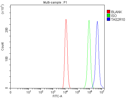
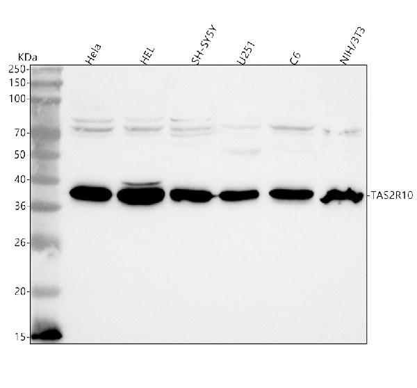
TAS2R10, Polyclonal Antibody (Cat# AAA127686)
Full Name
Anti-TAS2R10 Antibody Picoband
Gene Names
TAS2R10; TRB2; T2R10
Reactivity
Human, Mouse, Rat
Applications
Western Blot, Immunohistochemistry, Immunofluorescence, Flow Cytometry
Purity
Immunogen affinity purified.
Pricing
 Application Data
COVID 19 Spike S1 (delta69-70, N439K, D614G) Coronavirus, Recombinant Protein (Cat# AAA73325)
Full Name
2019-nCoV Spike protein S1 (delta69-70, N439K, D614G)
Pricing

 Bioactivity
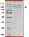
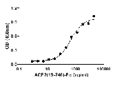
(Activity: Sample activity data. for lot-specific data please refer to the specific datasheet.)
Bioactivity
(Activity: Sample activity data. for lot-specific data please refer to the specific datasheet.)
COVID 19 Spike RBD (N439K) Coronavirus, Recombinant Protein (Cat# AAA73344)
Full Name
2019-nCoV Spike protein RBD (N439K)
Pricing
 Standard Curve (Sample)
GRO gamma, ELISA Kit (Cat# AAA263112)
Full Name
Mouse GRO gamma ELISA Kit
Gene Names
Cxcl3; Dcip1; Gm1960
Reactivity
Mouse
Pricing
 Bioactivity
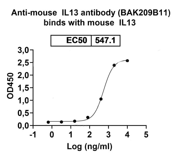
(Detects Mouse IL13 Antibody (BAK209B11) in indirect ELISAs.)
Bioactivity
(Detects Mouse IL13 Antibody (BAK209B11) in indirect ELISAs.)
IL13, Recombinant Protein (Cat# AAA120643)
Full Name
Recombinant Mouse IL13 Protein, C-Fc
Gene Names
Il13; Il-13
Applications
Western Blot
Purity
>90% as determined by SDS-PAGE
Pricing
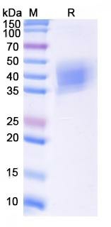
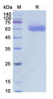
 SDS-PAGE
(The purity was determined to be >90% by desitometry.Approx. MW 65kDa.)
SDS-PAGE
(The purity was determined to be >90% by desitometry.Approx. MW 65kDa.)
IRS1, Recombinant Protein (Cat# AAA72896)
Full Name
IRS1 recombinant protein
Gene Names
IRS1; HIRS-1
Applications
Western Blot
Purity
>90%
Pricing
Natural Cytotoxicity Receptor NKp46, Recombinant Protein (Cat# AAA38489)
Full Name
Recombinant Human Natural Cytotoxicity Receptor NKp46
Gene Names
NCR1; LY94; CD335; NKP46; NK-p46
Purity
Greater than 95.0% as determined by SDS-PAGE.
Pricing
 Bioactivity
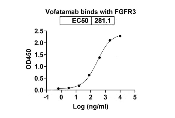
(Detects Vofatamab in indirect ELISAs.)
Bioactivity
(Detects Vofatamab in indirect ELISAs.)
CD333/FGFR3, Recombinant Protein (Cat# AAA120465)
Full Name
Recombinant Human CD333/FGFR3, C-His
Gene Names
FGFR3; ACH; CEK2; JTK4; CD333; HSFGFR3EX
Applications
Western Blot
Purity
>90% as determined by SDS-PAGE
Pricing

 Amino Acid Info
(Amino Acid info)
Amino Acid Info
(Amino Acid info)
Serum Amyloid A (SAA), Recombinant Protein (Cat# AAA153400)
Full Name
Recombinant Serum Amyloid A (SAA)
Applications
Western Blot
Purity
Greater than 90% by SDS-PAGE
Pricing
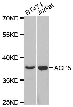
 IF (Immunofluorescence)
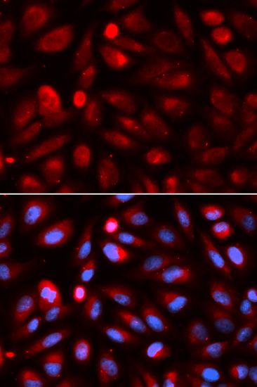
(Immunofluorescence analysis of U2OS cells using ACP5 antibody. Blue: DAPI for nuclear staining.)
IF (Immunofluorescence)
(Immunofluorescence analysis of U2OS cells using ACP5 antibody. Blue: DAPI for nuclear staining.)
ACP5, Polyclonal Antibody (Cat# AAA37515)
Full Name
ACP5 Polyclonal Antibody
Gene Names
ACP5; TRAP; SPENCDI
Reactivity
Human, Mouse
Applications
Immunofluorescence, Immunohistochemistry, Western Blot
Purity
Affinity Purification
Pricing
 SDS-PAGE
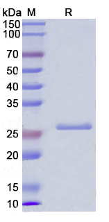
(SDS PAGE for Recombinant Human IL27A / IL30 protein)
SDS-PAGE
(SDS PAGE for Recombinant Human IL27A / IL30 protein)
IL27A/IL30, Recombinant Protein (Cat# AAA120506)
Full Name
Recombinant Human IL27A/IL30, N-His
Gene Names
IL27; p28; IL30; IL-27; IL27A; IL-27A; IL27p28
Applications
Western Blot
Purity
>90% as determined by SDS-PAGE
Pricing
 Amino Acid Info
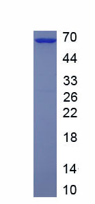
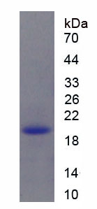
MANSC Domain Containing Protein 1 (MANSC1), Recombinant Protein (Cat# AAA153752)
Full Name
Recombinant MANSC Domain Containing Protein 1 (MANSC1)
Gene Names
MANSC1; LOH12CR3; 9130403P13Rik
Applications
Western Blot
Purity
Greater than 95% by SDS-PAGE
Pricing
 Amino Acid Info
Neuroplastin (NPTN), Recombinant Protein (Cat# AAA153538)
Full Name
Recombinant Neuroplastin (NPTN)
Gene Names
NPTN; GP55; GP65; SDR1; np55; np65; SDFR1
Applications
Western Blot
Purity
Greater than 90% by SDS-PAGE
Pricing
Amyloid A, Serum (SAA), Monoclonal Antibody (Cat# AAA57783)
Full Name
MAb to Serum Amyloid A
Applications
Western Blot
Purity
> 90% pure (SDS-PAGE)
Protein A Chromatography
Protein A Chromatography
Pricing
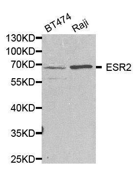
Actin, Recombinant Protein (Cat# AAA39121)
Full Name
Recombinant Human Actin, Alpha 2, Smooth Muscle, Aorta
Gene Names
ACTA2; AAT6; ACTSA; MYMY5
Purity
Greater than 85% as determined by SDS-PAGE.
Pricing
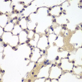 IHC (Immunohiostchemistry)
(Immunohistochemistry of paraffin-embedded mouse lung using ESR2 antibody at dilution of 1:200 (40x lens).)
IHC (Immunohiostchemistry)
(Immunohistochemistry of paraffin-embedded mouse lung using ESR2 antibody at dilution of 1:200 (40x lens).)
ESR2, Polyclonal Antibody (Cat# AAA37331)
Full Name
ESR2 Polyclonal Antibody
Gene Names
ESR2; Erb; ESRB; ESTRB; NR3A2; ER-BETA; ESR-BETA
Reactivity
Human, Mouse
Applications
Immunohistochemistry, Western Blot
Purity
Affinity Purification
Pricing
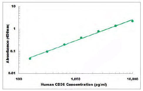 Standard Curve (Sample)
CD36, ELISA Kit (Cat# AAA105556)
Full Name
Human CD36 ELISA Kit
Gene Names
CD36; FAT; GP4; GP3B; GPIV; CHDS7; PASIV; SCARB3; BDPLT10
Reactivity
Human
Applications
Sandwich ELISA
Pricing
HIV1 Nef, Recombinant Protein (Cat# AAA75308)
Full Name
HIV1 Nef protein
Purity
> 95% by SDS-PAGE.
Pricing
 Application Data
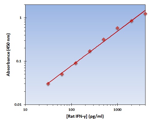
(The OmniKine Rat IFN-gamma ELISA Kit allows for the detection and quantification of endogenous levels of natural and/or recombinant Rat IFN-gamma proteins within the range of 32-4000 pg/ml.)
Application Data
(The OmniKine Rat IFN-gamma ELISA Kit allows for the detection and quantification of endogenous levels of natural and/or recombinant Rat IFN-gamma proteins within the range of 32-4000 pg/ml.)
IFN-gamma, ELISA Kit (Cat# AAA315694)
Full Name
IFN-gamma (Rat) OmniKine ELISA Kit
Gene Names
Ifng; IFNG2
Reactivity
Rat
Pricing
 WB (Western Blot)
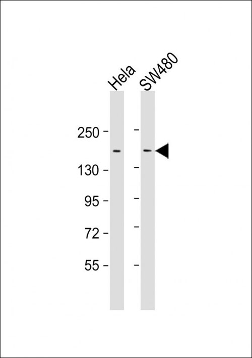
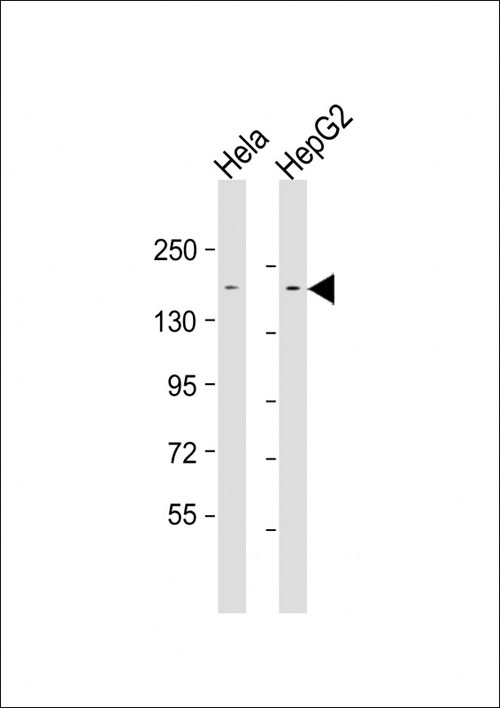
(All lanes : Anti-LAP2 Antibody at 1:1000-1:2000 dilutionLane 1: Hela whole cell lysateLane 2: HepG2 whole cell lysateLysates/proteins at 20 ug per lane.SecondaryGoat Anti-mouse IgG, (H+L), Peroxidase conjugated at 1/10000 dilution.Predicted band size : 158 kDaBlocking/Dilution buffer: 5% NFDM/TBST.)
WB (Western Blot)
(All lanes : Anti-LAP2 Antibody at 1:1000-1:2000 dilutionLane 1: Hela whole cell lysateLane 2: HepG2 whole cell lysateLysates/proteins at 20 ug per lane.SecondaryGoat Anti-mouse IgG, (H+L), Peroxidase conjugated at 1/10000 dilution.Predicted band size : 158 kDaBlocking/Dilution buffer: 5% NFDM/TBST.)
LAP2, Monoclonal Antibody (Cat# AAA290618)
Full Name
LAP2 Antibody
Gene Names
ERBB2IP; LAP2; ERBIN; HEL-S-78
Reactivity
Human
Applications
Western Blot
Pricing
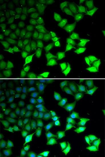 IF (Immunofluorescence)
(Immunofluorescence analysis of U2OS cells using KDM4B antibody. Blue: DAPI for nuclear staining.)
IF (Immunofluorescence)
(Immunofluorescence analysis of U2OS cells using KDM4B antibody. Blue: DAPI for nuclear staining.)
KDM4B, Polyclonal Antibody (Cat# AAA37589)
Full Name
KDM4B Polyclonal Antibody
Gene Names
KDM4B; JMJD2B; TDRD14B
Reactivity
Human, Mouse, Rat
Applications
Immunofluorescence, Immunohistochemistry, Western Blot
Purity
Affinity Purification
Pricing