
Filters
Clonality
Type
Reactivity
Gene Name
Isotype
Host
Application
Clone
10000 results for "Recombinant Proteins" - showing 9950-10000
 WB (Western Blot)
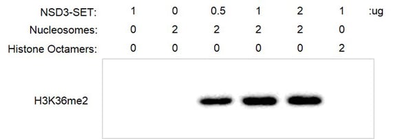
(Recombinant NSD3 (WHSC1L1)-SET activity assay. 2 ug recombinant nucleosomes were incubated with NSD3 (WHSC1L1)-SET protein in 30 ul reaction system containing 50 mM Tris-HCl, pH 8.6, 0.02% Triton X-100, 2 mM MgCl2, 1 mM TCEP, 50 uM SAM for 3 h at RT. 5 ul reaction products were loaded on a 12.5% SDS-PAGE gel. Western Blot was used to detect reaction products (H3K36me2). NSD3 (WHSC1L1)-SET only was used as negative control. Results show that the activity of NSD3 (WHSC1L1)-SET with nucleosomes as substrate is much better than its activity with histone octamers as substrate.)
WB (Western Blot)
(Recombinant NSD3 (WHSC1L1)-SET activity assay. 2 ug recombinant nucleosomes were incubated with NSD3 (WHSC1L1)-SET protein in 30 ul reaction system containing 50 mM Tris-HCl, pH 8.6, 0.02% Triton X-100, 2 mM MgCl2, 1 mM TCEP, 50 uM SAM for 3 h at RT. 5 ul reaction products were loaded on a 12.5% SDS-PAGE gel. Western Blot was used to detect reaction products (H3K36me2). NSD3 (WHSC1L1)-SET only was used as negative control. Results show that the activity of NSD3 (WHSC1L1)-SET with nucleosomes as substrate is much better than its activity with histone octamers as substrate.)
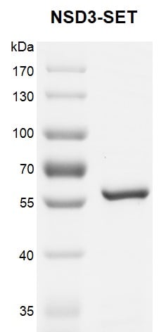
NSD3, Recombinant Protein (Cat# AAA59707)
Full Name
Recombinant NSD3 (WHSC1L1)-SET protein
Gene Names
NSD3; KMT3F; KMT3G; WHISTLE; WHSC1L1; pp14328
Pricing
 ELISA
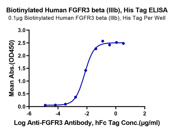
(Biotinylated Human FGFR3 beta (IIIb) Protein ELISA DataImmobilized Biotinylated Human FGFR3 beta (lllb) at 1?g/ml (100?l/Well) on the plate. Dose response curve for Anti-FGFR3 Antibody, hFc Tag with the EC50 of 6.9ng/ml determined by ELISA.)
ELISA
(Biotinylated Human FGFR3 beta (IIIb) Protein ELISA DataImmobilized Biotinylated Human FGFR3 beta (lllb) at 1?g/ml (100?l/Well) on the plate. Dose response curve for Anti-FGFR3 Antibody, hFc Tag with the EC50 of 6.9ng/ml determined by ELISA.)
FGFR3 beta (IIIb), Recombinant Protein (Cat# AAA76326)
Full Name
Biotinylated Human FGFR3 beta (IIIb) Protein Recombinant
Gene Names
FGFR3; ACH; CEK2; JTK4; CD333; HSFGFR3EX
Purity
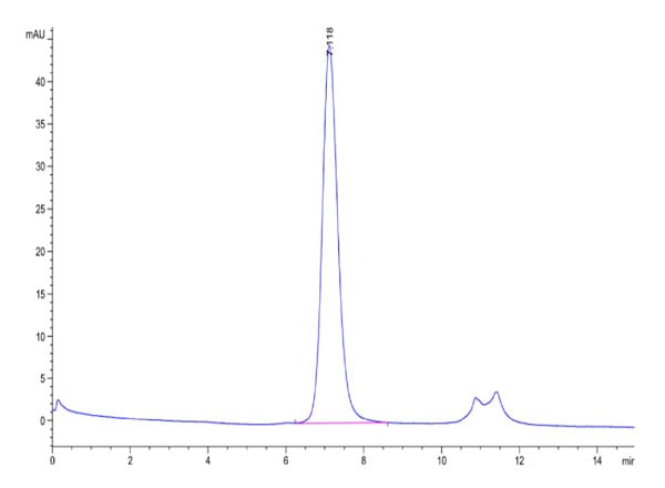
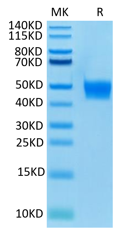
>95%, as determined by SDS-PAGE and HPLC
Pricing
 Bioactivity
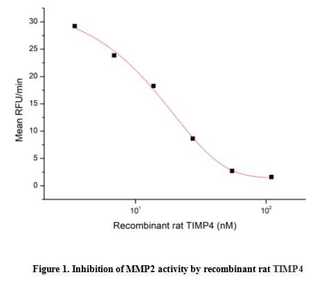
(Tissue Inhibitors of Metalloproteinase 4 (TIMP4) is an enzyme that in humans is encoded by the TIMP4 gene. This gene belongs to the tissue inhibitor of metalloproteinases gene family. The proteins encoded by this gene family are inhibitors of the matrix metalloproteinases, a group of peptidases involved in degradation of the extracellular matrix. The secreted, netrin domain-containing protein encoded by this gene is involved in regulation of platelet aggregation and recruitment and may play role in hormonal regulation and endometrial tissue remodeling. The activity of recombinant rat TIMP4 was measured by its ability to inhibit rhMMP2 cleavage of a fluorogenic peptide substrate MCA-Pro-Leu-Gly-Leu-DPA-Ala-Arg-NH2 in the assay buffer 50 mM Tris, 10 mM CaCl2, 150 mM NaCl, 0.05% (w/v) Brij-35, pH 7.5. rhMMP2 was diluted to 100 ug/ml and activated with 1 mM APMA at 37 degree C for 1 hour and rrTIMP4 (MW: 22.79 KD) was diluted to different concentrations with the assay buffer. Mix 8 ul of rrTIMP4 curve dilutions, 12.8 ul of activated rhMMP-2, and 59.2 ul of assay buffer, including a control containing assay buffer and the diluted rhMMP-2 and incubate the reactions for 2 hours at 37 degree C. Loading 50 ul of the incubated mixtures which were diluted five-fold in assay buffer into empty wells of a plate, and start the reaction by adding 50 ul of 20 uM substrate. Include a substrate blank containing 50 ul of assay buffer and 50 ul of 20 uM substrate. Then read at excitiation and emission wavelengths of 320 nm and 405 nm, respectively, in kinetic mode for 5 minutes. The result was shown in Figure 1 and it was obvious that recombinant rat TIMP4 significantly decreased rhMMP2 activity. The inhibition IC50 was )
Bioactivity
(Tissue Inhibitors of Metalloproteinase 4 (TIMP4) is an enzyme that in humans is encoded by the TIMP4 gene. This gene belongs to the tissue inhibitor of metalloproteinases gene family. The proteins encoded by this gene family are inhibitors of the matrix metalloproteinases, a group of peptidases involved in degradation of the extracellular matrix. The secreted, netrin domain-containing protein encoded by this gene is involved in regulation of platelet aggregation and recruitment and may play role in hormonal regulation and endometrial tissue remodeling. The activity of recombinant rat TIMP4 was measured by its ability to inhibit rhMMP2 cleavage of a fluorogenic peptide substrate MCA-Pro-Leu-Gly-Leu-DPA-Ala-Arg-NH2 in the assay buffer 50 mM Tris, 10 mM CaCl2, 150 mM NaCl, 0.05% (w/v) Brij-35, pH 7.5. rhMMP2 was diluted to 100 ug/ml and activated with 1 mM APMA at 37 degree C for 1 hour and rrTIMP4 (MW: 22.79 KD) was diluted to different concentrations with the assay buffer. Mix 8 ul of rrTIMP4 curve dilutions, 12.8 ul of activated rhMMP-2, and 59.2 ul of assay buffer, including a control containing assay buffer and the diluted rhMMP-2 and incubate the reactions for 2 hours at 37 degree C. Loading 50 ul of the incubated mixtures which were diluted five-fold in assay buffer into empty wells of a plate, and start the reaction by adding 50 ul of 20 uM substrate. Include a substrate blank containing 50 ul of assay buffer and 50 ul of 20 uM substrate. Then read at excitiation and emission wavelengths of 320 nm and 405 nm, respectively, in kinetic mode for 5 minutes. The result was shown in Figure 1 and it was obvious that recombinant rat TIMP4 significantly decreased rhMMP2 activity. The inhibition IC50 was )
Tissue Inhibitors Of Metalloproteinase 4 (TIMP4), Active Protein (Cat# AAA161703)
Full Name
Active Tissue Inhibitors Of Metalloproteinase 4 (TIMP4)
Applications
Activity Assay, Cell Culture
Purity
Greater than 95% by SDS-PAGE
Pricing
 FCM/FACS (Flow Cytometry)
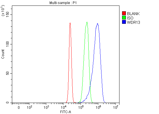
(Figure 2. Flow Cytometry analysis of HepG2 cells using anti-WDR13 antibody (AAA127777).Overlay histogram showing HepG2 cells stained with AAA127777 (Blue line). To facilitate intracellular staining, cells were fixed with 4% paraformaldehyde and permeabilized with permeabilization buffer. The cells were blocked with 10% normal goat serum. And then incubated with rabbit anti-WDR13 Antibody (AAA127777, 1ug/1x106 cells) for 30 min at 20 degree C. DyLight488 conjugated goat anti-rabbit IgG was used as secondary antibody for 30 minutes at 20 degree C. Isotype control antibody (Green line) was rabbit IgG (1ug/1x106) used under the same conditions. Unlabelled sample without incubation with primary antibody and secondary antibody (Red line) was used as a blank control.)
FCM/FACS (Flow Cytometry)
(Figure 2. Flow Cytometry analysis of HepG2 cells using anti-WDR13 antibody (AAA127777).Overlay histogram showing HepG2 cells stained with AAA127777 (Blue line). To facilitate intracellular staining, cells were fixed with 4% paraformaldehyde and permeabilized with permeabilization buffer. The cells were blocked with 10% normal goat serum. And then incubated with rabbit anti-WDR13 Antibody (AAA127777, 1ug/1x106 cells) for 30 min at 20 degree C. DyLight488 conjugated goat anti-rabbit IgG was used as secondary antibody for 30 minutes at 20 degree C. Isotype control antibody (Green line) was rabbit IgG (1ug/1x106) used under the same conditions. Unlabelled sample without incubation with primary antibody and secondary antibody (Red line) was used as a blank control.)
WDR13, Polyclonal Antibody (Cat# AAA127777)
Full Name
Anti-WDR13 Antibody Picoband
Gene Names
WDR13; MG21
Reactivity
Human
Applications
Flow Cytometry, Western Blot
Purity
Immunogen affinity purified.
Pricing
 Application Data
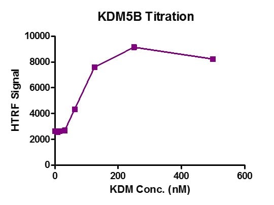
(Recombinant JARID1B / KDM5B protein activity assay. 1 ?M H3K4me3 peptide was incubated with different concentrations of JARID1B / KDM5B protein in 10 ?l reaction system, then 10 ?l anti-H3K4me2 antibody and SA-XL665 mixture (each 1:100 dilution in HTRF Detection Buffer) was added to each reaction system and incubated for 30 min. All the operations and reactions were performed at room temperature. HTRF assay was used for detection.)
Application Data
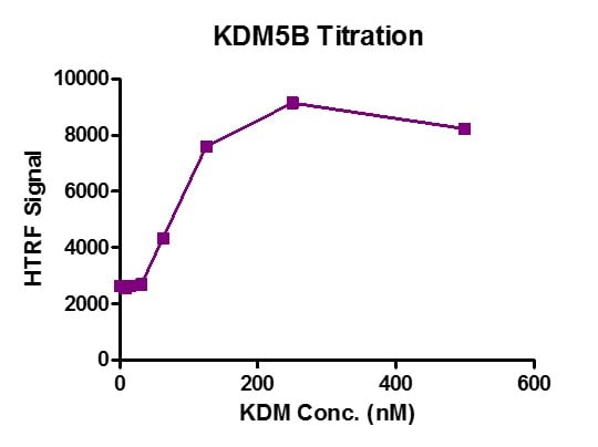
(Recombinant JARID1B / KDM5B protein activity assay. 1 ?M H3K4me3 peptide was incubated with different concentrations of JARID1B / KDM5B protein in 10 ?l reaction system, then 10 ?l anti-H3K4me2 antibody and SA-XL665 mixture (each 1:100 dilution in HTRF Detection Buffer) was added to each reaction system and incubated for 30 min. All the operations and reactions were performed at room temperature. HTRF assay was used for detection.)
JARID1B/KDM5B, Recombinant Protein (Cat# AAA59680)
Full Name
Recombinant JARID1B/KDM5B protein
Gene Names
KDM5B; CT31; PLU1; PUT1; PLU-1; JARID1B; FLJ10538; FLJ12459; FLJ12491; FLJ16281; FLJ23670; RBBP2H1A
Purity
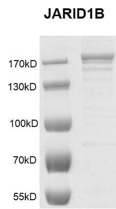
The recombinant protein is >75% pure by SDS-PAGE.
Pricing
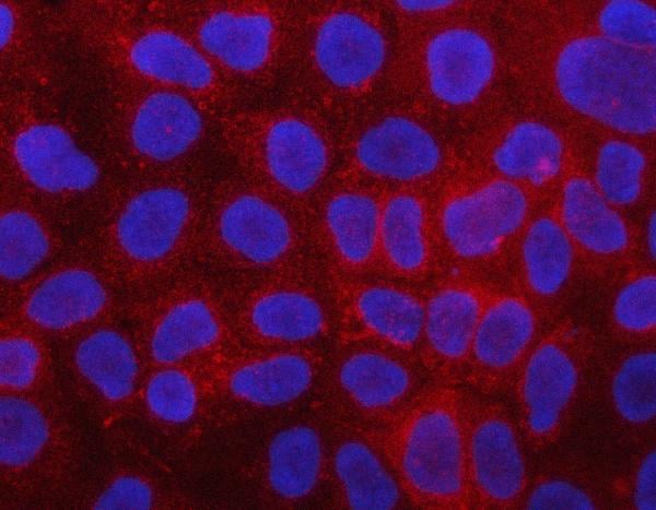
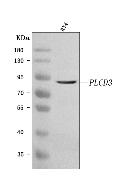
 FCM/FACS (Flow Cytometry)
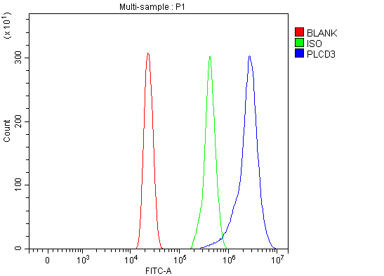
(Figure 3. Flow Cytometry analysis of HL-60 cells using anti-PLCD3 antibody (AAA127403).Overlay histogram showing HL-60 cells stained with AAA127403 (Blue line). To facilitate intracellular staining, cells were fixed with 4% paraformaldehyde and permeabilized with permeabilization buffer. The cells were blocked with 10% normal goat serum. And then incubated with rabbit anti-PLCD3 Antibody (AAA127403, 1ug/1x106 cells) for 30 min at 20 degree C. DyLight488 conjugated goat anti-rabbit IgG was used as secondary antibody for 30 minutes at 20 degree C. Isotype control antibody (Green line) was rabbit IgG (1ug/1x106) used under the same conditions. Unlabelled sample without incubation with primary antibody and secondary antibody (Red line) was used as a blank control.)
FCM/FACS (Flow Cytometry)
(Figure 3. Flow Cytometry analysis of HL-60 cells using anti-PLCD3 antibody (AAA127403).Overlay histogram showing HL-60 cells stained with AAA127403 (Blue line). To facilitate intracellular staining, cells were fixed with 4% paraformaldehyde and permeabilized with permeabilization buffer. The cells were blocked with 10% normal goat serum. And then incubated with rabbit anti-PLCD3 Antibody (AAA127403, 1ug/1x106 cells) for 30 min at 20 degree C. DyLight488 conjugated goat anti-rabbit IgG was used as secondary antibody for 30 minutes at 20 degree C. Isotype control antibody (Green line) was rabbit IgG (1ug/1x106) used under the same conditions. Unlabelled sample without incubation with primary antibody and secondary antibody (Red line) was used as a blank control.)
PLCD3, Polyclonal Antibody (Cat# AAA127403)
Full Name
Anti-PLCD3 Antibody Picoband
Gene Names
PLCD3; PLC-delta-3
Reactivity
Human
Applications
Flow Cytometry, Immunofluorescence, Immunocytochemistry, Western Blot
Purity
Immunogen affinity purified.
Pricing
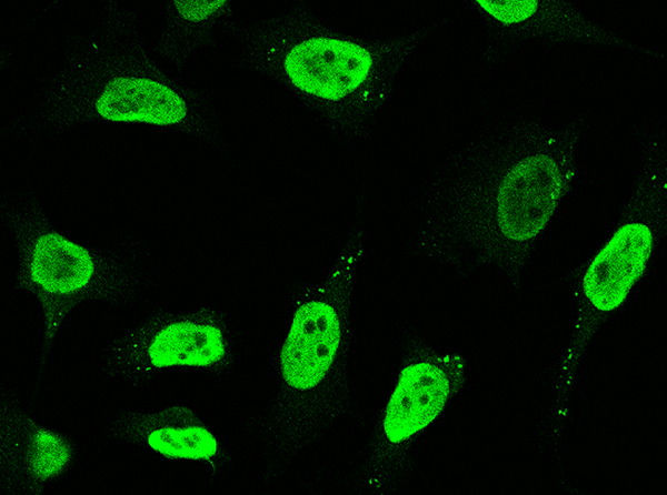
 IP (Immunoprecipitation)
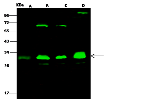
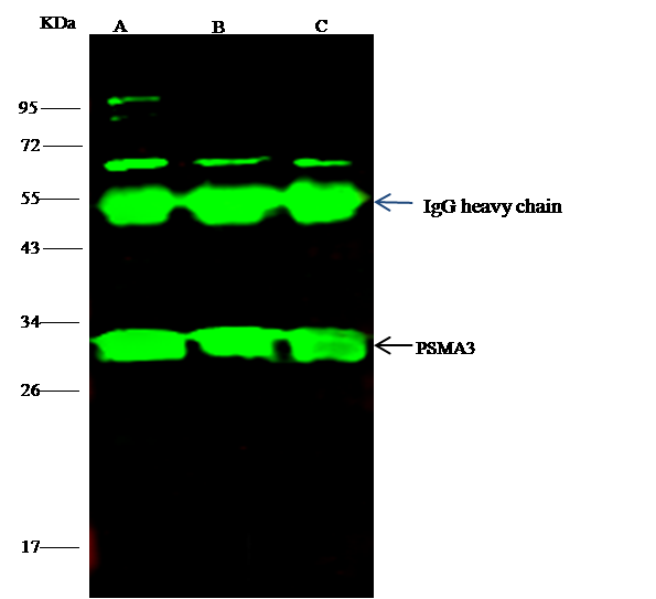
(PSMA3 was immunoprecipitated using:Lane A:0.5 mg MOLT-4 Whole Cell LysateLane B:0.5 mg NIH-3T3 Whole Cell LysateLane C:0.5 mg A549 Whole Cell Lysate2 uL anti-PSMA3 rabbit polyclonal antibody and 15 ul of 50 % Protein G agarose.Primary antibody:Anti-PSMA3 rabbit polyclonal antibody,at 1:100 dilutionSecondary antibody:Dylight 800-labeled antibody to rabbit IgG (H+L), at 1:5000 dilutionDeveloped using the odssey technique.Performed under reducing conditions.Predicted band size: 28 kDaObserved band size: 28 kDa)
IP (Immunoprecipitation)
(PSMA3 was immunoprecipitated using:Lane A:0.5 mg MOLT-4 Whole Cell LysateLane B:0.5 mg NIH-3T3 Whole Cell LysateLane C:0.5 mg A549 Whole Cell Lysate2 uL anti-PSMA3 rabbit polyclonal antibody and 15 ul of 50 % Protein G agarose.Primary antibody:Anti-PSMA3 rabbit polyclonal antibody,at 1:100 dilutionSecondary antibody:Dylight 800-labeled antibody to rabbit IgG (H+L), at 1:5000 dilutionDeveloped using the odssey technique.Performed under reducing conditions.Predicted band size: 28 kDaObserved band size: 28 kDa)
PSMA3, Polyclonal Antibody (Cat# AAA255811)
Full Name
Anti-PSMA3 Antibody, Rabbit Polyclonal
Reactivity
Human
Applications
Immunoprecipitation, Immunofluorescence, Immunocytochemistry, Western Blot
Purity
Protein A & Antigen Affinity
Pricing
 IF (Immunofluorescence)
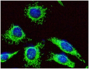
(ICC/IF analysis of HSP60 in HeLa cells line, stained with DAPI (Blue) for nucleus staining and monoclonal anti-human HSP60 antibody (1:100) with goat anti-mouse IgG-Alexa fluor 488 conjugate (Green).)
IF (Immunofluorescence)
(ICC/IF analysis of HSP60 in HeLa cells line, stained with DAPI (Blue) for nucleus staining and monoclonal anti-human HSP60 antibody (1:100) with goat anti-mouse IgG-Alexa fluor 488 conjugate (Green).)
HSP60, Monoclonal Antibody (Cat# AAA48012)
Full Name
HSP60 antibody
Gene Names
HSPD1; HLD4; CPN60; GROEL; HSP60; HSP65; SPG13; HSP-60; HuCHA60
Reactivity
Human
Applications
Flow Cytometry, Immunofluorescence, Immunocytochemistry, Immunohistochemistry, Western Blot
Purity
By protein-G affinity chromatography
Pricing
 Application Data
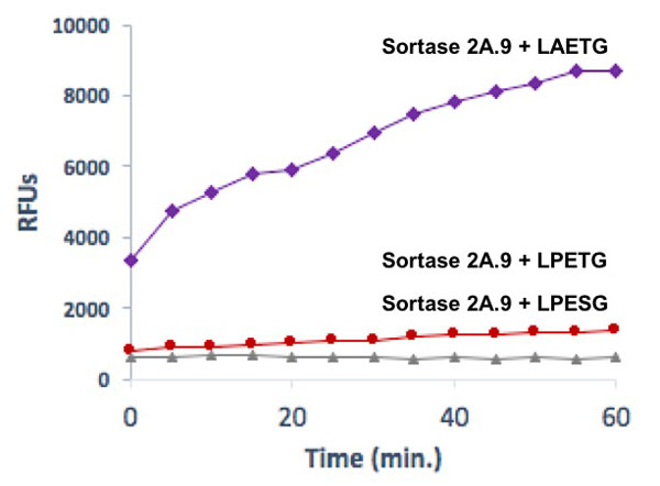
(Recombinant Sortase (2A.9) protein specificity for LAETG sequence Sortase (2A.9) at 12 ng/ul was incubated with 0.4 mM LAETG, LPETG or LPESG substrates in reaction buffer, followed by fluorescence detection upon cleavage. Results demonstrate the high specificity of Sortase (2A.9) for LAETG. The 2A.9 shows minimal activity to the WT substrate LPETG and no activity towards the mutated substrate LPESG.)
Application Data
(Recombinant Sortase (2A.9) protein specificity for LAETG sequence Sortase (2A.9) at 12 ng/ul was incubated with 0.4 mM LAETG, LPETG or LPESG substrates in reaction buffer, followed by fluorescence detection upon cleavage. Results demonstrate the high specificity of Sortase (2A.9) for LAETG. The 2A.9 shows minimal activity to the WT substrate LPETG and no activity towards the mutated substrate LPESG.)
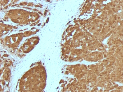
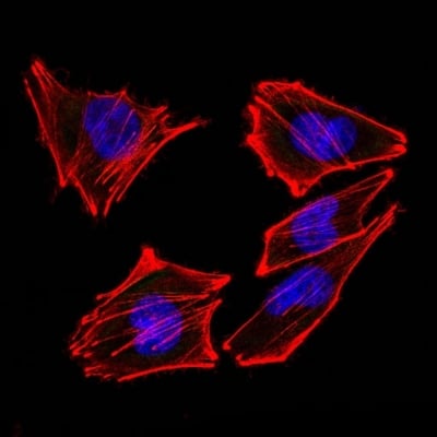 IF (Immunofluorescence)
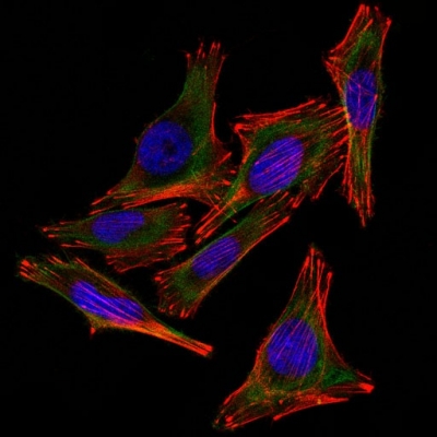
(Confocal Immunofluorescent analysis of A2058 cells using AF488-labeled Isotype Control Monoclonal Antibody (IgG2a) (Green). F-actin filaments were labeled with DyLight 554 Phalloidin (red). DAPI was used to stain the cell nuclei (blue). (Negative Control))
IF (Immunofluorescence)
(Confocal Immunofluorescent analysis of A2058 cells using AF488-labeled Isotype Control Monoclonal Antibody (IgG2a) (Green). F-actin filaments were labeled with DyLight 554 Phalloidin (red). DAPI was used to stain the cell nuclei (blue). (Negative Control))
S100B, Monoclonal Antibody (Cat# AAA214511)
Full Name
S100B (Astrocyte and Melanoma Marker)
Gene Names
S100B; NEF; S100; S100-B; S100beta
Reactivity
Human, Mouse, Rat, Cow. Others not known.
Applications
Immunohistochemistry, Western Blot, Immunofluorescence, Flow Cytometry
Pricing
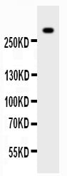
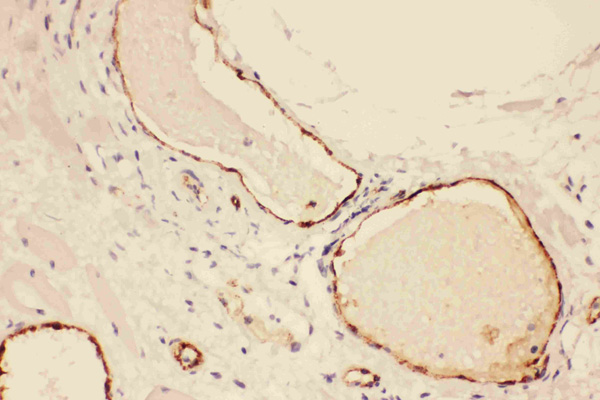
 IHC (Immunohistochemisry)
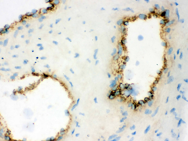
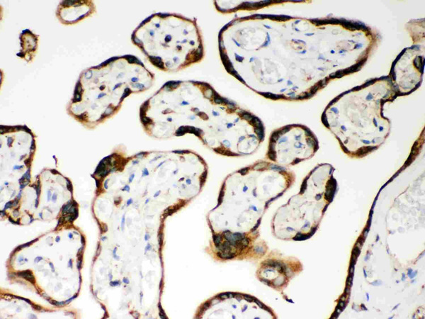
(Figure 3. IHC analysis of VWF using anti-VWF antibody (AAA45673).VWF was detected in frozen section of human placenta tissue. Heat mediated antigen retrieval was performed in citrate buffer (pH6, epitope retrieval solution) for 20 mins. The tissue section was blocked with 10% goat serum. The tissue section was then incubated with 1ug/ml rabbit anti-VWF Antibody (AAA45673) overnight at 4 degree C. Biotinylated goat anti-rabbit IgG was used as secondary antibody and incubated for 30 minutes at 37 degree C. The tissue section was developed using Strepavidin-Biotin-Complex (SABC) with DAB as the chromogen.)
IHC (Immunohistochemisry)
(Figure 3. IHC analysis of VWF using anti-VWF antibody (AAA45673).VWF was detected in frozen section of human placenta tissue. Heat mediated antigen retrieval was performed in citrate buffer (pH6, epitope retrieval solution) for 20 mins. The tissue section was blocked with 10% goat serum. The tissue section was then incubated with 1ug/ml rabbit anti-VWF Antibody (AAA45673) overnight at 4 degree C. Biotinylated goat anti-rabbit IgG was used as secondary antibody and incubated for 30 minutes at 37 degree C. The tissue section was developed using Strepavidin-Biotin-Complex (SABC) with DAB as the chromogen.)
VWF, Polyclonal Antibody (Cat# AAA45673)
Full Name
Anti-VWF Antibody
Gene Names
VWF; VWD; F8VWF
Reactivity
Human
Applications
Immunohistochemistry, Western Blot
Purity
Immunogen affinity purified.
Pricing
 WB (Western Blot)
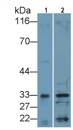
(Western Blot; Sample: Lane1: Rat Lung lysate; Lane2: Rat Small intestine lysate Primary Ab: 0.4ug/ml Mouse AntiRat IL33 Antibody Second Ab: 0.2ug/mL HRPLinked Caprine AntiMouse IgG Polyclonal Antibody (Catalog: SAA544Mu19))
WB (Western Blot)
(Western Blot; Sample: Lane1: Rat Lung lysate; Lane2: Rat Small intestine lysate Primary Ab: 0.4ug/ml Mouse AntiRat IL33 Antibody Second Ab: 0.2ug/mL HRPLinked Caprine AntiMouse IgG Polyclonal Antibody (Catalog: SAA544Mu19))
Interleukin 33 (IL33), Monoclonal Antibody (Cat# AAA151776)
Full Name
Monoclonal Antibody to Interleukin 33 (IL33)
Gene Names
Il33; RGD1311155
Reactivity
Human, Rat
Applications
Immunoprecipitation, Immunocytochemistry, Immunohistochemistry, Western Blot
Purity
Protein A + Protein G affinity chromatography
Pricing
 IHC (Immunohistochemisry)
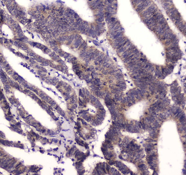
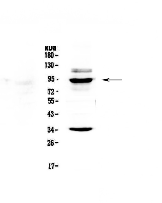
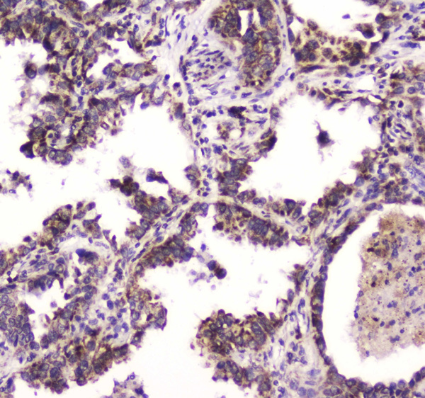
(Figure 3. IHC analysis of BRD7 using anti-BRD7 antibody (AAA124637).BRD7 was detected in paraffin-embedded section of human lung cancer tissue. Heat mediated antigen retrieval was performed in citrate buffer (pH6, epitope retrieval solution) for 20 mins. The tissue section was blocked with 10% goat serum. The tissue section was then incubated with 1ug/ml rabbit anti-BRD7 Antibody (AAA124637) overnight at 4 degree C. Biotinylated goat anti-rabbit IgG was used as secondary antibody and incubated for 30 minutes at 37 degree C. The tissue section was developed using Strepavidin-Biotin-Complex (SABC) with DAB as the chromogen.)
IHC (Immunohistochemisry)
(Figure 3. IHC analysis of BRD7 using anti-BRD7 antibody (AAA124637).BRD7 was detected in paraffin-embedded section of human lung cancer tissue. Heat mediated antigen retrieval was performed in citrate buffer (pH6, epitope retrieval solution) for 20 mins. The tissue section was blocked with 10% goat serum. The tissue section was then incubated with 1ug/ml rabbit anti-BRD7 Antibody (AAA124637) overnight at 4 degree C. Biotinylated goat anti-rabbit IgG was used as secondary antibody and incubated for 30 minutes at 37 degree C. The tissue section was developed using Strepavidin-Biotin-Complex (SABC) with DAB as the chromogen.)
BRD7, Polyclonal Antibody (Cat# AAA124637)
Full Name
Anti-BRD7 Picoband antibody
Gene Names
BRD7; BP75; NAG4; CELTIX1
Reactivity
Human, Mouse, Rat
No cross reactivity with other proteins.
No cross reactivity with other proteins.
Applications
Western Blot, Immunohistochemistry
Pricing
 FCM/FACS (Flow Cytometry)
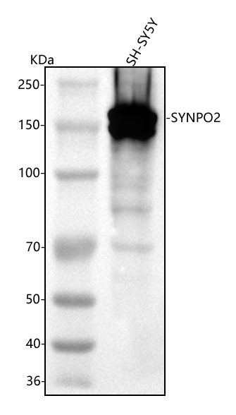
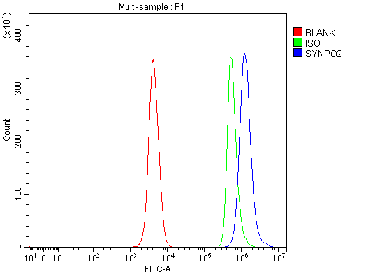
(Figure 2. Flow Cytometry analysis of SH-SY5Y cells using anti-SYNPO2 antibody (AAA127462).Overlay histogram showing SH-SY5Y cells stained with AAA127462 (Blue line). To facilitate intracellular staining, cells were fixed with 4% paraformaldehyde and permeabilized with permeabilization buffer. The cells were blocked with 10% normal goat serum. And then incubated with rabbit anti-SYNPO2 Antibody (AAA127462, 1ug/1x106 cells) for 30 min at 20 degree C. DyLight488 conjugated goat anti-rabbit IgG was used as secondary antibody for 30 minutes at 20 degree C. Isotype control antibody (Green line) was rabbit IgG (1ug/1x106) used under the same conditions. Unlabelled sample (Red line) was also used as a control.)
FCM/FACS (Flow Cytometry)
(Figure 2. Flow Cytometry analysis of SH-SY5Y cells using anti-SYNPO2 antibody (AAA127462).Overlay histogram showing SH-SY5Y cells stained with AAA127462 (Blue line). To facilitate intracellular staining, cells were fixed with 4% paraformaldehyde and permeabilized with permeabilization buffer. The cells were blocked with 10% normal goat serum. And then incubated with rabbit anti-SYNPO2 Antibody (AAA127462, 1ug/1x106 cells) for 30 min at 20 degree C. DyLight488 conjugated goat anti-rabbit IgG was used as secondary antibody for 30 minutes at 20 degree C. Isotype control antibody (Green line) was rabbit IgG (1ug/1x106) used under the same conditions. Unlabelled sample (Red line) was also used as a control.)
SYNPO2, Polyclonal Antibody (Cat# AAA127462)
Full Name
Anti-SYNPO2 Antibody Picoband
Reactivity
Human
Applications
Flow Cytometry, Western Blot
Purity
Immunogen affinity purified.
Pricing
 IP (Immunoprecipitation)
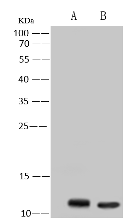
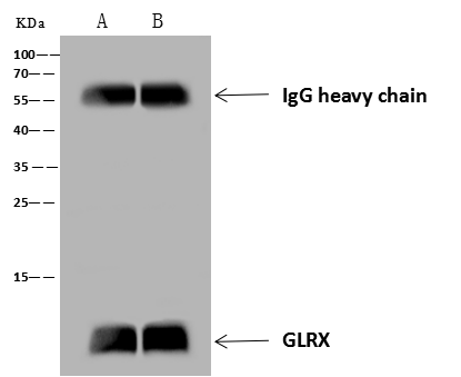
(GLRX was immunoprecipitated using:Lane A:0.5 mg HepG2 Whole Cell LysateLane B:0.5 mg Jurkat Whole Cell Lysate4 uL anti-GLRX rabbit polyclonal antibody and 60 ug of Immunomagnetic beads Protein A/G.Primary antibody:Anti-GLRX rabbit polyclonal antibody,at 1:100 dilutionSecondary antibody:Goat Anti-Rabbit IgG (H+L)/HRP at 1/10000 dilutionDeveloped using the ECL technique.Performed under reducing conditions.Predicted band size: 12 kDaObserved band size :12 kDa)
IP (Immunoprecipitation)
(GLRX was immunoprecipitated using:Lane A:0.5 mg HepG2 Whole Cell LysateLane B:0.5 mg Jurkat Whole Cell Lysate4 uL anti-GLRX rabbit polyclonal antibody and 60 ug of Immunomagnetic beads Protein A/G.Primary antibody:Anti-GLRX rabbit polyclonal antibody,at 1:100 dilutionSecondary antibody:Goat Anti-Rabbit IgG (H+L)/HRP at 1/10000 dilutionDeveloped using the ECL technique.Performed under reducing conditions.Predicted band size: 12 kDaObserved band size :12 kDa)
Glutaredoxin 1/GRX1, Polyclonal Antibody (Cat# AAA258919)
Full Name
Anti-Glutaredoxin 1/GRX1 Antibody, Rabbit Polyclonal
Reactivity
Human
Applications
Immunoprecipitation, Western Blot
Purity
Protein A & Antigen Affinity
Pricing
 ICC (Immunocytochemistry)
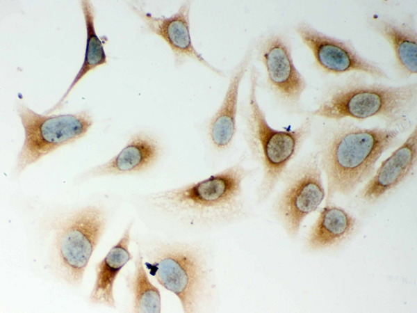
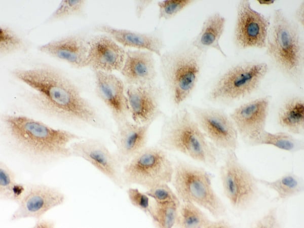
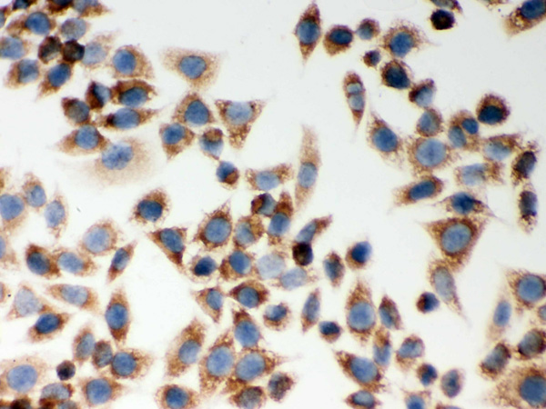
(Figure 5. IHC analysis of CARD4 using anti-CARD4 antibody (AAA46346).CARD4 was detected in immunocytochemical section of SMMC-7721 cell. Heat mediated antigen retrieval was performed in citrate buffer (pH6, epitope retrieval solution) for 20 mins. The tissue section was blocked with 10% goat serum. The tissue section was then incubated with 1ug/ml rabbit anti-CARD4 Antibody (AAA46346) overnight at 4 degree C. Biotinylated goat anti-rabbit IgG was used as secondary antibody and incubated for 30 minutes at 37 degree C. The tissue section was developed using Strepavidin-Biotin-Complex (SABC) with DAB as the chromogen.)
ICC (Immunocytochemistry)
(Figure 5. IHC analysis of CARD4 using anti-CARD4 antibody (AAA46346).CARD4 was detected in immunocytochemical section of SMMC-7721 cell. Heat mediated antigen retrieval was performed in citrate buffer (pH6, epitope retrieval solution) for 20 mins. The tissue section was blocked with 10% goat serum. The tissue section was then incubated with 1ug/ml rabbit anti-CARD4 Antibody (AAA46346) overnight at 4 degree C. Biotinylated goat anti-rabbit IgG was used as secondary antibody and incubated for 30 minutes at 37 degree C. The tissue section was developed using Strepavidin-Biotin-Complex (SABC) with DAB as the chromogen.)
CARD4, Polyclonal Antibody (Cat# AAA46346)
Full Name
Anti-CARD4 Antibody
Gene Names
NOD1; CARD4; NLRC1; CLR7.1
Reactivity
Human, Rat
Applications
Immunohistochemistry, Western Blot
Purity
Immunogen Affinity Purified
Pricing
 IHC (Immunohistochemistry)
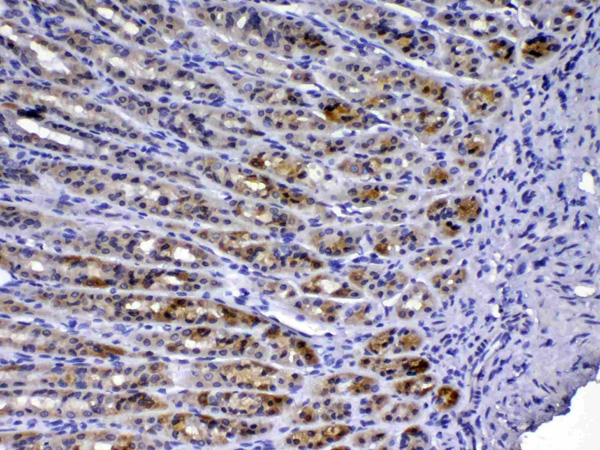
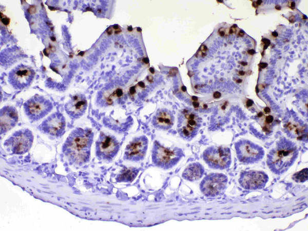
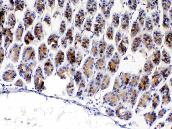
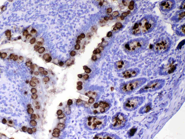
(Figure 5. IHC analysis of TFF3 using anti-TFF3 antibody (AAA124663).TFF3 was detected in paraffin-embedded section of rat small intestine tissue. Heat mediated antigen retrieval was performed in citrate buffer (pH6, epitope retrieval solution) for 20 mins. The tissue section was blocked with 10% goat serum. The tissue section was then incubated with 1ug/ml rabbit anti-TFF3 Antibody (AAA124663) overnight at 4 degree C. Biotinylated goat anti-rabbit IgG was used as secondary antibody and incubated for 30 minutes at 37 degree C. The tissue section was developed using Strepavidin-Biotin-Complex (SABC) with DAB as the chromogen.)
IHC (Immunohistochemistry)
(Figure 5. IHC analysis of TFF3 using anti-TFF3 antibody (AAA124663).TFF3 was detected in paraffin-embedded section of rat small intestine tissue. Heat mediated antigen retrieval was performed in citrate buffer (pH6, epitope retrieval solution) for 20 mins. The tissue section was blocked with 10% goat serum. The tissue section was then incubated with 1ug/ml rabbit anti-TFF3 Antibody (AAA124663) overnight at 4 degree C. Biotinylated goat anti-rabbit IgG was used as secondary antibody and incubated for 30 minutes at 37 degree C. The tissue section was developed using Strepavidin-Biotin-Complex (SABC) with DAB as the chromogen.)
TFF3/Itf, Polyclonal Antibody (Cat# AAA124663)
Full Name
Anti-TFF3/Itf Picoband Antibody
Gene Names
Tff3; ITF
Reactivity
Mouse, Rat
No cross reactivity with other proteins.
No cross reactivity with other proteins.
Applications
Western Blot, Immunohistochemistry
Purity
Immunogen affinity purified
Pricing
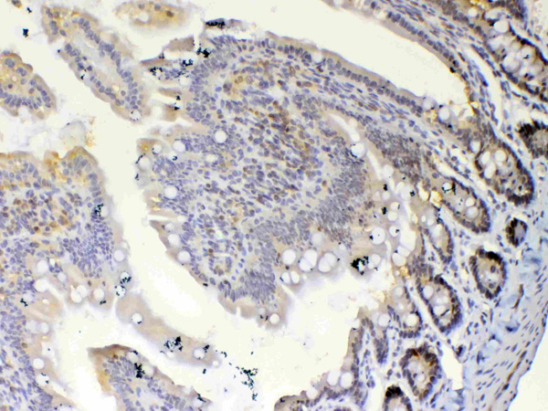 IHC (Immunohistochemistry)
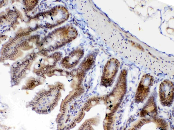
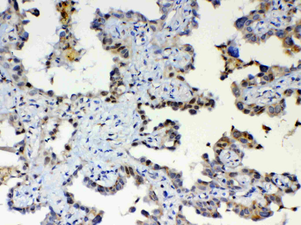
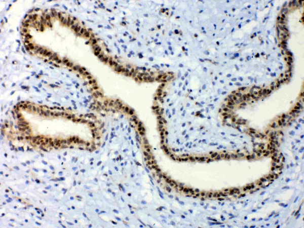
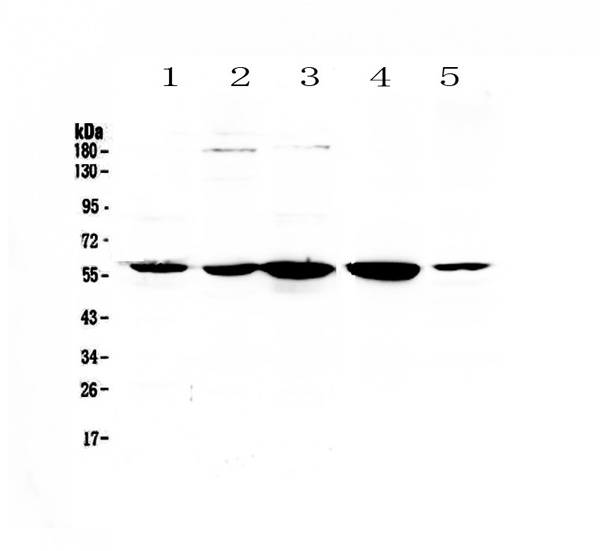
(Figure 5. IHC analysis of ETV6/Tel using anti-ETV6/Tel antibody (AAA124566).ETV6/Tel was detected in paraffin-embedded section of rat small intestine tissue. Heat mediated antigen retrieval was performed in citrate buffer (pH6, epitope retrieval solution) for 20 mins. The tissue section was blocked with 10% goat serum. The tissue section was then incubated with 1ug/ml rabbit anti-ETV6/Tel Antibody (AAA124566) overnight at 4 degree C. Biotinylated goat anti-rabbit IgG was used as secondary antibody and incubated for 30 minutes at 37 degree C. The tissue section was developed using Strepavidin-Biotin-Complex (SABC) with DAB as the chromogen.)
IHC (Immunohistochemistry)
(Figure 5. IHC analysis of ETV6/Tel using anti-ETV6/Tel antibody (AAA124566).ETV6/Tel was detected in paraffin-embedded section of rat small intestine tissue. Heat mediated antigen retrieval was performed in citrate buffer (pH6, epitope retrieval solution) for 20 mins. The tissue section was blocked with 10% goat serum. The tissue section was then incubated with 1ug/ml rabbit anti-ETV6/Tel Antibody (AAA124566) overnight at 4 degree C. Biotinylated goat anti-rabbit IgG was used as secondary antibody and incubated for 30 minutes at 37 degree C. The tissue section was developed using Strepavidin-Biotin-Complex (SABC) with DAB as the chromogen.)
ETV6/Tel, Polyclonal Antibody (Cat# AAA124566)
Full Name
Anti-ETV6/Tel Picoband antibody
Gene Names
ETV6; TEL; THC5; TEL/ABL
Reactivity
Human, Mouse, Rat
No cross reactivity with other proteins.
No cross reactivity with other proteins.
Applications
Western Blot, Immunohistochemistry
Pricing
 ICC (Immunocytochemistry)
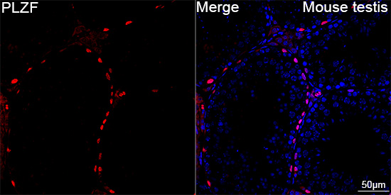
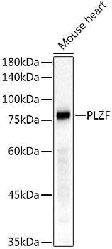
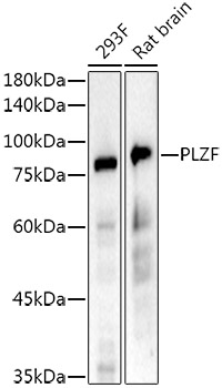
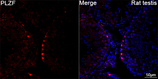
(Confocal imaging of paraffin-embedded Rat testis tissue using PLZF Rabbit mAb (AAA282902, dilution 1:200) followed by a further incubation with Cy3 Goat Anti-Rabbit IgG (H+L) (AS007, dilution 1:500) (Red). DAPI was used for nuclear staining (Blue). High pressure antigen retrieval performed with 0.01M Citrate Buffer (pH 6.0) prior to IF staining. Objective: 40x.)
ICC (Immunocytochemistry)
(Confocal imaging of paraffin-embedded Rat testis tissue using PLZF Rabbit mAb (AAA282902, dilution 1:200) followed by a further incubation with Cy3 Goat Anti-Rabbit IgG (H+L) (AS007, dilution 1:500) (Red). DAPI was used for nuclear staining (Blue). High pressure antigen retrieval performed with 0.01M Citrate Buffer (pH 6.0) prior to IF staining. Objective: 40x.)
PLZF, Monoclonal Antibody (Cat# AAA282902)
Full Name
PLZF Rabbit mAb
Reactivity
Human
Applications
Immunocytochemistry, Immunofluorescence, Western Blot
Purity
Affinity purification
Pricing
 FCM/FACS (Flow Cytometry)
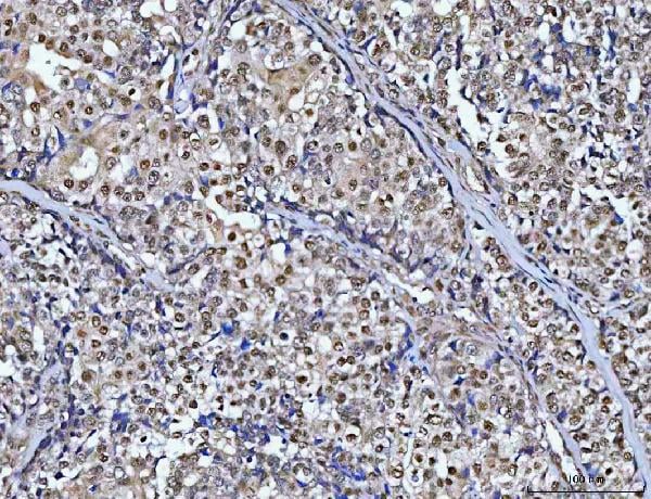
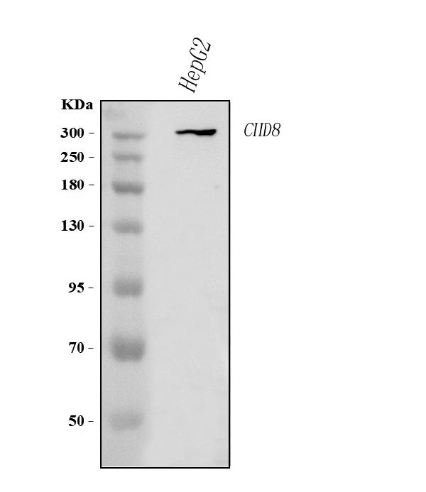
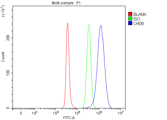
(Figure 3. Flow Cytometry analysis of SH-SY5Y cells using anti-CHD8 antibody (AAA127053).Overlay histogram showing SH-SY5Y cells stained with AAA127053 (Blue line). To facilitate intracellular staining, cells were fixed with 4% paraformaldehyde and permeabilized with permeabilization buffer. The cells were blocked with 10% normal goat serum. And then incubated with rabbit anti-CHD8 Antibody (AAA127053, 1ug/1x106 cells) for 30 min at 20 degree C. DyLight488 conjugated goat anti-rabbit IgG was used as secondary antibody for 30 minutes at 20 degree C. Isotype control antibody (Green line) was rabbit IgG (1ug/1x106) used under the same conditions. Unlabelled sample (Red line) was also used as a control.)
FCM/FACS (Flow Cytometry)
(Figure 3. Flow Cytometry analysis of SH-SY5Y cells using anti-CHD8 antibody (AAA127053).Overlay histogram showing SH-SY5Y cells stained with AAA127053 (Blue line). To facilitate intracellular staining, cells were fixed with 4% paraformaldehyde and permeabilized with permeabilization buffer. The cells were blocked with 10% normal goat serum. And then incubated with rabbit anti-CHD8 Antibody (AAA127053, 1ug/1x106 cells) for 30 min at 20 degree C. DyLight488 conjugated goat anti-rabbit IgG was used as secondary antibody for 30 minutes at 20 degree C. Isotype control antibody (Green line) was rabbit IgG (1ug/1x106) used under the same conditions. Unlabelled sample (Red line) was also used as a control.)
CHD8, Polyclonal Antibody (Cat# AAA127053)
Full Name
Anti-CHD8 Antibody Picoband
Gene Names
CHD8; HELSNF1; KIAA1564; DKFZp686N17164
Reactivity
Human
Applications
Flow Cytometry, Immunohistochemistry, Western Blot
Purity
Immunogen affinity purified.
Pricing
 FCM/FACS (Flow Cytometry)
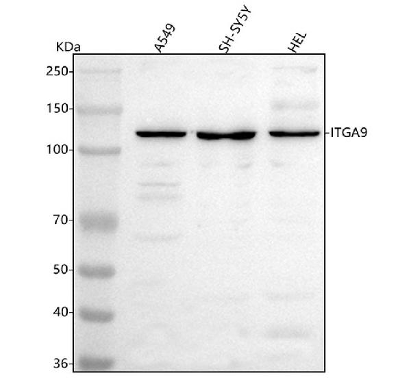
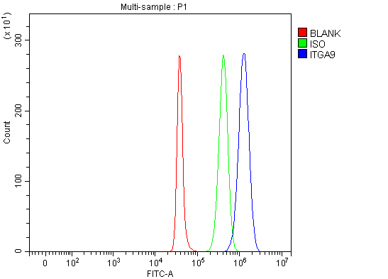
(Figure 2. Flow Cytometry analysis of SH-SY5Y cells using anti-Integrin Alpha-9/ITGA9 antibody (AAA127410).Overlay histogram showing SH-SY5Y cells stained with AAA127410 (Blue line). The cells were fixed with 4% paraformaldehyde and blocked with 10% normal goat serum. And then incubated with rabbit anti-Integrin Alpha-9/ITGA9 Antibody (AAA127410, 1ug/1x106 cells) for 30 min at 20 degree C. DyLight488 conjugated goat anti-rabbit IgG was used as secondary antibody for 30 minutes at 20 degree C. Isotype control antibody (Green line) was rabbit IgG (1ug/1x106) used under the same conditions. Unlabelled sample (Red line) was also used as a control.)
FCM/FACS (Flow Cytometry)
(Figure 2. Flow Cytometry analysis of SH-SY5Y cells using anti-Integrin Alpha-9/ITGA9 antibody (AAA127410).Overlay histogram showing SH-SY5Y cells stained with AAA127410 (Blue line). The cells were fixed with 4% paraformaldehyde and blocked with 10% normal goat serum. And then incubated with rabbit anti-Integrin Alpha-9/ITGA9 Antibody (AAA127410, 1ug/1x106 cells) for 30 min at 20 degree C. DyLight488 conjugated goat anti-rabbit IgG was used as secondary antibody for 30 minutes at 20 degree C. Isotype control antibody (Green line) was rabbit IgG (1ug/1x106) used under the same conditions. Unlabelled sample (Red line) was also used as a control.)
Integrin alpha-9/ITGA9, Polyclonal Antibody (Cat# AAA127410)
Full Name
Anti-Integrin alpha-9/ITGA9 Antibody Picoband
Gene Names
ITGA9; RLC; ITGA4L; ALPHA-RLC
Reactivity
Human
Applications
Flow Cytometry, Western Blot
Purity
Immunogen affinity purified.
Pricing
 FCM/FACS (Flow Cytometry)
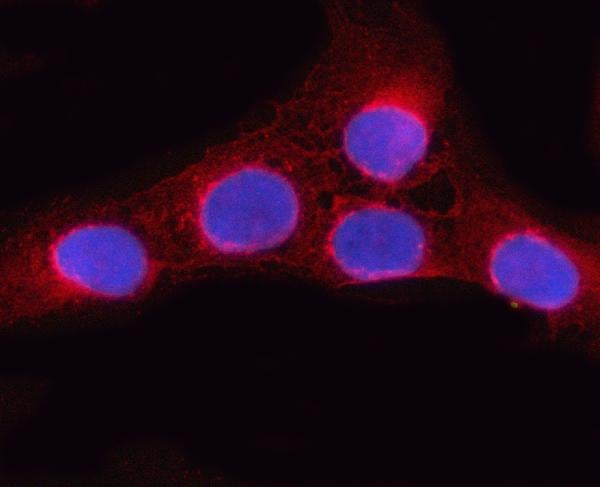
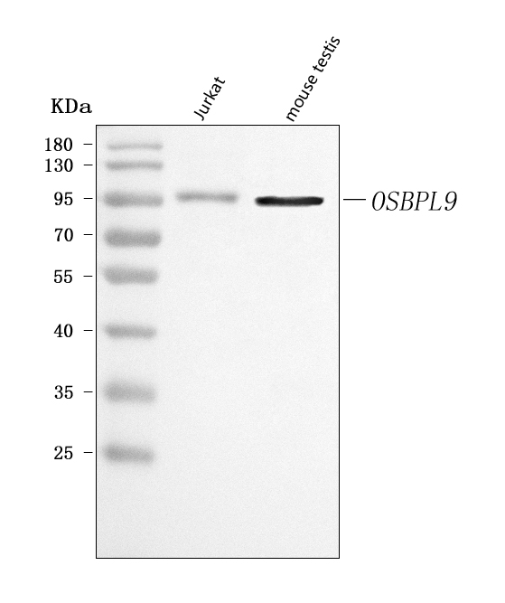
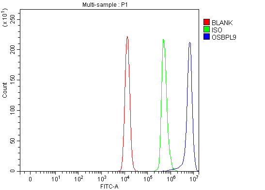
(Figure 3. Flow Cytometry analysis of Raji cells using anti-OSBPL9 antibody (AAA127586).Overlay histogram showing Raji cells stained with AAA127586 (Blue line). To facilitate intracellular staining, cells were fixed with 4% paraformaldehyde and permeabilized with permeabilization buffer. The cells were blocked with 10% normal goat serum. And then incubated with rabbit anti-OSBPL9 Antibody (AAA127586, 1ug/1x106 cells) for 30 min at 20 degree C. DyLight488 conjugated goat anti-rabbit IgG was used as secondary antibody for 30 minutes at 20 degree C. Isotype control antibody (Green line) was rabbit IgG (1ug/1x106) used under the same conditions. Unlabelled sample without incubation with primary antibody and secondary antibody (Red line) was used as a blank control.)
FCM/FACS (Flow Cytometry)
(Figure 3. Flow Cytometry analysis of Raji cells using anti-OSBPL9 antibody (AAA127586).Overlay histogram showing Raji cells stained with AAA127586 (Blue line). To facilitate intracellular staining, cells were fixed with 4% paraformaldehyde and permeabilized with permeabilization buffer. The cells were blocked with 10% normal goat serum. And then incubated with rabbit anti-OSBPL9 Antibody (AAA127586, 1ug/1x106 cells) for 30 min at 20 degree C. DyLight488 conjugated goat anti-rabbit IgG was used as secondary antibody for 30 minutes at 20 degree C. Isotype control antibody (Green line) was rabbit IgG (1ug/1x106) used under the same conditions. Unlabelled sample without incubation with primary antibody and secondary antibody (Red line) was used as a blank control.)
OSBPL9, Polyclonal Antibody (Cat# AAA127586)
Full Name
Anti-OSBPL9 Antibody Picoband
Gene Names
OSBPL9; ORP9; ORP-9
Reactivity
Human, Mouse
Applications
Flow Cytometry, Immunofluorescence, Immunocytochemistry, Western Blot
Purity
Immunogen affinity purified.
Pricing
 IHC (Immunohiostchemistry)
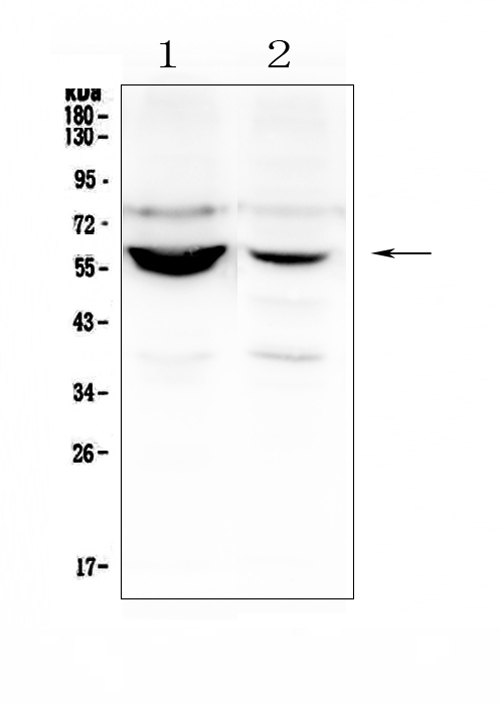
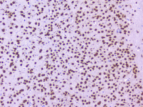
(Figure 3. IHC analysis of Nova1 using anti-Nova1 antibody.Nova1 was detected in paraffin-embedded section of rat brain tissue. Heat mediated antigen retrieval was performed in citrate buffer (pH6, epitope retrieval solution) for 20 mins. The tissue section was blocked with 10% goat serum. The tissue section was then incubated with 1ug/ml rabbit anti-Nova1 antibody.Nova1 was detected in paraffin-embedded section of rat brain tissue. Heat mediated antigen retrieval was performed in citrate buffer (pH6, epitope retrieval solution) for 20 mins. The tissue section was blocked with 10% goat serum. The tissue section was then incubated with 1ug/ml rabbit anti-Nova1 antibody.Nova1 was detected in paraffin-embedded section of mouse brain tissue. Heat mediated antigen retrieval was performed in citrate buffer (pH6, epitope retrieval solution) for 20 mins. The tissue section was blocked with 10% goat serum. The tissue section was then incubated with 1ug/ml rabbit anti-Nova1 Antibody overnight at 4 degree C. Biotinylated goat anti-rabbit IgG was used as secondary antibody and incubated for 30 minutes at 37 degree C. The tissue section was developed using Strepavidin-Biotin-Complex (SABC) with DAB as the chromogen.)
IHC (Immunohiostchemistry)
(Figure 3. IHC analysis of Nova1 using anti-Nova1 antibody.Nova1 was detected in paraffin-embedded section of rat brain tissue. Heat mediated antigen retrieval was performed in citrate buffer (pH6, epitope retrieval solution) for 20 mins. The tissue section was blocked with 10% goat serum. The tissue section was then incubated with 1ug/ml rabbit anti-Nova1 antibody.Nova1 was detected in paraffin-embedded section of rat brain tissue. Heat mediated antigen retrieval was performed in citrate buffer (pH6, epitope retrieval solution) for 20 mins. The tissue section was blocked with 10% goat serum. The tissue section was then incubated with 1ug/ml rabbit anti-Nova1 antibody.Nova1 was detected in paraffin-embedded section of mouse brain tissue. Heat mediated antigen retrieval was performed in citrate buffer (pH6, epitope retrieval solution) for 20 mins. The tissue section was blocked with 10% goat serum. The tissue section was then incubated with 1ug/ml rabbit anti-Nova1 Antibody overnight at 4 degree C. Biotinylated goat anti-rabbit IgG was used as secondary antibody and incubated for 30 minutes at 37 degree C. The tissue section was developed using Strepavidin-Biotin-Complex (SABC) with DAB as the chromogen.)
Nova1, Polyclonal Antibody (Cat# AAA124964)
Full Name
Anti-Nova1 Antibody
Gene Names
NOVA1; Nova-1
Reactivity
Human, Mouse, Rat
Applications
Direct ELISA, Immunofluorescence, Immunocytochemistry, Immunohistochemistry, Western Blot
Purity
Immunogen Affinity Purified
Pricing
 WB (Western Blot)
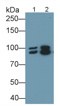
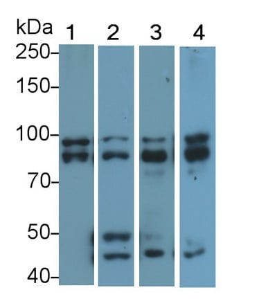
(Western Blot; Sample: Lane1: MCF7 cell lysate; Lane2: Rat Cerebrum lysate; Lane3: Mouse Cerebrum lysate; Lane4: Porcine Cerebrum lysatePrimary Ab: 0.2ug/ml Rabbit AntiHuman OPA1 AntibodySecond Ab: 0.2ug/mL HRPLinked Caprine AntiRabbit IgG Polyclonal Antibody(Catalog: SAA544Rb19))
WB (Western Blot)
(Western Blot; Sample: Lane1: MCF7 cell lysate; Lane2: Rat Cerebrum lysate; Lane3: Mouse Cerebrum lysate; Lane4: Porcine Cerebrum lysatePrimary Ab: 0.2ug/ml Rabbit AntiHuman OPA1 AntibodySecond Ab: 0.2ug/mL HRPLinked Caprine AntiRabbit IgG Polyclonal Antibody(Catalog: SAA544Rb19))
Optic Atrophy 1, Autosomal Dominant (OPA1), Polyclonal Antibody (Cat# AAA151968)
Full Name
Polyclonal Antibody to Optic Atrophy 1, Autosomal Dominant (OPA1)
Gene Names
OPA1; NPG; NTG; MGM1; BERHS; largeG; MTDPS14
Reactivity
Human, Mouse, Rat, Pig
Applications
Immunoprecipitation, Immunocytochemistry, Immunohistochemistry, Western Blot
Purity
Antigenspecific affinity chromatography followed by Protein A affinity chromatography
Pricing
 FCM/FACS (Flow Cytometry)
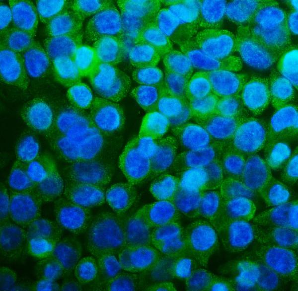
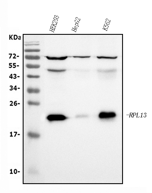
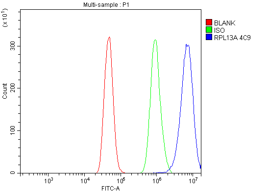
(Figure 3. Flow Cytometry analysis of A431 cells using anti-RPL13A antibody (AAA125929).Overlay histogram showing A431 cells stained with AAA125929 (Blue line). The cells were blocked with 10% normal goat serum. And then incubated with mouse anti-RPL13A Antibody (AAA125929, 1μg/1x106 cells) for 30 min at 20 degree C. DyLight®488 conjugated goat anti-mouse IgG (BA1126, 5-10μg/1x106 cells) was used as secondary antibody for 30 minutes at 20 degree C. Isotype control antibody (Green line) was mouse IgG (1μg/1x106) used under the same conditions. Unlabelled sample (Red line) was also used as a control.)
FCM/FACS (Flow Cytometry)
(Figure 3. Flow Cytometry analysis of A431 cells using anti-RPL13A antibody (AAA125929).Overlay histogram showing A431 cells stained with AAA125929 (Blue line). The cells were blocked with 10% normal goat serum. And then incubated with mouse anti-RPL13A Antibody (AAA125929, 1μg/1x106 cells) for 30 min at 20 degree C. DyLight®488 conjugated goat anti-mouse IgG (BA1126, 5-10μg/1x106 cells) was used as secondary antibody for 30 minutes at 20 degree C. Isotype control antibody (Green line) was mouse IgG (1μg/1x106) used under the same conditions. Unlabelled sample (Red line) was also used as a control.)
RPL13A, Monoclonal Antibody (Cat# AAA125929)
Full Name
Anti-RPL13A Antibody (monoclonal, 4C9)
Gene Names
RPL13A; L13A; TSTA1
Reactivity
Human
Applications
Flow Cytometry, Immunofluorescence, Immunocytochemistry, Western Blot
Purity
Immunogen affinity purified.
Pricing
 WB (Western Blot)
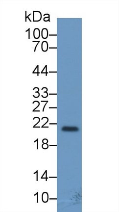
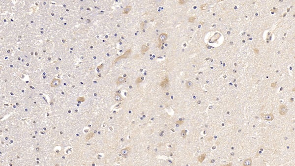
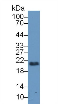
(Western Blot; Sample: Rat Cerebrum lysate; Primary Ab: 2ug/ml Mouse AntiHuman FGF13 AntibodySecond Ab: 0.2ug/mL HRPLinked Caprine AntiMouse IgG Polyclonal Antibody(Catalog: SAA544Mu19))
WB (Western Blot)
(Western Blot; Sample: Rat Cerebrum lysate; Primary Ab: 2ug/ml Mouse AntiHuman FGF13 AntibodySecond Ab: 0.2ug/mL HRPLinked Caprine AntiMouse IgG Polyclonal Antibody(Catalog: SAA544Mu19))
Fibroblast Growth Factor 13 (FGF13), Monoclonal Antibody (Cat# AAA151819)
Full Name
Monoclonal Antibody to Fibroblast Growth Factor 13 (FGF13)
Gene Names
FGF13; FGF2; FHF2; FHF-2; FGF-13
Reactivity
Human, Rat, Pig
Applications
Immunoprecipitation, Immunocytochemistry, Immunohistochemistry, Western Blot
Purity
Protein A + Protein G affinity chromatography
Pricing
 IF (Immunofluorescence)
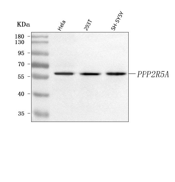
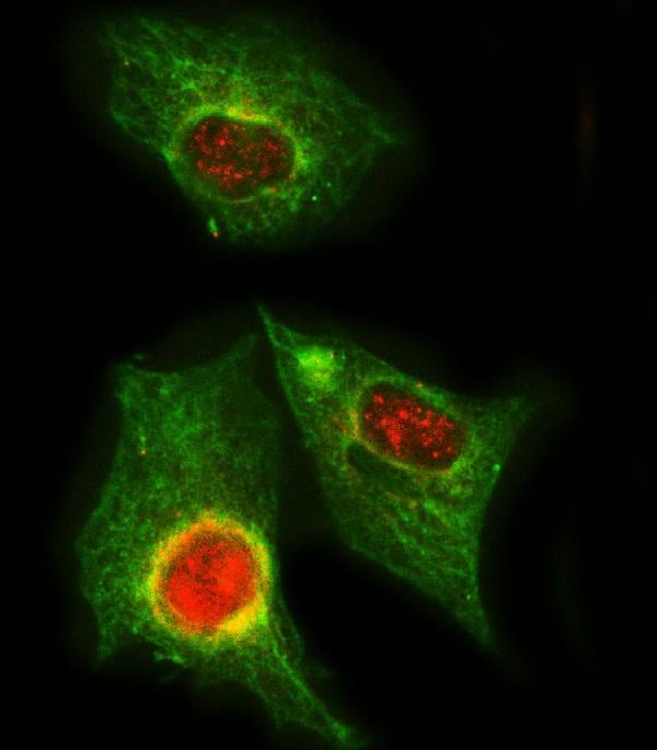
(Figure 2. IF analysis of PPP2R5A using anti-PPP2R5A antibody (AAA127337) and anti-Beta Tubulin antibody (M01857-3).PPP2R5A was detected in immunocytochemical section of A549 cell. Enzyme antigen retrieval was performed using IHC enzyme antigen retrieval reagent for 15 mins. The cells were blocked with 10% goat serum. And then incubated with 5ug/mL rabbit anti-PPP2R5A Antibody (AAA127337) and mouse anti-Beta Tubulin antibody (M01857-3) overnight at 4 degree C. Cy3 Conjugated Goat Anti-Rabbit IgG (BA1032) and DyLight488 Conjugated Goat Anti-Mouse IgG (BA1126) were used as secondary antibody at 1:500 dilution and incubated for 30 minutes at 37 degree C. Visualize using a fluorescence microscope and filter sets appropriate for the label used.)
IF (Immunofluorescence)
(Figure 2. IF analysis of PPP2R5A using anti-PPP2R5A antibody (AAA127337) and anti-Beta Tubulin antibody (M01857-3).PPP2R5A was detected in immunocytochemical section of A549 cell. Enzyme antigen retrieval was performed using IHC enzyme antigen retrieval reagent for 15 mins. The cells were blocked with 10% goat serum. And then incubated with 5ug/mL rabbit anti-PPP2R5A Antibody (AAA127337) and mouse anti-Beta Tubulin antibody (M01857-3) overnight at 4 degree C. Cy3 Conjugated Goat Anti-Rabbit IgG (BA1032) and DyLight488 Conjugated Goat Anti-Mouse IgG (BA1126) were used as secondary antibody at 1:500 dilution and incubated for 30 minutes at 37 degree C. Visualize using a fluorescence microscope and filter sets appropriate for the label used.)
PPP2R5A, Polyclonal Antibody (Cat# AAA127337)
Full Name
Anti-PPP2R5A Antibody Picoband
Gene Names
PPP2R5A; B56A; PR61A
Reactivity
Human
Applications
Immunofluorescence, Immunocytochemistry, Western Blot
Purity
Immunogen affinity purified.
Pricing
 FCM/FACS (Flow Cytometry)
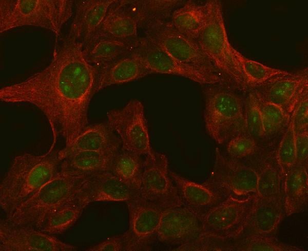
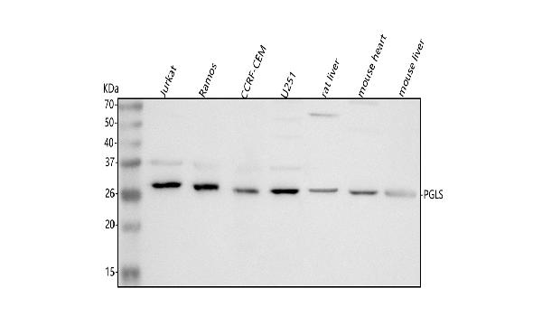
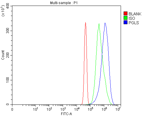
(Figure 3. Flow Cytometry analysis of JK cells using anti-PGLS antibody (AAA127127).Overlay histogram showing JK cells stained with AAA127127 (Blue line). To facilitate intracellular staining, cells were fixed with 4% paraformaldehyde and permeabilized with permeabilization buffer. The cells were blocked with 10% normal goat serum. And then incubated with rabbit anti-PGLS Antibody (AAA127127, 1ug/1x106 cells) for 30 min at 20 degree C. DyLight488 conjugated goat anti-rabbit IgG was used as secondary antibody for 30 minutes at 20 degree C. Isotype control antibody (Green line) was rabbit IgG (1ug/1x106) used under the same conditions. Unlabelled sample (Red line) was also used as a control.)
FCM/FACS (Flow Cytometry)
(Figure 3. Flow Cytometry analysis of JK cells using anti-PGLS antibody (AAA127127).Overlay histogram showing JK cells stained with AAA127127 (Blue line). To facilitate intracellular staining, cells were fixed with 4% paraformaldehyde and permeabilized with permeabilization buffer. The cells were blocked with 10% normal goat serum. And then incubated with rabbit anti-PGLS Antibody (AAA127127, 1ug/1x106 cells) for 30 min at 20 degree C. DyLight488 conjugated goat anti-rabbit IgG was used as secondary antibody for 30 minutes at 20 degree C. Isotype control antibody (Green line) was rabbit IgG (1ug/1x106) used under the same conditions. Unlabelled sample (Red line) was also used as a control.)
PGLS, Polyclonal Antibody (Cat# AAA127127)
Full Name
Anti-PGLS Antibody Picoband
Gene Names
PGLS; 6PGL
Reactivity
Human, Mouse, Rat
Applications
Flow Cytometry, Immunofluorescence, Immunocytochemistry, Western Blot
Purity
Immunogen affinity purified.
Pricing
 FCM/FACS (Flow Cytometry)
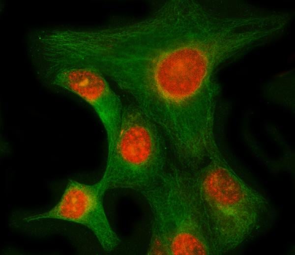
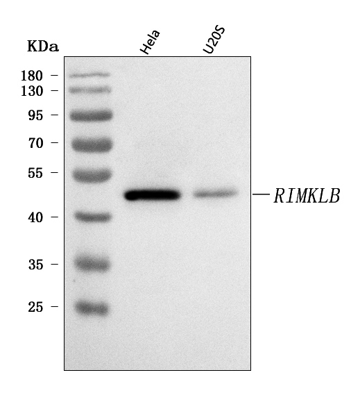
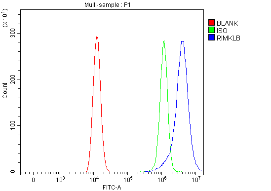
(Figure 3. Flow Cytometry analysis of HEL cells using anti-RIMKLB antibody (AAA127817).Overlay histogram showing HEL cells stained with AAA127817 (Blue line). To facilitate intracellular staining, cells were fixed with 4% paraformaldehyde and permeabilized with permeabilization buffer. The cells were blocked with 10% normal goat serum. And then incubated with rabbit anti-RIMKLB Antibody (AAA127817, 1ug/1x106 cells) for 30 min at 20 degree C. DyLight488 conjugated goat anti-rabbit IgG was used as secondary antibody for 30 minutes at 20 degree C. Isotype control antibody (Green line) was rabbit IgG (1ug/1x106) used under the same conditions. Unlabelled sample without incubation with primary antibody and secondary antibody (Red line) was used as a blank control.)
FCM/FACS (Flow Cytometry)
(Figure 3. Flow Cytometry analysis of HEL cells using anti-RIMKLB antibody (AAA127817).Overlay histogram showing HEL cells stained with AAA127817 (Blue line). To facilitate intracellular staining, cells were fixed with 4% paraformaldehyde and permeabilized with permeabilization buffer. The cells were blocked with 10% normal goat serum. And then incubated with rabbit anti-RIMKLB Antibody (AAA127817, 1ug/1x106 cells) for 30 min at 20 degree C. DyLight488 conjugated goat anti-rabbit IgG was used as secondary antibody for 30 minutes at 20 degree C. Isotype control antibody (Green line) was rabbit IgG (1ug/1x106) used under the same conditions. Unlabelled sample without incubation with primary antibody and secondary antibody (Red line) was used as a blank control.)
RIMKLB, Polyclonal Antibody (Cat# AAA127817)
Full Name
Anti-RIMKLB Antibody Picoband
Gene Names
RIMKLB; NAAGS; FAM80B; NAAGS-I
Reactivity
Human
Applications
Flow Cytometry, Immunofluorescence, Immunocytochemistry, Western Blot
Purity
Immunogen affinity purified.
Pricing
TGF-beta RII, Active Protein (Cat# AAA48997)
Full Name
Recombinant human TGF-beta RII protein
Gene Names
TGFBR2; AAT3; FAA3; LDS2; MFS2; RIIC; LDS1B; LDS2B; TAAD2; TGFR-2; TGFbeta-RII
Applications
Bioactivity, SDS-PAGE
Purity
> 95% by SDS-PAGE
Pricing
 Application Data
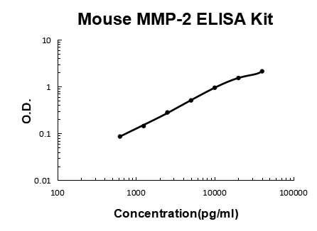
MMP-2, ELISA Kit (Cat# AAA125859)
Full Name
Mouse MMP-2 Quick ELISA Kit
Gene Names
Mmp2; GelA; Clg4a; MMP-2
Reactivity
Mouse
Pricing
 IF (Immunofluorescence)
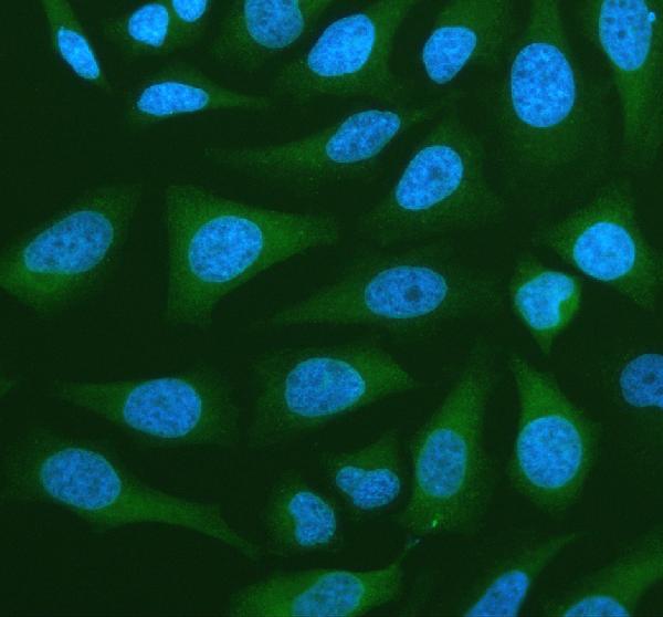
(Figure 2. IF analysis of NANS using anti-NANS antibody (AAA128217).NANS was detected in an immunocytochemical section of Hela cells. Enzyme antigen retrieval was performed using IHC enzyme antigen retrieval reagent for 15 mins. The cells were blocked with 10% goat serum. And then incubated with 4ug/mL rabbit anti-NANS Antibody (AAA128217) overnight at 4 degree C. DyLight®488 Conjugated Goat Anti-Rabbit IgG was used as secondary antibody at 1:500 dilution and incubated for 30 minutes at 37 degree C. The section was counterstained with DAPI. Visualize using a fluorescence microscope and filter sets appropriate for the label used.)
IF (Immunofluorescence)
(Figure 2. IF analysis of NANS using anti-NANS antibody (AAA128217).NANS was detected in an immunocytochemical section of Hela cells. Enzyme antigen retrieval was performed using IHC enzyme antigen retrieval reagent for 15 mins. The cells were blocked with 10% goat serum. And then incubated with 4ug/mL rabbit anti-NANS Antibody (AAA128217) overnight at 4 degree C. DyLight®488 Conjugated Goat Anti-Rabbit IgG was used as secondary antibody at 1:500 dilution and incubated for 30 minutes at 37 degree C. The section was counterstained with DAPI. Visualize using a fluorescence microscope and filter sets appropriate for the label used.)
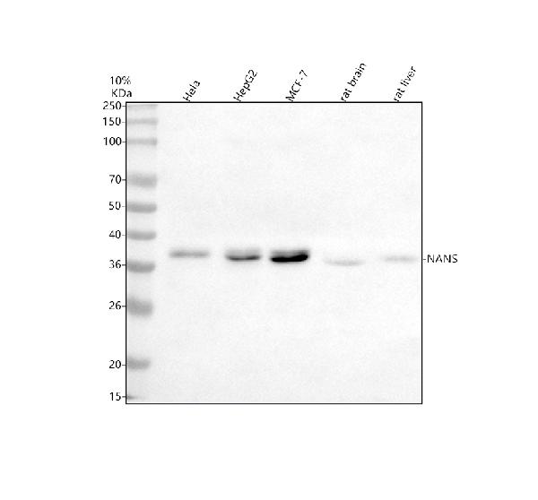
NANS, Polyclonal Antibody (Cat# AAA128217)
Full Name
Anti-NANS Antibody Picoband
Gene Names
NANS; SAS; HEL-S-100
Reactivity
Human, Rat
Applications
Immunofluorescence, Immunocytochemistry, Western Blot
Purity
Immunogen affinity purified.
Pricing
 IHC (Immunohistochemisry)
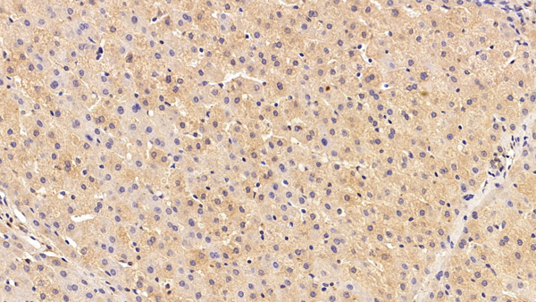
(DAB staining on IHCP;Sample: Human Liver Tissue; Primary Ab: 20ug/ml Rabbit AntiHuman CYP4F2 AntibodySecond Ab: 2ug/mL HRPLinked Caprine AntiRabbit IgG Polyclonal Antibody(Catalog: SAA544Rb19))
IHC (Immunohistochemisry)
(DAB staining on IHCP;Sample: Human Liver Tissue; Primary Ab: 20ug/ml Rabbit AntiHuman CYP4F2 AntibodySecond Ab: 2ug/mL HRPLinked Caprine AntiRabbit IgG Polyclonal Antibody(Catalog: SAA544Rb19))
Cytochrome P450 4F2 (CYP4F2), Polyclonal Antibody (Cat# AAA151974)
Full Name
Polyclonal Antibody to Cytochrome P450 4F2 (CYP4F2)
Gene Names
CYP4F2; CPF2
Reactivity
Human, Pig
Applications
Immunoprecipitation, Immunocytochemistry, Immunohistochemistry, Western Blot
Purity
Antigenspecific affinity chromatography followed by Protein A affinity chromatography
Pricing
 FCM/FACS (Flow Cytometry)
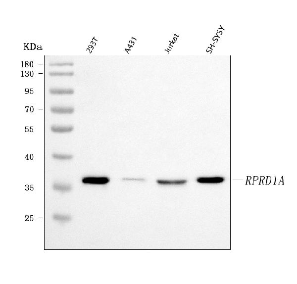
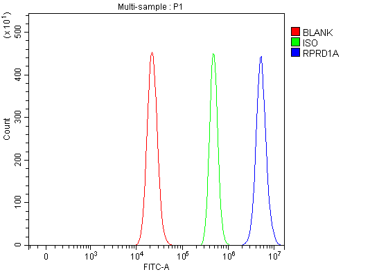
(Figure 2. Flow Cytometry analysis of RT4 cells using anti-P15RS/RPRD1A antibody (AAA127626).Overlay histogram showing RT4 cells stained with AAA127626 (Blue line). To facilitate intracellular staining, cells were fixed with 4% paraformaldehyde and permeabilized with permeabilization buffer. The cells were blocked with 10% normal goat serum. And then incubated with rabbit anti-P15RS/RPRD1A Antibody (AAA127626, 1ug/1x106 cells) for 30 min at 20 degree C. DyLight488 conjugated goat anti-rabbit IgG was used as secondary antibody for 30 minutes at 20 degree C. Isotype control antibody (Green line) was rabbit IgG (1ug/1x106) used under the same conditions. Unlabelled sample without incubation with primary antibody and secondary antibody (Red line) was used as a blank control.)
FCM/FACS (Flow Cytometry)
(Figure 2. Flow Cytometry analysis of RT4 cells using anti-P15RS/RPRD1A antibody (AAA127626).Overlay histogram showing RT4 cells stained with AAA127626 (Blue line). To facilitate intracellular staining, cells were fixed with 4% paraformaldehyde and permeabilized with permeabilization buffer. The cells were blocked with 10% normal goat serum. And then incubated with rabbit anti-P15RS/RPRD1A Antibody (AAA127626, 1ug/1x106 cells) for 30 min at 20 degree C. DyLight488 conjugated goat anti-rabbit IgG was used as secondary antibody for 30 minutes at 20 degree C. Isotype control antibody (Green line) was rabbit IgG (1ug/1x106) used under the same conditions. Unlabelled sample without incubation with primary antibody and secondary antibody (Red line) was used as a blank control.)
P15RS/RPRD1A, Polyclonal Antibody (Cat# AAA127626)
Full Name
Anti-P15RS/RPRD1A Antibody Picoband
Gene Names
RPRD1A; P15RS; HsT3101
Reactivity
Human
Applications
Flow Cytometry, Western Blot
Purity
Immunogen affinity purified.
Pricing
 IF (Immunofluorescence)
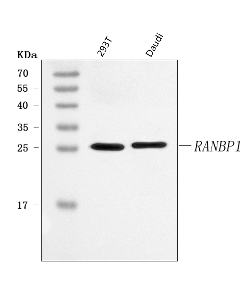
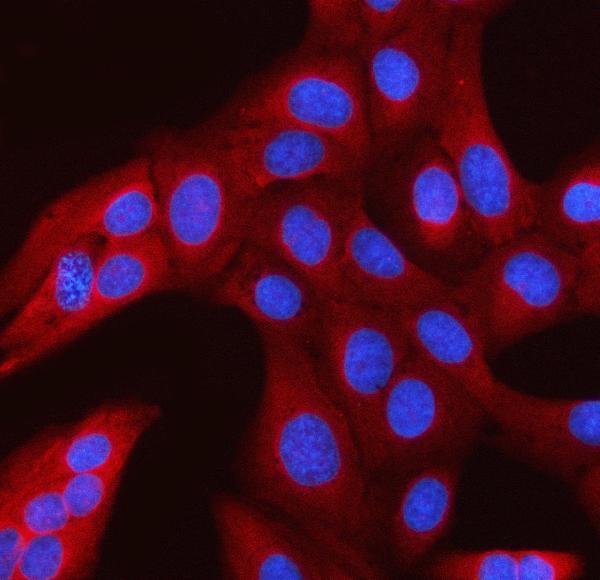
(Figure 2. IF analysis of RANBP1 using anti-RANBP1 antibody (AAA127208).RANBP1 was detected in an immunocytochemical section of U2OS cells. Enzyme antigen retrieval was performed using IHC enzyme antigen retrieval reagent for 15 mins. The cells were blocked with 10% goat serum. And then incubated with 5ug/mL rabbit anti-RANBP1 Antibody (AAA127208) overnight at 4 degree C. Cy3 Conjugated Goat Anti-Rabbit IgG (BA1032) was used as secondary antibody at 1:500 dilution and incubated for 30 minutes at 37 degree C. The section was counterstained with DAPI. Visualize using a fluorescence microscope and filter sets appropriate for the label used.)
IF (Immunofluorescence)
(Figure 2. IF analysis of RANBP1 using anti-RANBP1 antibody (AAA127208).RANBP1 was detected in an immunocytochemical section of U2OS cells. Enzyme antigen retrieval was performed using IHC enzyme antigen retrieval reagent for 15 mins. The cells were blocked with 10% goat serum. And then incubated with 5ug/mL rabbit anti-RANBP1 Antibody (AAA127208) overnight at 4 degree C. Cy3 Conjugated Goat Anti-Rabbit IgG (BA1032) was used as secondary antibody at 1:500 dilution and incubated for 30 minutes at 37 degree C. The section was counterstained with DAPI. Visualize using a fluorescence microscope and filter sets appropriate for the label used.)
RANBP1, Polyclonal Antibody (Cat# AAA127208)
Full Name
Anti-RANBP1 Antibody Picoband
Gene Names
RANBP1; HTF9A
Reactivity
Human
Applications
Immunofluorescence, Immunocytochemistry, Western Blot
Purity
Immunogen affinity purified.
Pricing
 FCM/FACS (Flow Cytometry)
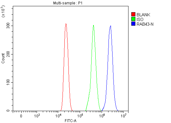
(Figure 3. Flow Cytometry analysis of HL-60 cells using anti-RAB43 antibody (AAA127578).Overlay histogram showing HL-60 cells stained with AAA127578 (Blue line). To facilitate intracellular staining, cells were fixed with 4% paraformaldehyde and permeabilized with permeabilization buffer. The cells were blocked with 10% normal goat serum. And then incubated with rabbit anti-RAB43 Antibody (AAA127578, 1ug/1x106 cells) for 30 min at 20 degree C. DyLight488 conjugated goat anti-rabbit IgG was used as secondary antibody for 30 minutes at 20 degree C. Isotype control antibody (Green line) was rabbit IgG (1ug/1x106) used under the same conditions. Unlabelled sample (Red line) was also used as a control.)
FCM/FACS (Flow Cytometry)
(Figure 3. Flow Cytometry analysis of HL-60 cells using anti-RAB43 antibody (AAA127578).Overlay histogram showing HL-60 cells stained with AAA127578 (Blue line). To facilitate intracellular staining, cells were fixed with 4% paraformaldehyde and permeabilized with permeabilization buffer. The cells were blocked with 10% normal goat serum. And then incubated with rabbit anti-RAB43 Antibody (AAA127578, 1ug/1x106 cells) for 30 min at 20 degree C. DyLight488 conjugated goat anti-rabbit IgG was used as secondary antibody for 30 minutes at 20 degree C. Isotype control antibody (Green line) was rabbit IgG (1ug/1x106) used under the same conditions. Unlabelled sample (Red line) was also used as a control.)
RAB43, Polyclonal Antibody (Cat# AAA127578)
Full Name
Anti-RAB43 Antibody Picoband
Gene Names
RAB43; RAB41; RAB11B; MGC90481
Reactivity
Human
Applications
Flow Cytometry, Immunofluorescence, Immunocytochemistry, Western Blot
Purity
Immunogen affinity purified.
Pricing
 FCM/FACS (Flow Cytometry)
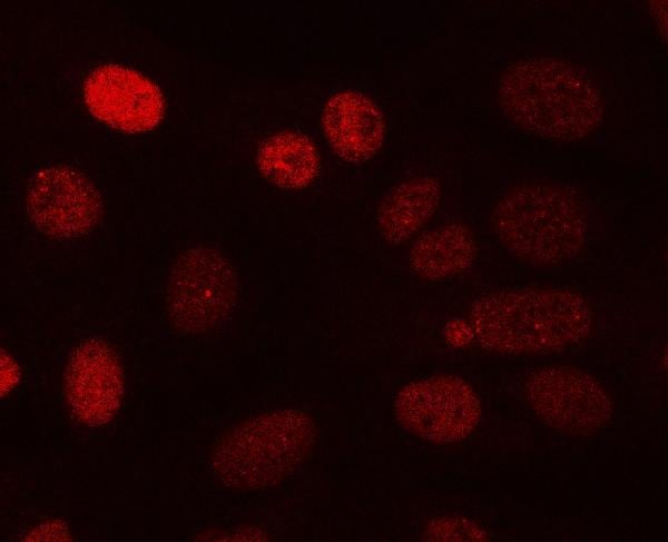
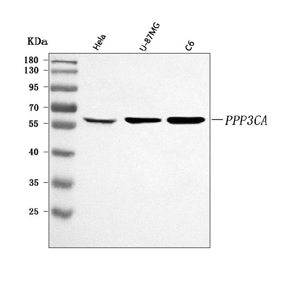
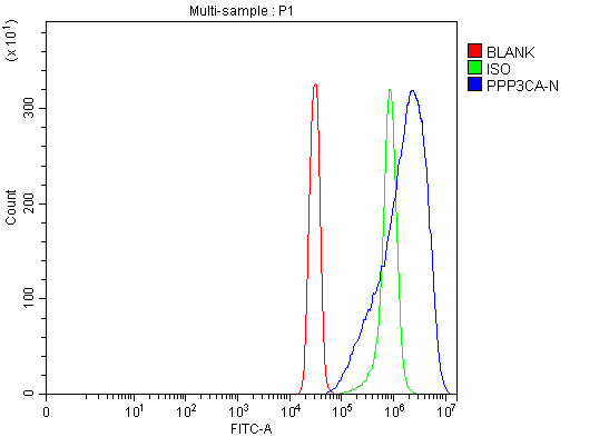
(Figure 3. Flow Cytometry analysis of SiHa cells using anti-PPP3CA antibody (AAA127158).Overlay histogram showing SiHa cells stained with AAA127158 (Blue line). To facilitate intracellular staining, cells were fixed with 4% paraformaldehyde and permeabilized with permeabilization buffer. The cells were blocked with 10% normal goat serum. And then incubated with rabbit anti-PPP3CA Antibody (AAA127158, 1ug/1x106 cells) for 30 min at 20 degree C. DyLight488 conjugated goat anti-rabbit IgG was used as secondary antibody for 30 minutes at 20 degree C. Isotype control antibody (Green line) was rabbit IgG (1ug/1x106) used under the same conditions. Unlabelled sample (Red line) was also used as a control.)
FCM/FACS (Flow Cytometry)
(Figure 3. Flow Cytometry analysis of SiHa cells using anti-PPP3CA antibody (AAA127158).Overlay histogram showing SiHa cells stained with AAA127158 (Blue line). To facilitate intracellular staining, cells were fixed with 4% paraformaldehyde and permeabilized with permeabilization buffer. The cells were blocked with 10% normal goat serum. And then incubated with rabbit anti-PPP3CA Antibody (AAA127158, 1ug/1x106 cells) for 30 min at 20 degree C. DyLight488 conjugated goat anti-rabbit IgG was used as secondary antibody for 30 minutes at 20 degree C. Isotype control antibody (Green line) was rabbit IgG (1ug/1x106) used under the same conditions. Unlabelled sample (Red line) was also used as a control.)
PPP3CA, Polyclonal Antibody (Cat# AAA127158)
Full Name
Anti-PPP3CA Antibody Picoband
Gene Names
PPP3CA; CALN; CCN1; CNA1; CALNA; PPP2B; CALNA1
Reactivity
Human, Rat
Applications
Flow Cytometry, Immunofluorescence, Immunocytochemistry, Western Blot
Purity
Immunogen affinity purified.
Pricing
 IHC (Immunohistochemistry)
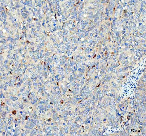
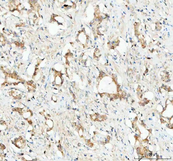
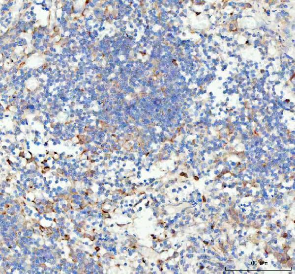
(Figure 4. IHC analysis of GPX1 using anti-GPX1 antibody (AAA127028).GPX1 was detected in a paraffin-embedded section of human spleen tissue. Heat mediated antigen retrieval was performed in EDTA buffer (pH 8.0, epitope retrieval solution). The tissue section was blocked with 10% goat serum. The tissue section was then incubated with 2ug/ml rabbit anti-GPX1 Antibody (AAA127028) overnight at 4 degree C. Peroxidase Conjugated Goat Anti-rabbit IgG was used as secondary antibody and incubated for 30 minutes at 37 degree C. The tissue section was developed using HRP Conjugated Rabbit IgG Super Vision Assay Kit with DAB as the chromogen.)
IHC (Immunohistochemistry)
(Figure 4. IHC analysis of GPX1 using anti-GPX1 antibody (AAA127028).GPX1 was detected in a paraffin-embedded section of human spleen tissue. Heat mediated antigen retrieval was performed in EDTA buffer (pH 8.0, epitope retrieval solution). The tissue section was blocked with 10% goat serum. The tissue section was then incubated with 2ug/ml rabbit anti-GPX1 Antibody (AAA127028) overnight at 4 degree C. Peroxidase Conjugated Goat Anti-rabbit IgG was used as secondary antibody and incubated for 30 minutes at 37 degree C. The tissue section was developed using HRP Conjugated Rabbit IgG Super Vision Assay Kit with DAB as the chromogen.)
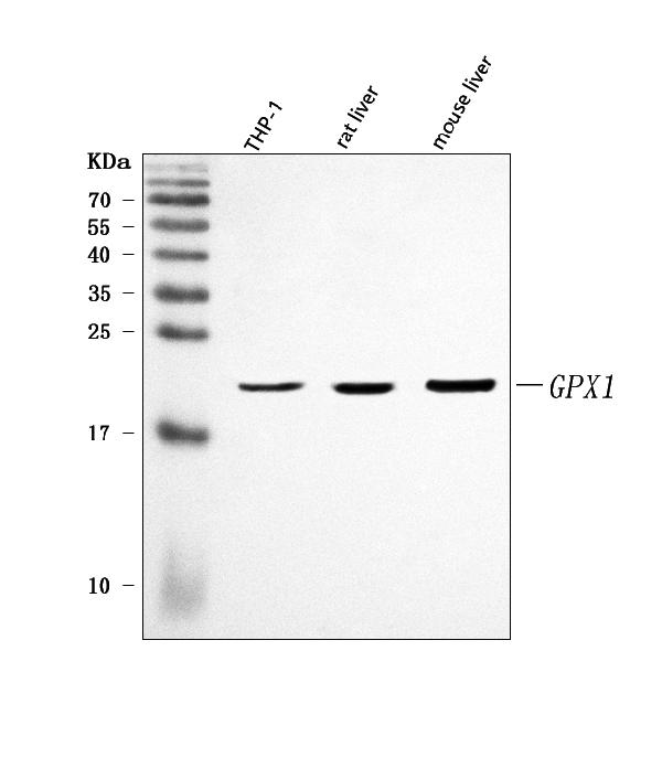
GPX1, Polyclonal Antibody (Cat# AAA127028)
Full Name
Anti-GPX1 Antibody Picoband
Gene Names
GPX1; GPXD; GSHPX1
Reactivity
Human, Mouse, Rat
Applications
Western Blot, Immunohistochemistry
Purity
Immunogen affinity purified.
Pricing
 FCM/FACS (Flow Cytometry)
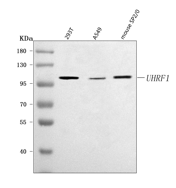
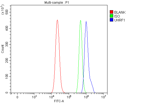
(Figure 3. Flow Cytometry analysis of RT4 cells using anti-UHRF1 antibody (AAA127040).Overlay histogram showing RT4 cells stained with AAA127040 (Blue line). To facilitate intracellular staining, cells were fixed with 4% paraformaldehyde and permeabilized with permeabilization buffer. The cells were blocked with 10% normal goat serum. And then incubated with rabbit anti-UHRF1 Antibody (AAA127040, 1ug/1x106 cells) for 30 min at 20 degree C. DyLight488 conjugated goat anti-rabbit IgG was used as secondary antibody for 30 minutes at 20 degree C. Isotype control antibody (Green line) was rabbit IgG (1ug/1x106) used under the same conditions. Unlabelled sample without incubation with primary antibody and secondary antibody (Red line) was used as a blank control.)
FCM/FACS (Flow Cytometry)
(Figure 3. Flow Cytometry analysis of RT4 cells using anti-UHRF1 antibody (AAA127040).Overlay histogram showing RT4 cells stained with AAA127040 (Blue line). To facilitate intracellular staining, cells were fixed with 4% paraformaldehyde and permeabilized with permeabilization buffer. The cells were blocked with 10% normal goat serum. And then incubated with rabbit anti-UHRF1 Antibody (AAA127040, 1ug/1x106 cells) for 30 min at 20 degree C. DyLight488 conjugated goat anti-rabbit IgG was used as secondary antibody for 30 minutes at 20 degree C. Isotype control antibody (Green line) was rabbit IgG (1ug/1x106) used under the same conditions. Unlabelled sample without incubation with primary antibody and secondary antibody (Red line) was used as a blank control.)
UHRF1, Polyclonal Antibody (Cat# AAA127040)
Full Name
Anti-UHRF1 Antibody Picoband
Gene Names
UHRF1; Np95; hNP95; ICBP90; RNF106; hUHRF1
Reactivity
Human, Mouse
Applications
Flow Cytometry, Immunofluorescence, Immunocytochemistry, Western Blot
Purity
Immunogen affinity purified.
Pricing
 FCM/FACS (Flow Cytometry)
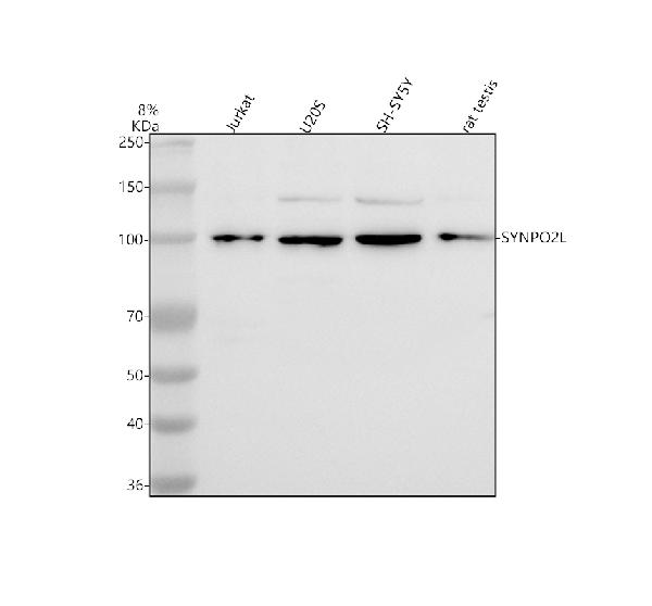
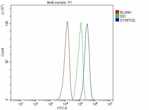
(Figure 2. Flow Cytometry analysis of SH-SY5Y cells using anti-SYNPO2L antibody (AAA127825).Overlay histogram showing SH-SY5Y cells stained with AAA127825 (Blue line). To facilitate intracellular staining, cells were fixed with 4% paraformaldehyde and permeabilized with permeabilization buffer. The cells were blocked with 10% normal goat serum. And then incubated with rabbit anti-SYNPO2L Antibody (AAA127825, 1ug/1x106 cells) for 30 min at 20 degree C. DyLight488 conjugated goat anti-rabbit IgG was used as secondary antibody for 30 minutes at 20 degree C. Isotype control antibody (Green line) was rabbit IgG (1ug/1x106) used under the same conditions. Unlabelled sample without incubation with primary antibody and secondary antibody (Red line) was used as a blank control.)
FCM/FACS (Flow Cytometry)
(Figure 2. Flow Cytometry analysis of SH-SY5Y cells using anti-SYNPO2L antibody (AAA127825).Overlay histogram showing SH-SY5Y cells stained with AAA127825 (Blue line). To facilitate intracellular staining, cells were fixed with 4% paraformaldehyde and permeabilized with permeabilization buffer. The cells were blocked with 10% normal goat serum. And then incubated with rabbit anti-SYNPO2L Antibody (AAA127825, 1ug/1x106 cells) for 30 min at 20 degree C. DyLight488 conjugated goat anti-rabbit IgG was used as secondary antibody for 30 minutes at 20 degree C. Isotype control antibody (Green line) was rabbit IgG (1ug/1x106) used under the same conditions. Unlabelled sample without incubation with primary antibody and secondary antibody (Red line) was used as a blank control.)
SYNPO2L, Polyclonal Antibody (Cat# AAA127825)
Full Name
Anti-SYNPO2L Antibody Picoband
Reactivity
Human, Rat
Applications
Flow Cytometry, Western Blot
Purity
Immunogen affinity purified.
Pricing
 FCM/FACS (Flow Cytometry)
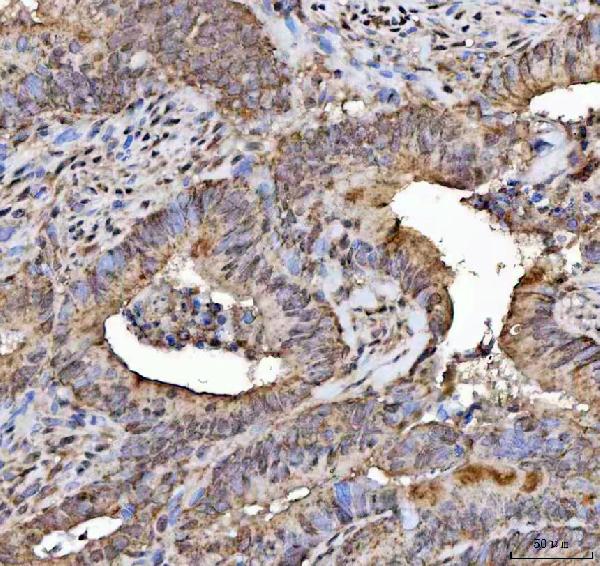
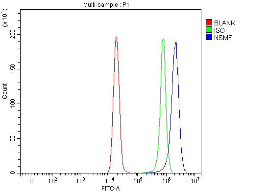
(Figure 3. Flow Cytometry analysis of Hela cells using anti-NELF/NSMF antibody (AAA127521).Overlay histogram showing Hela cells stained with AAA127521 (Blue line). To facilitate intracellular staining, cells were fixed with 4% paraformaldehyde and permeabilized with permeabilization buffer. The cells were blocked with 10% normal goat serum. And then incubated with rabbit anti-NELF/NSMF Antibody (AAA127521, 1ug/1x106 cells) for 30 min at 20 degree C. DyLight488 conjugated goat anti-rabbit IgG was used as secondary antibody for 30 minutes at 20 degree C. Isotype control antibody (Green line) was rabbit IgG (1ug/1x106) used under the same conditions. Unlabelled sample (Red line) was also used as a control.)
FCM/FACS (Flow Cytometry)
(Figure 3. Flow Cytometry analysis of Hela cells using anti-NELF/NSMF antibody (AAA127521).Overlay histogram showing Hela cells stained with AAA127521 (Blue line). To facilitate intracellular staining, cells were fixed with 4% paraformaldehyde and permeabilized with permeabilization buffer. The cells were blocked with 10% normal goat serum. And then incubated with rabbit anti-NELF/NSMF Antibody (AAA127521, 1ug/1x106 cells) for 30 min at 20 degree C. DyLight488 conjugated goat anti-rabbit IgG was used as secondary antibody for 30 minutes at 20 degree C. Isotype control antibody (Green line) was rabbit IgG (1ug/1x106) used under the same conditions. Unlabelled sample (Red line) was also used as a control.)
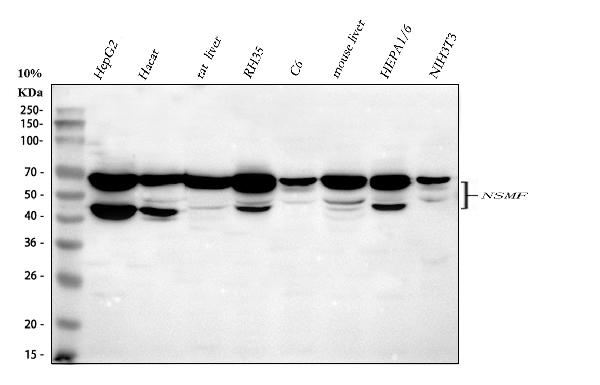
NELF/NSMF, Polyclonal Antibody (Cat# AAA127521)
Full Name
Anti-NELF/NSMF Antibody Picoband
Gene Names
NSMF; HH9; NELF
Reactivity
Human, Mouse, Rat
Applications
Flow Cytometry, Immunohistochemistry, Western Blot
Purity
Immunogen affinity purified.
Pricing
 FCM/FACS (Flow Cytometry)
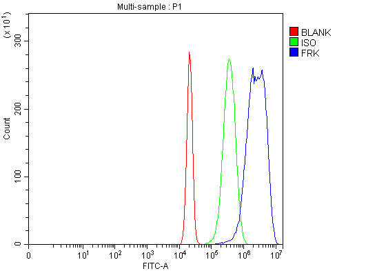
(Figure 2. Flow Cytometry analysis of U2OS cells using anti-FRK antibody (AAA126349).Overlay histogram showing U2OS cells stained with AAA126349 (Blue line). The cells were blocked with 10% normal goat serum. And then incubated with rabbit anti-FRK Antibody (AAA126349, 1 ug/1x10^6 cells) for 30 min at 20 degree C. DyLight488 conjugated goat anti-rabbit IgG was used as secondary antibody for 30 minutes at 20 degree C. Isotype control antibody (Green line) was rabbit IgG (1 ug/1x10^6) used under the same conditions. Unlabelled sample (Red line) was also used as a control.)
FCM/FACS (Flow Cytometry)
(Figure 2. Flow Cytometry analysis of U2OS cells using anti-FRK antibody (AAA126349).Overlay histogram showing U2OS cells stained with AAA126349 (Blue line). The cells were blocked with 10% normal goat serum. And then incubated with rabbit anti-FRK Antibody (AAA126349, 1 ug/1x10^6 cells) for 30 min at 20 degree C. DyLight488 conjugated goat anti-rabbit IgG was used as secondary antibody for 30 minutes at 20 degree C. Isotype control antibody (Green line) was rabbit IgG (1 ug/1x10^6) used under the same conditions. Unlabelled sample (Red line) was also used as a control.)
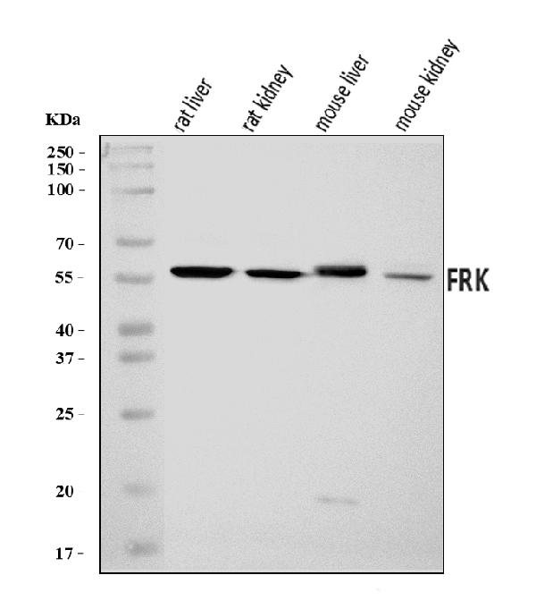
FRK, Polyclonal Antibody (Cat# AAA126349)
Full Name
Anti-FRK Antibody Picoband
Gene Names
FRK; GTK; RAK; PTK5
Reactivity
Human, Mouse, Rat
Applications
Flow Cytometry, Western Blot
Purity
Immunogen affinity purified.
Pricing
 FCM/FACS (Flow Cytometry)
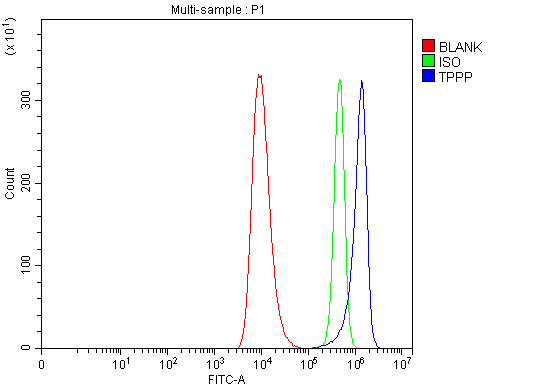
(Figure 2. Flow Cytometry analysis of HEL cells using anti-TPPP antibody (AAA126379).Overlay histogram showing HEL cells stained with AAA126379 (Blue line). The cells were blocked with 10% normal goat serum. And then incubated with rabbit anti-TPPP Antibody (AAA126379, 1 ug/1x10^6 cells) for 30 min at 20 degree C. DyLight488 conjugated goat anti-rabbit IgG was used as secondary antibody for 30 minutes at 20 degree C. Isotype control antibody (Green line) was rabbit IgG (1 ug/1x10^6) used under the same conditions. Unlabelled sample (Red line) was also used as a control.)
FCM/FACS (Flow Cytometry)
(Figure 2. Flow Cytometry analysis of HEL cells using anti-TPPP antibody (AAA126379).Overlay histogram showing HEL cells stained with AAA126379 (Blue line). The cells were blocked with 10% normal goat serum. And then incubated with rabbit anti-TPPP Antibody (AAA126379, 1 ug/1x10^6 cells) for 30 min at 20 degree C. DyLight488 conjugated goat anti-rabbit IgG was used as secondary antibody for 30 minutes at 20 degree C. Isotype control antibody (Green line) was rabbit IgG (1 ug/1x10^6) used under the same conditions. Unlabelled sample (Red line) was also used as a control.)
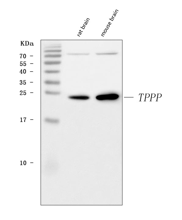
TPPP, Polyclonal Antibody (Cat# AAA126379)
Full Name
Anti-TPPP Antibody Picoband
Gene Names
TPPP; p24; p25; TPPP1; TPPP/p25; p25alpha
Reactivity
Human, Mouse, Rat
Applications
Western Blot, Flow Cytometry
Purity
Immunogen affinity purified.
Pricing
 FCM/FACS (Flow Cytometry)
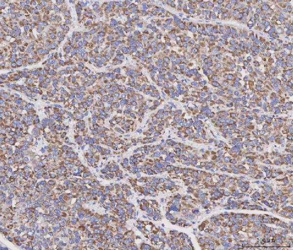
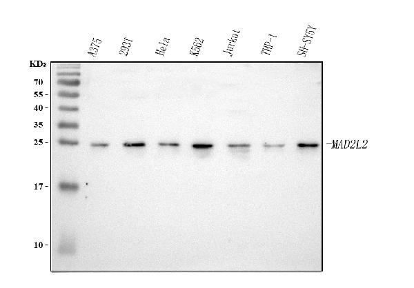
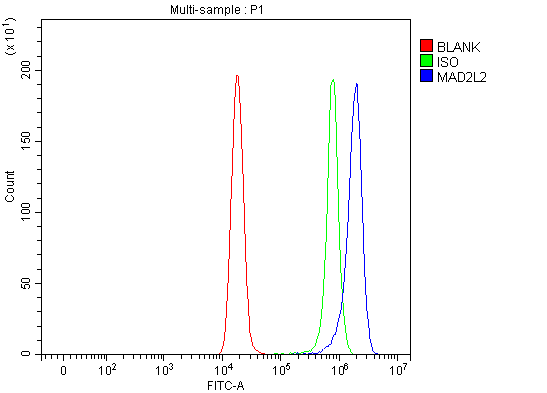
(Figure 4. Flow Cytometry analysis of Hela cells using anti-MAD2L2 antibody (AAA127109).Overlay histogram showing Hela cells stained with AAA127109 (Blue line). To facilitate intracellular staining, cells were fixed with 4% paraformaldehyde and permeabilized with permeabilization buffer. The cells were blocked with 10% normal goat serum. And then incubated with rabbit anti-MAD2L2 Antibody (AAA127109, 1ug/1x106 cells) for 30 min at 20 degree C. DyLight488 conjugated goat anti-rabbit IgG was used as secondary antibody for 30 minutes at 20 degree C. Isotype control antibody (Green line) was rabbit IgG (1ug/1x106) used under the same conditions. Unlabelled sample (Red line) was also used as a control.)
FCM/FACS (Flow Cytometry)
(Figure 4. Flow Cytometry analysis of Hela cells using anti-MAD2L2 antibody (AAA127109).Overlay histogram showing Hela cells stained with AAA127109 (Blue line). To facilitate intracellular staining, cells were fixed with 4% paraformaldehyde and permeabilized with permeabilization buffer. The cells were blocked with 10% normal goat serum. And then incubated with rabbit anti-MAD2L2 Antibody (AAA127109, 1ug/1x106 cells) for 30 min at 20 degree C. DyLight488 conjugated goat anti-rabbit IgG was used as secondary antibody for 30 minutes at 20 degree C. Isotype control antibody (Green line) was rabbit IgG (1ug/1x106) used under the same conditions. Unlabelled sample (Red line) was also used as a control.)
MAD2L2, Polyclonal Antibody (Cat# AAA127109)
Full Name
Anti-MAD2L2 Antibody Picoband
Gene Names
MAD2L2; REV7; MAD2B; POLZ2
Reactivity
Human
Applications
Flow Cytometry, Western Blot, Immunocytochemistry, Immunohistochemistry, Immunofluorescence
Purity
Immunogen affinity purified.
Pricing
 FCM/FACS (Flow Cytometry)
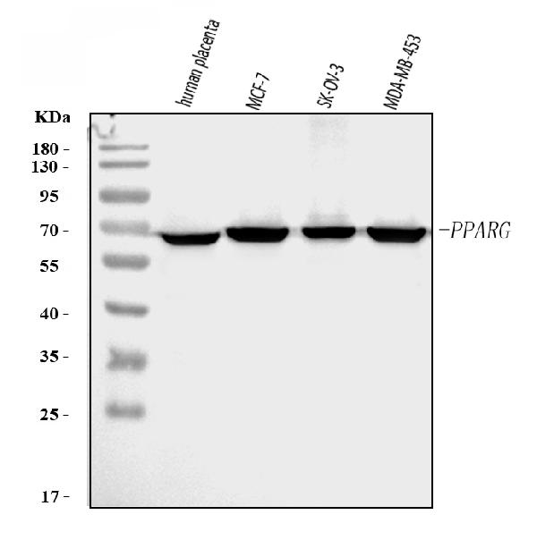
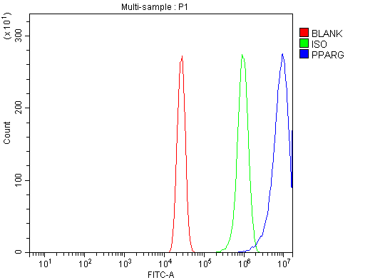
(Figure 2. Flow Cytometry analysis of A431 cells using anti-PPAR Gamma/PPARG antibody (AAA126027).Overlay histogram showing A431 cells stained with AAA126027 (Blue line). The cells were blocked with 10% normal goat serum. And then incubated with rabbit anti-PPAR Gamma/PPARG Antibody (AAA126027, 1 ug/1x10^6 cells) for 30 min at 20 degree C. DyLight488 conjugated goat anti-rabbit IgG was used as secondary antibody for 30 minutes at 20 degree C. Isotype control antibody (Green line) was rabbit IgG (1 ug/1x10^6) used under the same conditions. Unlabelled sample (Red line) was also used as a control.)
FCM/FACS (Flow Cytometry)
(Figure 2. Flow Cytometry analysis of A431 cells using anti-PPAR Gamma/PPARG antibody (AAA126027).Overlay histogram showing A431 cells stained with AAA126027 (Blue line). The cells were blocked with 10% normal goat serum. And then incubated with rabbit anti-PPAR Gamma/PPARG Antibody (AAA126027, 1 ug/1x10^6 cells) for 30 min at 20 degree C. DyLight488 conjugated goat anti-rabbit IgG was used as secondary antibody for 30 minutes at 20 degree C. Isotype control antibody (Green line) was rabbit IgG (1 ug/1x10^6) used under the same conditions. Unlabelled sample (Red line) was also used as a control.)
PPAR gamma/PPARG, Polyclonal Antibody (Cat# AAA126027)
Full Name
Anti-PPAR gamma/PPARG Antibody Picoband
Gene Names
PPARG; GLM1; CIMT1; NR1C3; PPARG1; PPARG2; PPARgamma
Reactivity
Human
Applications
Flow Cytometry, Western Blot
Purity
Immunogen affinity purified.
Pricing
 FCM/FACS (Flow Cytometry)
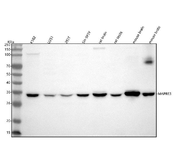
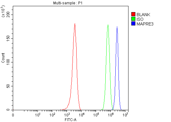
(Figure 2. Flow Cytometry analysis of U251 cells using anti-EB3/MAPRE3 antibody (AAA127328).Overlay histogram showing U251 cells stained with AAA127328 (Blue line). To facilitate intracellular staining, cells were fixed with 4% paraformaldehyde and permeabilized with permeabilization buffer. The cells were blocked with 10% normal goat serum. And then incubated with rabbit anti-EB3/MAPRE3 Antibody (AAA127328, 1ug/1x106 cells) for 30 min at 20 degree C. DyLight488 conjugated goat anti-rabbit IgG was used as secondary antibody for 30 minutes at 20 degree C. Isotype control antibody (Green line) was rabbit IgG (1ug/1x106) used under the same conditions. Unlabelled sample (Red line) was also used as a control.)
FCM/FACS (Flow Cytometry)
(Figure 2. Flow Cytometry analysis of U251 cells using anti-EB3/MAPRE3 antibody (AAA127328).Overlay histogram showing U251 cells stained with AAA127328 (Blue line). To facilitate intracellular staining, cells were fixed with 4% paraformaldehyde and permeabilized with permeabilization buffer. The cells were blocked with 10% normal goat serum. And then incubated with rabbit anti-EB3/MAPRE3 Antibody (AAA127328, 1ug/1x106 cells) for 30 min at 20 degree C. DyLight488 conjugated goat anti-rabbit IgG was used as secondary antibody for 30 minutes at 20 degree C. Isotype control antibody (Green line) was rabbit IgG (1ug/1x106) used under the same conditions. Unlabelled sample (Red line) was also used as a control.)
EB3/MAPRE3, Polyclonal Antibody (Cat# AAA127328)
Full Name
Anti-EB3/MAPRE3 Antibody Picoband
Gene Names
MAPRE3; EB3; RP3; EBF3; EBF3-S
Reactivity
Human, Mouse, Rat
Applications
Flow Cytometry, Western Blot
Purity
Immunogen affinity purified.
Pricing
 FCM/FACS (Flow Cytometry)
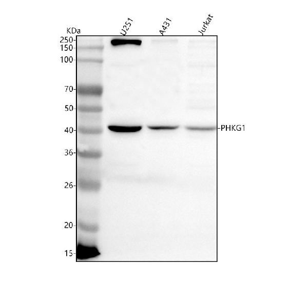
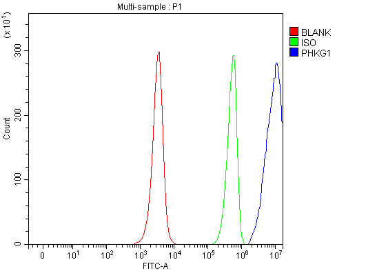
(Figure 2. Flow Cytometry analysis of JK cells using anti-PHKG1 antibody (AAA127749).Overlay histogram showing JK cells stained with AAA127749 (Blue line). To facilitate intracellular staining, cells were fixed with 4% paraformaldehyde and permeabilized with permeabilization buffer. The cells were blocked with 10% normal goat serum. And then incubated with rabbit anti-PHKG1 Antibody (AAA127749, 1ug/1x106 cells) for 30 min at 20 degree C. DyLight488 conjugated goat anti-rabbit IgG was used as secondary antibody for 30 minutes at 20 degree C. Isotype control antibody (Green line) was rabbit IgG (1ug/1x106) used under the same conditions. Unlabelled sample (Red line) was also used as a control.)
FCM/FACS (Flow Cytometry)
(Figure 2. Flow Cytometry analysis of JK cells using anti-PHKG1 antibody (AAA127749).Overlay histogram showing JK cells stained with AAA127749 (Blue line). To facilitate intracellular staining, cells were fixed with 4% paraformaldehyde and permeabilized with permeabilization buffer. The cells were blocked with 10% normal goat serum. And then incubated with rabbit anti-PHKG1 Antibody (AAA127749, 1ug/1x106 cells) for 30 min at 20 degree C. DyLight488 conjugated goat anti-rabbit IgG was used as secondary antibody for 30 minutes at 20 degree C. Isotype control antibody (Green line) was rabbit IgG (1ug/1x106) used under the same conditions. Unlabelled sample (Red line) was also used as a control.)
PHKG1, Polyclonal Antibody (Cat# AAA127749)
Full Name
Anti-PHKG1 Antibody Picoband
Gene Names
PHKG1; PHKG
Reactivity
Human
Applications
Flow Cytometry, Western Blot
Purity
Immunogen affinity purified.
Pricing
 FCM/FACS (Flow Cytometry)
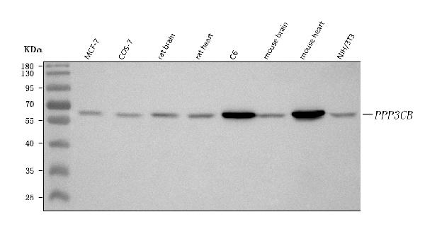
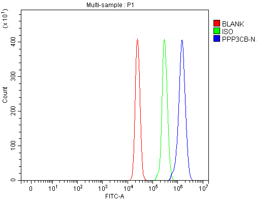
(Figure 5. Flow Cytometry analysis of U937 cells using anti-PPP3CB antibody (AAA127434).Overlay histogram showing U937 cells stained with AAA127434 (Blue line). To facilitate intracellular staining, cells were fixed with 4% paraformaldehyde and permeabilized with permeabilization buffer. The cells were blocked with 10% normal goat serum. And then incubated with rabbit anti-PPP3CB Antibody (AAA127434, 1ug/1x106 cells) for 30 min at 20 degree C. DyLight488 conjugated goat anti-rabbit IgG was used as secondary antibody for 30 minutes at 20 degree C. Isotype control antibody (Green line) was rabbit IgG (1ug/1x106) used under the same conditions. Unlabelled sample (Red line) was also used as a control.)
FCM/FACS (Flow Cytometry)
(Figure 5. Flow Cytometry analysis of U937 cells using anti-PPP3CB antibody (AAA127434).Overlay histogram showing U937 cells stained with AAA127434 (Blue line). To facilitate intracellular staining, cells were fixed with 4% paraformaldehyde and permeabilized with permeabilization buffer. The cells were blocked with 10% normal goat serum. And then incubated with rabbit anti-PPP3CB Antibody (AAA127434, 1ug/1x106 cells) for 30 min at 20 degree C. DyLight488 conjugated goat anti-rabbit IgG was used as secondary antibody for 30 minutes at 20 degree C. Isotype control antibody (Green line) was rabbit IgG (1ug/1x106) used under the same conditions. Unlabelled sample (Red line) was also used as a control.)
PPP3CB, Polyclonal Antibody (Cat# AAA127434)
Full Name
Anti-PPP3CB Antibody Picoband
Gene Names
PPP3CB; CNA2; CALNB; CALNA2; PP2Bbeta
Reactivity
Human, Monkey, Mouse, Rat
Applications
Flow Cytometry, Immunohistochemistry, Western Blot
Purity
Immunogen affinity purified.
Pricing
 FCM/FACS (Flow Cytometry)
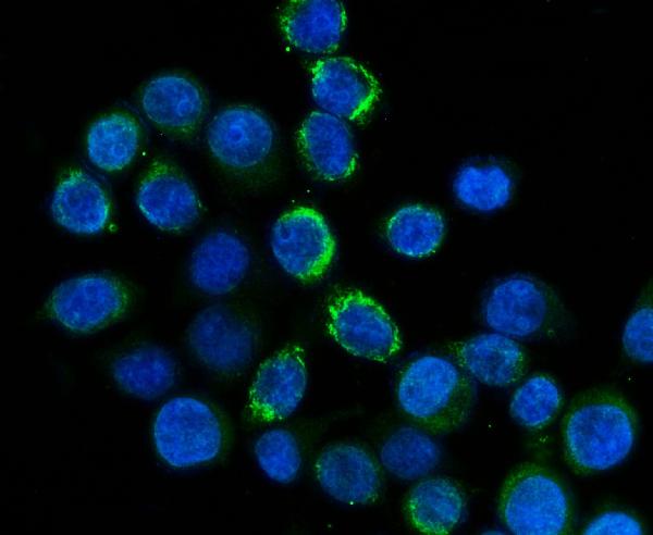
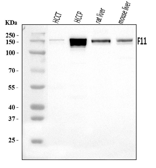
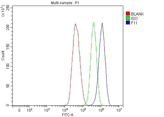
(Figure 3. Flow Cytometry analysis of HepG2 cells using anti-FXI/F11 antibody (AAA126099).Overlay histogram showing HepG2 cells stained with AAA126099 (Blue line). The cells were blocked with 10% normal goat serum. And then incubated with rabbit anti-FXI/F11 Antibody (AAA126099, 1 ug/1x10^6 cells) for 30 min at 20 degree C. DyLight488 conjugated goat anti-rabbit IgG was used as secondary antibody for 30 minutes at 20 degree C. Isotype control antibody (Green line) was rabbit IgG (1 ug/1x10^6) used under the same conditions. Unlabelled sample (Red line) was also used as a control.)
FCM/FACS (Flow Cytometry)
(Figure 3. Flow Cytometry analysis of HepG2 cells using anti-FXI/F11 antibody (AAA126099).Overlay histogram showing HepG2 cells stained with AAA126099 (Blue line). The cells were blocked with 10% normal goat serum. And then incubated with rabbit anti-FXI/F11 Antibody (AAA126099, 1 ug/1x10^6 cells) for 30 min at 20 degree C. DyLight488 conjugated goat anti-rabbit IgG was used as secondary antibody for 30 minutes at 20 degree C. Isotype control antibody (Green line) was rabbit IgG (1 ug/1x10^6) used under the same conditions. Unlabelled sample (Red line) was also used as a control.)
FXI/F11, Polyclonal Antibody (Cat# AAA126099)
Full Name
Anti-FXI/F11 Antibody Picoband
Gene Names
F11; FXI
Reactivity
Human, Mouse, Rat
Applications
Flow Cytometry, Immunofluorescence, Immunocytochemistry, Western Blot
Purity
Immunogen affinity purified.
Pricing
 FCM/FACS (Flow Cytometry)
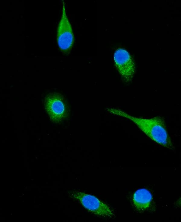
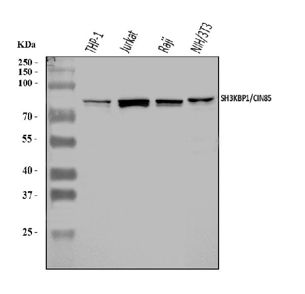
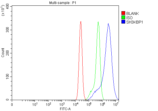
(Figure 3. Flow Cytometry analysis of CACO-2 cells using anti-SH3KBP1 antibody (AAA126261).Overlay histogram showing CACO-2 cells stained with AAA126261 (Blue line). The cells were blocked with 10% normal goat serum. And then incubated with rabbit anti-SH3KBP1 Antibody (AAA126261, 1 ug/1x10^6 cells) for 30 min at 20 degree C. DyLight488 conjugated goat anti-rabbit IgG was used as secondary antibody for 30 minutes at 20 degree C. Isotype control antibody (Green line) was rabbit IgG (1 ug/1x10^6) used under the same conditions. Unlabelled sample (Red line) was also used as a control.)
FCM/FACS (Flow Cytometry)
(Figure 3. Flow Cytometry analysis of CACO-2 cells using anti-SH3KBP1 antibody (AAA126261).Overlay histogram showing CACO-2 cells stained with AAA126261 (Blue line). The cells were blocked with 10% normal goat serum. And then incubated with rabbit anti-SH3KBP1 Antibody (AAA126261, 1 ug/1x10^6 cells) for 30 min at 20 degree C. DyLight488 conjugated goat anti-rabbit IgG was used as secondary antibody for 30 minutes at 20 degree C. Isotype control antibody (Green line) was rabbit IgG (1 ug/1x10^6) used under the same conditions. Unlabelled sample (Red line) was also used as a control.)
SH3KBP1, Polyclonal Antibody (Cat# AAA126261)
Full Name
Anti-SH3KBP1 Antibody Picoband
Gene Names
SH3KBP1; HSB1; CIN85; GIG10; HSB-1; MIG18; CD2BP3
Reactivity
Human, Mouse
Applications
Flow Cytometry, Immunofluorescence, Immunocytochemistry, Western Blot
Purity
Immunogen affinity purified.
Pricing