Filters
Clonality
Type
Reactivity
Gene Name
Isotype
Host
Application
Clone
1614 results for " Proteins" - showing 1300-1350


 WB (Western Blot)
(Western Blot Analysis: Representative lot data. Lysate from HEK-293 cells was resolved by electrophoresis, transferred to PVDF and probed with anti-PI3 Kinase, p110β (0.05 ug/mL). Proteins were visualized using donkey anti-rabbit secondary antibody conjugated to HRP and chemiluminescence detection. Arrow indicates PI3 Kinase, p110β (~110kD).)
WB (Western Blot)
(Western Blot Analysis: Representative lot data. Lysate from HEK-293 cells was resolved by electrophoresis, transferred to PVDF and probed with anti-PI3 Kinase, p110β (0.05 ug/mL). Proteins were visualized using donkey anti-rabbit secondary antibody conjugated to HRP and chemiluminescence detection. Arrow indicates PI3 Kinase, p110β (~110kD).)
Phosphoinositide 3 Kinase, p110 beta, Polyclonal Antibody (Cat# AAA14720)
Full Name
Phosphoinositide 3 Kinase, p110 beta (DKFZp779K1237, MGC133043, p110beta, Phosphatidylinositol 3 Kinase Catalytic beta Polypeptide, Phosphatidylinositol-4,5-bisphosphate 3-kinase Catalytic Subunit beta Isoform, Phosphoinositide 3 Kinase Catalytic beta Pol
Gene Names
PIK3CB; PI3K; PIK3C1; P110BETA; PI3KBETA; MGC133043; DKFZp779K1237
Reactivity
Human
Applications
EL/EIA, WB, IP, IHC, ICC
Purity
Affinity Purified
Purified by immunoaffinity chromatography.
Purified by immunoaffinity chromatography.
Pricing


 Application Data
(Analysis of Protein Array containing more than 19,000 full-length human proteins using Calretinin Mouse Monoclonal Antibody (CALB2/2786). Z- and S- Score: The Z-score represents the strength of a signal that a monoclonal antibody (MAb) (in combination with a fluorescently-tagged anti-IgG secondary antibody) produces when binding to a particular protein on the HuProtTM array. Z-scores are described in units of standard deviations (SD's) above the mean value of all signals generated on that array. If targets on HuProtTM are arranged in descending order of the Z-score, the S-score is the difference (also in units of SD's) between the Z-score. S-score therefore represents the relative target specificity of a MAb to its intended target. A MAb is considered to specific to its intended target, if the MAb has an S-score of at least 2.5. For example, if a MAb binds to protein X with a Z-score of 43 and to protein Y with a Z-score of 14, then the S-score for the binding of that MAb to protein X is equal to 29.)
Application Data
(Analysis of Protein Array containing more than 19,000 full-length human proteins using Calretinin Mouse Monoclonal Antibody (CALB2/2786). Z- and S- Score: The Z-score represents the strength of a signal that a monoclonal antibody (MAb) (in combination with a fluorescently-tagged anti-IgG secondary antibody) produces when binding to a particular protein on the HuProtTM array. Z-scores are described in units of standard deviations (SD's) above the mean value of all signals generated on that array. If targets on HuProtTM are arranged in descending order of the Z-score, the S-score is the difference (also in units of SD's) between the Z-score. S-score therefore represents the relative target specificity of a MAb to its intended target. A MAb is considered to specific to its intended target, if the MAb has an S-score of at least 2.5. For example, if a MAb binds to protein X with a Z-score of 43 and to protein Y with a Z-score of 14, then the S-score for the binding of that MAb to protein X is equal to 29.)
Calretinin/Calbindin 2, Monoclonal Antibody (Cat# AAA23939)
Full Name
Calretinin/Calbindin 2 (Mesothelioma Marker)
Gene Names
CALB2; CR; CAL2; CAB29
Reactivity
Human
Applications
WB, IHC
Purity
Purified Ab with BSA and Azide at 200ug/ml OR Purified Ab WITHOUT BSA at 1.0mg/ml
Pricing


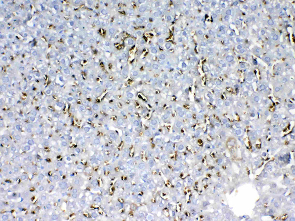
 IHC (Immunohistochemistry)
(Figure 7. IHC analysis of PARK7 / DJ1 using anti-PARK7 / DJ1 antibody (AAA19139).PARK7 / DJ1 was detected in paraffin-embedded section of rat testis tissue. Heat mediated antigen retrieval was performed in citrate buffer (pH6, epitope retrieval solution) for 20 mins. The tissue section was blocked with 10% goat serum. The tissue section was then incubated with 1ug/ml rabbit anti-PARK7 / DJ1 Antibody (AAA19139) overnight at 4 degree C. Biotinylated goat anti-rabbit IgG was used as secondary antibody and incubated for 30 minutes at 37 degree C. The tissue section was developed using Strepavidin-Biotin-Complex (SABC) with DAB as the chromogen.)
IHC (Immunohistochemistry)
(Figure 7. IHC analysis of PARK7 / DJ1 using anti-PARK7 / DJ1 antibody (AAA19139).PARK7 / DJ1 was detected in paraffin-embedded section of rat testis tissue. Heat mediated antigen retrieval was performed in citrate buffer (pH6, epitope retrieval solution) for 20 mins. The tissue section was blocked with 10% goat serum. The tissue section was then incubated with 1ug/ml rabbit anti-PARK7 / DJ1 Antibody (AAA19139) overnight at 4 degree C. Biotinylated goat anti-rabbit IgG was used as secondary antibody and incubated for 30 minutes at 37 degree C. The tissue section was developed using Strepavidin-Biotin-Complex (SABC) with DAB as the chromogen.)
PARK7/DJ1, Polyclonal Antibody (Cat# AAA19139)
Full Name
Anti-PARK7/DJ1 Picoband Antibody
Gene Names
Park7; Dj1; CAP1; DJ-1; SP22
Reactivity
Mouse, Rat
No cross reactivity with other proteins.
No cross reactivity with other proteins.
Applications
EIA, IHC, WB
Purity
Immunogen affinity purified
Pricing


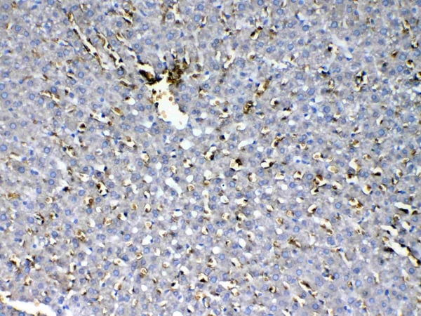 IHC (Immunohistochemistry)
(Figure 8. IHC analysis of HBD using anti-HBD antibody (AAA19142).HBD was detected in paraffin-embedded section of rat liver tissue. Heat mediated antigen retrieval was performed in citrate buffer (pH6, epitope retrieval solution) for 20 mins. The tissue section was blocked with 10% goat serum. The tissue section was then incubated with 1ug/ml rabbit anti-HBD Antibody (AAA19142) overnight at 4 degree C. Biotinylated goat anti-rabbit IgG was used as secondary antibody and incubated for 30 minutes at 37 degree C. The tissue section was developed using Strepavidin-Biotin-Complex (SABC) with DAB as the chromogen.)
IHC (Immunohistochemistry)
(Figure 8. IHC analysis of HBD using anti-HBD antibody (AAA19142).HBD was detected in paraffin-embedded section of rat liver tissue. Heat mediated antigen retrieval was performed in citrate buffer (pH6, epitope retrieval solution) for 20 mins. The tissue section was blocked with 10% goat serum. The tissue section was then incubated with 1ug/ml rabbit anti-HBD Antibody (AAA19142) overnight at 4 degree C. Biotinylated goat anti-rabbit IgG was used as secondary antibody and incubated for 30 minutes at 37 degree C. The tissue section was developed using Strepavidin-Biotin-Complex (SABC) with DAB as the chromogen.)
HBD, Polyclonal Antibody (Cat# AAA19142)
Full Name
Anti-HBD Picoband Antibody
Reactivity
Human, Mouse, Rat
No cross reactivity with other proteins.
No cross reactivity with other proteins.
Applications
EIA, IHC, WB
Purity
Immunogen affinity purified
Pricing


 IHC (Immunohistochemistry)
(Figure 7. IHC analysis of CD105 using anti-CD105 antibody (AAA19165).CD105 was detected in paraffin-embedded section of rat spleen tissue. Heat mediated antigen retrieval was performed in citrate buffer (pH6, epitope retrieval solution) for 20 mins. The tissue section was blocked with 10% goat serum. The tissue section was then incubated with 1ug/ml rabbit anti-CD105 Antibody (AAA19165) overnight at 4 degree C. Biotinylated goat anti-rabbit IgG was used as secondary antibody and incubated for 30 minutes at 37 degree C. The tissue section was developed using Strepavidin-Biotin-Complex (SABC) with DAB as the chromogen.)
IHC (Immunohistochemistry)
(Figure 7. IHC analysis of CD105 using anti-CD105 antibody (AAA19165).CD105 was detected in paraffin-embedded section of rat spleen tissue. Heat mediated antigen retrieval was performed in citrate buffer (pH6, epitope retrieval solution) for 20 mins. The tissue section was blocked with 10% goat serum. The tissue section was then incubated with 1ug/ml rabbit anti-CD105 Antibody (AAA19165) overnight at 4 degree C. Biotinylated goat anti-rabbit IgG was used as secondary antibody and incubated for 30 minutes at 37 degree C. The tissue section was developed using Strepavidin-Biotin-Complex (SABC) with DAB as the chromogen.)
CD105, Polyclonal Antibody (Cat# AAA19165)
Full Name
Anti-CD105 Picoband Antibody
Gene Names
Eng; Endo; CD105; AI528660; AI662476; S-endoglin
Reactivity
Mouse, Rat
No cross reactivity with other proteins.
No cross reactivity with other proteins.
Applications
EIA, IHC, WB
Purity
Immunogen affinity purified
Pricing


 IHC (Immunohistchemistry)
(Figure 6. IHC analysis of S100A10 using anti-S100A10 antibody (AAA19162).S100A10 was detected in paraffin-embedded section of rat spleen tissue. Heat mediated antigen retrieval was performed in citrate buffer (pH6, epitope retrieval solution) for 20 mins. The tissue section was blocked with 10% goat serum. The tissue section was then incubated with 1ug/ml rabbit anti-S100A10 Antibody (AAA19162) overnight at 4 degree C. Biotinylated goat anti-rabbit IgG was used as secondary antibody and incubated for 30 minutes at 37 degree C. The tissue section was developed using Strepavidin-Biotin-Complex (SABC) with DAB as the chromogen.)
IHC (Immunohistchemistry)
(Figure 6. IHC analysis of S100A10 using anti-S100A10 antibody (AAA19162).S100A10 was detected in paraffin-embedded section of rat spleen tissue. Heat mediated antigen retrieval was performed in citrate buffer (pH6, epitope retrieval solution) for 20 mins. The tissue section was blocked with 10% goat serum. The tissue section was then incubated with 1ug/ml rabbit anti-S100A10 Antibody (AAA19162) overnight at 4 degree C. Biotinylated goat anti-rabbit IgG was used as secondary antibody and incubated for 30 minutes at 37 degree C. The tissue section was developed using Strepavidin-Biotin-Complex (SABC) with DAB as the chromogen.)
S100A10, Polyclonal Antibody (Cat# AAA19162)
Full Name
Anti-S100A10 Picoband antibody
Gene Names
S100A10; 42C; P11; p10; GP11; ANX2L; CAL1L; CLP11; Ca[1]; ANX2LG
Reactivity
Human, Mouse, Rat
No cross reactivity with other proteins.
No cross reactivity with other proteins.
Applications
EIA, IHC, WB
Pricing


 IHC (Immunohistchemistry)
(Figure 6. IHC analysis of AMD1 using anti-AMD1 antibody (AAA19177).AMD1 was detected in paraffin-embedded section of human mammary cancer tissue. Heat mediated antigen retrieval was performed in citrate buffer (pH6, epitope retrieval solution) for 20 mins. The tissue section was blocked with 10% goat serum. The tissue section was then incubated with 1ug/ml rabbit anti-AMD1 Antibody (AAA19177) overnight at 4 degree C. Biotinylated goat anti-rabbit IgG was used as secondary antibody and incubated for 30 minutes at 37 degree C. The tissue section was developed using Strepavidin-Biotin-Complex (SABC) with DAB as the chromogen.)
IHC (Immunohistchemistry)
(Figure 6. IHC analysis of AMD1 using anti-AMD1 antibody (AAA19177).AMD1 was detected in paraffin-embedded section of human mammary cancer tissue. Heat mediated antigen retrieval was performed in citrate buffer (pH6, epitope retrieval solution) for 20 mins. The tissue section was blocked with 10% goat serum. The tissue section was then incubated with 1ug/ml rabbit anti-AMD1 Antibody (AAA19177) overnight at 4 degree C. Biotinylated goat anti-rabbit IgG was used as secondary antibody and incubated for 30 minutes at 37 degree C. The tissue section was developed using Strepavidin-Biotin-Complex (SABC) with DAB as the chromogen.)
AMD1/Adometdc, Polyclonal Antibody (Cat# AAA19177)
Full Name
Anti-AMD1/Adometdc Antibody
Gene Names
AMD1; AMD; SAMDC; ADOMETDC
Reactivity
Human, Mouse, Rat
No cross reactivity with other proteins.
No cross reactivity with other proteins.
Applications
IHC, WB
Purity
Immunogen affinity purified
Pricing


 IHC (Immunohistochemistry)
(Figure 8. IHC analysis of MED18 using anti-MED18 antibody (AAA19179).MED18 was detected in paraffin-embedded section of human mammary cancer tissue. Heat mediated antigen retrieval was performed in citrate buffer (pH6, epitope retrieval solution) for 20 mins. The tissue section was blocked with 10% goat serum. The tissue section was then incubated with 1ug/ml rabbit anti-MED18 Antibody (AAA19179) overnight at 4 degree C. Biotinylated goat anti-rabbit IgG was used as secondary antibody and incubated for 30 minutes at 37 degree C. The tissue section was developed using Strepavidin-Biotin-Complex (SABC) with DAB as the chromogen.)
IHC (Immunohistochemistry)
(Figure 8. IHC analysis of MED18 using anti-MED18 antibody (AAA19179).MED18 was detected in paraffin-embedded section of human mammary cancer tissue. Heat mediated antigen retrieval was performed in citrate buffer (pH6, epitope retrieval solution) for 20 mins. The tissue section was blocked with 10% goat serum. The tissue section was then incubated with 1ug/ml rabbit anti-MED18 Antibody (AAA19179) overnight at 4 degree C. Biotinylated goat anti-rabbit IgG was used as secondary antibody and incubated for 30 minutes at 37 degree C. The tissue section was developed using Strepavidin-Biotin-Complex (SABC) with DAB as the chromogen.)
MED18, Polyclonal Antibody (Cat# AAA19179)
Full Name
Anti-MED18 Picoband Antibody
Gene Names
MED18; SRB5; p28b
Reactivity
Human, Mouse, Rat
No cross reactivity with other proteins.
No cross reactivity with other proteins.
Applications
EIA, IHC, WB
Purity
Immunogen affinity purified
Pricing


 IHC (Immunohistchemistry)
(Figure 6. IHC analysis of MED9 using anti-MED9 antibody (AAA19180).MED9 was detected in paraffin-embedded section of human mammary cancer tissue. Heat mediated antigen retrieval was performed in citrate buffer (pH6, epitope retrieval solution) for 20 mins. The tissue section was blocked with 10% goat serum. The tissue section was then incubated with 1ug/ml rabbit anti-MED9 Antibody (AAA19180) overnight at 4 degree C. Biotinylated goat anti-rabbit IgG was used as secondary antibody and incubated for 30 minutes at 37 degree C. The tissue section was developed using Strepavidin-Biotin-Complex (SABC) with DAB as the chromogen.)
IHC (Immunohistchemistry)
(Figure 6. IHC analysis of MED9 using anti-MED9 antibody (AAA19180).MED9 was detected in paraffin-embedded section of human mammary cancer tissue. Heat mediated antigen retrieval was performed in citrate buffer (pH6, epitope retrieval solution) for 20 mins. The tissue section was blocked with 10% goat serum. The tissue section was then incubated with 1ug/ml rabbit anti-MED9 Antibody (AAA19180) overnight at 4 degree C. Biotinylated goat anti-rabbit IgG was used as secondary antibody and incubated for 30 minutes at 37 degree C. The tissue section was developed using Strepavidin-Biotin-Complex (SABC) with DAB as the chromogen.)
MED9, Polyclonal Antibody (Cat# AAA19180)
Full Name
Anti-MED9 Picoband Antibody
Gene Names
MED9; MED25
Reactivity
Human, Mouse, Rat
No cross reactivity with other proteins.
No cross reactivity with other proteins.
Applications
EIA, IHC, WB
Purity
Immunogen affinity purified
Pricing


 ICC (Immunocytochemistry)
(Figure 10 Immunocytochemistry Validation of PD-L1Immunocytochemical analysis of 4% paraformaldehyde-fixed PD-L1 transfected 293 cells labeling PD-L1 with at 1 μg/ml, followed by Goat anti-mouse IgG secondary antibody at 1/250 dilution (red). Lower left: Use mouse IgG antibody at 1 μg/ml as control.)
ICC (Immunocytochemistry)
(Figure 10 Immunocytochemistry Validation of PD-L1Immunocytochemical analysis of 4% paraformaldehyde-fixed PD-L1 transfected 293 cells labeling PD-L1 with at 1 μg/ml, followed by Goat anti-mouse IgG secondary antibody at 1/250 dilution (red). Lower left: Use mouse IgG antibody at 1 μg/ml as control.)
PDL1, Monoclonal Antibody (Cat# AAA10992)
Full Name
PDL1 Antibody [6H10]
Gene Names
CD274; B7-H; B7H1; PDL1; PD-L1; PDCD1L1; PDCD1LG1
Reactivity
Human
Applications
EIA, WB, IHC, ICC, IF
Purity
Protein A purified
Pricing


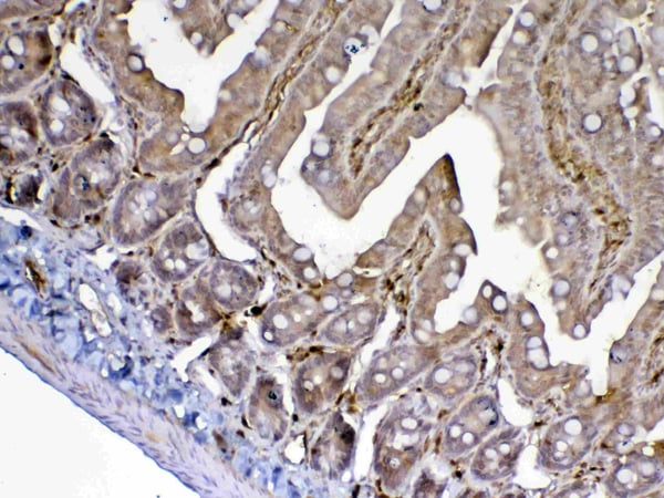
 IHC (Immunohistchemistry)
(Figure 6. IHC analysis of CD13/ANPEP using anti-CD13/ANPEP antibody (AAA19371).CD13/ANPEP was detected in paraffin-embedded section of rat kidney tissue. Heat mediated antigen retrieval was performed in EDTA buffer (pH8. 0, epitope retrieval solution). The tissue section was blocked with 10% goat serum. The tissue section was then incubated with 2μg/ml mouse anti-CD13/ANPEP Antibody (AAA19371) overnight at 4 degree C. Biotinylated goat anti-mouse IgG was used as secondary antibody and incubated for 30 minutes at 37 degree C. The tissue section was developed using Strepavidin-Biotin-Complex (SABC) (Catalog # with DAB as the chromogen.)
IHC (Immunohistchemistry)
(Figure 6. IHC analysis of CD13/ANPEP using anti-CD13/ANPEP antibody (AAA19371).CD13/ANPEP was detected in paraffin-embedded section of rat kidney tissue. Heat mediated antigen retrieval was performed in EDTA buffer (pH8. 0, epitope retrieval solution). The tissue section was blocked with 10% goat serum. The tissue section was then incubated with 2μg/ml mouse anti-CD13/ANPEP Antibody (AAA19371) overnight at 4 degree C. Biotinylated goat anti-mouse IgG was used as secondary antibody and incubated for 30 minutes at 37 degree C. The tissue section was developed using Strepavidin-Biotin-Complex (SABC) (Catalog # with DAB as the chromogen.)
CD13/ANPEP, Monoclonal Antibody (Cat# AAA19371)
Full Name
Anti-CD13/ANPEP Antibody (monoclonal, 5B9)
Gene Names
ANPEP; APN; CD13; LAP1; P150; PEPN; GP150
Reactivity
Human, Rat, Monkey
Applications
WB, IHC-P
Purity
Immunogen affinity purified.
Pricing


 IP (Immunoprecipitation)
(Figure 11 Immunoprecipitation Validation in HEK293 cells (Kawai et al., 2004)HEK293 cells were transiently transfected with DYKDDDDK-IRF7. Ccell lysates were immunoprecipitated with control rabbit anti-mouse immunoglobulin serum (IgG) or anti-MyD88 (Ab1 and Ab2), followed by immunoblotting with anti-DYKDDDDK.)
IP (Immunoprecipitation)
(Figure 11 Immunoprecipitation Validation in HEK293 cells (Kawai et al., 2004)HEK293 cells were transiently transfected with DYKDDDDK-IRF7. Ccell lysates were immunoprecipitated with control rabbit anti-mouse immunoglobulin serum (IgG) or anti-MyD88 (Ab1 and Ab2), followed by immunoblotting with anti-DYKDDDDK.)
MYD88, Polyclonal Antibody (Cat# AAA10944)
Full Name
MYD88 Antibody
Gene Names
MYD88; MYD88D
Reactivity
Human, Mouse, Rat
Applications
EIA, WB
Purity
MYD88 Antibody is affinity chromatography purified via peptide column.
Pricing


 IHC (Immunohistchemistry)
(Figure 6. IHC analysis of Cytoglobin using anti- Cytoglobin antibody (AAA11681).Cytoglobin was detected in paraffin-embedded section of human lung cancer tissues. Heat mediated antigen retrieval was performed in citrate buffer (pH6, epitope retrieval solution) for 20 mins. The tissue section was blocked with 10% goat serum. The tissue section was then incubated with 1ug/ml rabbit anti- Cytoglobin Antibody (AAA11681) overnight at 4 degree C. Biotinylated goat anti-rabbit IgG was used as secondary antibody and incubated for 30 minutes at 37 degree C. The tissue section was developed using Strepavidin-Biotin-Complex (SABC) with DAB as the chromogen.)
IHC (Immunohistchemistry)
(Figure 6. IHC analysis of Cytoglobin using anti- Cytoglobin antibody (AAA11681).Cytoglobin was detected in paraffin-embedded section of human lung cancer tissues. Heat mediated antigen retrieval was performed in citrate buffer (pH6, epitope retrieval solution) for 20 mins. The tissue section was blocked with 10% goat serum. The tissue section was then incubated with 1ug/ml rabbit anti- Cytoglobin Antibody (AAA11681) overnight at 4 degree C. Biotinylated goat anti-rabbit IgG was used as secondary antibody and incubated for 30 minutes at 37 degree C. The tissue section was developed using Strepavidin-Biotin-Complex (SABC) with DAB as the chromogen.)
Cytoglobin, Polyclonal Antibody (Cat# AAA11681)
Full Name
Anti-Cytoglobin Antibody
Gene Names
CYGB; HGB; STAP
Reactivity
Human, Mouse, Rat
Applications
WB, IHC
Purity
Immunogen affinity purified.
Pricing


 Application Data
(Formalin fixed, paraffin embedded human breast cancer biopsy stained with Mouse anti Human estrogen receptor beta5 antibody followed by HRP polymer detection and DAB substrate development following heat mediated antigen retrieval using citrate buffer at pH6.2 (low power))
Application Data
(Formalin fixed, paraffin embedded human breast cancer biopsy stained with Mouse anti Human estrogen receptor beta5 antibody followed by HRP polymer detection and DAB substrate development following heat mediated antigen retrieval using citrate buffer at pH6.2 (low power))
ESTROGEN RECEPTOR BETA 5, Monoclonal Antibody (Cat# AAA12216)
Full Name
MOUSE ANTI HUMAN ESTROGEN RECEPTOR BETA 5
Gene Names
ESR2; Erb; ESRB; ESTRB; NR3A2; ER-BETA; ESR-BETA
Applications
WB
Pricing


 IHC (Immunohistchemistry)
(Figure 6. IHC analysis of ADA using anti-ADA antibody (AAA19140).ADA was detected in paraffin-embedded section of rat spleen tissue. Heat mediated antigen retrieval was performed in citrate buffer (pH6, epitope retrieval solution) for 20 mins. The tissue section was blocked with 10% goat serum. The tissue section was then incubated with 1ug/ml rabbit anti-ADA Antibody (AAA19140) overnight at 4 degree C. Biotinylated goat anti-rabbit IgG was used as secondary antibody and incubated for 30 minutes at 37 degree C. The tissue section was developed using Strepavidin-Biotin-Complex (SABC) with DAB as the chromogen.)
IHC (Immunohistchemistry)
(Figure 6. IHC analysis of ADA using anti-ADA antibody (AAA19140).ADA was detected in paraffin-embedded section of rat spleen tissue. Heat mediated antigen retrieval was performed in citrate buffer (pH6, epitope retrieval solution) for 20 mins. The tissue section was blocked with 10% goat serum. The tissue section was then incubated with 1ug/ml rabbit anti-ADA Antibody (AAA19140) overnight at 4 degree C. Biotinylated goat anti-rabbit IgG was used as secondary antibody and incubated for 30 minutes at 37 degree C. The tissue section was developed using Strepavidin-Biotin-Complex (SABC) with DAB as the chromogen.)
ADA/Adenosine Deaminase, Polyclonal Antibody (Cat# AAA19140)
Full Name
Anti-ADA/Adenosine Deaminase Picoband antibody
Reactivity
Mouse, Rat
No cross reactivity with other proteins.
No cross reactivity with other proteins.
Applications
EIA, IHC, WB
Pricing


 Application Data
(Analysis of Protein Array containing more than 19, 000 full-length human proteins using Mouse Emerin Monoclonal Antibody (EMD/2168) Z- and S- Score: The Z-score represents the strength of a signal that a monoclonal antibody (MAb) (in combination with a fluorescently-tagged anti-IgG secondary antibody) produces when binding to a particular protein on the HuProtTM array. Z-scores are described in units of standard deviations (SD's) above the mean value of all signals generated on that array. If targets on HuProtTM are arranged in descending order of the Z-score, the S-score is the difference (also in units of SD's) between the Z-score. S-score therefore represents the relative target specificity of a MAb to its intended target. A MAb is considered to specific to its intended target, if the MAb has an S-score of at least 2.5. For example, if a MAb binds to protein X with a Z-score of 43 and to protein Y with a Z-score of 14, then the S-score for the binding of that MAb to protein X is equal to 29.)
Application Data
(Analysis of Protein Array containing more than 19, 000 full-length human proteins using Mouse Emerin Monoclonal Antibody (EMD/2168) Z- and S- Score: The Z-score represents the strength of a signal that a monoclonal antibody (MAb) (in combination with a fluorescently-tagged anti-IgG secondary antibody) produces when binding to a particular protein on the HuProtTM array. Z-scores are described in units of standard deviations (SD's) above the mean value of all signals generated on that array. If targets on HuProtTM are arranged in descending order of the Z-score, the S-score is the difference (also in units of SD's) between the Z-score. S-score therefore represents the relative target specificity of a MAb to its intended target. A MAb is considered to specific to its intended target, if the MAb has an S-score of at least 2.5. For example, if a MAb binds to protein X with a Z-score of 43 and to protein Y with a Z-score of 14, then the S-score for the binding of that MAb to protein X is equal to 29.)
Emerin, Monoclonal Antibody (Cat# AAA23899)
Full Name
Emerin (Papillary Thyroid Carcinoma and EDMD Marker)
Gene Names
EMD; STA; EDMD; LEMD5
Reactivity
Human. Others not known.
Applications
IHC
Pricing


 IF (Immunofluorescence)
(Immunofluorescent analysis of 4% paraformaldehyde-fixed, 0.1% Triton X-100 permeabilized MCF-7 (human breast cancer cell line) cells labeling Pdx1 with at 1:25 dilution, followed by DyLight 488-conjugated IgG goat anti-rabbit secondary antibody at 1:200 dilution (green). Immunofluorescence image showing cytoplasm staining on MCF-7 cell line. Cytoplasmic actin is detected with DyLight 554 Phalloidin (PD18466410) at 1:100 dilution (red). The nuclear counter stain is DAPI (blue).)
IF (Immunofluorescence)
(Immunofluorescent analysis of 4% paraformaldehyde-fixed, 0.1% Triton X-100 permeabilized MCF-7 (human breast cancer cell line) cells labeling Pdx1 with at 1:25 dilution, followed by DyLight 488-conjugated IgG goat anti-rabbit secondary antibody at 1:200 dilution (green). Immunofluorescence image showing cytoplasm staining on MCF-7 cell line. Cytoplasmic actin is detected with DyLight 554 Phalloidin (PD18466410) at 1:100 dilution (red). The nuclear counter stain is DAPI (blue).)
OPN-a/b, Polyclonal Antibody (Cat# AAA26857)
Full Name
OPN-a/b, NT (SPP1, BNSP, OPN, Osteopontin, Bone sialoprotein 1, Nephropontin, Secreted phosphoprotein 1, Urinary stone protein, Uropontin) (APC)
Gene Names
SPP1; OPN; BNSP; BSPI; ETA-1
Reactivity
Human
Applications
WB, IHC, IF
Purity
Purified by Protein A and Peptide Affinity Chromatography.
Pricing


 IF (Immunofluorescence)
(Immunofluorescent analysis of 4% paraformaldehyde-fixed, 0.1% Triton X-100 permeabilized MCF-7 (human breast cancer cell line) cells labeling Pdx1 with at 1:25 dilution, followed by DyLight 488-conjugated IgG goat anti-rabbit secondary antibody at 1:200 dilution (green). Immunofluorescence image showing cytoplasm staining on MCF-7 cell line. Cytoplasmic actin is detected with DyLight 554 Phalloidin (PD18466410) at 1:100 dilution (red). The nuclear counter stain is DAPI (blue).)
IF (Immunofluorescence)
(Immunofluorescent analysis of 4% paraformaldehyde-fixed, 0.1% Triton X-100 permeabilized MCF-7 (human breast cancer cell line) cells labeling Pdx1 with at 1:25 dilution, followed by DyLight 488-conjugated IgG goat anti-rabbit secondary antibody at 1:200 dilution (green). Immunofluorescence image showing cytoplasm staining on MCF-7 cell line. Cytoplasmic actin is detected with DyLight 554 Phalloidin (PD18466410) at 1:100 dilution (red). The nuclear counter stain is DAPI (blue).)
OPN-a/b, Polyclonal Antibody (Cat# AAA26860)
Full Name
OPN-a/b, NT (SPP1, BNSP, OPN, Osteopontin, Bone sialoprotein 1, Nephropontin, Secreted phosphoprotein 1, Urinary stone protein, Uropontin) (MaxLight 405)
Gene Names
SPP1; OPN; BNSP; BSPI; ETA-1
Reactivity
Human
Applications
WB, IHC, IF
Purity
Purified by Protein A and Peptide Affinity Chromatography.
Pricing


 IHC (Immunohistchemistry)
(Figure 6. IHC analysis of CPI17 alpha using anti- CPI17 alpha antibody (AAA19176).CPI17 alpha was detected in paraffin-embedded section of human placenta tissues. Heat mediated antigen retrieval was performed in citrate buffer (pH6, epitope retrieval solution) for 20 mins. The tissue section was blocked with 10% goat serum. The tissue section was then incubated with 1ug/ml rabbit anti- CPI17 alpha Antibody (AAA19176) overnight at 4 degree C. Biotinylated goat anti-rabbit IgG was used as secondary antibody and incubated for 30 minutes at 37 degree C. The tissue section was developed using Strepavidin-Biotin-Complex (SABC) with DAB as the chromogen.)
IHC (Immunohistchemistry)
(Figure 6. IHC analysis of CPI17 alpha using anti- CPI17 alpha antibody (AAA19176).CPI17 alpha was detected in paraffin-embedded section of human placenta tissues. Heat mediated antigen retrieval was performed in citrate buffer (pH6, epitope retrieval solution) for 20 mins. The tissue section was blocked with 10% goat serum. The tissue section was then incubated with 1ug/ml rabbit anti- CPI17 alpha Antibody (AAA19176) overnight at 4 degree C. Biotinylated goat anti-rabbit IgG was used as secondary antibody and incubated for 30 minutes at 37 degree C. The tissue section was developed using Strepavidin-Biotin-Complex (SABC) with DAB as the chromogen.)
CPI17 alpha, Polyclonal Antibody (Cat# AAA19176)
Full Name
Anti-CPI17 alpha Picoband Antibody
Gene Names
PPP1R14A; CPI17; CPI-17; PPP1INL
Reactivity
Human, Mouse, Rat
No cross reactivity with other proteins
No cross reactivity with other proteins
Applications
IHC, WB
Purity
Immunogen affinity purified
Pricing


 IF (Immunofluorescence)
(Immunofluorescent analysis of 4% paraformaldehyde-fixed, 0.1% Triton X-100 permeabilized MCF-7 (human breast cancer cell line) cells labeling Pdx1 with at 1:25 dilution, followed by DyLight 488-conjugated IgG goat anti-rabbit secondary antibody at 1:200 dilution (green). Immunofluorescence image showing cytoplasm staining on MCF-7 cell line. Cytoplasmic actin is detected with DyLight 554 Phalloidin (PD18466410) at 1:100 dilution (red). The nuclear counter stain is DAPI (blue).)
IF (Immunofluorescence)
(Immunofluorescent analysis of 4% paraformaldehyde-fixed, 0.1% Triton X-100 permeabilized MCF-7 (human breast cancer cell line) cells labeling Pdx1 with at 1:25 dilution, followed by DyLight 488-conjugated IgG goat anti-rabbit secondary antibody at 1:200 dilution (green). Immunofluorescence image showing cytoplasm staining on MCF-7 cell line. Cytoplasmic actin is detected with DyLight 554 Phalloidin (PD18466410) at 1:100 dilution (red). The nuclear counter stain is DAPI (blue).)
OPN-a/b, Polyclonal Antibody (Cat# AAA26859)
Full Name
OPN-a/b, NT (SPP1, BNSP, OPN, Osteopontin, Bone sialoprotein 1, Nephropontin, Secreted phosphoprotein 1, Urinary stone protein, Uropontin) (FITC)
Gene Names
SPP1; OPN; BNSP; BSPI; ETA-1
Reactivity
Human
Applications
WB, IHC, IF
Purity
Purified by Protein A and Peptide Affinity Chromatography.
Pricing


 IF (Immunofluorescence)
(Immunofluorescent analysis of 4% paraformaldehyde-fixed, 0.1% Triton X-100 permeabilized MCF-7 (human breast cancer cell line) cells labeling Pdx1 with at 1:25 dilution, followed by DyLight 488-conjugated IgG goat anti-rabbit secondary antibody at 1:200 dilution (green). Immunofluorescence image showing cytoplasm staining on MCF-7 cell line. Cytoplasmic actin is detected with DyLight 554 Phalloidin (PD18466410) at 1:100 dilution (red). The nuclear counter stain is DAPI (blue).)
IF (Immunofluorescence)
(Immunofluorescent analysis of 4% paraformaldehyde-fixed, 0.1% Triton X-100 permeabilized MCF-7 (human breast cancer cell line) cells labeling Pdx1 with at 1:25 dilution, followed by DyLight 488-conjugated IgG goat anti-rabbit secondary antibody at 1:200 dilution (green). Immunofluorescence image showing cytoplasm staining on MCF-7 cell line. Cytoplasmic actin is detected with DyLight 554 Phalloidin (PD18466410) at 1:100 dilution (red). The nuclear counter stain is DAPI (blue).)
OPN-a/b, Polyclonal Antibody (Cat# AAA26864)
Full Name
OPN-a/b, NT (SPP1, BNSP, OPN, Osteopontin, Bone sialoprotein 1, Nephropontin, Secreted phosphoprotein 1, Urinary stone protein, Uropontin) (MaxLight 750)
Gene Names
SPP1; OPN; BNSP; BSPI; ETA-1
Reactivity
Human
Applications
WB, IHC, IF
Purity
Purified by Protein A and Peptide Affinity Chromatography.
Pricing


 IF (Immunofluorescence)
(Immunofluorescent analysis of 4% paraformaldehyde-fixed, 0.1% Triton X-100 permeabilized MCF-7 (human breast cancer cell line) cells labeling Pdx1 with at 1:25 dilution, followed by DyLight 488-conjugated IgG goat anti-rabbit secondary antibody at 1:200 dilution (green). Immunofluorescence image showing cytoplasm staining on MCF-7 cell line. Cytoplasmic actin is detected with DyLight 554 Phalloidin (PD18466410) at 1:100 dilution (red). The nuclear counter stain is DAPI (blue).)
IF (Immunofluorescence)
(Immunofluorescent analysis of 4% paraformaldehyde-fixed, 0.1% Triton X-100 permeabilized MCF-7 (human breast cancer cell line) cells labeling Pdx1 with at 1:25 dilution, followed by DyLight 488-conjugated IgG goat anti-rabbit secondary antibody at 1:200 dilution (green). Immunofluorescence image showing cytoplasm staining on MCF-7 cell line. Cytoplasmic actin is detected with DyLight 554 Phalloidin (PD18466410) at 1:100 dilution (red). The nuclear counter stain is DAPI (blue).)
OPN-a/b, Polyclonal Antibody (Cat# AAA26863)
Full Name
OPN-a/b, NT (SPP1, BNSP, OPN, Osteopontin, Bone sialoprotein 1, Nephropontin, Secreted phosphoprotein 1, Urinary stone protein, Uropontin) (MaxLight 650)
Gene Names
SPP1; OPN; BNSP; BSPI; ETA-1
Reactivity
Human
Applications
WB, IHC, IF
Purity
Purified by Protein A and Peptide Affinity Chromatography.
Pricing


 IF (Immunofluorescence)
(Immunofluorescent analysis of 4% paraformaldehyde-fixed, 0.1% Triton X-100 permeabilized MCF-7 (human breast cancer cell line) cells labeling Pdx1 with at 1:25 dilution, followed by DyLight 488-conjugated IgG goat anti-rabbit secondary antibody at 1:200 dilution (green). Immunofluorescence image showing cytoplasm staining on MCF-7 cell line. Cytoplasmic actin is detected with DyLight 554 Phalloidin (PD18466410) at 1:100 dilution (red). The nuclear counter stain is DAPI (blue).)
IF (Immunofluorescence)
(Immunofluorescent analysis of 4% paraformaldehyde-fixed, 0.1% Triton X-100 permeabilized MCF-7 (human breast cancer cell line) cells labeling Pdx1 with at 1:25 dilution, followed by DyLight 488-conjugated IgG goat anti-rabbit secondary antibody at 1:200 dilution (green). Immunofluorescence image showing cytoplasm staining on MCF-7 cell line. Cytoplasmic actin is detected with DyLight 554 Phalloidin (PD18466410) at 1:100 dilution (red). The nuclear counter stain is DAPI (blue).)
OPN-a/b, Polyclonal Antibody (Cat# AAA26862)
Full Name
OPN-a/b, NT (SPP1, BNSP, OPN, Osteopontin, Bone sialoprotein 1, Nephropontin, Secreted phosphoprotein 1, Urinary stone protein, Uropontin) (MaxLight 550)
Gene Names
SPP1; OPN; BNSP; BSPI; ETA-1
Reactivity
Human
Applications
WB, IHC, IF
Purity
Purified by Protein A and Peptide Affinity Chromatography.
Pricing


 IHC (Immunohistchemistry)
(Immunohistochemical analysis of paraffin-embedded bladder cancer tissues using UBE2I mouse mAb with DAB staining.)
IHC (Immunohistchemistry)
(Immunohistochemical analysis of paraffin-embedded bladder cancer tissues using UBE2I mouse mAb with DAB staining.)
Ubc9, Monoclonal Antibody (Cat# AAA14109)
Full Name
Anti-Ubc9 Mouse mAb
Gene Names
UBE2I; P18; UBC9; C358B7.1
Reactivity
Human, Mouse, Rat
Applications
WB
Pricing


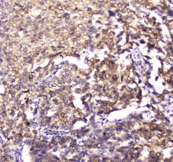
 IHC (Immunohistchemistry)
(At 1/100 staining Human ovarian cancer and adjacent normal tissues by IHC-P. The sample was formaldehyde fixed and a heat mediated antigen retrieval step in citrate buffer was performed. The sample was then blocked and incubated with the primary antibody at 4 degree C overnight. An HRP conjugated anti-Rabbit antibody was used as the secondary antibody.)
IHC (Immunohistchemistry)
(At 1/100 staining Human ovarian cancer and adjacent normal tissues by IHC-P. The sample was formaldehyde fixed and a heat mediated antigen retrieval step in citrate buffer was performed. The sample was then blocked and incubated with the primary antibody at 4 degree C overnight. An HRP conjugated anti-Rabbit antibody was used as the secondary antibody.)
alpha 1 Catenin, Polyclonal Antibody (Cat# AAA31319)
Full Name
Phospho-alpha 1 Catenin (Ser655/Thr658) Antibody
Gene Names
CTNNA1; CAP102
Reactivity
Human, Mouse, Rat
Applications
WB, IHC, EIA
Purity
The antibody is from purified rabbit serum by affinity purification via sequential chromatography on phospho-peptide and non-phospho-peptide affinity columns.
Pricing


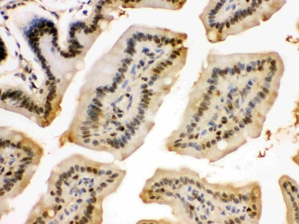
 IHC (Immunohistochemistry)
(Figure 8. IHC analysis of PRDM1/Blimp1 using anti-PRDM1/Blimp1 antibody (AAA19133).PRDM1/Blimp1 was detected in paraffin-embedded section of rat small intestine tissue. Heat mediated antigen retrieval was performed in citrate buffer (pH6, epitope retrieval solution) for 20 mins. The tissue section was blocked with 10% goat serum. The tissue section was then incubated with 1ug/ml rabbit anti-PRDM1/Blimp1 Antibody (AAA19133) overnight at 4 degree C. Biotinylated goat anti-rabbit IgG was used as secondary antibody and incubated for 30 minutes at 37 degree C. The tissue section was developed using Strepavidin-Biotin-Complex (SABC) with DAB as the chromogen.)
IHC (Immunohistochemistry)
(Figure 8. IHC analysis of PRDM1/Blimp1 using anti-PRDM1/Blimp1 antibody (AAA19133).PRDM1/Blimp1 was detected in paraffin-embedded section of rat small intestine tissue. Heat mediated antigen retrieval was performed in citrate buffer (pH6, epitope retrieval solution) for 20 mins. The tissue section was blocked with 10% goat serum. The tissue section was then incubated with 1ug/ml rabbit anti-PRDM1/Blimp1 Antibody (AAA19133) overnight at 4 degree C. Biotinylated goat anti-rabbit IgG was used as secondary antibody and incubated for 30 minutes at 37 degree C. The tissue section was developed using Strepavidin-Biotin-Complex (SABC) with DAB as the chromogen.)
PRDM1/Blimp1, Polyclonal Antibody (Cat# AAA19133)
Full Name
Anti-PRDM1/Blimp1 Picoband antibody
Gene Names
PRDM1; BLIMP1; PRDI-BF1
Reactivity
Human, Mouse, Rat
No cross reactivity with other proteins.
No cross reactivity with other proteins.
Applications
EIA, IHC, WB
Pricing


 IHC (Immunohistchemistry)
(Figure 6. IHC analysis of NSF using anti-NSF antibody (AAA19136).NSF was detected in paraffin-embedded section of human mammary cancer tissue. Heat mediated antigen retrieval was performed in citrate buffer (pH6, epitope retrieval solution) for 20 mins. The tissue section was blocked with 10% goat serum. The tissue section was then incubated with 1ug/ml rabbit anti-NSF Antibody (AAA19136) overnight at 4 degree C. Biotinylated goat anti-rabbit IgG was used as secondary antibody and incubated for 30 minutes at 37 degree C. The tissue section was developed using Strepavidin-Biotin-Complex (SABC) with DAB as the chromogen.)
IHC (Immunohistchemistry)
(Figure 6. IHC analysis of NSF using anti-NSF antibody (AAA19136).NSF was detected in paraffin-embedded section of human mammary cancer tissue. Heat mediated antigen retrieval was performed in citrate buffer (pH6, epitope retrieval solution) for 20 mins. The tissue section was blocked with 10% goat serum. The tissue section was then incubated with 1ug/ml rabbit anti-NSF Antibody (AAA19136) overnight at 4 degree C. Biotinylated goat anti-rabbit IgG was used as secondary antibody and incubated for 30 minutes at 37 degree C. The tissue section was developed using Strepavidin-Biotin-Complex (SABC) with DAB as the chromogen.)
NSF, Polyclonal Antibody (Cat# AAA19136)
Full Name
Anti-NSF Picoband antibody
Gene Names
NSF; SKD2; SEC18
Reactivity
Human, Mouse, Rat
No cross reactivity with other proteins.
No cross reactivity with other proteins.
Applications
EIA, IHC, WB
Pricing


 WB (Western Blot)
(Figure 9 KD Validation of PUMA (Han et al., 2010)Immunoblot analyses of Tet-induced p53 cells treated with NOXA, Puma, Bim or non-targeting siRNAs that were utilized in this experiment. PUMA protein levels were markedly reduced in PUMA KD cells detected by anti-PUMA antibodies (3041).)
WB (Western Blot)
(Figure 9 KD Validation of PUMA (Han et al., 2010)Immunoblot analyses of Tet-induced p53 cells treated with NOXA, Puma, Bim or non-targeting siRNAs that were utilized in this experiment. PUMA protein levels were markedly reduced in PUMA KD cells detected by anti-PUMA antibodies (3041).)
PUMA, Polyclonal Antibody (Cat# AAA10943)
Full Name
PUMA Antibody
Gene Names
BBC3; JFY1; PUMA; JFY-1
Reactivity
Human, Mouse
Applications
EIA, WB, ICC, IF
Purity
PUMA Antibody is affinity chromatography purified via peptide column.
Pricing


 IHC (Immunohistchemistry)
(Figure 6. IHC analysis of 14-3-3 zeta/delta using anti-14-3-3 zeta/delta antibody (AAA19144).14-3-3 zeta/delta was detected in paraffin-embedded section of rat kidney tissue. Heat mediated antigen retrieval was performed in citrate buffer (pH6, epitope retrieval solution) for 20 mins. The tissue section was blocked with 10% goat serum. The tissue section was then incubated with 1ug/ml rabbit anti-14-3-3 zeta/delta Antibody (AAA19144) overnight at 4 degree C. Biotinylated goat anti-rabbit IgG was used as secondary antibody and incubated for 30 minutes at 37 degree C. The tissue section was developed using Strepavidin-Biotin-Complex (SABC) with DAB as the chromogen.)
IHC (Immunohistchemistry)
(Figure 6. IHC analysis of 14-3-3 zeta/delta using anti-14-3-3 zeta/delta antibody (AAA19144).14-3-3 zeta/delta was detected in paraffin-embedded section of rat kidney tissue. Heat mediated antigen retrieval was performed in citrate buffer (pH6, epitope retrieval solution) for 20 mins. The tissue section was blocked with 10% goat serum. The tissue section was then incubated with 1ug/ml rabbit anti-14-3-3 zeta/delta Antibody (AAA19144) overnight at 4 degree C. Biotinylated goat anti-rabbit IgG was used as secondary antibody and incubated for 30 minutes at 37 degree C. The tissue section was developed using Strepavidin-Biotin-Complex (SABC) with DAB as the chromogen.)
14-3-3 zeta/delta, Polyclonal Antibody (Cat# AAA19144)
Full Name
Anti-14-3-3 zeta/delta Picoband antibody
Gene Names
YWHAZ; HEL4; YWHAD; KCIP-1; HEL-S-3; HEL-S-93; 14-3-3-zeta
Reactivity
Human, Mouse, Rat
No cross reactivity with other proteins.
No cross reactivity with other proteins.
Applications
IHC, WB
Pricing


 IHC (Immunohistochemistry)
(Figure 7. IHC analysis of splicing factor 1 using anti-splicing factor 1 antibody (AAA19141).splicing factor 1 was detected in paraffin-embedded section of rat small intestine tissue. Heat mediated antigen retrieval was performed in citrate buffer (pH6, epitope retrieval solution) for 20 mins. The tissue section was blocked with 10% goat serum. The tissue section was then incubated with 1ug/ml rabbit anti-splicing factor 1 Antibody (AAA19141) overnight at 4 degree C. Biotinylated goat anti-rabbit IgG was used as secondary antibody and incubated for 30 minutes at 37 degree C. The tissue section was developed using Strepavidin-Biotin-Complex (SABC) with DAB as the chromogen.)
IHC (Immunohistochemistry)
(Figure 7. IHC analysis of splicing factor 1 using anti-splicing factor 1 antibody (AAA19141).splicing factor 1 was detected in paraffin-embedded section of rat small intestine tissue. Heat mediated antigen retrieval was performed in citrate buffer (pH6, epitope retrieval solution) for 20 mins. The tissue section was blocked with 10% goat serum. The tissue section was then incubated with 1ug/ml rabbit anti-splicing factor 1 Antibody (AAA19141) overnight at 4 degree C. Biotinylated goat anti-rabbit IgG was used as secondary antibody and incubated for 30 minutes at 37 degree C. The tissue section was developed using Strepavidin-Biotin-Complex (SABC) with DAB as the chromogen.)
splicing factor 1, Polyclonal Antibody (Cat# AAA19141)
Full Name
Anti-splicing factor 1 Picoband antibody
Gene Names
SF1; BBP; MBBP; ZFM1; ZNF162; D11S636; ZCCHC25
Reactivity
Human, Mouse, Rat
No cross reactivity with other proteins.
No cross reactivity with other proteins.
Applications
EIA, IHC, WB
Pricing


 IHC (Immunohistchemistry)
(Figure 6. IHC analysis of MAP1LC3A using anti-MAP1LC3A antibody (AAA19151).MAP1LC3A was detected in paraffin-embedded section of rat brain tissue. Heat mediated antigen retrieval was performed in citrate buffer (pH6, epitope retrieval solution) for 20 mins. The tissue section was blocked with 10% goat serum. The tissue section was then incubated with 1ug/ml rabbit anti-MAP1LC3A Antibody (AAA19151) overnight at 4 degree C. Biotinylated goat anti-rabbit IgG was used as secondary antibody and incubated for 30 minutes at 37 degree C. The tissue section was developed using Strepavidin-Biotin-Complex (SABC) with DAB as the chromogen.)
IHC (Immunohistchemistry)
(Figure 6. IHC analysis of MAP1LC3A using anti-MAP1LC3A antibody (AAA19151).MAP1LC3A was detected in paraffin-embedded section of rat brain tissue. Heat mediated antigen retrieval was performed in citrate buffer (pH6, epitope retrieval solution) for 20 mins. The tissue section was blocked with 10% goat serum. The tissue section was then incubated with 1ug/ml rabbit anti-MAP1LC3A Antibody (AAA19151) overnight at 4 degree C. Biotinylated goat anti-rabbit IgG was used as secondary antibody and incubated for 30 minutes at 37 degree C. The tissue section was developed using Strepavidin-Biotin-Complex (SABC) with DAB as the chromogen.)
MAP1LC3A, Polyclonal Antibody (Cat# AAA19151)
Full Name
Anti-MAP1LC3A Picoband antibody
Gene Names
MAP1LC3A; LC3; LC3A; ATG8E; MAP1ALC3; MAP1BLC3
Reactivity
Human, Mouse, Rat
No cross reactivity with other proteins.
No cross reactivity with other proteins.
Applications
EIA, IHC, WB
Pricing


 ICC (Immunocytochemistry)
(Figure 6. IHC analysis of LYRIC using anti-LYRIC antibody (AAA11650).LYRIC was detected in immunocytochemical section of SMMC-7721 cell. Heat mediated antigen retrieval was performed in citrate buffer (pH6, epitope retrieval solution) for 20 mins. The tissue section was blocked with 10% goat serum. The tissue section was then incubated with 1ug/ml rabbit anti-LYRIC Antibody (AAA11650) overnight at 4 degree C. Biotinylated goat anti-rabbit IgG was used as secondary antibody and incubated for 30 minutes at 37 degree C. The tissue section was developed using Strepavidin-Biotin-Complex (SABC) with DAB as the chromogen.)
ICC (Immunocytochemistry)
(Figure 6. IHC analysis of LYRIC using anti-LYRIC antibody (AAA11650).LYRIC was detected in immunocytochemical section of SMMC-7721 cell. Heat mediated antigen retrieval was performed in citrate buffer (pH6, epitope retrieval solution) for 20 mins. The tissue section was blocked with 10% goat serum. The tissue section was then incubated with 1ug/ml rabbit anti-LYRIC Antibody (AAA11650) overnight at 4 degree C. Biotinylated goat anti-rabbit IgG was used as secondary antibody and incubated for 30 minutes at 37 degree C. The tissue section was developed using Strepavidin-Biotin-Complex (SABC) with DAB as the chromogen.)
LYRIC, Polyclonal Antibody (Cat# AAA11650)
Full Name
Anti-LYRIC Antibody
Gene Names
MTDH; 3D3; AEG1; AEG-1; LYRIC; LYRIC/3D3
Reactivity
Human, Mouse, Rat
Applications
WB, IHC
Purity
Immunogen Affinity Purified
Pricing


 ICC (Immunocytochemistry)
(Figure 6. IHC analysis of EIF6 using anti-EIF6 antibody (AAA11655).EIF6 was detected in immunocytochemical section of SW480 cell. Heat mediated antigen retrieval was performed in citrate buffer (pH6, epitope retrieval solution) for 20 mins. The tissue section was blocked with 10% goat serum. The tissue section was then incubated with 1ug/ml rabbit anti-EIF6 Antibody (AAA11655) overnight at 4 degree C. Biotinylated goat anti-rabbit IgG was used as secondary antibody and incubated for 30 minutes at 37 degree C. The tissue section was developed using Strepavidin-Biotin-Complex (SABC) with DAB as the chromogen.)
ICC (Immunocytochemistry)
(Figure 6. IHC analysis of EIF6 using anti-EIF6 antibody (AAA11655).EIF6 was detected in immunocytochemical section of SW480 cell. Heat mediated antigen retrieval was performed in citrate buffer (pH6, epitope retrieval solution) for 20 mins. The tissue section was blocked with 10% goat serum. The tissue section was then incubated with 1ug/ml rabbit anti-EIF6 Antibody (AAA11655) overnight at 4 degree C. Biotinylated goat anti-rabbit IgG was used as secondary antibody and incubated for 30 minutes at 37 degree C. The tissue section was developed using Strepavidin-Biotin-Complex (SABC) with DAB as the chromogen.)
EIF6, Polyclonal Antibody (Cat# AAA11655)
Full Name
Anti-EIF6 Antibody
Gene Names
EIF6; CAB; EIF3A; eIF-6; p27BBP; ITGB4BP; b(2)gcn; p27(BBP)
Reactivity
Human, Mouse, Rat
Applications
WB, IHC
Purity
Immunogen Affinity Purified
Pricing


 IHC (Immunohistochemistry)
(Figure 8. IHC analysis of DDB1 using anti-DDB1 antibody (AAA11660).DDB1 was detected in frozen section of human placenta tissue. Heat mediated antigen retrieval was performed in citrate buffer (pH6, epitope retrieval solution) for 20 mins. The tissue section was blocked with 10% goat serum. The tissue section was then incubated with 1ug/ml rabbit anti-DDB1 Antibody (AAA11660) overnight at 4 degree C. Biotinylated goat anti-rabbit IgG was used as secondary antibody and incubated for 30 minutes at 37 degree C. The tissue section was developed using Strepavidin-Biotin-Complex (SABC) with DAB as the chromogen.)
IHC (Immunohistochemistry)
(Figure 8. IHC analysis of DDB1 using anti-DDB1 antibody (AAA11660).DDB1 was detected in frozen section of human placenta tissue. Heat mediated antigen retrieval was performed in citrate buffer (pH6, epitope retrieval solution) for 20 mins. The tissue section was blocked with 10% goat serum. The tissue section was then incubated with 1ug/ml rabbit anti-DDB1 Antibody (AAA11660) overnight at 4 degree C. Biotinylated goat anti-rabbit IgG was used as secondary antibody and incubated for 30 minutes at 37 degree C. The tissue section was developed using Strepavidin-Biotin-Complex (SABC) with DAB as the chromogen.)
DDB1, Polyclonal Antibody (Cat# AAA11660)
Full Name
Anti-DDB1 Antibody
Gene Names
DDB1; XPE; DDBA; XAP1; XPCE; XPE-BF; UV-DDB1
Reactivity
Human, Mouse, Rat
Applications
WB, IHC
Purity
Immunogen Affinity Purified
Pricing


 Application Data
(Analysis of Protein Array containing more than 19, 000 full-length human proteins using Mouse Emerin Monoclonal Antibody (EMD/2167) Z- and S- Score: The Z-score represents the strength of a signal that a monoclonal antibody (MAb) (in combination with a fluorescently-tagged anti-IgG secondary antibody) produces when binding to a particular protein on the HuProtTM array. Z-scores are described in units of standard deviations (SD's) above the mean value of all signals generated on that array. If targets on HuProtTM are arranged in descending order of the Z-score, the S-score is the difference (also in units of SD's) between the Z-score. S-score therefore represents the relative target specificity of a MAb to its intended target. A MAb is considered to specific to its intended target, if the MAb has an S-score of at least 2.5. For example, if a MAb binds to protein X with a Z-score of 43 and to protein Y with a Z-score of 14, then the S-score for the binding of that MAb to protein X is equal to 29.)
Application Data
(Analysis of Protein Array containing more than 19, 000 full-length human proteins using Mouse Emerin Monoclonal Antibody (EMD/2167) Z- and S- Score: The Z-score represents the strength of a signal that a monoclonal antibody (MAb) (in combination with a fluorescently-tagged anti-IgG secondary antibody) produces when binding to a particular protein on the HuProtTM array. Z-scores are described in units of standard deviations (SD's) above the mean value of all signals generated on that array. If targets on HuProtTM are arranged in descending order of the Z-score, the S-score is the difference (also in units of SD's) between the Z-score. S-score therefore represents the relative target specificity of a MAb to its intended target. A MAb is considered to specific to its intended target, if the MAb has an S-score of at least 2.5. For example, if a MAb binds to protein X with a Z-score of 43 and to protein Y with a Z-score of 14, then the S-score for the binding of that MAb to protein X is equal to 29.)
Emerin, Monoclonal Antibody (Cat# AAA23898)
Full Name
Emerin (Papillary Thyroid Carcinoma and EDMD Marker)
Gene Names
EMD; STA; EDMD; LEMD5
Reactivity
Human. Others not known.
Applications
IHC
Pricing


 SDS-PAGE
(SDS-PAGE Analysis Purified HSP60 Mouse Monoclonal Antibody (CPTC-HSPD1-1). Confirmation of Purity and Integrity of Antibody.)
SDS-PAGE
(SDS-PAGE Analysis Purified HSP60 Mouse Monoclonal Antibody (CPTC-HSPD1-1). Confirmation of Purity and Integrity of Antibody.)
HSP60 (Heat Shock Protein 60), Monoclonal Antibody (Cat# AAA23919)
Full Name
HSP60 (Heat Shock Protein 60) (Mitochondrial Marker)
Gene Names
HSPD1; HLD4; CPN60; GROEL; HSP60; HSP65; SPG13; HSP-60; HuCHA60
Reactivity
Human
Applications
FC/FACS, IF, WB, IHC
Purity
Purified Ab with BSA and Azide at 200ug/ml OR Purified Ab WITHOUT BSA and Azide at 1.0mg/ml
Pricing


 IHC (Immunohistochemistry)
(Figure 10. IHC analysis of Lamin B1 using anti-Lamin B1 antibody (AAA11669).Lamin B1 was detected in frozen section of mouse small intestine tissue. Heat mediated antigen retrieval was performed in citrate buffer (pH6, epitope retrieval solution) for 20 mins. The tissue section was blocked with 10% goat serum. The tissue section was then incubated with 1ug/ml rabbit anti-Lamin B1 Antibody (AAA11669) overnight at 4 degree C. Biotinylated goat anti-rabbit IgG was used as secondary antibody and incubated for 30 minutes at 37 degree C. The tissue section was developed using Strepavidin-Biotin-Complex (SABC) with DAB as the chromogen.)
IHC (Immunohistochemistry)
(Figure 10. IHC analysis of Lamin B1 using anti-Lamin B1 antibody (AAA11669).Lamin B1 was detected in frozen section of mouse small intestine tissue. Heat mediated antigen retrieval was performed in citrate buffer (pH6, epitope retrieval solution) for 20 mins. The tissue section was blocked with 10% goat serum. The tissue section was then incubated with 1ug/ml rabbit anti-Lamin B1 Antibody (AAA11669) overnight at 4 degree C. Biotinylated goat anti-rabbit IgG was used as secondary antibody and incubated for 30 minutes at 37 degree C. The tissue section was developed using Strepavidin-Biotin-Complex (SABC) with DAB as the chromogen.)
Lamin B1, Polyclonal Antibody (Cat# AAA11669)
Full Name
Anti-Lamin B1 Antibody
Gene Names
LMNB1; LMN; ADLD; LMN2; LMNB
Reactivity
Human, Mouse, Rat
Applications
WB, IHC
Purity
Immunogen Affinity Purified
Pricing


 IF (Immunofluorescence)
(Immunofluorescent analysis of 4% paraformaldehyde-fixed, 0.1% Triton X-100 permeabilized MCF-7 (human breast cancer cell line) cells labeling Pdx1 with at 1:25 dilution, followed by DyLight 488-conjugated IgG goat anti-rabbit secondary antibody at 1:200 dilution (green). Immunofluorescence image showing cytoplasm staining on MCF-7 cell line. Cytoplasmic actin is detected with DyLight 554 Phalloidin (PD18466410) at 1:100 dilution (red). The nuclear counter stain is DAPI (blue).)
IF (Immunofluorescence)
(Immunofluorescent analysis of 4% paraformaldehyde-fixed, 0.1% Triton X-100 permeabilized MCF-7 (human breast cancer cell line) cells labeling Pdx1 with at 1:25 dilution, followed by DyLight 488-conjugated IgG goat anti-rabbit secondary antibody at 1:200 dilution (green). Immunofluorescence image showing cytoplasm staining on MCF-7 cell line. Cytoplasmic actin is detected with DyLight 554 Phalloidin (PD18466410) at 1:100 dilution (red). The nuclear counter stain is DAPI (blue).)
OPN-a/b, Polyclonal Antibody (Cat# AAA26861)
Full Name
OPN-a/b, NT (SPP1, BNSP, OPN, Osteopontin, Bone sialoprotein 1, Nephropontin, Secreted phosphoprotein 1, Urinary stone protein, Uropontin) (MaxLight 490)
Gene Names
SPP1; OPN; BNSP; BSPI; ETA-1
Reactivity
Human
Applications
WB, IHC, IF
Purity
Purified by Protein A and Peptide Affinity Chromatography.
Pricing


 IF (Immunofluorescence)
(Immunofluorescent analysis of 4% paraformaldehyde-fixed, 0.1% Triton X-100 permeabilized MCF-7 (human breast cancer cell line) cells labeling Pdx1 with at 1:25 dilution, followed by DyLight 488-conjugated IgG goat anti-rabbit secondary antibody at 1:200 dilution (green). Immunofluorescence image showing cytoplasm staining on MCF-7 cell line. Cytoplasmic actin is detected with DyLight 554 Phalloidin (PD18466410) at 1:100 dilution (red). The nuclear counter stain is DAPI (blue).)
IF (Immunofluorescence)
(Immunofluorescent analysis of 4% paraformaldehyde-fixed, 0.1% Triton X-100 permeabilized MCF-7 (human breast cancer cell line) cells labeling Pdx1 with at 1:25 dilution, followed by DyLight 488-conjugated IgG goat anti-rabbit secondary antibody at 1:200 dilution (green). Immunofluorescence image showing cytoplasm staining on MCF-7 cell line. Cytoplasmic actin is detected with DyLight 554 Phalloidin (PD18466410) at 1:100 dilution (red). The nuclear counter stain is DAPI (blue).)
OPN-a/b, Polyclonal Antibody (Cat# AAA26865)
Full Name
OPN-a/b, NT (SPP1, BNSP, OPN, Osteopontin, Bone sialoprotein 1, Nephropontin, Secreted phosphoprotein 1, Urinary stone protein, Uropontin) (PE)
Gene Names
SPP1; OPN; BNSP; BSPI; ETA-1
Reactivity
Human
Applications
WB, IHC, IF
Purity
Purified by Protein A and Peptide Affinity Chromatography.
Pricing


 IHC (Immunohistochemistry)
(Figure 8. IHC analysis of FOXP1 using anti-FOXP1 antibody (AAA19227).FOXP1 was detected in paraffin-embedded section of rat brain tissue. Heat mediated antigen retrieval was performed in EDTA buffer (pH8. 0, epitope retrieval solution). The tissue section was blocked with 10% goat serum. The tissue section was then incubated with 2μg/ml rabbit anti-FOXP1 Antibody (AAA19227) overnight at 4 degree C. Biotinylated goat anti-rabbit IgG was used as secondary antibody and incubated for 30 minutes at 37 degree C. The tissue section was developed using Strepavidin-Biotin-Complex (SABC) (Catalog # with DAB as the chromogen.)
IHC (Immunohistochemistry)
(Figure 8. IHC analysis of FOXP1 using anti-FOXP1 antibody (AAA19227).FOXP1 was detected in paraffin-embedded section of rat brain tissue. Heat mediated antigen retrieval was performed in EDTA buffer (pH8. 0, epitope retrieval solution). The tissue section was blocked with 10% goat serum. The tissue section was then incubated with 2μg/ml rabbit anti-FOXP1 Antibody (AAA19227) overnight at 4 degree C. Biotinylated goat anti-rabbit IgG was used as secondary antibody and incubated for 30 minutes at 37 degree C. The tissue section was developed using Strepavidin-Biotin-Complex (SABC) (Catalog # with DAB as the chromogen.)
FOXP1, Polyclonal Antibody (Cat# AAA19227)
Full Name
Anti-FOXP1 Antibody
Gene Names
FOXP1; QRF1; 12CC4; hFKH1B; HSPC215
Reactivity
Human, Mouse, Rat, Monkey
Applications
WB, IHC-P
Purity
Immunogen affinity purified.
Pricing


 FCM (Flow Cytometry)
(Figure 6. Flow Cytometry analysis of PC-3 cells using anti-ATG14L antibody (AAA11658).Overlay histogram showing PC-3 cells stained with AAA11658 (Blue line).The cells were blocked with 10% normal goat serum. And then incubated with rabbit anti-ATG14L Antibody (AAA11658,1ug/1x10^6 cells) for 30 min at 20 degree C. DyLight®488 conjugated goat anti-rabbit IgG (5-10ug/1x10^6 cells) was used as secondary antibody for 30 minutes at 20 degree C. Isotype control antibody (Green line) was rabbit IgG (1ug/1x106) used under the same conditions. Unlabelled sample (Red line) was also used as a control.)
FCM (Flow Cytometry)
(Figure 6. Flow Cytometry analysis of PC-3 cells using anti-ATG14L antibody (AAA11658).Overlay histogram showing PC-3 cells stained with AAA11658 (Blue line).The cells were blocked with 10% normal goat serum. And then incubated with rabbit anti-ATG14L Antibody (AAA11658,1ug/1x10^6 cells) for 30 min at 20 degree C. DyLight®488 conjugated goat anti-rabbit IgG (5-10ug/1x10^6 cells) was used as secondary antibody for 30 minutes at 20 degree C. Isotype control antibody (Green line) was rabbit IgG (1ug/1x106) used under the same conditions. Unlabelled sample (Red line) was also used as a control.)
ATG14L, Polyclonal Antibody (Cat# AAA11658)
Full Name
Anti-ATG14L Antibody
Gene Names
ATG14; ATG14L; BARKOR; KIAA0831
Reactivity
Human, Rat
Applications
WB, IHC
Purity
Immunogen Affinity Purified
Pricing


 WB (Western Blot)
(Western Blot Analysis of human liver tissue lysate using Prohibitin Mouse Monoclonal Antibody (PHB/3194).)
WB (Western Blot)
(Western Blot Analysis of human liver tissue lysate using Prohibitin Mouse Monoclonal Antibody (PHB/3194).)
Prohibitin, Monoclonal Antibody (Cat# AAA23932)
Full Name
Prohibitin (Mitochondrial Marker)
Gene Names
PHB; PHB1; HEL-215; HEL-S-54e
Reactivity
Human
Applications
WB, IF, IHC
Purity
Purified Ab with BSA and Azide at 200ug/ml OR Purified Ab WITHOUT BSA and Azide at 1.0mg/ml
Pricing


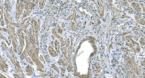
 IHC (Immunohistochemistry)
(Figure 8. IHC analysis of VEGF Receptor 3 using anti-VEGF Receptor 3 antibody (AAA19146).VEGF Receptor 3 was detected in paraffin-embedded section of human mammary cancer tissue. Heat mediated antigen retrieval was performed in citrate buffer (pH6, epitope retrieval solution) for 20 mins. The tissue section was blocked with 10% goat serum. The tissue section was then incubated with 1ug/ml rabbit anti-VEGF Receptor 3 Antibody (AAA19146) overnight at 4 degree C. Biotinylated goat anti-rabbit IgG was used as secondary antibody and incubated for 30 minutes at 37 degree C. The tissue section was developed using Strepavidin-Biotin-Complex (SABC) with DAB as the chromogen.)
IHC (Immunohistochemistry)
(Figure 8. IHC analysis of VEGF Receptor 3 using anti-VEGF Receptor 3 antibody (AAA19146).VEGF Receptor 3 was detected in paraffin-embedded section of human mammary cancer tissue. Heat mediated antigen retrieval was performed in citrate buffer (pH6, epitope retrieval solution) for 20 mins. The tissue section was blocked with 10% goat serum. The tissue section was then incubated with 1ug/ml rabbit anti-VEGF Receptor 3 Antibody (AAA19146) overnight at 4 degree C. Biotinylated goat anti-rabbit IgG was used as secondary antibody and incubated for 30 minutes at 37 degree C. The tissue section was developed using Strepavidin-Biotin-Complex (SABC) with DAB as the chromogen.)
VEGF Receptor 3, Polyclonal Antibody (Cat# AAA19146)
Full Name
Anti-VEGF Receptor 3 Picoband Antibody
Gene Names
FLT4; PCL; FLT-4; FLT41; LMPH1A; VEGFR3; VEGFR-3
Reactivity
Human, Mouse, Rat
No cross reactivity with other proteins.
No cross reactivity with other proteins.
Applications
IHC, WB
Purity
Immunogen affinity purified
Pricing


 IHC (Immunohistchemistry)
(Figure 6. IHC analysis of SAE2/UBA2 using anti-SAE2/UBA2 antibody (AAA19171).SAE2/UBA2 was detected in paraffin-embedded section of mouse testis tissue. Heat mediated antigen retrieval was performed in citrate buffer (pH6, epitope retrieval solution) for 20 mins. The tissue section was blocked with 10% goat serum. The tissue section was then incubated with 1ug/ml rabbit anti-SAE2/UBA2 Antibody (AAA19171) overnight at 4 degree C. Biotinylated goat anti-rabbit IgG was used as secondary antibody and incubated for 30 minutes at 37 degree C. The tissue section was developed using Strepavidin-Biotin-Complex (SABC) with DAB as the chromogen.)
IHC (Immunohistchemistry)
(Figure 6. IHC analysis of SAE2/UBA2 using anti-SAE2/UBA2 antibody (AAA19171).SAE2/UBA2 was detected in paraffin-embedded section of mouse testis tissue. Heat mediated antigen retrieval was performed in citrate buffer (pH6, epitope retrieval solution) for 20 mins. The tissue section was blocked with 10% goat serum. The tissue section was then incubated with 1ug/ml rabbit anti-SAE2/UBA2 Antibody (AAA19171) overnight at 4 degree C. Biotinylated goat anti-rabbit IgG was used as secondary antibody and incubated for 30 minutes at 37 degree C. The tissue section was developed using Strepavidin-Biotin-Complex (SABC) with DAB as the chromogen.)
SAE2/UBA2, Polyclonal Antibody (Cat# AAA19171)
Full Name
Anti-SAE2/UBA2 Picoband antibody
Gene Names
UBA2; ARX; SAE2; HRIHFB2115
Reactivity
Human, Mouse, Rat
No cross reactivity with other proteins.
No cross reactivity with other proteins.
Applications
EIA, IHC, WB
Pricing


 IHC (Immunohistchemistry)
(Figure 6. IHC analysis of DYNLT1 using anti-DYNLT1 antibody (AAA19175).DYNLT1 was detected in paraffin-embedded section of rat lung tissue. Heat mediated antigen retrieval was performed in citrate buffer (pH6, epitope retrieval solution) for 20 mins. The tissue section was blocked with 10% goat serum. The tissue section was then incubated with 1ug/ml rabbit anti-DYNLT1 Antibody (AAA19175) overnight at 4 degree C. Biotinylated goat anti-rabbit IgG was used as secondary antibody and incubated for 30 minutes at 37 degree C. The tissue section was developed using Strepavidin-Biotin-Complex (SABC) with DAB as the chromogen.)
IHC (Immunohistchemistry)
(Figure 6. IHC analysis of DYNLT1 using anti-DYNLT1 antibody (AAA19175).DYNLT1 was detected in paraffin-embedded section of rat lung tissue. Heat mediated antigen retrieval was performed in citrate buffer (pH6, epitope retrieval solution) for 20 mins. The tissue section was blocked with 10% goat serum. The tissue section was then incubated with 1ug/ml rabbit anti-DYNLT1 Antibody (AAA19175) overnight at 4 degree C. Biotinylated goat anti-rabbit IgG was used as secondary antibody and incubated for 30 minutes at 37 degree C. The tissue section was developed using Strepavidin-Biotin-Complex (SABC) with DAB as the chromogen.)
DYNLT1, Polyclonal Antibody (Cat# AAA19175)
Full Name
Anti-DYNLT1 Picoband antibody
Gene Names
DYNLT1; CW-1; TCTEL1; tctex-1
Reactivity
Human, Mouse, Rat
No cross reactivity with other proteins.
No cross reactivity with other proteins.
Applications
EIA, IHC, WB
Pricing


 IHC (Immunohistochemistry)
(Figure 7. IHC analysis of GALNS using anti-GALNS antibody (AAA19164).GALNS was detected in paraffin-embedded section of rat small intestine tissue. Heat mediated antigen retrieval was performed in citrate buffer (pH6, epitope retrieval solution) for 20 mins. The tissue section was blocked with 10% goat serum. The tissue section was then incubated with 1ug/ml rabbit anti-GALNS Antibody (AAA19164) overnight at 4 degree C. Biotinylated goat anti-rabbit IgG was used as secondary antibody and incubated for 30 minutes at 37 degree C. The tissue section was developed using Strepavidin-Biotin-Complex (SABC) with DAB as the chromogen.)
IHC (Immunohistochemistry)
(Figure 7. IHC analysis of GALNS using anti-GALNS antibody (AAA19164).GALNS was detected in paraffin-embedded section of rat small intestine tissue. Heat mediated antigen retrieval was performed in citrate buffer (pH6, epitope retrieval solution) for 20 mins. The tissue section was blocked with 10% goat serum. The tissue section was then incubated with 1ug/ml rabbit anti-GALNS Antibody (AAA19164) overnight at 4 degree C. Biotinylated goat anti-rabbit IgG was used as secondary antibody and incubated for 30 minutes at 37 degree C. The tissue section was developed using Strepavidin-Biotin-Complex (SABC) with DAB as the chromogen.)
GALNS, Antibody (Cat# AAA19164)
Full Name
Anti-GALNS Picoband antibody
Gene Names
GALNS; GAS; MPS4A; GalN6S; GALNAC6S
Reactivity
Reacts with: Human, Mouse, Rat
Applications
WB, EIA
Purity
Immunogen affinity purified
Pricing

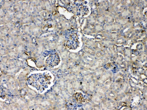


 IHC (Immunohistchemistry)
(Figure 6. IHC analysis of Thrombopoietin using anti- Thrombopoietin antibody (AAA19166).Thrombopoietin was detected in paraffin-embedded section of rat kidney tissues. Heat mediated antigen retrieval was performed in citrate buffer (pH6, epitope retrieval solution) for 20 mins. The tissue section was blocked with 10% goat serum. The tissue section was then incubated with 1ug/ml rabbit anti- Thrombopoietin Antibody (AAA19166) overnight at 4 degree C. Biotinylated goat anti-rabbit IgG was used as secondary antibody and incubated for 30 minutes at 37 degree C. The tissue section was developed using Strepavidin-Biotin-Complex (SABC) with DAB as the chromogen.)
IHC (Immunohistchemistry)
(Figure 6. IHC analysis of Thrombopoietin using anti- Thrombopoietin antibody (AAA19166).Thrombopoietin was detected in paraffin-embedded section of rat kidney tissues. Heat mediated antigen retrieval was performed in citrate buffer (pH6, epitope retrieval solution) for 20 mins. The tissue section was blocked with 10% goat serum. The tissue section was then incubated with 1ug/ml rabbit anti- Thrombopoietin Antibody (AAA19166) overnight at 4 degree C. Biotinylated goat anti-rabbit IgG was used as secondary antibody and incubated for 30 minutes at 37 degree C. The tissue section was developed using Strepavidin-Biotin-Complex (SABC) with DAB as the chromogen.)
Thrombopoietin, Polyclonal Antibody (Cat# AAA19166)
Full Name
Anti-Thrombopoietin Picoband Antibody
Gene Names
Thpo; Ml; Tpo; Mgdf; Mpllg
Reactivity
Mouse, Rat
No cross reactivity with other proteins
No cross reactivity with other proteins
Applications
EIA, IHC, WB
Purity
Immunogen affinity purified
Pricing


 FCM (Flow Cytometry)
(Figure 6. Flow Cytometry analysis of K562 cells using anti-D2AP antibody (M01756). Overlay histogram showing K562 cells stained with M01756 (Blue line).The cells were blocked with 10% normal goat serum. And then incubated with mouse anti-D2AP Antibody (M01756, 1ug/1x106 cells) for 30 min at 20 degree C. DyLight488 conjugated goat anti-mouse IgG (BA1126, 5-10ug/1x106 cells) was used as secondary antibody for 30 minutes at 20 degree C. Isotype control antibody (Green line) was mouse IgG (1ug/1x106) used under the same conditions. Unlabelled sample (Red line) was also used as a control.)
FCM (Flow Cytometry)
(Figure 6. Flow Cytometry analysis of K562 cells using anti-D2AP antibody (M01756). Overlay histogram showing K562 cells stained with M01756 (Blue line).The cells were blocked with 10% normal goat serum. And then incubated with mouse anti-D2AP Antibody (M01756, 1ug/1x106 cells) for 30 min at 20 degree C. DyLight488 conjugated goat anti-mouse IgG (BA1126, 5-10ug/1x106 cells) was used as secondary antibody for 30 minutes at 20 degree C. Isotype control antibody (Green line) was mouse IgG (1ug/1x106) used under the same conditions. Unlabelled sample (Red line) was also used as a control.)
CD2AP, Monoclonal Antibody (Cat# AAA19185)
Full Name
Anti-CD2AP Antibody (monoclonal, 5F8)
Gene Names
CD2AP; CMS
Reactivity
Human, Mouse, Rat
Applications
WB, IHC, FC/FACS
Purity
Immunogen Affinity Purified
Pricing


 IHC (Immunohistchemistry)
(Figure 5. IHC analysis of GNG4 using anti-GNG4 antibody (AAA19341).GNG4 was detected in paraffin-embedded section of human placenta tissue. Heat mediated antigen retrieval was performed in EDTA buffer (pH8. 0, epitope retrieval solution). The tissue section was blocked with 10% goat serum. The tissue section was then incubated with 2μg/ml rabbit anti-GNG4 Antibody (AAA19341) overnight at 4 degree C. Biotinylated goat anti-rabbit IgG was used as secondary antibody and incubated for 30 minutes at 37 degree C. The tissue section was developed using Strepavidin-Biotin-Complex (SABC) (Catalog # with DAB as the chromogen.)
IHC (Immunohistchemistry)
(Figure 5. IHC analysis of GNG4 using anti-GNG4 antibody (AAA19341).GNG4 was detected in paraffin-embedded section of human placenta tissue. Heat mediated antigen retrieval was performed in EDTA buffer (pH8. 0, epitope retrieval solution). The tissue section was blocked with 10% goat serum. The tissue section was then incubated with 2μg/ml rabbit anti-GNG4 Antibody (AAA19341) overnight at 4 degree C. Biotinylated goat anti-rabbit IgG was used as secondary antibody and incubated for 30 minutes at 37 degree C. The tissue section was developed using Strepavidin-Biotin-Complex (SABC) (Catalog # with DAB as the chromogen.)
GNG4, Polyclonal Antibody (Cat# AAA19341)
Full Name
Anti-GNG4 Antibody
Reactivity
Human, Mouse, Rat
Applications
WB, IHC-P, FC/FACS/FCM, EIA
Purity
Immunogen affinity purified.
Pricing


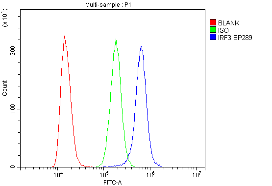 FCM (Flow Cytometry)
(Figure 6. Flow Cytometry analysis of K562 cells using anti-IRF3 antibody (AAA19216).Overlay histogram showing K562 cells stained with AAA19216 (Blue line). The cells were blocked with 10% normal goat serum. And then incubated with rabbit anti-IRF3 Antibody (AAA19216, 1μg/1x106 cells) for 30 min at 20 degree C. DyLight®488 conjugated goat anti-rabbit IgG (5-10μg/1x106 cells) was used as secondary antibody for 30 minutes at 20 degree C. Isotype control antibody (Green line) was rabbit IgG (1μg/1x106) used under the same conditions. Unlabelled sample (Red line) was also used as a control.)
FCM (Flow Cytometry)
(Figure 6. Flow Cytometry analysis of K562 cells using anti-IRF3 antibody (AAA19216).Overlay histogram showing K562 cells stained with AAA19216 (Blue line). The cells were blocked with 10% normal goat serum. And then incubated with rabbit anti-IRF3 Antibody (AAA19216, 1μg/1x106 cells) for 30 min at 20 degree C. DyLight®488 conjugated goat anti-rabbit IgG (5-10μg/1x106 cells) was used as secondary antibody for 30 minutes at 20 degree C. Isotype control antibody (Green line) was rabbit IgG (1μg/1x106) used under the same conditions. Unlabelled sample (Red line) was also used as a control.)
IRF3, Polyclonal Antibody (Cat# AAA19216)
Full Name
Anti-IRF3 Antibody
Reactivity
Human
Applications
WB, IHC-P, FC/FACS/FCM, EIA
Purity
Immunogen affinity purified.
Pricing