Filters
Clonality
Type
Reactivity
Gene Name
Isotype
Host
Application
Clone
1571 results for " Nuclear" - showing 1400-1450
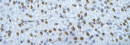


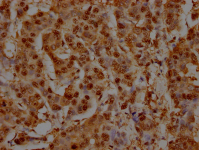
 IHC (Immunohistochemistry)
(Rabbit Anti-NONO Antibody Catalog Number: AAA23484 Paraffin Embedded Tissue: Human cardiac cell Cellular Data: Epithelial cells of renal tubule Antibody Concentration: 4.0-8.0 ug/ml Magnification: 400X)
IHC (Immunohistochemistry)
(Rabbit Anti-NONO Antibody Catalog Number: AAA23484 Paraffin Embedded Tissue: Human cardiac cell Cellular Data: Epithelial cells of renal tubule Antibody Concentration: 4.0-8.0 ug/ml Magnification: 400X)
NONO, Polyclonal Antibody (Cat# AAA23484)
Full Name
NONO antibody - C-terminal region
Gene Names
NONO; P54; NMT55; NRB54; MRXS34; P54NRB; PPP1R114
Reactivity
Tested Species Reactivity: Human (Predicted Species Reactivity: Cow, Dog, Guinea Pig, Horse, Human, Mouse, Rabbit, Rat)
Applications
IF, IHC, WB, IP
Purity
Protein A purified
Pricing


 Application Data
(C:FGFR2/isolectinB4 (C) and FGFR1/isolectinB4 (D) staining of apparent mesenchymal cells and the subpopulation of endothelial cells. Virtually all other dispersed apparent mesenchymal cells express FGFR1 and FGFR2 (merged image in E). F: FGFR2 (F) and FGFR1 (G) staining in clustered cells of epithelial origin (inferred by morphology here) demonstrating that epithelial cells express both FGFR1 and FGFR2 (merged image with DAPI staining in H).)
Application Data
(C:FGFR2/isolectinB4 (C) and FGFR1/isolectinB4 (D) staining of apparent mesenchymal cells and the subpopulation of endothelial cells. Virtually all other dispersed apparent mesenchymal cells express FGFR1 and FGFR2 (merged image in E). F: FGFR2 (F) and FGFR1 (G) staining in clustered cells of epithelial origin (inferred by morphology here) demonstrating that epithelial cells express both FGFR1 and FGFR2 (merged image with DAPI staining in H).)
FGFR2, Polyclonal Antibody (Cat# AAA14790)
Full Name
FGFR2, NT (FGFR2, BEK, KGFR, KSAM, Fibroblast growth factor receptor 2, K-sam, Keratinocyte growth factor receptor, CD332)
Gene Names
FGFR2; BEK; JWS; BBDS; CEK3; CFD1; ECT1; KGFR; TK14; TK25; BFR-1; CD332; K-SAM
Reactivity
Human, Monkey, Mouse, Rat
Applications
EL/EIA, WB, IHC, FC/FACS, IF
Purity
Affinity Purified
Purified by Protein A affinity chromatography.
Purified by Protein A affinity chromatography.
Pricing


 IHC (Immunohistochemistry)
(At 1/100 staining Mouse brain tissue by IHC-P. The sample was formaldehyde fixed and a heat mediated antigen retrieval step in citrate buffer was performed. The sample was then blocked and incubated with the primary antibody at 4 degree C overnight. An HRP conjugated anti-Rabbit antibody was used as the secondary antibody.)
IHC (Immunohistochemistry)
(At 1/100 staining Mouse brain tissue by IHC-P. The sample was formaldehyde fixed and a heat mediated antigen retrieval step in citrate buffer was performed. The sample was then blocked and incubated with the primary antibody at 4 degree C overnight. An HRP conjugated anti-Rabbit antibody was used as the secondary antibody.)
NACA, Polyclonal Antibody (Cat# AAA31427)
Full Name
Phospho-NACA (Thr159) Antibody
Gene Names
NACA; HSD48; NACA1; skNAC
Reactivity
Human, Mouse, Rat
Predicted Reactivity: Pig (100%), Zebrafish (100%), Bovine (100%), Sheep (100%), Chicken (100%), Xenopus (100%)
Predicted Reactivity: Pig (100%), Zebrafish (100%), Bovine (100%), Sheep (100%), Chicken (100%), Xenopus (100%)
Applications
WB, IHC, EIA
Purity
The antibody is from purified rabbit serum by affinity purification via sequential chromatography on phospho-peptide and non-phospho-peptide affinity columns.
Pricing


 FCM (Flow Cytometry)
(Flow cytometric analysis of MCF-7 cells with MUC1 antibody at 1/50 dilution (red) compared with an unlabelled control (cells without incubation with primary antibody; black). Alexa Fluor 488-conjugated goat anti rabbit IgG was used as the secondary antibody.)
FCM (Flow Cytometry)
(Flow cytometric analysis of MCF-7 cells with MUC1 antibody at 1/50 dilution (red) compared with an unlabelled control (cells without incubation with primary antibody; black). Alexa Fluor 488-conjugated goat anti rabbit IgG was used as the secondary antibody.)
MUC1, Monoclonal Antibody (Cat# AAA30155)
Full Name
MUC1 Antibody
Gene Names
MUC1; EMA; PEM; PUM; KL-6; MAM6; PEMT; CD227; H23AG; MUC-1; CA 15-3; MUC-1/X; MUC1/ZD; MUC-1/SEC
Reactivity
Human
Applications
WB, ICC, IF, IHC, IP, FC/FACS
Purity
ProA affinity purified
Pricing


 WB (Western Blot)
(Anti-ANP32A rabbit polyclonal antibody at 1:500 dilution Lane A: ANP32A konckout Hela Whole Cell Lysate Lane B: Hela Whole Cell Lysate Lysates/proteins at 20 ug per lane. Secondary Goat Anti-Rabbit IgG (H+L)/HRP at 1/10000 dilution. Developed using the ECL technique. Performed under reducing conditions. Predicted band size:28 kDa Observed band size:28 kDa)
WB (Western Blot)
(Anti-ANP32A rabbit polyclonal antibody at 1:500 dilution Lane A: ANP32A konckout Hela Whole Cell Lysate Lane B: Hela Whole Cell Lysate Lysates/proteins at 20 ug per lane. Secondary Goat Anti-Rabbit IgG (H+L)/HRP at 1/10000 dilution. Developed using the ECL technique. Performed under reducing conditions. Predicted band size:28 kDa Observed band size:28 kDa)
ANP32A, Polyclonal Antibody (Cat# AAA27748)
Full Name
Anti-ANP32A Antibody, Rabbit Polyclonal
Gene Names
ANP32A; LANP; MAPM; PP32; HPPCn; PHAP1; PHAPI; I1PP2A; C15orf1
Reactivity
Human
Applications
WB, EIA, IHC-P, ICC, IF, IP
Purity
Protein A & Antigen Affinity
Pricing


 Application Data
(Analysis of Protein Array containing more than 19,000 full-length human proteins using Calretinin Mouse Monoclonal Antibody (CALB2/2786). Z- and S- Score: The Z-score represents the strength of a signal that a monoclonal antibody (MAb) (in combination with a fluorescently-tagged anti-IgG secondary antibody) produces when binding to a particular protein on the HuProtTM array. Z-scores are described in units of standard deviations (SD's) above the mean value of all signals generated on that array. If targets on HuProtTM are arranged in descending order of the Z-score, the S-score is the difference (also in units of SD's) between the Z-score. S-score therefore represents the relative target specificity of a MAb to its intended target. A MAb is considered to specific to its intended target, if the MAb has an S-score of at least 2.5. For example, if a MAb binds to protein X with a Z-score of 43 and to protein Y with a Z-score of 14, then the S-score for the binding of that MAb to protein X is equal to 29.)
Application Data
(Analysis of Protein Array containing more than 19,000 full-length human proteins using Calretinin Mouse Monoclonal Antibody (CALB2/2786). Z- and S- Score: The Z-score represents the strength of a signal that a monoclonal antibody (MAb) (in combination with a fluorescently-tagged anti-IgG secondary antibody) produces when binding to a particular protein on the HuProtTM array. Z-scores are described in units of standard deviations (SD's) above the mean value of all signals generated on that array. If targets on HuProtTM are arranged in descending order of the Z-score, the S-score is the difference (also in units of SD's) between the Z-score. S-score therefore represents the relative target specificity of a MAb to its intended target. A MAb is considered to specific to its intended target, if the MAb has an S-score of at least 2.5. For example, if a MAb binds to protein X with a Z-score of 43 and to protein Y with a Z-score of 14, then the S-score for the binding of that MAb to protein X is equal to 29.)
Calretinin/Calbindin 2, Monoclonal Antibody (Cat# AAA23939)
Full Name
Calretinin/Calbindin 2 (Mesothelioma Marker)
Gene Names
CALB2; CR; CAL2; CAB29
Reactivity
Human
Applications
WB, IHC
Purity
Purified Ab with BSA and Azide at 200ug/ml OR Purified Ab WITHOUT BSA at 1.0mg/ml
Pricing


 Application Data
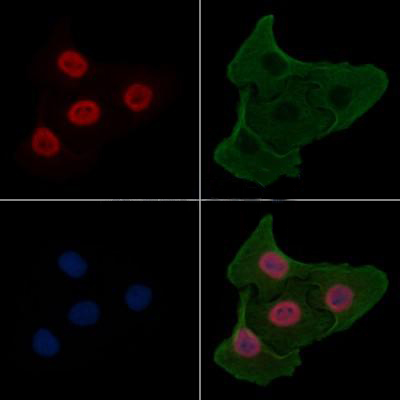
(At 25 degree C. Samples were then incubated with primary Ab(At 37 degree C. An AlexaFluor594 conjugated goat anti-rabbit IgG(H+L) Ab(Red) and an AlexaFluor488 conjugated goat anti-mouse IgG(H+L) Ab(Green) were used as the secondary antibody.The nuclear counter stain is DAPI(blue).)
Application Data
(At 25 degree C. Samples were then incubated with primary Ab(At 37 degree C. An AlexaFluor594 conjugated goat anti-rabbit IgG(H+L) Ab(Red) and an AlexaFluor488 conjugated goat anti-mouse IgG(H+L) Ab(Green) were used as the secondary antibody.The nuclear counter stain is DAPI(blue).)
KIF1C, Polyclonal Antibody (Cat# AAA31396)
Full Name
Phospho-KIF1C (Ser1092) Antibody
Gene Names
KIF1C; SAX2; LTXS1; SATX2; SPAX2; SPG58
Reactivity
Human, Mouse, Rat
Predicted Reactivity: Pig (100%), Bovine (100%), Horse (100%), Sheep (100%), Rabbit (100%), Dog (100%)
Predicted Reactivity: Pig (100%), Bovine (100%), Horse (100%), Sheep (100%), Rabbit (100%), Dog (100%)
Applications
WB, IHC, IF, ICC, EIA
Purity
The antibody is from purified rabbit serum by affinity purification via sequential chromatography on phospho-peptide and non-phospho-peptide affinity columns.
Pricing


 Application Data
(At 25 degree C. Samples were then incubated with primary Ab(At 37 degree C. An AlexaFluor594 conjugated goat anti-rabbit IgG(H+L) Ab(Red) and an AlexaFluor488 conjugated goat anti-mouse IgG(H+L) Ab(Green) were used as the secondary antibody.The nuclear counter stain is DAPI(blue).)
Application Data
(At 25 degree C. Samples were then incubated with primary Ab(At 37 degree C. An AlexaFluor594 conjugated goat anti-rabbit IgG(H+L) Ab(Red) and an AlexaFluor488 conjugated goat anti-mouse IgG(H+L) Ab(Green) were used as the secondary antibody.The nuclear counter stain is DAPI(blue).)
USP28, Polyclonal Antibody (Cat# AAA31438)
Full Name
Phospho-USP28 (Ser714) Antibody
Reactivity
Human, Mouse, Monkey
Applications
WB, IHC, IF, ICC, EIA
Purity
The antibody is from purified rabbit serum by affinity purification via sequential chromatography on phospho-peptide and non-phospho-peptide affinity columns.
Pricing


 IHC (Immunohistochemistry)
(Figure 7. IHC analysis of CD105 using anti-CD105 antibody (AAA19165).CD105 was detected in paraffin-embedded section of rat spleen tissue. Heat mediated antigen retrieval was performed in citrate buffer (pH6, epitope retrieval solution) for 20 mins. The tissue section was blocked with 10% goat serum. The tissue section was then incubated with 1ug/ml rabbit anti-CD105 Antibody (AAA19165) overnight at 4 degree C. Biotinylated goat anti-rabbit IgG was used as secondary antibody and incubated for 30 minutes at 37 degree C. The tissue section was developed using Strepavidin-Biotin-Complex (SABC) with DAB as the chromogen.)
IHC (Immunohistochemistry)
(Figure 7. IHC analysis of CD105 using anti-CD105 antibody (AAA19165).CD105 was detected in paraffin-embedded section of rat spleen tissue. Heat mediated antigen retrieval was performed in citrate buffer (pH6, epitope retrieval solution) for 20 mins. The tissue section was blocked with 10% goat serum. The tissue section was then incubated with 1ug/ml rabbit anti-CD105 Antibody (AAA19165) overnight at 4 degree C. Biotinylated goat anti-rabbit IgG was used as secondary antibody and incubated for 30 minutes at 37 degree C. The tissue section was developed using Strepavidin-Biotin-Complex (SABC) with DAB as the chromogen.)
CD105, Polyclonal Antibody (Cat# AAA19165)
Full Name
Anti-CD105 Picoband Antibody
Gene Names
Eng; Endo; CD105; AI528660; AI662476; S-endoglin
Reactivity
Mouse, Rat
No cross reactivity with other proteins.
No cross reactivity with other proteins.
Applications
EIA, IHC, WB
Purity
Immunogen affinity purified
Pricing


 IHC (Immunohistochemistry)
(Figure 8. IHC analysis of MED18 using anti-MED18 antibody (AAA19179).MED18 was detected in paraffin-embedded section of human mammary cancer tissue. Heat mediated antigen retrieval was performed in citrate buffer (pH6, epitope retrieval solution) for 20 mins. The tissue section was blocked with 10% goat serum. The tissue section was then incubated with 1ug/ml rabbit anti-MED18 Antibody (AAA19179) overnight at 4 degree C. Biotinylated goat anti-rabbit IgG was used as secondary antibody and incubated for 30 minutes at 37 degree C. The tissue section was developed using Strepavidin-Biotin-Complex (SABC) with DAB as the chromogen.)
IHC (Immunohistochemistry)
(Figure 8. IHC analysis of MED18 using anti-MED18 antibody (AAA19179).MED18 was detected in paraffin-embedded section of human mammary cancer tissue. Heat mediated antigen retrieval was performed in citrate buffer (pH6, epitope retrieval solution) for 20 mins. The tissue section was blocked with 10% goat serum. The tissue section was then incubated with 1ug/ml rabbit anti-MED18 Antibody (AAA19179) overnight at 4 degree C. Biotinylated goat anti-rabbit IgG was used as secondary antibody and incubated for 30 minutes at 37 degree C. The tissue section was developed using Strepavidin-Biotin-Complex (SABC) with DAB as the chromogen.)
MED18, Polyclonal Antibody (Cat# AAA19179)
Full Name
Anti-MED18 Picoband Antibody
Gene Names
MED18; SRB5; p28b
Reactivity
Human, Mouse, Rat
No cross reactivity with other proteins.
No cross reactivity with other proteins.
Applications
EIA, IHC, WB
Purity
Immunogen affinity purified
Pricing


 Application Data
(C:FGFR2/isolectinB4 (C) and FGFR1/isolectinB4 (D) staining of apparent mesenchymal cells and the subpopulation of endothelial cells. Virtually all other dispersed apparent mesenchymal cells express FGFR1 and FGFR2 (merged image in E). F: FGFR2 (F) and FGFR1 (G) staining in clustered cells of epithelial origin (inferred by morphology here) demonstrating that epithelial cells express both FGFR1 and FGFR2 (merged image with DAPI staining in H).)
Application Data
(C:FGFR2/isolectinB4 (C) and FGFR1/isolectinB4 (D) staining of apparent mesenchymal cells and the subpopulation of endothelial cells. Virtually all other dispersed apparent mesenchymal cells express FGFR1 and FGFR2 (merged image in E). F: FGFR2 (F) and FGFR1 (G) staining in clustered cells of epithelial origin (inferred by morphology here) demonstrating that epithelial cells express both FGFR1 and FGFR2 (merged image with DAPI staining in H).)
FGFR2, Polyclonal Antibody (Cat# AAA26852)
Full Name
FGFR2, NT (FGFR2, BEK, KGFR, KSAM, Fibroblast growth factor receptor 2, K-sam, Keratinocyte growth factor receptor, CD332) (APC)
Gene Names
FGFR2; BEK; JWS; BBDS; CEK3; CFD1; ECT1; KGFR; TK14; TK25; BFR-1; CD332; K-SAM
Reactivity
Human, Monkey, Mouse, Rat
Applications
FC/FACS, IF, IHC, WB
Purity
Purified by Protein G Affinity Chromatography.
Pricing


 IHC (Immunohistchemistry)
(Figure 6. IHC analysis of MED9 using anti-MED9 antibody (AAA19180).MED9 was detected in paraffin-embedded section of human mammary cancer tissue. Heat mediated antigen retrieval was performed in citrate buffer (pH6, epitope retrieval solution) for 20 mins. The tissue section was blocked with 10% goat serum. The tissue section was then incubated with 1ug/ml rabbit anti-MED9 Antibody (AAA19180) overnight at 4 degree C. Biotinylated goat anti-rabbit IgG was used as secondary antibody and incubated for 30 minutes at 37 degree C. The tissue section was developed using Strepavidin-Biotin-Complex (SABC) with DAB as the chromogen.)
IHC (Immunohistchemistry)
(Figure 6. IHC analysis of MED9 using anti-MED9 antibody (AAA19180).MED9 was detected in paraffin-embedded section of human mammary cancer tissue. Heat mediated antigen retrieval was performed in citrate buffer (pH6, epitope retrieval solution) for 20 mins. The tissue section was blocked with 10% goat serum. The tissue section was then incubated with 1ug/ml rabbit anti-MED9 Antibody (AAA19180) overnight at 4 degree C. Biotinylated goat anti-rabbit IgG was used as secondary antibody and incubated for 30 minutes at 37 degree C. The tissue section was developed using Strepavidin-Biotin-Complex (SABC) with DAB as the chromogen.)
MED9, Polyclonal Antibody (Cat# AAA19180)
Full Name
Anti-MED9 Picoband Antibody
Gene Names
MED9; MED25
Reactivity
Human, Mouse, Rat
No cross reactivity with other proteins.
No cross reactivity with other proteins.
Applications
EIA, IHC, WB
Purity
Immunogen affinity purified
Pricing


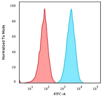
 FCM (Flow Cytometry)
(IDH1 Antibody (Center) flow cytometric analysis of 293 cells (right histogram) compared to a negative control cell (left histogram).FITC-conjugated goat-anti-rabbit secondary antibodies were used for the analysis.)
FCM (Flow Cytometry)
(IDH1 Antibody (Center) flow cytometric analysis of 293 cells (right histogram) compared to a negative control cell (left histogram).FITC-conjugated goat-anti-rabbit secondary antibodies were used for the analysis.)
IDH1, Polyclonal Antibody (Cat# AAA28676)
Full Name
IDH1 Antibody (Center)
Gene Names
IDH1; IDH; IDP; IDCD; IDPC; PICD; HEL-216; HEL-S-26
Reactivity
Human, mouse (Predicted Reactivity: Bovine, Rat)
Applications
EIA, IHC, IF, WB, FC/FACS
Purity
Purified Rabbit Polyclonal Antibody (Pab)
Pricing


 Application Data
(At 25 degree C. Samples were then incubated with primary Ab(At 37 degree C. An AlexaFluor594 conjugated goat anti-rabbit IgG(H+L) Ab(Red) and an AlexaFluor488 conjugated goat anti-mouse IgG(H+L) Ab(Green) were used as the secondary antibody.The nuclear counter stain is DAPI (blue).)
Application Data
(At 25 degree C. Samples were then incubated with primary Ab(At 37 degree C. An AlexaFluor594 conjugated goat anti-rabbit IgG(H+L) Ab(Red) and an AlexaFluor488 conjugated goat anti-mouse IgG(H+L) Ab(Green) were used as the secondary antibody.The nuclear counter stain is DAPI (blue).)
PFKFB3, Polyclonal Antibody (Cat# AAA31288)
Full Name
Phospho-PFKFB3 (Ser461) Antibody
Gene Names
PFKFB3; PFK2; IPFK2
Reactivity
Human, Rat
Applications
WB, IHC, IF, ICC, EIA
Purity
The antibody is from purified rabbit serum by affinity purification via sequential chromatography on phospho-peptide and non-phospho-peptide affinity columns.
Pricing


 IHC (Immunohistochemistry)
(At 1/100 staining Human gastric cancer and adjacent normal tissues by IHC-P. The sample was formaldehyde fixed and a heat mediated antigen retrieval step in citrate buffer was performed. The sample was then blocked and incubated with the primary antibody at 4 degree C overnight. An HRP conjugated anti-Rabbit antibody was used as the secondary antibody.)
IHC (Immunohistochemistry)
(At 1/100 staining Human gastric cancer and adjacent normal tissues by IHC-P. The sample was formaldehyde fixed and a heat mediated antigen retrieval step in citrate buffer was performed. The sample was then blocked and incubated with the primary antibody at 4 degree C overnight. An HRP conjugated anti-Rabbit antibody was used as the secondary antibody.)
SRF, Polyclonal Antibody (Cat# AAA31361)
Full Name
Phospho-SRF (Ser103) Antibody
Gene Names
SRF; MCM1
Reactivity
Human, Mouse, Rat
Predicted Reactivity: Pig (100%), Bovine (100%), Rabbit (89%), Dog (100%)
Predicted Reactivity: Pig (100%), Bovine (100%), Rabbit (89%), Dog (100%)
Applications
WB, IHC, EIA
Purity
The antibody is from purified rabbit serum by affinity purification via sequential chromatography on phospho-peptide and non-phospho-peptide affinity columns.
Pricing


 WB (Western Blot)
(Formalin-fixed and paraffin-embedded human brain tissue reacted with PDIA6 Antibody (Center K159), which was peroxidase-conjugated to the secondary antibody, followed by DAB staining. This data demonstrates the use of this antibody for immunohistochemistry; clinical relevance has not been evaluated.)
WB (Western Blot)
(Formalin-fixed and paraffin-embedded human brain tissue reacted with PDIA6 Antibody (Center K159), which was peroxidase-conjugated to the secondary antibody, followed by DAB staining. This data demonstrates the use of this antibody for immunohistochemistry; clinical relevance has not been evaluated.)
PDIA6, Polyclonal Antibody (Cat# AAA28792)
Full Name
PDIA6 Antibody (Center K159)
Gene Names
PDIA6; P5; ERP5; TXNDC7
Reactivity
Human, mouse, rat
Applications
EIA, IHC, IF, WB, FC/FACS
Purity
Purified Rabbit Polyclonal Antibody (Pab)
Pricing


 FCM (Flow Cytometry)
(Overlay histogram showing Raji cells stained with AAA28060 (red line) at 1:500. The cells were incubated in 10% normal goat serum to block non-specific protein-protein interactions followed by the antibody (1ug/1*106cells) for 1 h at 4 degree C. The secondary antibody used was FITC-conjugated Goat Anti-Mouse IgG(H+L) at 1/100 dilution for 30min at 4 degree C. Isotype control antibody (green line) was mouse IgG2b (1ug/1*106cells) used under the same conditions. Acquisition of >10,000 events was performed.)
FCM (Flow Cytometry)
(Overlay histogram showing Raji cells stained with AAA28060 (red line) at 1:500. The cells were incubated in 10% normal goat serum to block non-specific protein-protein interactions followed by the antibody (1ug/1*106cells) for 1 h at 4 degree C. The secondary antibody used was FITC-conjugated Goat Anti-Mouse IgG(H+L) at 1/100 dilution for 30min at 4 degree C. Isotype control antibody (green line) was mouse IgG2b (1ug/1*106cells) used under the same conditions. Acquisition of >10,000 events was performed.)
CD45, Monoclonal Antibody (Cat# AAA28060)
Full Name
CD45 Monoclonal Antibody
Gene Names
PTPRC; LCA; LY5; B220; CD45; L-CA; T200; CD45R; GP180
Reactivity
Human
Applications
EIA, WB, IHC, IF, FC/FACS
Purity
>95%, Protein A purified
Pricing


 ChIP (Chromatin Immunoprecipitation)
(Chromatin immunoprecipitation analysis of extracts of HeLa cells, using Acetyl-Histone H3-K9/K14/K18/K23/K27 antibody and rabbit IgG.The amount of immunoprecipitated DNA was checked by quantitative PCR. Histogram was constructed by the ratios of the immunoprecipitated DNA to the input.)
ChIP (Chromatin Immunoprecipitation)
(Chromatin immunoprecipitation analysis of extracts of HeLa cells, using Acetyl-Histone H3-K9/K14/K18/K23/K27 antibody and rabbit IgG.The amount of immunoprecipitated DNA was checked by quantitative PCR. Histogram was constructed by the ratios of the immunoprecipitated DNA to the input.)
Acetyl-Histone H3-K9/K14/K18/K23/K27, Polyclonal Antibody (Cat# AAA28357)
Full Name
Acetyl-Histone H3-K9/K14/K18/K23/K27 Rabbit pAb
Gene Names
HIST1H3A; H3/A; H3FA
Reactivity
Human, Mouse, Rat, Other (Wide Range)
Applications
WB, IHC, IF, ChIP
Purity
Affinity purification
Pricing


 Application Data
(C:FGFR2/isolectinB4 (C) and FGFR1/isolectinB4 (D) staining of apparent mesenchymal cells and the subpopulation of endothelial cells. Virtually all other dispersed apparent mesenchymal cells express FGFR1 and FGFR2 (merged image in E). F: FGFR2 (F) and FGFR1 (G) staining in clustered cells of epithelial origin (inferred by morphology here) demonstrating that epithelial cells express both FGFR1 and FGFR2 (merged image with DAPI staining in H).)
Application Data
(C:FGFR2/isolectinB4 (C) and FGFR1/isolectinB4 (D) staining of apparent mesenchymal cells and the subpopulation of endothelial cells. Virtually all other dispersed apparent mesenchymal cells express FGFR1 and FGFR2 (merged image in E). F: FGFR2 (F) and FGFR1 (G) staining in clustered cells of epithelial origin (inferred by morphology here) demonstrating that epithelial cells express both FGFR1 and FGFR2 (merged image with DAPI staining in H).)
FGFR2, Polyclonal Antibody (Cat# AAA26854)
Full Name
FGFR2, NT (FGFR2, BEK, KGFR, KSAM, Fibroblast growth factor receptor 2, K-sam, Keratinocyte growth factor receptor, CD332) (FITC)
Gene Names
FGFR2; BEK; JWS; BBDS; CEK3; CFD1; ECT1; KGFR; TK14; TK25; BFR-1; CD332; K-SAM
Reactivity
Human, Monkey, Mouse, Rat
Applications
WB, IHC, IF, FC/FACS
Purity
Purified by Protein G Affinity Chromatography.
Pricing


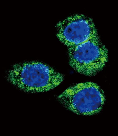
 IF (Immunofluorescence)
(Immunofluorescence analysis of HeLa cells using APP antibody at dilution of 1:100. Blue: DAPI for nuclear staining.)
IF (Immunofluorescence)
(Immunofluorescence analysis of HeLa cells using APP antibody at dilution of 1:100. Blue: DAPI for nuclear staining.)
APP, Polyclonal Antibody (Cat# AAA28326)
Full Name
APP Rabbit pAb
Gene Names
APP; AAA; AD1; PN2; ABPP; APPI; CVAP; ABETA; PN-II; CTFgamma
Reactivity
Human, Mouse, Rat
Applications
WB, IHC, IF
Purity
Affinity purification
Pricing


 IF (Immunofluorescence)
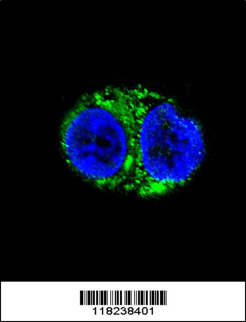
(Immunofluorescent analysis of 4% paraformaldehyde-fixed, 0.1% Triton X-100 permeabilized MCF-7 (human breast cancer cell line) cells labeling Pdx1 with AAA14796 at 1:25 dilution, followed by Dylight® 488-conjugated IgG goat anti-rabbit secondary antibody at 1:200 dilution (green). Immunofluorescence image showing cytoplasm staining on MCF-7 cell line. Cytoplasmic actin is detected with Dylight® 554 Phalloidin (PD18466410) at 1:100 dilution (red). The nuclear counter stain is DAPI (blue).)
IF (Immunofluorescence)
(Immunofluorescent analysis of 4% paraformaldehyde-fixed, 0.1% Triton X-100 permeabilized MCF-7 (human breast cancer cell line) cells labeling Pdx1 with AAA14796 at 1:25 dilution, followed by Dylight® 488-conjugated IgG goat anti-rabbit secondary antibody at 1:200 dilution (green). Immunofluorescence image showing cytoplasm staining on MCF-7 cell line. Cytoplasmic actin is detected with Dylight® 554 Phalloidin (PD18466410) at 1:100 dilution (red). The nuclear counter stain is DAPI (blue).)
OPN-a/b, Polyclonal Antibody (Cat# AAA14796)
Full Name
OPN-a/b, NT (SPP1, BNSP, OPN, Osteopontin, Bone sialoprotein 1, Nephropontin, Secreted phosphoprotein 1, Urinary stone protein, Uropontin)
Reactivity
Human
Applications
EL/EIA, WB, IHC, IF
Purity
Affinity Purified
Purified by Protein A affinity chromatography.
Purified by Protein A affinity chromatography.
Pricing


 SDS-PAGE
(SDS-PAGE Analysis Purified with Nucleolin Monoclonal Antibody (364-5). Confirmation of Integrity and Purity of Antibody)
SDS-PAGE
(SDS-PAGE Analysis Purified with Nucleolin Monoclonal Antibody (364-5). Confirmation of Integrity and Purity of Antibody)
Nucleolin, Monoclonal Antibody (Cat# AAA13835)
Full Name
Nucleolin (Marker of Human Cells) Mouse Monoclonal Antibody
Gene Names
NCL; C23
Reactivity
Does not react with Mouse, Human, Rat and Cow
Applications
FC/FACS, IF, WB, IHC
Pricing


 FCM (Flow Cytometry)
(Overlay histogram showing HepG2 cells stained with AAA28061 (red line) at 1:100. The cells were fixed in 4% formaldehyde and permeated by 0.2% TritonX-100. Then 10% normal goat serum was Incubated to block non-specific protein-protein interactions followed by the antibody (1ug/1*106cells) for 1 h at 4 degree C. The secondary antibody used was FITC-conjugated Goat Anti-Mouse IgG(H+L) at 1/100 dilution for 30min at 4 degree C. Isotype control antibody (green line) was mouse IgG2b (1ug/1*106cells) used under the same conditions. Acquisition of >10,000 events was performed.)
FCM (Flow Cytometry)
(Overlay histogram showing HepG2 cells stained with AAA28061 (red line) at 1:100. The cells were fixed in 4% formaldehyde and permeated by 0.2% TritonX-100. Then 10% normal goat serum was Incubated to block non-specific protein-protein interactions followed by the antibody (1ug/1*106cells) for 1 h at 4 degree C. The secondary antibody used was FITC-conjugated Goat Anti-Mouse IgG(H+L) at 1/100 dilution for 30min at 4 degree C. Isotype control antibody (green line) was mouse IgG2b (1ug/1*106cells) used under the same conditions. Acquisition of >10,000 events was performed.)
CD63, Monoclonal Antibody (Cat# AAA28061)
Full Name
CD63 Monoclonal Antibody
Gene Names
Cd63; ME491; C75951; Tspan30
Reactivity
Human
Applications
EIA, WB, IHC, IF, FC/FACS
Purity
>95%, Protein A purified
Pricing


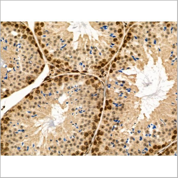
 IHC (Immunohistchemistry)
(At 1/100 staining Human gastric cancer by IHC-P. The sample was formaldehyde fixed and a heat mediated antigen retrieval step in citrate buffer was performed. The sample was then blocked and incubated with the primary antibody at 4 degree C overnight. An HRP conjugated anti-Rabbit antibody was used as the secondary antibody.)
IHC (Immunohistchemistry)
(At 1/100 staining Human gastric cancer by IHC-P. The sample was formaldehyde fixed and a heat mediated antigen retrieval step in citrate buffer was performed. The sample was then blocked and incubated with the primary antibody at 4 degree C overnight. An HRP conjugated anti-Rabbit antibody was used as the secondary antibody.)
PPAR alpha, Polyclonal Antibody (Cat# AAA31448)
Full Name
Phospho-PPAR alpha (Ser12) Antibody
Gene Names
PPARA; PPAR; NR1C1; hPPAR; PPARalpha
Reactivity
Human, Mouse, Rat, Monkey
Predicted Reactivity: Bovine (100%), Horse (100%), Sheep (100%), Dog (100%), Chicken (83%)
Predicted Reactivity: Bovine (100%), Horse (100%), Sheep (100%), Dog (100%), Chicken (83%)
Applications
WB, IHC, EIA
Purity
The antibody is from purified rabbit serum by affinity purification via sequential chromatography on phospho-peptide and non-phospho-peptide affinity columns.
Pricing


 WB (Western Blot)
(Western Blot Analysis of Human Spleen cell lysate using HLA-Pan Mouse Monoclonal Antibody (CR3/43).)
WB (Western Blot)
(Western Blot Analysis of Human Spleen cell lysate using HLA-Pan Mouse Monoclonal Antibody (CR3/43).)
HLA-DP/-DQ/-DR, Monoclonal Antibody (Cat# AAA23889)
Full Name
HLA-DP/-DQ/-DR (MHC II)
Gene Names
HLA-DPB1; DPB1; HLA-DP; HLA-DPB; HLA-DP1B
Reactivity
Human
Applications
IF, IHC
Pricing


 Application Data
(Analysis of Protein Array containing more than 19,000 full-length human proteins using ERCC1 Mouse Monoclonal Antibody (ERCC1/2318). Z- and S- Score: The Z-score represents the strength of a signal that a monoclonal antibody (MAb) (in combination with a fluorescently-tagged anti-IgG secondary antibody) produces when binding to a particular protein on the HuProtTM array. Z-scores are described in units of standard deviations (SD's) above the mean value of all signals generated on that array. If targets on HuProtTM are arranged in descending order of the Z-score, the S-score is the difference (also in units of SD's) between the Z-score. S-score therefore represents the relative target specificity of a MAb to its intended target. A MAb is considered to specific to its intended target, if the MAb has an S-score of at least 2.5. For example, if a MAb binds to protein X with a Z-score of 43 and to protein Y with a Z-score of 14, then the S-score for the binding of that MAb to protein X is equal to 29.)
Application Data
(Analysis of Protein Array containing more than 19,000 full-length human proteins using ERCC1 Mouse Monoclonal Antibody (ERCC1/2318). Z- and S- Score: The Z-score represents the strength of a signal that a monoclonal antibody (MAb) (in combination with a fluorescently-tagged anti-IgG secondary antibody) produces when binding to a particular protein on the HuProtTM array. Z-scores are described in units of standard deviations (SD's) above the mean value of all signals generated on that array. If targets on HuProtTM are arranged in descending order of the Z-score, the S-score is the difference (also in units of SD's) between the Z-score. S-score therefore represents the relative target specificity of a MAb to its intended target. A MAb is considered to specific to its intended target, if the MAb has an S-score of at least 2.5. For example, if a MAb binds to protein X with a Z-score of 43 and to protein Y with a Z-score of 14, then the S-score for the binding of that MAb to protein X is equal to 29.)
ERCC1/RAD10, Monoclonal Antibody (Cat# AAA23913)
Full Name
ERCC1/RAD10 (Tumor Progression Marker)
Gene Names
ERCC1; UV20; COFS4; RAD10
Reactivity
Human
Applications
IHC
Purity
Purified Ab with BSA and Azide at 200ug/ml OR Purified Ab WITHOUT BSA and Azide at 1.0mg/ml
Pricing


 IP (Immunoprecipitation)
(Immunoprecipitating GAPDH in Hela whole cell lysateLane 1: Mouse control IgG instead of in Hela whole cell lysate. Lane 2: (5ul) + Hela whole cell lysate (500ug)Lane 3: Hela whole cell lysate (10ug)For western blotting, the blot was detected at 1:5000, and a HRP-conjugated Protein G antibody was used as the secondary antibody at 1:2000)
IP (Immunoprecipitation)
(Immunoprecipitating GAPDH in Hela whole cell lysateLane 1: Mouse control IgG instead of in Hela whole cell lysate. Lane 2: (5ul) + Hela whole cell lysate (500ug)Lane 3: Hela whole cell lysate (10ug)For western blotting, the blot was detected at 1:5000, and a HRP-conjugated Protein G antibody was used as the secondary antibody at 1:2000)
GAPDH, Monoclonal Antibody (Cat# AAA27042)
Full Name
GAPDH Monoclonal Antibody
Reactivity
Human, Mouse, Rabbit
Applications
EIA, WB, IHC, IP, IF
Purity
>95%, Protein G purified
Pricing


 FCM (Flow Cytometry)
(Overlay histogram showing MCF-7 cells stained with (red line). The cells were fixed with 70% Ethylalcohol (18h) and then incubated in 10% normal goat serum to block non-specific protein-protein interactions followed by the primary antibody at 1/200 for 1 h at 4 degree C. The secondary antibody used was FITC goat anti-mouse IgG(H+L) at 1/100 dilution for 30min at 4 degree C. Isotype control antibody (green line) was mouse IgG1 used under the same conditions. Acquisition of >10,000 events was performed.)
FCM (Flow Cytometry)
(Overlay histogram showing MCF-7 cells stained with (red line). The cells were fixed with 70% Ethylalcohol (18h) and then incubated in 10% normal goat serum to block non-specific protein-protein interactions followed by the primary antibody at 1/200 for 1 h at 4 degree C. The secondary antibody used was FITC goat anti-mouse IgG(H+L) at 1/100 dilution for 30min at 4 degree C. Isotype control antibody (green line) was mouse IgG1 used under the same conditions. Acquisition of >10,000 events was performed.)
HSPA8, Monoclonal Antibody (Cat# AAA27046)
Full Name
HSPA8 Monoclonal Antibody
Gene Names
HSPA8; LAP1; HSC54; HSC70; HSC71; HSP71; HSP73; NIP71; HSPA10
Reactivity
Human
Applications
EIA, WB, IHC, IF, FC/FACS
Purity
>95%, Protein G purified
Pricing


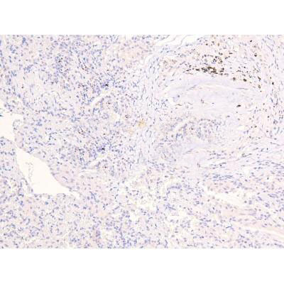
 IHC (Immunohistochemistry)
(Figure 7. IHC analysis of MED4 using anti-MED4 antibody (AAA19178).MED4 was detected in paraffin-embedded section of human rectal cancer tissue. Heat mediated antigen retrieval was performed in citrate buffer (pH6, epitope retrieval solution) for 20 mins. The tissue section was blocked with 10% goat serum. The tissue section was then incubated with 1ug/ml rabbit anti-MED4 Antibody (AAA19178) overnight at 4 degree C. Biotinylated goat anti-rabbit IgG was used as secondary antibody and incubated for 30 minutes at 37 degree C. The tissue section was developed using Strepavidin-Biotin-Complex (SABC) with DAB as the chromogen.)
IHC (Immunohistochemistry)
(Figure 7. IHC analysis of MED4 using anti-MED4 antibody (AAA19178).MED4 was detected in paraffin-embedded section of human rectal cancer tissue. Heat mediated antigen retrieval was performed in citrate buffer (pH6, epitope retrieval solution) for 20 mins. The tissue section was blocked with 10% goat serum. The tissue section was then incubated with 1ug/ml rabbit anti-MED4 Antibody (AAA19178) overnight at 4 degree C. Biotinylated goat anti-rabbit IgG was used as secondary antibody and incubated for 30 minutes at 37 degree C. The tissue section was developed using Strepavidin-Biotin-Complex (SABC) with DAB as the chromogen.)
MED4, Polyclonal Antibody (Cat# AAA19178)
Full Name
Anti-MED4 Picoband Antibody
Gene Names
MED4; ARC36; VDRIP; DRIP36; TRAP36; HSPC126
Reactivity
Human, Mouse, Rat
No cross reactivity with other proteins.
No cross reactivity with other proteins.
Applications
EIA, IHC, WB
Purity
Immunogen affinity purified
Pricing


 IF (Immunofluorescence)
(Immunofluorescent analysis of 4% paraformaldehyde-fixed, 0.1% Triton X-100 permeabilized MCF-7 (human breast cancer cell line) cells labeling Pdx1 with at 1:25 dilution, followed by DyLight 488-conjugated IgG goat anti-rabbit secondary antibody at 1:200 dilution (green). Immunofluorescence image showing cytoplasm staining on MCF-7 cell line. Cytoplasmic actin is detected with DyLight 554 Phalloidin (PD18466410) at 1:100 dilution (red). The nuclear counter stain is DAPI (blue).)
IF (Immunofluorescence)
(Immunofluorescent analysis of 4% paraformaldehyde-fixed, 0.1% Triton X-100 permeabilized MCF-7 (human breast cancer cell line) cells labeling Pdx1 with at 1:25 dilution, followed by DyLight 488-conjugated IgG goat anti-rabbit secondary antibody at 1:200 dilution (green). Immunofluorescence image showing cytoplasm staining on MCF-7 cell line. Cytoplasmic actin is detected with DyLight 554 Phalloidin (PD18466410) at 1:100 dilution (red). The nuclear counter stain is DAPI (blue).)
OPN-a/b, Polyclonal Antibody (Cat# AAA26858)
Full Name
OPN-a/b, NT (SPP1, BNSP, OPN, Osteopontin, Bone sialoprotein 1, Nephropontin, Secreted phosphoprotein 1, Urinary stone protein, Uropontin) (Biotin)
Gene Names
SPP1; OPN; BNSP; BSPI; ETA-1
Reactivity
Human
Applications
WB, IHC, IF, EIA
Purity
Purified by Protein A and Peptide Affinity Chromatography.
Pricing


 IHC (Immunohistchemistry)
(Figure 6. IHC analysis of Musashi 1/Msi1 using anti- Musashi 1/Msi1 antibody (AAA19172).Musashi 1/Msi1 was detected in paraffin-embedded section of human mammary cancer tissues. Heat mediated antigen retrieval was performed in citrate buffer (pH6, epitope retrieval solution) for 20 mins. The tissue section was blocked with 10% goat serum. The tissue section was then incubated with 1ug/ml rabbit anti- Musashi 1/Msi1 Antibody (AAA19172) overnight at 4 degree C. Biotinylated goat anti-rabbit IgG was used as secondary antibody and incubated for 30 minutes at 37 degree C. The tissue section was developed using Strepavidin-Biotin-Complex (SABC) with DAB as the chromogen.)
IHC (Immunohistchemistry)
(Figure 6. IHC analysis of Musashi 1/Msi1 using anti- Musashi 1/Msi1 antibody (AAA19172).Musashi 1/Msi1 was detected in paraffin-embedded section of human mammary cancer tissues. Heat mediated antigen retrieval was performed in citrate buffer (pH6, epitope retrieval solution) for 20 mins. The tissue section was blocked with 10% goat serum. The tissue section was then incubated with 1ug/ml rabbit anti- Musashi 1/Msi1 Antibody (AAA19172) overnight at 4 degree C. Biotinylated goat anti-rabbit IgG was used as secondary antibody and incubated for 30 minutes at 37 degree C. The tissue section was developed using Strepavidin-Biotin-Complex (SABC) with DAB as the chromogen.)
Musashi 1/Msi1, Polyclonal Antibody (Cat# AAA19172)
Full Name
Anti-Musashi 1/Msi1 Picoband Antibody
Reactivity
Human, Mouse, Rat
No cross reactivity with other proteins
No cross reactivity with other proteins
Applications
IHC, WB
Purity
Immunogen affinity purified
Pricing


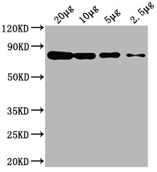
 WB (Western Blot)
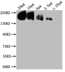
(WB Suggested Anti-GSR Antibody Titration: 0.2-1 ug/mlELISA Titer: 1:312500Positive Control: Hela cell lysate)
WB (Western Blot)
(WB Suggested Anti-GSR Antibody Titration: 0.2-1 ug/mlELISA Titer: 1:312500Positive Control: Hela cell lysate)
GSR, Polyclonal Antibody (Cat# AAA23553)
Full Name
GSR antibody - N-terminal region
Gene Names
GSR; GR; HEL-75; HEL-S-122m
Reactivity
Tested Reactivity: Human, Mouse, Zebrafish
Predicted Reactivity: Cow, Dog, Guinea Pig, Horse, Human, Mouse, Rabbit, Rat
Predicted Reactivity: Cow, Dog, Guinea Pig, Horse, Human, Mouse, Rabbit, Rat
Applications
IHC, WB
Purity
Affinity Purified
Pricing


 WB (Western Blot)
(Western blot analysis of Human Lysates showing detection of Hsp90 protein using Mouse Anti-Hsp90 Monoclonal Antibody, Clone H9010 . Primary Antibody: Mouse Anti-Hsp90 Monoclonal Antibody at 1:1000. Comparison of clone H9010 behavior with Hsp90 human beta (1) and Hsp90 human alpha (2). Courtesy of: David Toft, Mayo Clinic.)
WB (Western Blot)
(Western blot analysis of Human Lysates showing detection of Hsp90 protein using Mouse Anti-Hsp90 Monoclonal Antibody, Clone H9010 . Primary Antibody: Mouse Anti-Hsp90 Monoclonal Antibody at 1:1000. Comparison of clone H9010 behavior with Hsp90 human beta (1) and Hsp90 human alpha (2). Courtesy of: David Toft, Mayo Clinic.)
HSP90, Monoclonal Antibody (Cat# AAA27660)
Full Name
HSP90 Antibody
Gene Names
HSP90AB1; HSP84; HSPC2; HSPCB; D6S182; HSP90B
Reactivity
Human, Mouse, Rat, Rabbit, Chicken, Dog, Fish, Shark, Hamster
Applications
WB, IHC, ICC, IF, IP, EIA, AM
Purity
Protein G Purified
Pricing


 SDS-PAGE
(Purified ER-beta Mouse Monoclonal Antibody (ESR2/686). Confirmation of Integrity and Purity of Antibody.)
SDS-PAGE
(Purified ER-beta Mouse Monoclonal Antibody (ESR2/686). Confirmation of Integrity and Purity of Antibody.)
ER-beta1 (Estrogen Receptor beta-1), Monoclonal Antibody (Cat# AAA13846)
Full Name
ER-beta1 (Estrogen Receptor beta-1) Mouse Monoclonal Antibody
Gene Names
ESR2; Erb; ESRB; ESTRB; NR3A2; ER-BETA; ESR-BETA
Reactivity
Human
Applications
FC/FACS, IF, WB, IHC
Pricing


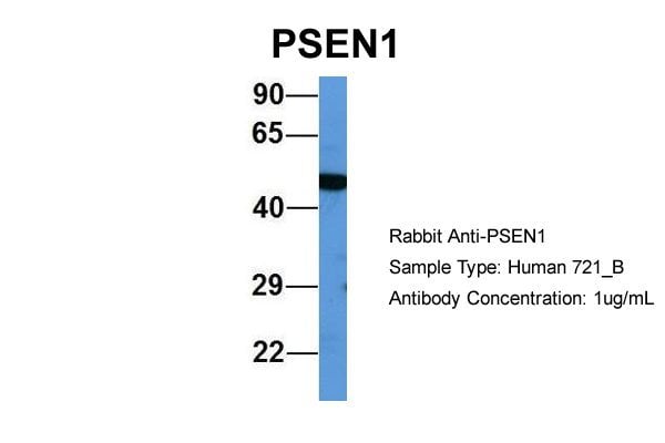


 WB (Western Blot)
(WB Suggested Anti-PSEN1 Antibody Titration: 0.2-1 ug/mlELISA Titer: 1:312500Positive Control: Human brain)
WB (Western Blot)
(WB Suggested Anti-PSEN1 Antibody Titration: 0.2-1 ug/mlELISA Titer: 1:312500Positive Control: Human brain)
PSEN1, Polyclonal Antibody (Cat# AAA23585)
Full Name
PSEN1 antibody - middle region
Gene Names
PSEN1; AD3; FAD; PS1; PS-1; S182; ACNINV3
Reactivity
Cow, Dog, Guinea Pig, Horse, Human, Mouse, Rabbit, Rat
Applications
IHC, IP, WB
Purity
Affinity Purified
Pricing


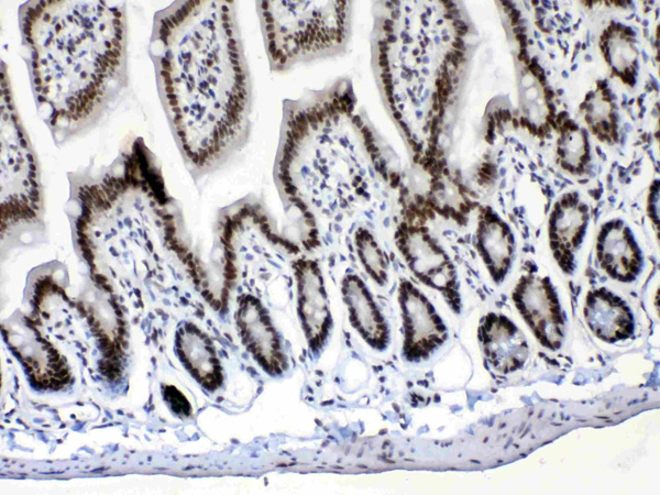
 WB (Western Blot)
(Western Blot (WB)Host: RabbitTarget: SOX10Positive control (+): Rat brain (R-BR)Negative control (-): Mouse intestine (M-IN) Antibody concentration: 1ug/ml)
WB (Western Blot)
(Western Blot (WB)Host: RabbitTarget: SOX10Positive control (+): Rat brain (R-BR)Negative control (-): Mouse intestine (M-IN) Antibody concentration: 1ug/ml)
SOX10, Polyclonal Antibody (Cat# AAA23423)
Full Name
SOX10 Antibody - middle region
Gene Names
SOX10; DOM; WS4; PCWH; WS2E; WS4C
Reactivity
Reactivity: Human
Predicted: Cow, Dog, Guinea Pig, Horse, Human, Mouse, Rabbit, Rat
Predicted: Cow, Dog, Guinea Pig, Horse, Human, Mouse, Rabbit, Rat
Applications
IHC, WB
Purity
Affinity Purified
Pricing


 Application Data
(Analysis of Protein Array containing more than 19,000 full-length human proteins using Calretinin Mouse Monoclonal Antibody (CALB2/2602). Z- and S- Score: The Z-score represents the strength of a signal that a monoclonal antibody (MAb) (in combination with a fluorescently-tagged anti-IgG secondary antibody) produces when binding to a particular protein on the HuProtTM array. Z-scores are described in units of standard deviations (SD's) above the mean value of all signals generated on that array. If targets on HuProtTM are arranged in descending order of the Z-score, the S-score is the difference (also in units of SD's) between the Z-score. S-score therefore represents the relative target specificity of a MAb to its intended target. A MAb is considered to specific to its intended target, if the MAb has an S-score of at least 2.5. For example, if a MAb binds to protein X with a Z-score of 43 and to protein Y with a Z-score of 14, then the S-score for the binding of that MAb to protein X is equal to 29.)
Application Data
(Analysis of Protein Array containing more than 19,000 full-length human proteins using Calretinin Mouse Monoclonal Antibody (CALB2/2602). Z- and S- Score: The Z-score represents the strength of a signal that a monoclonal antibody (MAb) (in combination with a fluorescently-tagged anti-IgG secondary antibody) produces when binding to a particular protein on the HuProtTM array. Z-scores are described in units of standard deviations (SD's) above the mean value of all signals generated on that array. If targets on HuProtTM are arranged in descending order of the Z-score, the S-score is the difference (also in units of SD's) between the Z-score. S-score therefore represents the relative target specificity of a MAb to its intended target. A MAb is considered to specific to its intended target, if the MAb has an S-score of at least 2.5. For example, if a MAb binds to protein X with a Z-score of 43 and to protein Y with a Z-score of 14, then the S-score for the binding of that MAb to protein X is equal to 29.)
Calretinin/Calbindin 2, Monoclonal Antibody (Cat# AAA23938)
Full Name
Calretinin/Calbindin 2 (Mesothelioma Marker)
Gene Names
CALB2; CR; CAL2; CAB29
Reactivity
Human
Applications
WB, IHC
Purity
Purified Ab with BSA and Azide at 200ug/ml OR Purified Ab WITHOUT BSA at 1.0mg/ml
Pricing


 Application Data
(At 25 degree C. Samples were then incubated with primary Ab(At 37 degree C. An AlexaFluor594 conjugated goat anti-rabbit IgG(H+L) Ab(Red) and an AlexaFluor488 conjugated goat anti-mouse IgG(H+L) Ab(Green) were used as the secondary antibody.The nuclear counter stain is DAPI(blue).)
Application Data
(At 25 degree C. Samples were then incubated with primary Ab(At 37 degree C. An AlexaFluor594 conjugated goat anti-rabbit IgG(H+L) Ab(Red) and an AlexaFluor488 conjugated goat anti-mouse IgG(H+L) Ab(Green) were used as the secondary antibody.The nuclear counter stain is DAPI(blue).)
TLK1, Polyclonal Antibody (Cat# AAA31385)
Full Name
Phospho-TLK1 (Ser741) Antibody
Gene Names
TLK1; PKU-beta
Reactivity
Human, Mouse, Rat
Predicted Reactivity: Pig (100%), Bovine (100%), Horse (100%), Sheep (100%), Rabbit (100%), Dog (100%), Chicken (100%)
Predicted Reactivity: Pig (100%), Bovine (100%), Horse (100%), Sheep (100%), Rabbit (100%), Dog (100%), Chicken (100%)
Applications
WB, IHC, IF, ICC, EIA
Purity
The antibody is from purified rabbit serum by affinity purification via sequential chromatography on phospho-peptide and non-phospho-peptide affinity columns.
Pricing


 Application Data
(C:FGFR2/isolectinB4 (C) and FGFR1/isolectinB4 (D) staining of apparent mesenchymal cells and the subpopulation of endothelial cells. Virtually all other dispersed apparent mesenchymal cells express FGFR1 and FGFR2 (merged image in E). F: FGFR2 (F) and FGFR1 (G) staining in clustered cells of epithelial origin (inferred by morphology here) demonstrating that epithelial cells express both FGFR1 and FGFR2 (merged image with DAPI staining in H).)
Application Data
(C:FGFR2/isolectinB4 (C) and FGFR1/isolectinB4 (D) staining of apparent mesenchymal cells and the subpopulation of endothelial cells. Virtually all other dispersed apparent mesenchymal cells express FGFR1 and FGFR2 (merged image in E). F: FGFR2 (F) and FGFR1 (G) staining in clustered cells of epithelial origin (inferred by morphology here) demonstrating that epithelial cells express both FGFR1 and FGFR2 (merged image with DAPI staining in H).)
FGFR2, Polyclonal Antibody (Cat# AAA26855)
Full Name
FGFR2, NT (FGFR2, BEK, KGFR, KSAM, Fibroblast growth factor receptor 2, K-sam, Keratinocyte growth factor receptor, CD332) (Azide free) (HRP)
Gene Names
FGFR2; BEK; JWS; BBDS; CEK3; CFD1; ECT1; KGFR; TK14; TK25; BFR-1; CD332; K-SAM
Reactivity
Human, Monkey, Mouse, Rat
Applications
IHC, EIA, WB
Purity
Purified by Protein G Affinity Chromatography.
Pricing


 Application Data
(C:FGFR2/isolectinB4 (C) and FGFR1/isolectinB4 (D) staining of apparent mesenchymal cells and the subpopulation of endothelial cells. Virtually all other dispersed apparent mesenchymal cells express FGFR1 and FGFR2 (merged image in E). F: FGFR2 (F) and FGFR1 (G) staining in clustered cells of epithelial origin (inferred by morphology here) demonstrating that epithelial cells express both FGFR1 and FGFR2 (merged image with DAPI staining in H).)
Application Data
(C:FGFR2/isolectinB4 (C) and FGFR1/isolectinB4 (D) staining of apparent mesenchymal cells and the subpopulation of endothelial cells. Virtually all other dispersed apparent mesenchymal cells express FGFR1 and FGFR2 (merged image in E). F: FGFR2 (F) and FGFR1 (G) staining in clustered cells of epithelial origin (inferred by morphology here) demonstrating that epithelial cells express both FGFR1 and FGFR2 (merged image with DAPI staining in H).)
FGFR2, Polyclonal Antibody (Cat# AAA26853)
Full Name
FGFR2, NT (FGFR2, BEK, KGFR, KSAM, Fibroblast growth factor receptor 2, K-sam, Keratinocyte growth factor receptor, CD332) (Biotin)
Gene Names
FGFR2; BEK; JWS; BBDS; CEK3; CFD1; ECT1; KGFR; TK14; TK25; BFR-1; CD332; K-SAM
Reactivity
Human, Monkey, Mouse, Rat
Applications
FC/FACS, EIA, IF, IHC, WB
Purity
Purified by Protein G Affinity Chromatography.
Pricing


 Application Data
(C:FGFR2/isolectinB4 (C) and FGFR1/isolectinB4 (D) staining of apparent mesenchymal cells and the subpopulation of endothelial cells. Virtually all other dispersed apparent mesenchymal cells express FGFR1 and FGFR2 (merged image in E). F: FGFR2 (F) and FGFR1 (G) staining in clustered cells of epithelial origin (inferred by morphology here) demonstrating that epithelial cells express both FGFR1 and FGFR2 (merged image with DAPI staining in H).)
Application Data
(C:FGFR2/isolectinB4 (C) and FGFR1/isolectinB4 (D) staining of apparent mesenchymal cells and the subpopulation of endothelial cells. Virtually all other dispersed apparent mesenchymal cells express FGFR1 and FGFR2 (merged image in E). F: FGFR2 (F) and FGFR1 (G) staining in clustered cells of epithelial origin (inferred by morphology here) demonstrating that epithelial cells express both FGFR1 and FGFR2 (merged image with DAPI staining in H).)
FGFR2, Polyclonal Antibody (Cat# AAA26851)
Full Name
FGFR2, NT (FGFR2, BEK, KGFR, KSAM, Fibroblast growth factor receptor 2, K-sam, Keratinocyte growth factor receptor, CD332) (AP)
Gene Names
FGFR2; BEK; JWS; BBDS; CEK3; CFD1; ECT1; KGFR; TK14; TK25; BFR-1; CD332; K-SAM
Reactivity
Human, Monkey, Mouse, Rat
Applications
IF, EIA, IHC, WB
Purity
Purified by Protein G Affinity Chromatography.
Pricing


 FCM (Flow Cytometry)
(Overlay histogram showing HepG2 cells stained with AAA28062 (red line) at 1:100. The cells were fixed in 4% formaldehyde and permeated by 0.2% TritonX-100. Then 10% normal goat serum was Incubated to block non-specific protein-protein interactions followed by the antibody (1ug/1*106cells) for 1 h at 4 degree C. The secondary antibody used was FITC-conjugated Goat Anti-Mouse IgG(H+L) at 1/100 dilution for 30min at 4 degree C. Isotype control antibody (green line) was mouse IgG2b (1ug/1*106cells) used under the same conditions. Acquisition of >10,000 events was performed.)
FCM (Flow Cytometry)
(Overlay histogram showing HepG2 cells stained with AAA28062 (red line) at 1:100. The cells were fixed in 4% formaldehyde and permeated by 0.2% TritonX-100. Then 10% normal goat serum was Incubated to block non-specific protein-protein interactions followed by the antibody (1ug/1*106cells) for 1 h at 4 degree C. The secondary antibody used was FITC-conjugated Goat Anti-Mouse IgG(H+L) at 1/100 dilution for 30min at 4 degree C. Isotype control antibody (green line) was mouse IgG2b (1ug/1*106cells) used under the same conditions. Acquisition of >10,000 events was performed.)
CD63, Monoclonal Antibody (Cat# AAA28062)
Full Name
CD63 Monoclonal Antibody
Gene Names
Cd63; ME491; C75951; Tspan30
Reactivity
Human, Rabbit
Applications
EIA, WB, IHC, IF, FC/FACS
Purity
>95%, Protein G purified
Pricing


 FCM (Flow Cytometry)
(Flow cytometric analysis of Hela cells with Hsp70 antibody at 1/50 dilution (blue) compared with an unlabelled control (cells without incubation with primary antibody; red). Alexa Fluor 488-conjugated goat anti rabbit IgG was used as the secondary antibody)
FCM (Flow Cytometry)
(Flow cytometric analysis of Hela cells with Hsp70 antibody at 1/50 dilution (blue) compared with an unlabelled control (cells without incubation with primary antibody; red). Alexa Fluor 488-conjugated goat anti rabbit IgG was used as the secondary antibody)
Hsp70, Monoclonal Antibody (Cat# AAA29972)
Full Name
Hsp70 Antibody
Gene Names
HSPA1B; HSP72; HSPA1; HSX70; HSP70-1; HSP70-2; HSP70.1; HSP70.2; HSP70-1B
Reactivity
Human, Mouse, Rat
Applications
WB, ICC, IF, IHC, FC/FACS
Purity
ProA affinity purified
Pricing


 Application Data
(Analysis of Protein Array containing more than 19, 000 full-length human proteins using Mouse Emerin Monoclonal Antibody (EMD/2167) Z- and S- Score: The Z-score represents the strength of a signal that a monoclonal antibody (MAb) (in combination with a fluorescently-tagged anti-IgG secondary antibody) produces when binding to a particular protein on the HuProtTM array. Z-scores are described in units of standard deviations (SD's) above the mean value of all signals generated on that array. If targets on HuProtTM are arranged in descending order of the Z-score, the S-score is the difference (also in units of SD's) between the Z-score. S-score therefore represents the relative target specificity of a MAb to its intended target. A MAb is considered to specific to its intended target, if the MAb has an S-score of at least 2.5. For example, if a MAb binds to protein X with a Z-score of 43 and to protein Y with a Z-score of 14, then the S-score for the binding of that MAb to protein X is equal to 29.)
Application Data
(Analysis of Protein Array containing more than 19, 000 full-length human proteins using Mouse Emerin Monoclonal Antibody (EMD/2167) Z- and S- Score: The Z-score represents the strength of a signal that a monoclonal antibody (MAb) (in combination with a fluorescently-tagged anti-IgG secondary antibody) produces when binding to a particular protein on the HuProtTM array. Z-scores are described in units of standard deviations (SD's) above the mean value of all signals generated on that array. If targets on HuProtTM are arranged in descending order of the Z-score, the S-score is the difference (also in units of SD's) between the Z-score. S-score therefore represents the relative target specificity of a MAb to its intended target. A MAb is considered to specific to its intended target, if the MAb has an S-score of at least 2.5. For example, if a MAb binds to protein X with a Z-score of 43 and to protein Y with a Z-score of 14, then the S-score for the binding of that MAb to protein X is equal to 29.)
Emerin, Monoclonal Antibody (Cat# AAA23898)
Full Name
Emerin (Papillary Thyroid Carcinoma and EDMD Marker)
Gene Names
EMD; STA; EDMD; LEMD5
Reactivity
Human. Others not known.
Applications
IHC
Pricing


 SDS-PAGE
(SDS-PAGE Analysis Purified HSP60 Mouse Monoclonal Antibody (CPTC-HSPD1-1). Confirmation of Purity and Integrity of Antibody.)
SDS-PAGE
(SDS-PAGE Analysis Purified HSP60 Mouse Monoclonal Antibody (CPTC-HSPD1-1). Confirmation of Purity and Integrity of Antibody.)
HSP60 (Heat Shock Protein 60), Monoclonal Antibody (Cat# AAA23919)
Full Name
HSP60 (Heat Shock Protein 60) (Mitochondrial Marker)
Gene Names
HSPD1; HLD4; CPN60; GROEL; HSP60; HSP65; SPG13; HSP-60; HuCHA60
Reactivity
Human
Applications
FC/FACS, IF, WB, IHC
Purity
Purified Ab with BSA and Azide at 200ug/ml OR Purified Ab WITHOUT BSA and Azide at 1.0mg/ml
Pricing


 IP (Immunoprecipitation)
(Immunoprecipitating PD-L2 in Hela whole cell lysate Lane 1: Mouse control IgG instead of in Hela whole cell lysate. Lane 2: (2ul) + Hela whole cell lysate (500ug) Lane 3: Hela whole cell lysate (20ug) For western blotting, the blot was detected at 1:2000, and a HRP-conjugated Protein G antibody was used as the secondary antibody at 1:2000 )
IP (Immunoprecipitation)
(Immunoprecipitating PD-L2 in Hela whole cell lysate Lane 1: Mouse control IgG instead of in Hela whole cell lysate. Lane 2: (2ul) + Hela whole cell lysate (500ug) Lane 3: Hela whole cell lysate (20ug) For western blotting, the blot was detected at 1:2000, and a HRP-conjugated Protein G antibody was used as the secondary antibody at 1:2000 )
PD-L2, Monoclonal Antibody (Cat# AAA27047)
Full Name
PD-L2 Monoclonal Antibody
Gene Names
PDCD1LG2; B7DC; Btdc; PDL2; CD273; PD-L2; PDCD1L2; bA574F11.2
Reactivity
Human
Applications
EIA, WB, IHC, IF, FC/FACS, IP
Purity
>95%, Protein A purified
Pricing


 IHC (Immunohistochemistry)
(At 1/100 staining Human gastric cancer and adjacent normal tissues by IHC-P. The sample was formaldehyde fixed and a heat mediated antigen retrieval step in citrate buffer was performed. The sample was then blocked and incubated with the primary antibody at 4 degree C overnight. An HRP conjugated anti-Rabbit antibody was used as the secondary antibody.)
IHC (Immunohistochemistry)
(At 1/100 staining Human gastric cancer and adjacent normal tissues by IHC-P. The sample was formaldehyde fixed and a heat mediated antigen retrieval step in citrate buffer was performed. The sample was then blocked and incubated with the primary antibody at 4 degree C overnight. An HRP conjugated anti-Rabbit antibody was used as the secondary antibody.)
CAMKK1/2, Polyclonal Antibody (Cat# AAA31402)
Full Name
Phospho-CAMKK1/2 (Ser458/Ser495) Antibody
Gene Names
CAMKK1; CAMKKA
Reactivity
Human, Mouse, Rat
Predicted Reactivity: Pig (100%), Bovine (100%), Sheep (100%), Rabbit (100%), Dog (100%), Chicken (100%), Xenopus (100%)
Predicted Reactivity: Pig (100%), Bovine (100%), Sheep (100%), Rabbit (100%), Dog (100%), Chicken (100%), Xenopus (100%)
Applications
WB, IHC, EIA
Purity
The antibody is from purified rabbit serum by affinity purification via sequential chromatography on phospho-peptide and non-phospho-peptide affinity columns.
Pricing


 Application Data
(At 25 degree C. The primary antibody was diluted at 1/200 and incubated with the sample for 1 hour at 37 degree C. An Alexa Fluor 594 conjugated goat anti-rabbit IgG (H+L) Ab, diluted at 1/600, was used as the secondary antibody.)
Application Data
(At 25 degree C. The primary antibody was diluted at 1/200 and incubated with the sample for 1 hour at 37 degree C. An Alexa Fluor 594 conjugated goat anti-rabbit IgG (H+L) Ab, diluted at 1/600, was used as the secondary antibody.)
SRF, Polyclonal Antibody (Cat# AAA31435)
Full Name
Phospho-SRF (Thr159) Antibody
Gene Names
SRF; MCM1
Reactivity
Human, Mouse, Rat
Predicted Reactivity: Pig (100%), Bovine (100%), Horse (100%), Rabbit (100%), Dog (100%), Chicken (100%), Xenopus (100%)
Predicted Reactivity: Pig (100%), Bovine (100%), Horse (100%), Rabbit (100%), Dog (100%), Chicken (100%), Xenopus (100%)
Applications
WB, IHC, IF, ICC, EIA
Purity
The antibody is from purified rabbit serum by affinity purification via sequential chromatography on phospho-peptide and non-phospho-peptide affinity columns.
Pricing


 IF (Immunofluorescence)
(Immunofluorescent analysis of 4% paraformaldehyde-fixed, 0.1% Triton X-100 permeabilized MCF-7 (human breast cancer cell line) cells labeling Pdx1 with at 1:25 dilution, followed by DyLight 488-conjugated IgG goat anti-rabbit secondary antibody at 1:200 dilution (green). Immunofluorescence image showing cytoplasm staining on MCF-7 cell line. Cytoplasmic actin is detected with DyLight 554 Phalloidin (PD18466410) at 1:100 dilution (red). The nuclear counter stain is DAPI (blue).)
IF (Immunofluorescence)
(Immunofluorescent analysis of 4% paraformaldehyde-fixed, 0.1% Triton X-100 permeabilized MCF-7 (human breast cancer cell line) cells labeling Pdx1 with at 1:25 dilution, followed by DyLight 488-conjugated IgG goat anti-rabbit secondary antibody at 1:200 dilution (green). Immunofluorescence image showing cytoplasm staining on MCF-7 cell line. Cytoplasmic actin is detected with DyLight 554 Phalloidin (PD18466410) at 1:100 dilution (red). The nuclear counter stain is DAPI (blue).)
OPN-a/b, Polyclonal Antibody (Cat# AAA26861)
Full Name
OPN-a/b, NT (SPP1, BNSP, OPN, Osteopontin, Bone sialoprotein 1, Nephropontin, Secreted phosphoprotein 1, Urinary stone protein, Uropontin) (MaxLight 490)
Gene Names
SPP1; OPN; BNSP; BSPI; ETA-1
Reactivity
Human
Applications
WB, IHC, IF
Purity
Purified by Protein A and Peptide Affinity Chromatography.
Pricing


 IF (Immunofluorescence)
(Immunofluorescent analysis of 4% paraformaldehyde-fixed, 0.1% Triton X-100 permeabilized MCF-7 (human breast cancer cell line) cells labeling Pdx1 with at 1:25 dilution, followed by DyLight 488-conjugated IgG goat anti-rabbit secondary antibody at 1:200 dilution (green). Immunofluorescence image showing cytoplasm staining on MCF-7 cell line. Cytoplasmic actin is detected with DyLight 554 Phalloidin (PD18466410) at 1:100 dilution (red). The nuclear counter stain is DAPI (blue).)
IF (Immunofluorescence)
(Immunofluorescent analysis of 4% paraformaldehyde-fixed, 0.1% Triton X-100 permeabilized MCF-7 (human breast cancer cell line) cells labeling Pdx1 with at 1:25 dilution, followed by DyLight 488-conjugated IgG goat anti-rabbit secondary antibody at 1:200 dilution (green). Immunofluorescence image showing cytoplasm staining on MCF-7 cell line. Cytoplasmic actin is detected with DyLight 554 Phalloidin (PD18466410) at 1:100 dilution (red). The nuclear counter stain is DAPI (blue).)
OPN-a/b, Polyclonal Antibody (Cat# AAA26865)
Full Name
OPN-a/b, NT (SPP1, BNSP, OPN, Osteopontin, Bone sialoprotein 1, Nephropontin, Secreted phosphoprotein 1, Urinary stone protein, Uropontin) (PE)
Gene Names
SPP1; OPN; BNSP; BSPI; ETA-1
Reactivity
Human
Applications
WB, IHC, IF
Purity
Purified by Protein A and Peptide Affinity Chromatography.
Pricing