Filters
Clonality
Type
Reactivity
Gene Name
Isotype
Host
Application
Clone
2045 results for " Antigen" - showing 1800-1850


 IHC (Immunohistchemistry)
(AAA31095 at 1/100 staining Human kidney tissue by IHC-P. The sample was formaldehyde fixed and a heat mediated antigen retrieval step in citrate buffer was performed. The sample was then blocked and incubated with the antibody for 1.5 hours at 22 degree C. An HRP conjugated goat anti-rabbit antibody was used as the secondary.)
IHC (Immunohistchemistry)
(AAA31095 at 1/100 staining Human kidney tissue by IHC-P. The sample was formaldehyde fixed and a heat mediated antigen retrieval step in citrate buffer was performed. The sample was then blocked and incubated with the antibody for 1.5 hours at 22 degree C. An HRP conjugated goat anti-rabbit antibody was used as the secondary.)
Histone H3, Polyclonal Antibody (Cat# AAA31095)
Full Name
Histone H3 Antibody
Gene Names
HIST1H3A; H3/A; H3FA
Reactivity
Human, Mouse, Rat
Applications
WB, IHC, IF, ICC, EIA
Purity
The antiserum was purified by peptide affinity chromatography using SulfoLink Coupling Resin.
Pricing


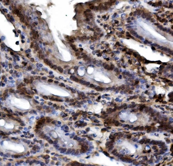
 IHC (Immunohistochemistry)
(At 1/100 staining Human gastric cancer and adjacent normal tissues by IHC-P. The sample was formaldehyde fixed and a heat mediated antigen retrieval step in citrate buffer was performed. The sample was then blocked and incubated with the primary antibody at 4 degree C overnight. An HRP conjugated anti-Rabbit antibody was used as the secondary antibody.)
IHC (Immunohistochemistry)
(At 1/100 staining Human gastric cancer and adjacent normal tissues by IHC-P. The sample was formaldehyde fixed and a heat mediated antigen retrieval step in citrate buffer was performed. The sample was then blocked and incubated with the primary antibody at 4 degree C overnight. An HRP conjugated anti-Rabbit antibody was used as the secondary antibody.)
H2A.Z, Polyclonal Antibody (Cat# AAA31357)
Full Name
Acetyl-H2A.Z (Lys4/7/11/13) Antibody
Gene Names
H2AFZ; H2AZ; H2A.z; H2A/z; H2A.Z-1
Reactivity
Human, Mouse, Rat
Predicted Reactivity: Bovine (100%), Sheep (100%), Dog (100%), Chicken (100%), Xenopus (91%)
Predicted Reactivity: Bovine (100%), Sheep (100%), Dog (100%), Chicken (100%), Xenopus (91%)
Applications
WB, IHC, EIA
Purity
The antiserum was purified by peptide affinity chromatography using SulfoLink Coupling Resin
Pricing


 FCM (Flow Cytometry)
(Figure 6. Flow Cytometry analysis of U-87 cells using anti-SSH3BP1 antibody (AAA11656).Overlay histogram showing U-87 cells stained with AAA11656 (Blue line).The cells were blocked with 10% normal goat serum. And then incubated with rabbit anti-SSH3BP1 Antibody (AAA11656,1ug/1x10^6 cells) for 30 min at 20 degree C. DyLight®488 conjugated goat anti-rabbit IgG (5-10ug/1x10^6 cells) was used as secondary antibody for 30 minutes at 20 degree C. Isotype control antibody (Green line) was rabbit IgG (1ug/1x106) used under the same conditions. Unlabelled sample (Red line) was also used as a control.)
FCM (Flow Cytometry)
(Figure 6. Flow Cytometry analysis of U-87 cells using anti-SSH3BP1 antibody (AAA11656).Overlay histogram showing U-87 cells stained with AAA11656 (Blue line).The cells were blocked with 10% normal goat serum. And then incubated with rabbit anti-SSH3BP1 Antibody (AAA11656,1ug/1x10^6 cells) for 30 min at 20 degree C. DyLight®488 conjugated goat anti-rabbit IgG (5-10ug/1x10^6 cells) was used as secondary antibody for 30 minutes at 20 degree C. Isotype control antibody (Green line) was rabbit IgG (1ug/1x106) used under the same conditions. Unlabelled sample (Red line) was also used as a control.)
SSH3BP1, Polyclonal Antibody (Cat# AAA11656)
Full Name
Anti-SSH3BP1 Antibody
Gene Names
ABI1; E3B1; ABI-1; ABLBP4; NAP1BP; SSH3BP; SSH3BP1
Reactivity
Human, Mouse, Rat
Applications
WB, IHC
Purity
Immunogen Affinity Purified
Pricing


 FCM (Flow Cytometry)
(Overlay histogram showing Hela cells stained with CSB-MA878942A1m (red line) at 1:300. The cells were incubated in 1x PBS /10% normal goat serum to block non-specific protein-protein interactions followed by primary antibody for 1 h at 4 degree C. The secondary antibody used was FITC goat anti-mouse IgG(H+L) at 1/200 dilution for 1 h at 4 degree C. Isotype control antibody (green line) was used under the same conditions. Acquisition of >10,000 events was performed.)
FCM (Flow Cytometry)
(Overlay histogram showing Hela cells stained with CSB-MA878942A1m (red line) at 1:300. The cells were incubated in 1x PBS /10% normal goat serum to block non-specific protein-protein interactions followed by primary antibody for 1 h at 4 degree C. The secondary antibody used was FITC goat anti-mouse IgG(H+L) at 1/200 dilution for 1 h at 4 degree C. Isotype control antibody (green line) was used under the same conditions. Acquisition of >10,000 events was performed.)
PD-L1, Monoclonal Antibody (Cat# AAA27017)
Full Name
PD-L1 Monoclonal Antibody
Gene Names
CD274; B7-H; B7H1; PDL1; PD-L1; PDCD1L1; PDCD1LG1
Reactivity
Human
Applications
EIA, WB, IHC, IF, FC/FACS
Purity
>95%
Protein G Purified
Protein G Purified
Pricing


 ICC (Immunocytochemistry)
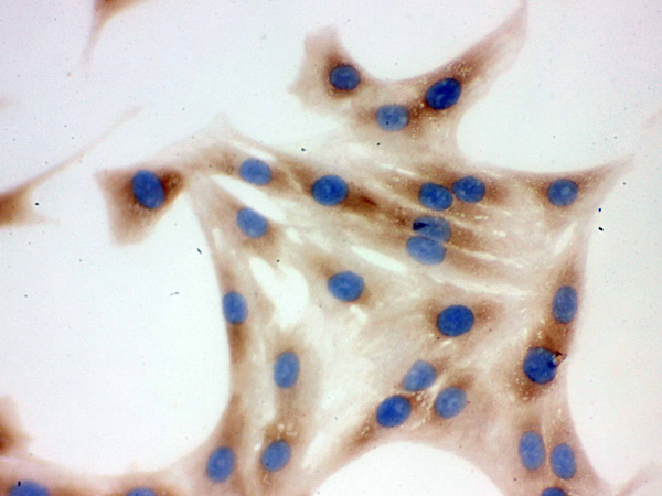
(Figure 7. IHC analysis of ERAB using anti-ERAB antibody (AAA11668).ERAB was detected in immunocytochemical section of SMMC-7721 cell. Heat mediated antigen retrieval was performed in citrate buffer (pH6, epitope retrieval solution) for 20 mins. The tissue section was blocked with 10% goat serum. The tissue section was then incubated with 1ug/ml rabbit anti-ERAB Antibody (AAA11668) overnight at 4 degree C. Biotinylated goat anti-rabbit IgG was used as secondary antibody and incubated for 30 minutes at 37 degree C. The tissue section was developed using Strepavidin-Biotin-Complex (SABC) with DAB as the chromogen.)
ICC (Immunocytochemistry)
(Figure 7. IHC analysis of ERAB using anti-ERAB antibody (AAA11668).ERAB was detected in immunocytochemical section of SMMC-7721 cell. Heat mediated antigen retrieval was performed in citrate buffer (pH6, epitope retrieval solution) for 20 mins. The tissue section was blocked with 10% goat serum. The tissue section was then incubated with 1ug/ml rabbit anti-ERAB Antibody (AAA11668) overnight at 4 degree C. Biotinylated goat anti-rabbit IgG was used as secondary antibody and incubated for 30 minutes at 37 degree C. The tissue section was developed using Strepavidin-Biotin-Complex (SABC) with DAB as the chromogen.)
ERAB, Polyclonal Antibody (Cat# AAA11668)
Full Name
Anti-ERAB Antibody
Gene Names
HSD17B10; ABAD; CAMR; ERAB; HCD2; MHBD; HADH2; MRPP2; MRX17; MRX31; SCHAD; MRXS10; SDR5C1; 17b-HSD10; DUPXp11.22
Reactivity
Human, Mouse
Applications
WB, IHC
Purity
Immunogen Affinity Purified
Pricing


 WB (Western Blot)
(Sample: Recombinant GAL3, Human; Antibody: Rabbit Anti-Human GAL3 Ab)
WB (Western Blot)
(Sample: Recombinant GAL3, Human; Antibody: Rabbit Anti-Human GAL3 Ab)
Galectin 3 (GAL3), Active Protein (Cat# AAA21140)
Full Name
Active Galectin 3 (GAL3)
Gene Names
LGALS3; L31; GAL3; MAC2; CBP35; GALBP; GALIG; LGALS2
Reactivity
Homo sapiens (Human)
Applications
Cell culture; Activity Assays.
Purity
>98%
Pricing


 Application Data
(Staining of J774 cells with Rat anti Mouse F4/80 antigen Biotin)
Application Data
(Staining of J774 cells with Rat anti Mouse F4/80 antigen Biotin)
F4/80, Monoclonal Antibody (Cat# AAA12174)
Full Name
RAT ANTI MOUSE F4/80
Gene Names
Emr1; Ly71; F4/80; Gpf480; TM7LN3; DD7A5-7; EGF-TM7
Applications
EM, FC/FACS, IF, IP, RIA, RE, WB
Pricing


 Application Data
TCP1 delta, Polyclonal Antibody (Cat# AAA11672)
Full Name
Anti-TCP1 delta Antibody
Gene Names
CCT4; SRB; Cctd; CCT-DELTA
Reactivity
Human, Mouse, Rat
No cross reactivity with other proteins.
No cross reactivity with other proteins.
Applications
WB, IHC
Purity
Immunogen affinity purified.
Pricing


 FCM (Flow Cytometry)
(Figure 9. Flow Cytometry analysis of MCF-7 cells using anti-CHRNA5 antibody (AAA11685).Overlay histogram showing MCF-7 cells stained with AAA11685 (Blue line).The cells were blocked with 10% normal goat serum. And then incubated with rabbit anti-CHRNA5 Antibody (AAA11685,1ug/1x10^6 cells) for 30 min at 20 degree C. DyLight®488 conjugated goat anti-rabbit IgG (5-10ug/1x10^6 cells) was used as secondary antibody for 30 minutes at 20 degree C. Isotype control antibody (Green line) was rabbit IgG (1ug/1x106) used under the same conditions. Unlabelled sample (Red line) was also used as a control.)
FCM (Flow Cytometry)
(Figure 9. Flow Cytometry analysis of MCF-7 cells using anti-CHRNA5 antibody (AAA11685).Overlay histogram showing MCF-7 cells stained with AAA11685 (Blue line).The cells were blocked with 10% normal goat serum. And then incubated with rabbit anti-CHRNA5 Antibody (AAA11685,1ug/1x10^6 cells) for 30 min at 20 degree C. DyLight®488 conjugated goat anti-rabbit IgG (5-10ug/1x10^6 cells) was used as secondary antibody for 30 minutes at 20 degree C. Isotype control antibody (Green line) was rabbit IgG (1ug/1x106) used under the same conditions. Unlabelled sample (Red line) was also used as a control.)
CHRNA5, Polyclonal Antibody (Cat# AAA11685)
Full Name
Anti-CHRNA5 Antibody
Gene Names
CHRNA5; LNCR2
Reactivity
Human, Mouse, Rat
Applications
WB, IHC
Purity
Immunogen affinity purified.
Pricing


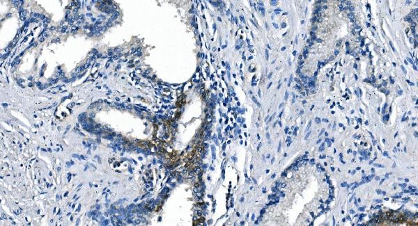
 IHC (Immunohistochemistry)
(At 1/200 staining Human lung cancer tissue sections by IHC-P. The tissue was formaldehyde fixed and a heat mediated antigen retrieval step in citrate buffer was performed. The tissue was then blocked and incubated with the antibody for 1.5 hours at 22 degree C. An HRP conjugated goat anti-rabbit antibody was used as the secondary antibody.)
IHC (Immunohistochemistry)
(At 1/200 staining Human lung cancer tissue sections by IHC-P. The tissue was formaldehyde fixed and a heat mediated antigen retrieval step in citrate buffer was performed. The tissue was then blocked and incubated with the antibody for 1.5 hours at 22 degree C. An HRP conjugated goat anti-rabbit antibody was used as the secondary antibody.)
Caveolin 2, Polyclonal Antibody (Cat# AAA31397)
Full Name
Phospho-Caveolin 2 (Ser36) Antibody
Gene Names
CAV2; CAV
Reactivity
Human, Mouse, Rat
Applications
WB, IHC, EIA
Purity
The antibody is from purified rabbit serum by affinity purification via sequential chromatography on phospho-peptide and non-phospho-peptide affinity columns.
Pricing


 Application Data
(At 25 degree C. The primary antibody was diluted at 1/200 and incubated with the sample for 1 hour at 37 degree C. An Alexa Fluor 594 conjugated goat anti-rabbit IgG (H+L) Ab, diluted at 1/600, was used as the secondary antibody.)
Application Data
(At 25 degree C. The primary antibody was diluted at 1/200 and incubated with the sample for 1 hour at 37 degree C. An Alexa Fluor 594 conjugated goat anti-rabbit IgG (H+L) Ab, diluted at 1/600, was used as the secondary antibody.)
LAT, Polyclonal Antibody (Cat# AAA31413)
Full Name
Phospho-LAT (Tyr161) Antibody
Gene Names
LAT; LAT1; pp36
Reactivity
Human, Mouse, Rat
Predicted Reactivity: Pig (91%), Zebrafish (83%), Bovine (100%), Horse (91%), Sheep (100%), Rabbit (80%), Dog (82%)
Predicted Reactivity: Pig (91%), Zebrafish (83%), Bovine (100%), Horse (91%), Sheep (100%), Rabbit (80%), Dog (82%)
Applications
WB, IHC, IF, ICC, EIA
Purity
The antibody is from purified rabbit serum by affinity purification via sequential chromatography on phospho-peptide and non-phospho-peptide affinity columns.
Pricing


 Application Data
(At 25 degree C. The primary antibody was diluted at 1/200 and incubated with the sample for 1 hour at 37 degree C. An Alexa Fluor 594 conjugated goat anti-rabbit IgG (H+L) Ab, diluted at 1/600, was used as the secondary antibody.)
Application Data
(At 25 degree C. The primary antibody was diluted at 1/200 and incubated with the sample for 1 hour at 37 degree C. An Alexa Fluor 594 conjugated goat anti-rabbit IgG (H+L) Ab, diluted at 1/600, was used as the secondary antibody.)
p70 S6 Kinase, Polyclonal Antibody (Cat# AAA31408)
Full Name
Phospho-p70 S6 Kinase (Ser427) Antibody
Gene Names
RPS6KB1; S6K; PS6K; S6K1; STK14A; p70-S6K; p70 S6KA; p70-alpha; S6K-beta-1; p70(S6K)-alpha
Reactivity
Human, Mouse, Rat
Predicted Reactivity: Pig (100%), Bovine (100%), Horse (100%), Sheep (100%), Rabbit (100%), Dog (100%), Chicken (100%), Xenopus (100%)
Predicted Reactivity: Pig (100%), Bovine (100%), Horse (100%), Sheep (100%), Rabbit (100%), Dog (100%), Chicken (100%), Xenopus (100%)
Applications
WB, IHC, IF, ICC, EIA
Purity
The antibody is from purified rabbit serum by affinity purification via sequential chromatography on phospho-peptide and non-phospho-peptide affinity columns.
Pricing


 FCM (Flow Cytometry)
(Figure 6. Flow Cytometry analysis of Hela cells using anti-PDE4D antibody (AAA19143).Overlay histogram showing Hela cells stained with AAA19143 (Blue line).The cells were blocked with 10% normal goat serum. And then incubated with rabbit anti-PDE4D Antibody (AAA19143,1ug/1x10^6 cells) for 30 min at 20 degree C. DyLight®488 conjugated goat anti-rabbit IgG (5-10ug/1x10^6 cells) was used as secondary antibody for 30 minutes at 20 degree C. Isotype control antibody (Green line) was rabbit IgG (1ug/1x106) used under the same conditions. Unlabelled sample (Red line) was also used as a control.)
FCM (Flow Cytometry)
(Figure 6. Flow Cytometry analysis of Hela cells using anti-PDE4D antibody (AAA19143).Overlay histogram showing Hela cells stained with AAA19143 (Blue line).The cells were blocked with 10% normal goat serum. And then incubated with rabbit anti-PDE4D Antibody (AAA19143,1ug/1x10^6 cells) for 30 min at 20 degree C. DyLight®488 conjugated goat anti-rabbit IgG (5-10ug/1x10^6 cells) was used as secondary antibody for 30 minutes at 20 degree C. Isotype control antibody (Green line) was rabbit IgG (1ug/1x106) used under the same conditions. Unlabelled sample (Red line) was also used as a control.)
PDE4D, Polyclonal Antibody (Cat# AAA19143)
Full Name
Anti-PDE4D Picoband antibody
Gene Names
PDE4D; DPDE3; PDE43; STRK1; ACRDYS2; HSPDE4D; PDE4DN2
Reactivity
Human, Mouse, Rat
No cross reactivity with other proteins.
No cross reactivity with other proteins.
Applications
EIA, FC/FACS, IHC, ICC, WB
Pricing


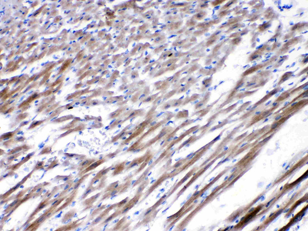
 IHC (Immunohistochemistry)
(Figure 7. IHC analysis of MYLK using anti-MYLK antibody (AAA19154).MYLK was detected in paraffin-embedded section of rat lung tissue. Heat mediated antigen retrieval was performed in citrate buffer (pH6, epitope retrieval solution) for 20 mins. The tissue section was blocked with 10% goat serum. The tissue section was then incubated with 1ug/ml rabbit anti-MYLK Antibody (AAA19154) overnight at 4 degree C. Biotinylated goat anti-rabbit IgG was used as secondary antibody and incubated for 30 minutes at 37 degree C. The tissue section was developed using Strepavidin-Biotin-Complex (SABC) with DAB as the chromogen. )
IHC (Immunohistochemistry)
(Figure 7. IHC analysis of MYLK using anti-MYLK antibody (AAA19154).MYLK was detected in paraffin-embedded section of rat lung tissue. Heat mediated antigen retrieval was performed in citrate buffer (pH6, epitope retrieval solution) for 20 mins. The tissue section was blocked with 10% goat serum. The tissue section was then incubated with 1ug/ml rabbit anti-MYLK Antibody (AAA19154) overnight at 4 degree C. Biotinylated goat anti-rabbit IgG was used as secondary antibody and incubated for 30 minutes at 37 degree C. The tissue section was developed using Strepavidin-Biotin-Complex (SABC) with DAB as the chromogen. )
MYLK, Polyclonal Antibody (Cat# AAA19154)
Full Name
Anti-MYLK Picoband antibody
Gene Names
MYLK; KRP; AAT7; MLCK; MLCK1; MYLK1; smMLCK; MLCK108; MLCK210; MSTP083
Reactivity
Human, Mouse, Rat
No cross reactivity with other proteins.
No cross reactivity with other proteins.
Applications
EIA, IHC, WB
Pricing


 Application Data
(At 25 degree C. The primary antibody was diluted at 1/200 and incubated with the sample for 1 hour at 37 degree C. An Alexa Fluor 594 conjugated goat anti-rabbit IgG (H+L) Ab, diluted at 1/600, was used as the secondary antibody.)
Application Data
(At 25 degree C. The primary antibody was diluted at 1/200 and incubated with the sample for 1 hour at 37 degree C. An Alexa Fluor 594 conjugated goat anti-rabbit IgG (H+L) Ab, diluted at 1/600, was used as the secondary antibody.)
Caspase 3, Polyclonal Antibody (Cat# AAA31443)
Full Name
Phospho-Caspase 3 (Ser26) Antibody
Gene Names
CASP3; CPP32; SCA-1; CPP32B
Reactivity
Human, Mouse, Rat
Predicted Reactivity: Rabbit (88%)
Predicted Reactivity: Rabbit (88%)
Applications
WB, IHC, IF, ICC, EIA
Purity
The antibody is from purified rabbit serum by affinity purification via sequential chromatography on phospho-peptide and non-phospho-peptide affinity columns.
Pricing


 IHC (Immunohistochemistry)
(At 1/100 staining Mouse brain tissue by IHC-P. The sample was formaldehyde fixed and a heat mediated antigen retrieval step in citrate buffer was performed. The sample was then blocked and incubated with the primary antibody at 4 degree C overnight. An HRP conjugated anti-Rabbit antibody was used as the secondary antibody.)
IHC (Immunohistochemistry)
(At 1/100 staining Mouse brain tissue by IHC-P. The sample was formaldehyde fixed and a heat mediated antigen retrieval step in citrate buffer was performed. The sample was then blocked and incubated with the primary antibody at 4 degree C overnight. An HRP conjugated anti-Rabbit antibody was used as the secondary antibody.)
IRF7, Polyclonal Antibody (Cat# AAA31458)
Full Name
Phospho-IRF7 (Ser477) Antibody
Gene Names
IRF7; IRF7A; IRF7B; IRF7C; IRF7H; IRF-7H
Reactivity
Human, Mouse, Rat
Predicted Reactivity: Pig (100%), Bovine (100%), Horse (100%), Sheep (100%), Rabbit (100%)
Predicted Reactivity: Pig (100%), Bovine (100%), Horse (100%), Sheep (100%), Rabbit (100%)
Applications
WB, IHC, EIA
Purity
The antibody is from purified rabbit serum by affinity purification via sequential chromatography on phospho-peptide and non-phospho-peptide affinity columns.
Pricing


 Application Data
(At 25 degree C. Samples were then incubated with primary Ab(At 37 degree C. An AlexaFluor594 conjugated goat anti-rabbit IgG(H+L) Ab(Red) and an AlexaFluor488 conjugated goat anti-mouse IgG(H+L) Ab(Green) were used as the secondary antibody.The nuclear counter stain is DAPI(blue).)
Application Data
(At 25 degree C. Samples were then incubated with primary Ab(At 37 degree C. An AlexaFluor594 conjugated goat anti-rabbit IgG(H+L) Ab(Red) and an AlexaFluor488 conjugated goat anti-mouse IgG(H+L) Ab(Green) were used as the secondary antibody.The nuclear counter stain is DAPI(blue).)
PDHA1/2, Polyclonal Antibody (Cat# AAA31464)
Full Name
Phospho-PDHA1/2 (Ser293/Ser291) Antibody
Gene Names
PDHA1; PDHA; PHE1A; PDHCE1A
Reactivity
Human, Mouse, Rat
Predicted Reactivity: Pig (100%), Bovine (100%), Horse (100%), Sheep (100%), Rabbit (100%), Chicken (100%), Xenopus (100%)
Predicted Reactivity: Pig (100%), Bovine (100%), Horse (100%), Sheep (100%), Rabbit (100%), Chicken (100%), Xenopus (100%)
Applications
WB, IHC, IF, ICC, EIA
Purity
The antibody is from purified rabbit serum by affinity purification via sequential chromatography on phospho-peptide and non-phospho-peptide affinity columns.
Pricing


 FCM (Flow Cytometry)
(Figure 11. Flow Cytometry analysis of U937 cells using anti-Coronin 1a/TACO/CORO1A antibody (AAA19291).Overlay histogram showing U937 cells stained with AAA19291 (Blue line). The cells were blocked with 10% normal goat serum. And then incubated with rabbit anti-Coronin 1a/TACO/CORO1A Antibody (AAA19291, 1μg/1x106 cells) for 30 min at 20 degree C. DyLight®488 conjugated goat anti-rabbit IgG (5-10μg/1x106 cells) was used as secondary antibody for 30 minutes at 20 degree C. Isotype control antibody (Green line) was rabbit IgG (1μg/1x106) used under the same conditions. Unlabelled sample (Red line) was also used as a control.)
FCM (Flow Cytometry)
(Figure 11. Flow Cytometry analysis of U937 cells using anti-Coronin 1a/TACO/CORO1A antibody (AAA19291).Overlay histogram showing U937 cells stained with AAA19291 (Blue line). The cells were blocked with 10% normal goat serum. And then incubated with rabbit anti-Coronin 1a/TACO/CORO1A Antibody (AAA19291, 1μg/1x106 cells) for 30 min at 20 degree C. DyLight®488 conjugated goat anti-rabbit IgG (5-10μg/1x106 cells) was used as secondary antibody for 30 minutes at 20 degree C. Isotype control antibody (Green line) was rabbit IgG (1μg/1x106) used under the same conditions. Unlabelled sample (Red line) was also used as a control.)
Coronin 1a/TACO/CORO1A, Polyclonal Antibody (Cat# AAA19291)
Full Name
Anti-Coronin 1a/TACO/CORO1A Antibody
Gene Names
CORO1A; p57; TACO; CLABP; HCORO1; CLIPINA
Reactivity
Human, Mouse, Rat
Applications
WB, IHC-P, FC/FACS/FCM, EIA
Purity
Immunogen affinity purified.
Pricing


 FCM (Flow Cytometry)
(Figure 7. Flow Cytometry analysis of A549 cells using anti-DR6/TNFRSF21 antibody (AAA19292).Overlay histogram showing A549 cells stained with AAA19292 (Blue line). The cells were blocked with 10% normal goat serum. And then incubated with rabbit anti-DR6/TNFRSF21 Antibody (AAA19292, 1μg/1x106 cells) for 30 min at 20 degree C. DyLight®488 conjugated goat anti-rabbit IgG (5-10μg/1x106 cells) was used as secondary antibody for 30 minutes at 20 degree C. Isotype control antibody (Green line) was rabbit IgG (1μg/1x106) used under the same conditions. Unlabelled sample (Red line) was also used as a control.)
FCM (Flow Cytometry)
(Figure 7. Flow Cytometry analysis of A549 cells using anti-DR6/TNFRSF21 antibody (AAA19292).Overlay histogram showing A549 cells stained with AAA19292 (Blue line). The cells were blocked with 10% normal goat serum. And then incubated with rabbit anti-DR6/TNFRSF21 Antibody (AAA19292, 1μg/1x106 cells) for 30 min at 20 degree C. DyLight®488 conjugated goat anti-rabbit IgG (5-10μg/1x106 cells) was used as secondary antibody for 30 minutes at 20 degree C. Isotype control antibody (Green line) was rabbit IgG (1μg/1x106) used under the same conditions. Unlabelled sample (Red line) was also used as a control.)
DR6/TNFRSF21, Polyclonal Antibody (Cat# AAA19292)
Full Name
Anti-DR6/TNFRSF21 Antibody
Gene Names
TNFRSF21; DR6; CD358; BM-018
Reactivity
Human, Mouse, Rat
Applications
WB, IHC-P, FC/FACS/FCM
Purity
Immunogen affinity purified.
Pricing


 IF (Immunofluorescence)
(Figure 6. IF analysis of SCG10/STMN2 using anti- SCG10/STMN2 antibody (AAA19299).SCG10/STMN2 was detected in immunocytochemical section of U20S cells. Enzyme antigen retrieval was performed using IHC enzyme antigen retrieval reagent for 15 mins. The cells were blocked with 10% goat serum. And then incubated with 5μg/mL rabbit anti- SCG10/STMN2 Antibody (AAA19299) overnight at 4 degree C. DyLight®488 Conjugated Goat Anti-Rabbit IgG was used as secondary antibody at 1:100 dilution and incubated for 30 minutes at 37 degree C. The section was counterstained with DAPI. Visualize using a fluorescence microscope and filter sets appropriate for the label used.)
IF (Immunofluorescence)
(Figure 6. IF analysis of SCG10/STMN2 using anti- SCG10/STMN2 antibody (AAA19299).SCG10/STMN2 was detected in immunocytochemical section of U20S cells. Enzyme antigen retrieval was performed using IHC enzyme antigen retrieval reagent for 15 mins. The cells were blocked with 10% goat serum. And then incubated with 5μg/mL rabbit anti- SCG10/STMN2 Antibody (AAA19299) overnight at 4 degree C. DyLight®488 Conjugated Goat Anti-Rabbit IgG was used as secondary antibody at 1:100 dilution and incubated for 30 minutes at 37 degree C. The section was counterstained with DAPI. Visualize using a fluorescence microscope and filter sets appropriate for the label used.)
SCG10/STMN2, Polyclonal Antibody (Cat# AAA19299)
Full Name
Anti-SCG10/STMN2 Antibody
Gene Names
STMN2; SCG10; SCGN10
Reactivity
Human, Mouse, Rat
Applications
WB, IHC-P, ICC, IF, FC/FACS/FCM, EIA
Purity
Immunogen affinity purified.
Pricing


 FCM (Flow Cytometry)
(Figure 7. Flow Cytometry analysis of CACO-2 cells using anti-Glycine decarboxylase/GLDC antibody (AAA19300).Overlay histogram showing CACO-2 cells stained with AAA19300 (Blue line). The cells were blocked with 10% normal goat serum. And then incubated with rabbit anti-Glycine decarboxylase/GLDC Antibody (AAA19300, 1μg/1x106 cells) for 30 min at 20 degree C. DyLight®488 conjugated goat anti-rabbit IgG (5-10μg/1x106 cells) was used as secondary antibody for 30 minutes at 20 degree C. Isotype control antibody (Green line) was rabbit IgG (1μg/1x106) used under the same conditions. Unlabelled sample (Red line) was also used as a control.)
FCM (Flow Cytometry)
(Figure 7. Flow Cytometry analysis of CACO-2 cells using anti-Glycine decarboxylase/GLDC antibody (AAA19300).Overlay histogram showing CACO-2 cells stained with AAA19300 (Blue line). The cells were blocked with 10% normal goat serum. And then incubated with rabbit anti-Glycine decarboxylase/GLDC Antibody (AAA19300, 1μg/1x106 cells) for 30 min at 20 degree C. DyLight®488 conjugated goat anti-rabbit IgG (5-10μg/1x106 cells) was used as secondary antibody for 30 minutes at 20 degree C. Isotype control antibody (Green line) was rabbit IgG (1μg/1x106) used under the same conditions. Unlabelled sample (Red line) was also used as a control.)
Glycine decarboxylase/GLDC, Polyclonal Antibody (Cat# AAA19300)
Full Name
Anti-Glycine decarboxylase/GLDC Antibody
Gene Names
GLDC; GCE; GCSP; HYGN1
Reactivity
Human, Mouse, Rat
Applications
WB, IHC-P, FC/FACS/FCM, EIA
Purity
Immunogen affinity purified.
Pricing


 FCM (Flow Cytometry)
(Figure 7. Flow Cytometry analysis of U20S cells using anti-VDAC3 antibody (AAA19301).Overlay histogram showing U20S cells stained with AAA19301 (Blue line). The cells were blocked with 10% normal goat serum. And then incubated with rabbit anti-VDAC3 Antibody (AAA19301, 1μg/1x106 cells) for 30 min at 20 degree C. DyLight®488 conjugated goat anti-rabbit IgG (5-10μg/1x106 cells) was used as secondary antibody for 30 minutes at 20 degree C. Isotype control antibody (Green line) was rabbit IgG (1μg/1x106) used under the same conditions. Unlabelled sample (Red line) was also used as a control.)
FCM (Flow Cytometry)
(Figure 7. Flow Cytometry analysis of U20S cells using anti-VDAC3 antibody (AAA19301).Overlay histogram showing U20S cells stained with AAA19301 (Blue line). The cells were blocked with 10% normal goat serum. And then incubated with rabbit anti-VDAC3 Antibody (AAA19301, 1μg/1x106 cells) for 30 min at 20 degree C. DyLight®488 conjugated goat anti-rabbit IgG (5-10μg/1x106 cells) was used as secondary antibody for 30 minutes at 20 degree C. Isotype control antibody (Green line) was rabbit IgG (1μg/1x106) used under the same conditions. Unlabelled sample (Red line) was also used as a control.)
VDAC3, Polyclonal Antibody (Cat# AAA19301)
Full Name
Anti-VDAC3 Antibody
Gene Names
VDAC3; VDAC-3; HD-VDAC3
Reactivity
Human, Mouse, Rat
Applications
WB, IHC-P, FC/FACS/FCM, EIA
Purity
Immunogen affinity purified.
Pricing


 FCM (Flow Cytometry)
(Figure 9. Flow Cytometry analysis of 293T cells using anti-CDC45L antibody (AAA19360).Overlay histogram showing 293T cells stained with AAA19360 (Blue line). The cells were blocked with 10% normal goat serum. And then incubated with mouse anti- CDC45L Antibody (AAA19360, 1μg/1x106 cells) for 30 min at 20 degree C. DyLight®488 conjugated goat anti-mouse IgG (BA1126, 5-10μg/1x106 cells) was used as secondary antibody for 30 minutes at 20 degree C. Isotype control antibody (Green line) was mouse IgG (1μg/1x106) used under the same conditions. Unlabelled sample (Red line) was also used as a control.)
FCM (Flow Cytometry)
(Figure 9. Flow Cytometry analysis of 293T cells using anti-CDC45L antibody (AAA19360).Overlay histogram showing 293T cells stained with AAA19360 (Blue line). The cells were blocked with 10% normal goat serum. And then incubated with mouse anti- CDC45L Antibody (AAA19360, 1μg/1x106 cells) for 30 min at 20 degree C. DyLight®488 conjugated goat anti-mouse IgG (BA1126, 5-10μg/1x106 cells) was used as secondary antibody for 30 minutes at 20 degree C. Isotype control antibody (Green line) was mouse IgG (1μg/1x106) used under the same conditions. Unlabelled sample (Red line) was also used as a control.)
CDC45L, Monoclonal Antibody (Cat# AAA19360)
Full Name
Anti-CDC45L Antibody (monoclonal, 6H6)
Gene Names
CDC45; CDC45L; CDC45L2; PORC-PI-1
Reactivity
Human, Mouse, Rat
Applications
WB, IHC-P, FC/FACS/FCM
Purity
Immunogen affinity purified.
Pricing


 IHC (Immunohistchemistry)
(Validation of PKR in Rat Lung Immunohistochemical analysis of paraffin-embedded rat lung tissue using anti-PKR antibody (AAA10917) at 2.5 ug/ml. Tissue was fixed with formaldehyde and blocked with 10% serum for 1h at RT; antigen retrieval was by heat mediation with a citrate buffer (pH6). Samples were incubated with primary antibody overnight at 4 degree C. A goat anti-rabbit IgG H&L (HRP) at 1/250 was used as secondary. Counter stained with Hematoxylin.)
IHC (Immunohistchemistry)
(Validation of PKR in Rat Lung Immunohistochemical analysis of paraffin-embedded rat lung tissue using anti-PKR antibody (AAA10917) at 2.5 ug/ml. Tissue was fixed with formaldehyde and blocked with 10% serum for 1h at RT; antigen retrieval was by heat mediation with a citrate buffer (pH6). Samples were incubated with primary antibody overnight at 4 degree C. A goat anti-rabbit IgG H&L (HRP) at 1/250 was used as secondary. Counter stained with Hematoxylin.)
PKR, Polyclonal Antibody (Cat# AAA10917)
Full Name
PKR Antibody
Gene Names
EIF2AK2; PKR; PRKR; EIF2AK1; PPP1R83
Reactivity
Human, Mouse, Rat
Applications
EIA, WB, IHC, IF
Purity
PKR Antibody is affinity chromatography purified via peptide column.
Pricing


 FCM (Flow Cytometry)
(Figure 8. Flow Cytometry analysis of SiHa cells using anti- ASS1 antibody (AAA19369).Overlay histogram showing SiHa cells stained with AAA19369 (Blue line). The cells were blocked with 10% normal goat serum. And then incubated with mouse anti-ASS1 Antibody (AAA19369, 1μg/1x106 cells) for 30 min at 20 degree C. DyLight®488 conjugated goat anti-mouse IgG (BA1126, 5-10μg/1x106 cells) was used as secondary antibody for 30 minutes at 20 degree C. Isotype control antibody (Green line) was mouse IgG (1μg/1x106) used under the same conditions. Unlabelled sample (Red line) was also used as a control.)
FCM (Flow Cytometry)
(Figure 8. Flow Cytometry analysis of SiHa cells using anti- ASS1 antibody (AAA19369).Overlay histogram showing SiHa cells stained with AAA19369 (Blue line). The cells were blocked with 10% normal goat serum. And then incubated with mouse anti-ASS1 Antibody (AAA19369, 1μg/1x106 cells) for 30 min at 20 degree C. DyLight®488 conjugated goat anti-mouse IgG (BA1126, 5-10μg/1x106 cells) was used as secondary antibody for 30 minutes at 20 degree C. Isotype control antibody (Green line) was mouse IgG (1μg/1x106) used under the same conditions. Unlabelled sample (Red line) was also used as a control.)
ASS1, Monoclonal Antibody (Cat# AAA19369)
Full Name
Anti-ASS1 Antibody (monoclonal, 5I5)
Gene Names
ASS1; ASS; CTLN1
Reactivity
Human, Mouse, Rat, Monkey
Applications
WB, IHC-P, ICC, IF, FC/FACS/FCM
Purity
Immunogen affinity purified.
Pricing


 FCM (Flow Cytometry)
(Figure 8. Flow Cytometry analysis of SiHa cells using anti- ASS1 antibody (AAA19370).Overlay histogram showing SiHa cells stained with AAA19370 (Blue line). The cells were blocked with 10% normal goat serum. And then incubated with mouse anti-ASS1 Antibody (AAA19370, 1μg/1x106 cells) for 30 min at 20 degree C. DyLight®488 conjugated goat anti-mouse IgG (BA1126, 5-10μg/1x106 cells) was used as secondary antibody for 30 minutes at 20 degree C. Isotype control antibody (Green line) was mouse IgG (1μg/1x106) used under the same conditions. Unlabelled sample (Red line) was also used as a control.)
FCM (Flow Cytometry)
(Figure 8. Flow Cytometry analysis of SiHa cells using anti- ASS1 antibody (AAA19370).Overlay histogram showing SiHa cells stained with AAA19370 (Blue line). The cells were blocked with 10% normal goat serum. And then incubated with mouse anti-ASS1 Antibody (AAA19370, 1μg/1x106 cells) for 30 min at 20 degree C. DyLight®488 conjugated goat anti-mouse IgG (BA1126, 5-10μg/1x106 cells) was used as secondary antibody for 30 minutes at 20 degree C. Isotype control antibody (Green line) was mouse IgG (1μg/1x106) used under the same conditions. Unlabelled sample (Red line) was also used as a control.)
ASS1, Monoclonal Antibody (Cat# AAA19370)
Full Name
Anti-ASS1 Antibody (monoclonal, 7I9)
Gene Names
ASS1; ASS; CTLN1
Reactivity
Human, Mouse, Rat, Monkey
Applications
WB, IHC-P, ICC, IF, FC/FACS/FCM
Purity
Immunogen affinity purified.
Pricing


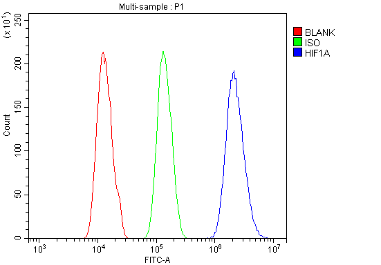 FCM (Flow Cytometry)
(Figure 6. Flow Cytometry analysis of A431 cells using anti-HIF-1 alpha/HIF1A antibody (AAA19213).Overlay histogram showing A431 cells stained with AAA19213 (Blue line). The cells were blocked with 10% normal goat serum. And then incubated with rabbit anti-HIF-1 alpha/HIF1A Antibody (AAA19213, 1μg/1x106 cells) for 30 min at 20 degree C. DyLight®488 conjugated goat anti-rabbit IgG (5-10μg/1x106 cells) was used as secondary antibody for 30 minutes at 20 degree C. Isotype control antibody (Green line) was rabbit IgG (1μg/1x106) used under the same conditions. Unlabelled sample (Red line) was also used as a control.)
FCM (Flow Cytometry)
(Figure 6. Flow Cytometry analysis of A431 cells using anti-HIF-1 alpha/HIF1A antibody (AAA19213).Overlay histogram showing A431 cells stained with AAA19213 (Blue line). The cells were blocked with 10% normal goat serum. And then incubated with rabbit anti-HIF-1 alpha/HIF1A Antibody (AAA19213, 1μg/1x106 cells) for 30 min at 20 degree C. DyLight®488 conjugated goat anti-rabbit IgG (5-10μg/1x106 cells) was used as secondary antibody for 30 minutes at 20 degree C. Isotype control antibody (Green line) was rabbit IgG (1μg/1x106) used under the same conditions. Unlabelled sample (Red line) was also used as a control.)
HIF-1 alpha/HIF1A, Polyclonal Antibody (Cat# AAA19213)
Full Name
Anti-HIF-1 alpha/HIF1A Antibody
Gene Names
HIF1A; HIF1; MOP1; PASD8; HIF-1A; bHLHe78; HIF-1alpha; HIF1-ALPHA
Reactivity
Human, Mouse, Rat
Applications
WB, IHC-P, FC/FACS/FCM, EIA
Purity
Immunogen affinity purified.
Pricing


 FCM (Flow Cytometry)
(Figure 6. Flow Cytometry analysis of Raji cells using anti-CD38 antibody (AAA19217).Overlay histogram showing Raji cells stained with AAA19217 (Blue line). The cells were blocked with 10% normal goat serum. And then incubated with rabbit anti-CD38 Antibody (AAA19217, 1μg/1x106 cells) for 30 min at 20 degree C. DyLight®488 conjugated goat anti-rabbit IgG (5-10μg/1x106 cells) was used as secondary antibody for 30 minutes at 20 degree C. Isotype control antibody (Green line) was rabbit IgG (1μg/1x106) used under the same conditions. Unlabelled sample (Red line) was also used as a control.)
FCM (Flow Cytometry)
(Figure 6. Flow Cytometry analysis of Raji cells using anti-CD38 antibody (AAA19217).Overlay histogram showing Raji cells stained with AAA19217 (Blue line). The cells were blocked with 10% normal goat serum. And then incubated with rabbit anti-CD38 Antibody (AAA19217, 1μg/1x106 cells) for 30 min at 20 degree C. DyLight®488 conjugated goat anti-rabbit IgG (5-10μg/1x106 cells) was used as secondary antibody for 30 minutes at 20 degree C. Isotype control antibody (Green line) was rabbit IgG (1μg/1x106) used under the same conditions. Unlabelled sample (Red line) was also used as a control.)
CD38, Polyclonal Antibody (Cat# AAA19217)
Full Name
Anti-CD38 Antibody
Gene Names
CD38; T10; ADPRC 1
Reactivity
Human
Applications
WB, IHC-P, ICC, IF, FC/FACS/FCM
Purity
Immunogen affinity purified.
Pricing


 FCM (Flow Cytometry)
(Figure 8. Flow Cytometry analysis of HepG2 cells using anti-MERTK antibody (AAA19219).Overlay histogram showing HepG2 cells stained with AAA19219 (Blue line). The cells were blocked with 10% normal goat serum. And then incubated with rabbit anti-MERTK Antibody (AAA19219, 1ug/1x106 cells) for 30 min at 20 degree C. DyLight488 conjugated goat anti-rabbit IgG (5-10ug/1x106 cells) was used as secondary antibody for 30 minutes at 20 degree C. Isotype control antibody (Green line) was rabbit IgG (1ug/1x106) used under the same conditions. Unlabelled sample (Red line) was also used as a control.)
FCM (Flow Cytometry)
(Figure 8. Flow Cytometry analysis of HepG2 cells using anti-MERTK antibody (AAA19219).Overlay histogram showing HepG2 cells stained with AAA19219 (Blue line). The cells were blocked with 10% normal goat serum. And then incubated with rabbit anti-MERTK Antibody (AAA19219, 1ug/1x106 cells) for 30 min at 20 degree C. DyLight488 conjugated goat anti-rabbit IgG (5-10ug/1x106 cells) was used as secondary antibody for 30 minutes at 20 degree C. Isotype control antibody (Green line) was rabbit IgG (1ug/1x106) used under the same conditions. Unlabelled sample (Red line) was also used as a control.)
MERTK, Polyclonal Antibody (Cat# AAA19219)
Full Name
Anti-MERTK Antibody
Gene Names
MERTK; MER; RP38; c-mer
Reactivity
Human, Mouse, Rat
Applications
WB, IHC-P, FC/FACS/FCM, EIA
Purity
Immunogen affinity purified.
Pricing


 FCM (Flow Cytometry)
(Figure 7. Flow Cytometry analysis of U20S cells using anti- ADA antibody (AAA19184). Overlay histogram showing U20S cells stained with AAA19184 (Blue line).The cells were blocked with 10% normal goat serum. And then incubated with mouse anti- ADA Antibody (AAA19184, 1ug/1x106 cells) for 30 min at 20 degree C. DyLight488 conjugated goat anti-mouse IgG (BA1126, 5-10ug/1x106 cells) was used as secondary antibody for 30 minutes at 20 degree C. Isotype control antibody (Green line) was mouse IgG (1ug/1x106) used under the same conditions. Unlabelled sample (Red line) was also used as a control.)
FCM (Flow Cytometry)
(Figure 7. Flow Cytometry analysis of U20S cells using anti- ADA antibody (AAA19184). Overlay histogram showing U20S cells stained with AAA19184 (Blue line).The cells were blocked with 10% normal goat serum. And then incubated with mouse anti- ADA Antibody (AAA19184, 1ug/1x106 cells) for 30 min at 20 degree C. DyLight488 conjugated goat anti-mouse IgG (BA1126, 5-10ug/1x106 cells) was used as secondary antibody for 30 minutes at 20 degree C. Isotype control antibody (Green line) was mouse IgG (1ug/1x106) used under the same conditions. Unlabelled sample (Red line) was also used as a control.)
ADA, Monoclonal Antibody (Cat# AAA19184)
Full Name
Anti-ADA Antibody (monoclonal, 6D4)
Reactivity
Human
Applications
WB, IHC, FC/FACS
Purity
Immunogen Affinity Purified
Pricing


 IF (Immunofluorescence)
(Figure 6. IF analysis of CD72 using anti- CD72 antibody (AAA19329).CD72 was detected in immunocytochemical section of HEPA1-6 cells. Enzyme antigen retrieval was performed using IHC enzyme antigen retrieval reagent for 15 mins. The cells were blocked with 10% goat serum. And then incubated with 5μg/mL rabbit anti- CD72 Antibody (AAA19329) overnight at 4 degree C. DyLight®488 Conjugated Goat Anti-Rabbit IgG was used as secondary antibody at 1:100 dilution and incubated for 30 minutes at 37 degree C. The section was counterstained with DAPI. Visualize using a fluorescence microscope and filter sets appropriate for the label used.)
IF (Immunofluorescence)
(Figure 6. IF analysis of CD72 using anti- CD72 antibody (AAA19329).CD72 was detected in immunocytochemical section of HEPA1-6 cells. Enzyme antigen retrieval was performed using IHC enzyme antigen retrieval reagent for 15 mins. The cells were blocked with 10% goat serum. And then incubated with 5μg/mL rabbit anti- CD72 Antibody (AAA19329) overnight at 4 degree C. DyLight®488 Conjugated Goat Anti-Rabbit IgG was used as secondary antibody at 1:100 dilution and incubated for 30 minutes at 37 degree C. The section was counterstained with DAPI. Visualize using a fluorescence microscope and filter sets appropriate for the label used.)
CD72, Polyclonal Antibody (Cat# AAA19329)
Full Name
Anti-CD72 Antibody
Gene Names
Cd72; CD72c; Ly-19; Ly-32; Lyb-2; Ly-m19
Reactivity
Mouse, Rat
Applications
WB, IHC-P, ICC, IF, FC/FACS/FCM, EIA
Purity
Immunogen affinity purified.
Pricing


 IF (Immunofluorescence)
(Figure 7. IF analysis of METTL1 using anti- METTL1 antibody (AAA19336).METTL1 was detected in immunocytochemical section of A431 cells. Enzyme antigen retrieval was performed using IHC enzyme antigen retrieval reagent for 15 mins. The cells were blocked with 10% goat serum. And then incubated with 5μg/mL rabbit anti-METTL1 Antibody (AAA19336) overnight at 4 degree C. DyLight®488 Conjugated Goat Anti-Rabbit IgG was used as secondary antibody at 1:100 dilution and incubated for 30 minutes at 37 degree C. The section was counterstained with DAPI. Visualize using a fluorescence microscope and filter sets appropriate for the label used.)
IF (Immunofluorescence)
(Figure 7. IF analysis of METTL1 using anti- METTL1 antibody (AAA19336).METTL1 was detected in immunocytochemical section of A431 cells. Enzyme antigen retrieval was performed using IHC enzyme antigen retrieval reagent for 15 mins. The cells were blocked with 10% goat serum. And then incubated with 5μg/mL rabbit anti-METTL1 Antibody (AAA19336) overnight at 4 degree C. DyLight®488 Conjugated Goat Anti-Rabbit IgG was used as secondary antibody at 1:100 dilution and incubated for 30 minutes at 37 degree C. The section was counterstained with DAPI. Visualize using a fluorescence microscope and filter sets appropriate for the label used.)
METTL1, Polyclonal Antibody (Cat# AAA19336)
Full Name
Anti-METTL1 Antibody
Gene Names
METTL1; TRM8; TRMT8; C12orf1; YDL201w
Reactivity
Human, Rat
Applications
WB, IHC-P, ICC, IF, EIA
Purity
Immunogen affinity purified.
Pricing


 FCM (Flow Cytometry)
(Figure 7. Flow Cytometry analysis of HEPA1-6 cells using anti-Rad9b antibody (AAA19340).Overlay histogram showing HEPA1-6 cells stained with AAA19340 (Blue line). The cells were blocked with 10% normal goat serum. And then incubated with rabbit anti-Rad9b Antibody (AAA19340, 1μg/1x106 cells) for 30 min at 20 degree C. DyLight®488 conjugated goat anti-rabbit IgG (5-10μg/1x106 cells) was used as secondary antibody for 30 minutes at 20 degree C. Isotype control antibody (Green line) was rabbit IgG (1μg/1x106) used under the same conditions. Unlabelled sample (Red line) was also used as a control.)
FCM (Flow Cytometry)
(Figure 7. Flow Cytometry analysis of HEPA1-6 cells using anti-Rad9b antibody (AAA19340).Overlay histogram showing HEPA1-6 cells stained with AAA19340 (Blue line). The cells were blocked with 10% normal goat serum. And then incubated with rabbit anti-Rad9b Antibody (AAA19340, 1μg/1x106 cells) for 30 min at 20 degree C. DyLight®488 conjugated goat anti-rabbit IgG (5-10μg/1x106 cells) was used as secondary antibody for 30 minutes at 20 degree C. Isotype control antibody (Green line) was rabbit IgG (1μg/1x106) used under the same conditions. Unlabelled sample (Red line) was also used as a control.)
Rad9b, Polyclonal Antibody (Cat# AAA19340)
Full Name
Anti-Rad9b Antibody
Gene Names
Rad9b; BC021784; A630082N15Rik
Reactivity
Mouse, Rat
Applications
WB, IHC-P, FC/FACS/FCM, EIA
Purity
Immunogen affinity purified.
Pricing


 FCM (Flow Cytometry)
(Figure 6. Flow Cytometry analysis of U87 cells using anti-GRB10 antibody (AAA19244).Overlay histogram showing U87 cells stained with AAA19244 (Blue line). The cells were blocked with 10% normal goat serum. And then incubated with rabbit anti-GRB10 Antibody (AAA19244,1μg/1x106 cells) for 30 min at 20 degree C. DyLight®488 conjugated goat anti-rabbit IgG (5-10μg/1x106 cells) was used as secondary antibody for 30 minutes at 20 degree C. Isotype control antibody (Green line) was rabbit IgG (1μg/1x106) used under the same conditions. Unlabelled sample (Red line) was also used as a control.)
FCM (Flow Cytometry)
(Figure 6. Flow Cytometry analysis of U87 cells using anti-GRB10 antibody (AAA19244).Overlay histogram showing U87 cells stained with AAA19244 (Blue line). The cells were blocked with 10% normal goat serum. And then incubated with rabbit anti-GRB10 Antibody (AAA19244,1μg/1x106 cells) for 30 min at 20 degree C. DyLight®488 conjugated goat anti-rabbit IgG (5-10μg/1x106 cells) was used as secondary antibody for 30 minutes at 20 degree C. Isotype control antibody (Green line) was rabbit IgG (1μg/1x106) used under the same conditions. Unlabelled sample (Red line) was also used as a control.)
GRB10, Polyclonal Antibody (Cat# AAA19244)
Full Name
Anti-GRB10 Antibody
Gene Names
GRB10; RSS; IRBP; MEG1; GRB-IR; Grb-10
Reactivity
Human, Mouse, Rat
Applications
WB, IHC-P, FC/FACS/FCM, EIA
Purity
Immunogen affinity purified.
Pricing


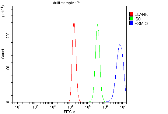 FCM (Flow Cytometry)
(Figure 7. Flow Cytometry analysis of 293T cells using anti-TBP-1/PSMC3 antibody (AAA19314).Overlay histogram showing 293T cells stained with AAA19314 (Blue line). The cells were blocked with 10% normal goat serum. And then incubated with rabbit anti-TBP-1/PSMC3 Antibody (AAA19314, 1μg/1x106 cells) for 30 min at 20 degree C. DyLight®488 conjugated goat anti-rabbit IgG (5-10μg/1x106 cells) was used as secondary antibody for 30 minutes at 20 degree C. Isotype control antibody (Green line) was rabbit IgG (1μg/1x106) used under the same conditions. Unlabelled sample (Red line) was also used as a control.)
FCM (Flow Cytometry)
(Figure 7. Flow Cytometry analysis of 293T cells using anti-TBP-1/PSMC3 antibody (AAA19314).Overlay histogram showing 293T cells stained with AAA19314 (Blue line). The cells were blocked with 10% normal goat serum. And then incubated with rabbit anti-TBP-1/PSMC3 Antibody (AAA19314, 1μg/1x106 cells) for 30 min at 20 degree C. DyLight®488 conjugated goat anti-rabbit IgG (5-10μg/1x106 cells) was used as secondary antibody for 30 minutes at 20 degree C. Isotype control antibody (Green line) was rabbit IgG (1μg/1x106) used under the same conditions. Unlabelled sample (Red line) was also used as a control.)
TBP-1/PSMC3, Polyclonal Antibody (Cat# AAA19314)
Full Name
Anti-TBP-1/PSMC3 Antibody
Gene Names
PSMC3; TBP1
Reactivity
Human, Mouse, Rat
Applications
WB, IHC-P, FC/FACS/FCM, EIA
Purity
Immunogen affinity purified.
Pricing


 IF (Immunofluorescence)
(Figure 6. IF analysis of GRSF1 using anti- GRSF1 antibody (AAA19315).GRSF1 was detected in immunocytochemical section of MCF-7 cells. Enzyme antigen retrieval was performed using IHC enzyme antigen retrieval reagent for 15 mins. The cells were blocked with 10% goat serum. And then incubated with 5μg/mL rabbit anti- GRSF1 Antibody (AAA19315) overnight at 4 degree C. DyLight®488 Conjugated Goat Anti-Rabbit IgG was used as secondary antibody at 1:100 dilution and incubated for 30 minutes at 37 degree C. The section was counterstained with DAPI. Visualize using a fluorescence microscope and filter sets appropriate for the label used.)
IF (Immunofluorescence)
(Figure 6. IF analysis of GRSF1 using anti- GRSF1 antibody (AAA19315).GRSF1 was detected in immunocytochemical section of MCF-7 cells. Enzyme antigen retrieval was performed using IHC enzyme antigen retrieval reagent for 15 mins. The cells were blocked with 10% goat serum. And then incubated with 5μg/mL rabbit anti- GRSF1 Antibody (AAA19315) overnight at 4 degree C. DyLight®488 Conjugated Goat Anti-Rabbit IgG was used as secondary antibody at 1:100 dilution and incubated for 30 minutes at 37 degree C. The section was counterstained with DAPI. Visualize using a fluorescence microscope and filter sets appropriate for the label used.)
GRSF1, Polyclonal Antibody (Cat# AAA19315)
Full Name
Anti-GRSF1 Antibody
Reactivity
Human
Applications
WB, IHC-P, ICC, IF, FC/FACS/FCM, EIA
Purity
Immunogen affinity purified.
Pricing


 FCM (Flow Cytometry)
(Figure 6. Flow Cytometry analysis of THP-1 cells using anti-PRMT8 antibody (AAA19321).Overlay histogram showing THP-1 cells stained with AAA19321 (Blue line). The cells were blocked with 10% normal goat serum. And then incubated with rabbit anti-PRMT8 Antibody (AAA19321,1μg/1x106 cells) for 30 min at 20 degree C. DyLight®488 conjugated goat anti-rabbit IgG (5-10μg/1x106 cells) was used as secondary antibody for 30 minutes at 20 degree C. Isotype control antibody (Green line) was rabbit IgG (1μg/1x106) used under the same conditions. Unlabelled sample (Red line) was also used as a control.)
FCM (Flow Cytometry)
(Figure 6. Flow Cytometry analysis of THP-1 cells using anti-PRMT8 antibody (AAA19321).Overlay histogram showing THP-1 cells stained with AAA19321 (Blue line). The cells were blocked with 10% normal goat serum. And then incubated with rabbit anti-PRMT8 Antibody (AAA19321,1μg/1x106 cells) for 30 min at 20 degree C. DyLight®488 conjugated goat anti-rabbit IgG (5-10μg/1x106 cells) was used as secondary antibody for 30 minutes at 20 degree C. Isotype control antibody (Green line) was rabbit IgG (1μg/1x106) used under the same conditions. Unlabelled sample (Red line) was also used as a control.)
PRMT8, Polyclonal Antibody (Cat# AAA19321)
Full Name
Anti-PRMT8 Antibody
Gene Names
PRMT8; HRMT1L3; HRMT1L4
Reactivity
Human, Rat
Applications
WB, IHC-P, FC/FACS/FCM, EIA
Purity
Immunogen affinity purified.
Pricing


 FCM (Flow Cytometry)
(Figure 8. Flow Cytometry analysis of HepG2 cells using anti-REA/PHB2 antibody (AAA19374).Overlay histogram showing HepG2 cells stained with AAA19374 (Blue line). The cells were blocked with 10% normal goat serum. And then incubated with mouse anti- REA/PHB2 Antibody (AAA19374, 1μg/1x106 cells) for 30 min at 20 degree C. DyLight®488 conjugated goat anti-mouse IgG (BA1126, 5-10μg/1x106 cells) was used as secondary antibody for 30 minutes at 20 degree C. Isotype control antibody (Green line) was mouse IgG (1μg/1x106) used under the same conditions. Unlabelled sample (Red line) was also used as a control.)
FCM (Flow Cytometry)
(Figure 8. Flow Cytometry analysis of HepG2 cells using anti-REA/PHB2 antibody (AAA19374).Overlay histogram showing HepG2 cells stained with AAA19374 (Blue line). The cells were blocked with 10% normal goat serum. And then incubated with mouse anti- REA/PHB2 Antibody (AAA19374, 1μg/1x106 cells) for 30 min at 20 degree C. DyLight®488 conjugated goat anti-mouse IgG (BA1126, 5-10μg/1x106 cells) was used as secondary antibody for 30 minutes at 20 degree C. Isotype control antibody (Green line) was mouse IgG (1μg/1x106) used under the same conditions. Unlabelled sample (Red line) was also used as a control.)
REA/PHB2, Monoclonal Antibody (Cat# AAA19374)
Full Name
Anti-REA/PHB2 Antibody (monoclonal, 2B5)
Gene Names
PHB2; BAP; REA; p22; Bap37; BCAP37; PNAS-141
Reactivity
Human, Mouse, Rat
Applications
WB, IHC-P, FC/FACS/FCM
Purity
Immunogen affinity purified.
Pricing


 FCM (Flow Cytometry)
(Figure 6. Flow Cytometry analysis of MCF-7 cells using anti-DDX1 antibody (AAA19379).Overlay histogram showing MCF-7 cells stained with AAA19379 (Blue line). The cells were blocked with 10% normal goat serum. And then incubated with mouse anti- DDX1 Antibody (AAA19379, 1μg/1x106 cells) for 30 min at 20 degree C. DyLight®488 conjugated goat anti-mouse IgG (BA1126, 5-10μg/1x106 cells) was used as secondary antibody for 30 minutes at 20 degree C. Isotype control antibody (Green line) was mouse IgG (1μg/1x106) used under the same conditions. Unlabelled sample (Red line) was also used as a control.)
FCM (Flow Cytometry)
(Figure 6. Flow Cytometry analysis of MCF-7 cells using anti-DDX1 antibody (AAA19379).Overlay histogram showing MCF-7 cells stained with AAA19379 (Blue line). The cells were blocked with 10% normal goat serum. And then incubated with mouse anti- DDX1 Antibody (AAA19379, 1μg/1x106 cells) for 30 min at 20 degree C. DyLight®488 conjugated goat anti-mouse IgG (BA1126, 5-10μg/1x106 cells) was used as secondary antibody for 30 minutes at 20 degree C. Isotype control antibody (Green line) was mouse IgG (1μg/1x106) used under the same conditions. Unlabelled sample (Red line) was also used as a control.)
DDX1, Monoclonal Antibody (Cat# AAA19379)
Full Name
Anti-DDX1 Antibody (monoclonal, 2D4)
Gene Names
DDX1; DBP-RB; UKVH5d
Reactivity
Human, Mouse, Rat
Applications
WB, IHC-P, FC/FACS/FCM
Purity
Immunogen affinity purified.
Pricing


 IHC (Immunohistochemistry)
(At 1/100 staining Mouse kidney tissue by IHC-P. The sample was formaldehyde fixed and a heat mediated antigen retrieval step in citrate buffer was performed. The sample was then blocked and incubated with the primary antibody at 4 degree C overnight. An HRP conjugated anti-Rabbit antibody was used as the secondary antibody.)
IHC (Immunohistochemistry)
(At 1/100 staining Mouse kidney tissue by IHC-P. The sample was formaldehyde fixed and a heat mediated antigen retrieval step in citrate buffer was performed. The sample was then blocked and incubated with the primary antibody at 4 degree C overnight. An HRP conjugated anti-Rabbit antibody was used as the secondary antibody.)
YB1, Polyclonal Antibody (Cat# AAA31294)
Full Name
Phospho-YB1 (Ser102) Antibody
Gene Names
YBX1; YB1; BP-8; CSDB; DBPB; YB-1; CSDA2; NSEP1; NSEP-1; MDR-NF1
Reactivity
Human, Mouse, Rat
Applications
IHC, EIA
Purity
The antibody is from purified rabbit serum by affinity purification via sequential chromatography on phospho-peptide and non-phospho-peptide affinity columns.
Pricing


 Application Data
(At 25 degree C. Samples were then incubated with primary Ab(At 37 degree C. An AlexaFluor594 conjugated goat anti-rabbit IgG(H+L) Ab(Red) and an AlexaFluor488 conjugated goat anti-mouse IgG(H+L) Ab(Green) were used as the secondary antibody.The nuclear counter stain is DAPI (blue).)
Application Data
(At 25 degree C. Samples were then incubated with primary Ab(At 37 degree C. An AlexaFluor594 conjugated goat anti-rabbit IgG(H+L) Ab(Red) and an AlexaFluor488 conjugated goat anti-mouse IgG(H+L) Ab(Green) were used as the secondary antibody.The nuclear counter stain is DAPI (blue).)
Bub1, Polyclonal Antibody (Cat# AAA31312)
Full Name
Phospho-Bub1 (Ser459) Antibody
Gene Names
BUB1; BUB1A; BUB1L; hBUB1
Reactivity
Human, Rat
Applications
IHC, IF, ICC, EIA
Purity
The antibody is from purified rabbit serum by affinity purification via sequential chromatography on phospho-peptide and non-phospho-peptide affinity columns.
Pricing


 IHC (Immunohistchemistry)
(At 1/100 staining Mouse kidney tissue by IHC-P. The sample was formaldehyde fixed and a heat mediated antigen retrieval step in citrate buffer was performed. The sample was then blocked and incubated with the primary antibody at 4 degree C overnight. An HRP conjugated anti-Rabbit antibody was used as the secondary antibody.)
IHC (Immunohistchemistry)
(At 1/100 staining Mouse kidney tissue by IHC-P. The sample was formaldehyde fixed and a heat mediated antigen retrieval step in citrate buffer was performed. The sample was then blocked and incubated with the primary antibody at 4 degree C overnight. An HRP conjugated anti-Rabbit antibody was used as the secondary antibody.)
ARRB1, Polyclonal Antibody (Cat# AAA31314)
Full Name
Phospho-ARRB1 (Ser412) Antibody
Gene Names
ARRB1; ARB1; ARR1
Reactivity
Human, Mouse, Rat
Applications
WB, IHC, EIA
Purity
The antibody is from purified rabbit serum by affinity purification via sequential chromatography on phospho-peptide and non-phospho-peptide affinity columns.
Pricing


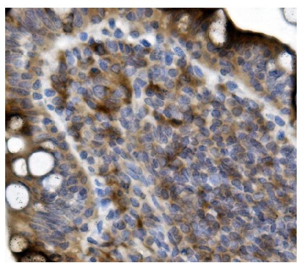
 IHC (Immunohistochemistry)
(At 1/100 staining Human gastric cancer and adjacent normal tissues by IHC-P. The sample was formaldehyde fixed and a heat mediated antigen retrieval step in citrate buffer was performed. The sample was then blocked and incubated with the primary antibody at 4 degree C overnight. An HRP conjugated anti-Rabbit antibody was used as the secondary antibody.)
IHC (Immunohistochemistry)
(At 1/100 staining Human gastric cancer and adjacent normal tissues by IHC-P. The sample was formaldehyde fixed and a heat mediated antigen retrieval step in citrate buffer was performed. The sample was then blocked and incubated with the primary antibody at 4 degree C overnight. An HRP conjugated anti-Rabbit antibody was used as the secondary antibody.)
AIRE, Polyclonal Antibody (Cat# AAA31324)
Full Name
Phospho-AIRE (Ser156) Antibody
Gene Names
AIRE; APS1; APSI; PGA1; AIRE1; APECED
Reactivity
Human, Mouse, Rat
Applications
IHC, EIA
Purity
The antibody is from purified rabbit serum by affinity purification via sequential chromatography on phospho-peptide and non-phospho-peptide affinity columns.
Pricing


 Application Data
(At 25 degree C. Samples were then incubated with primary Ab(At 37 degree C. An AlexaFluor594 conjugated goat anti-rabbit IgG(H+L) Ab(Red) and an AlexaFluor488 conjugated goat anti-mouse IgG(H+L) Ab(Green) were used as the secondary antibody.The nuclear counter stain is DAPI (blue).)
Application Data
(At 25 degree C. Samples were then incubated with primary Ab(At 37 degree C. An AlexaFluor594 conjugated goat anti-rabbit IgG(H+L) Ab(Red) and an AlexaFluor488 conjugated goat anti-mouse IgG(H+L) Ab(Green) were used as the secondary antibody.The nuclear counter stain is DAPI (blue).)
GSK3 beta, Polyclonal Antibody (Cat# AAA31297)
Full Name
Phospho-GSK3 beta (Thr390) Antibody
Reactivity
Human, Mouse, Rat
Applications
WB, IHC, IF, ICC, EIA
Purity
The antibody is from purified rabbit serum by affinity purification via sequential chromatography on phospho-peptide and non-phospho-peptide affinity columns.
Pricing


 Application Data
(At 25 degree C. The primary antibody was diluted at 1/200 and incubated with the sample for 1 hour at 37 degree C. An Alexa Fluor 594 conjugated goat anti-rabbit IgG (H+L) antibody(Red), diluted at 1/600, was used as secondary antibody.)
Application Data
(At 25 degree C. The primary antibody was diluted at 1/200 and incubated with the sample for 1 hour at 37 degree C. An Alexa Fluor 594 conjugated goat anti-rabbit IgG (H+L) antibody(Red), diluted at 1/600, was used as secondary antibody.)
RAD17, Polyclonal Antibody (Cat# AAA31431)
Full Name
Phospho-RAD17 (Ser646) Antibody
Gene Names
RAD17; CCYC; R24L; RAD24; HRAD17; RAD17SP
Reactivity
Human, Mouse, Rat
Predicted Reactivity: Pig (100%), Bovine (100%), Horse (100%), Sheep (100%)
Predicted Reactivity: Pig (100%), Bovine (100%), Horse (100%), Sheep (100%)
Applications
WB, IHC, IF, ICC, EIA
Purity
The antibody is from purified rabbit serum by affinity purification via sequential chromatography on phospho-peptide and non-phospho-peptide affinity columns.
Pricing


 Application Data
(Figure 10 KD Validation in HeLa cells (Horinaka et al., 2005)HeLa cells were transfected with DR4siRNA or LacZ control siRNA. At 24 h after transfection, the cells were treated with or without 20 ?M luteolin for 24 h. Western blot analysis was carried out with anti-DR4 antibodies (1139). DR4 expression was markedly reduced after DR4 knockdown.)
Application Data
(Figure 10 KD Validation in HeLa cells (Horinaka et al., 2005)HeLa cells were transfected with DR4siRNA or LacZ control siRNA. At 24 h after transfection, the cells were treated with or without 20 ?M luteolin for 24 h. Western blot analysis was carried out with anti-DR4 antibodies (1139). DR4 expression was markedly reduced after DR4 knockdown.)
DR4, Polyclonal Antibody (Cat# AAA10952)
Full Name
DR4 Antibody
Gene Names
TNFRSF10A; DR4; APO2; CD261; TRAILR1; TRAILR-1
Reactivity
Human
Applications
EIA, WB, ICC
Purity
DR4 Antibody is Antibody is affinity chromatography purified via peptide column.
Pricing


 Application Data
(At 25 degree C. The primary antibody was diluted at 1/200 and incubated with the sample for 1 hour at 37 degree C. An Alexa Fluor 594 conjugated goat anti-rabbit IgG (H+L) Ab, diluted at 1/600, was used as the secondary antibody.)
Application Data
(At 25 degree C. The primary antibody was diluted at 1/200 and incubated with the sample for 1 hour at 37 degree C. An Alexa Fluor 594 conjugated goat anti-rabbit IgG (H+L) Ab, diluted at 1/600, was used as the secondary antibody.)
FOXO4, Polyclonal Antibody (Cat# AAA31400)
Full Name
Phospho-FOXO4 (Thr455) Antibody
Gene Names
FOXO4; AFX; AFX1; MLLT7
Reactivity
Human, Mouse, Rat
Predicted Reactivity: Bovine (91%), Horse (83%), Sheep (91%), Dog (91%)
Predicted Reactivity: Bovine (91%), Horse (83%), Sheep (91%), Dog (91%)
Applications
WB, IHC, IF, ICC, EIA
Purity
The antibody is from purified rabbit serum by affinity purification via sequential chromatography on phospho-peptide and non-phospho-peptide affinity columns.
Pricing


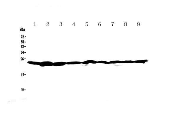 WB (Western Blot)
(Figure 1. Western blot analysis of Ran using anti-Ran antibody (AAA19131). Electrophoresis was performed on a 5-20% SDS-PAGE gel at 70V (Stacking gel) / 90V (Resolving gel) for 2-3 hours. The sample well of each lane was loaded with 50ug of sample under reducing conditions. Lane 1: rat brain tissue lysate,Lane 2: rat testis tissue lysate,Lane 3: rat thymus tissue lysate,Lane 4: mouse brain tissue lysate,Lane 5: mouse testis tissue lysate,Lane 6: mouse thymus tissue lysate,Lane 7: human A549 whole cell lysate,Lane 8: human 22RV1 whole cell lysate,Lane 9: human Hela whole cell lysate. After Electrophoresis, proteins were transferred to a Nitrocellulose membrane at 150mA for 50-90 minutes. Blocked the membrane with 5% Non-fat Milk/ TBS for 1.5 hour at RT. The membrane was incubated with rabbit anti-Ran antigen affinity purified polyclonal antibody at 0.5ug/mL overnight at 4 degree C, then washed with TBS-0.1%Tween 3 times with 5 minutes each and probed with a goat anti-rabbit IgG-HRP secondary antibody at a dilution of 1:10000 for 1.5 hour at RT. The signal is developed using an Enhanced Chemiluminescent detection (ECL) kit with Tanon 5200 system. A specific band was detected for Ran at approximately 24KD. The expected band size for Ran is at 24KD.)
WB (Western Blot)
(Figure 1. Western blot analysis of Ran using anti-Ran antibody (AAA19131). Electrophoresis was performed on a 5-20% SDS-PAGE gel at 70V (Stacking gel) / 90V (Resolving gel) for 2-3 hours. The sample well of each lane was loaded with 50ug of sample under reducing conditions. Lane 1: rat brain tissue lysate,Lane 2: rat testis tissue lysate,Lane 3: rat thymus tissue lysate,Lane 4: mouse brain tissue lysate,Lane 5: mouse testis tissue lysate,Lane 6: mouse thymus tissue lysate,Lane 7: human A549 whole cell lysate,Lane 8: human 22RV1 whole cell lysate,Lane 9: human Hela whole cell lysate. After Electrophoresis, proteins were transferred to a Nitrocellulose membrane at 150mA for 50-90 minutes. Blocked the membrane with 5% Non-fat Milk/ TBS for 1.5 hour at RT. The membrane was incubated with rabbit anti-Ran antigen affinity purified polyclonal antibody at 0.5ug/mL overnight at 4 degree C, then washed with TBS-0.1%Tween 3 times with 5 minutes each and probed with a goat anti-rabbit IgG-HRP secondary antibody at a dilution of 1:10000 for 1.5 hour at RT. The signal is developed using an Enhanced Chemiluminescent detection (ECL) kit with Tanon 5200 system. A specific band was detected for Ran at approximately 24KD. The expected band size for Ran is at 24KD.)
Ran, Polyclonal Antibody (Cat# AAA19131)
Full Name
Anti-Ran Picoband Antibody
Gene Names
RAN; TC4; Gsp1; ARA24
Reactivity
Human, Mouse, Rat
No cross reactivity with other proteins.
No cross reactivity with other proteins.
Applications
IHC, WB
Purity
Immunogen affinity purified
Pricing


 Application Data
(Published Customer Image:Rat anti Mouse Gr-1 antibody, clone RB6-8C5 used for the identification of neutrophils by immunofluorescence.Image caption:S. typhimurium-Infected Macrophages Containing Phagocytosed Neutrophils and T Cells Confocal fluorescence microscopy of 50-mum-thick liver sections from 1-wk-infected Slc11a1 wild-type mice. (A-C) S.Typhimurium (O-antigen, arrows) are red, macrophages (F4-80 and MOMA-2) are blue, DNA (DAPI) is gray, phalloidin is green, and neutrophils (Gr-1/Ly-6G/RB6-8C5) are pink (arrowheads). (A) Collapsed image from a 40-mum Z-stack. Scale bar is 20 mum. (B and C) Sections from (A) that are 4 mum apart. The video from which (A-C) were derived (Video S2) is available online. (D-G) T cells within multinucleate macrophages.Macrophages (F4-80 and MOMA-2) are blue (D, G, and H), T cells (CD3zeta) are red (D, G, arrowheads), DAPI is gray (E, G), actin-bound phalloidin is green (F, G). (G) Is a composite of (D, E, and F). Scale bars are 16 mum. (H) An image from a different mouse stained and labeled as described for (D-G). Scale bar is 8 mum. A video showing a T cell inside of a macrophage is available online (Video S3).From: Nix RN, Altschuler SE, Henson PM, Detweiler CS (2007) Hemophagocytic Macrophages Harbor Salmonella enterica during Persistent Infection.PLoS Pathog 3(12): e193.)
Application Data
(Published Customer Image:Rat anti Mouse Gr-1 antibody, clone RB6-8C5 used for the identification of neutrophils by immunofluorescence.Image caption:S. typhimurium-Infected Macrophages Containing Phagocytosed Neutrophils and T Cells Confocal fluorescence microscopy of 50-mum-thick liver sections from 1-wk-infected Slc11a1 wild-type mice. (A-C) S.Typhimurium (O-antigen, arrows) are red, macrophages (F4-80 and MOMA-2) are blue, DNA (DAPI) is gray, phalloidin is green, and neutrophils (Gr-1/Ly-6G/RB6-8C5) are pink (arrowheads). (A) Collapsed image from a 40-mum Z-stack. Scale bar is 20 mum. (B and C) Sections from (A) that are 4 mum apart. The video from which (A-C) were derived (Video S2) is available online. (D-G) T cells within multinucleate macrophages.Macrophages (F4-80 and MOMA-2) are blue (D, G, and H), T cells (CD3zeta) are red (D, G, arrowheads), DAPI is gray (E, G), actin-bound phalloidin is green (F, G). (G) Is a composite of (D, E, and F). Scale bars are 16 mum. (H) An image from a different mouse stained and labeled as described for (D-G). Scale bar is 8 mum. A video showing a T cell inside of a macrophage is available online (Video S3).From: Nix RN, Altschuler SE, Henson PM, Detweiler CS (2007) Hemophagocytic Macrophages Harbor Salmonella enterica during Persistent Infection.PLoS Pathog 3(12): e193.)
Gr-1, Monoclonal Antibody (Cat# AAA12257)
Full Name
Rat Anti Mouse Gr-1: FITC
Reactivity
Mouse
Applications
FC/FACS
Purity
Purified IgG prepared by affinity chromatography on Protein G from tissue culture supernatant
Pricing


 FCM (Flow Cytometry)
(Figure 8. Flow Cytometry analysis of U87 cells using anti-Transketolase/TKT antibody (AAA19254).Overlay histogram showing U87 cells stained with AAA19254 (Blue line). The cells were blocked with 10% normal goat serum. And then incubated with rabbit anti-Transketolase/TKT Antibody (AAA19254,1μg/1x106 cells) for 30 min at 20 degree C. DyLight®488 conjugated goat anti-rabbit IgG (5-10μg/1x106 cells) was used as secondary antibody for 30 minutes at 20 degree C. Isotype control antibody (Green line) was rabbit IgG (1μg/1x106) used under the same conditions. Unlabelled sample (Red line) was also used as a control.)
FCM (Flow Cytometry)
(Figure 8. Flow Cytometry analysis of U87 cells using anti-Transketolase/TKT antibody (AAA19254).Overlay histogram showing U87 cells stained with AAA19254 (Blue line). The cells were blocked with 10% normal goat serum. And then incubated with rabbit anti-Transketolase/TKT Antibody (AAA19254,1μg/1x106 cells) for 30 min at 20 degree C. DyLight®488 conjugated goat anti-rabbit IgG (5-10μg/1x106 cells) was used as secondary antibody for 30 minutes at 20 degree C. Isotype control antibody (Green line) was rabbit IgG (1μg/1x106) used under the same conditions. Unlabelled sample (Red line) was also used as a control.)
Transketolase/TKT, Polyclonal Antibody (Cat# AAA19254)
Full Name
Anti-Transketolase/TKT Antibody
Gene Names
TKT; TK; TKT1; HEL107
Reactivity
Human, Mouse, Rat
Applications
WB, IHC-P, ICC, IF, FC/FACS/FCM, EIA
Purity
Immunogen affinity purified.
Pricing