Filters
▼Clonality
▼Type
▼Reactivity
▼Gene Name
▼Isotype
▼Host
▼Application
▼Clone
▼Viewing 4100-4150 of 265407 product results
 FCM/FACS (Flow Cytometry)
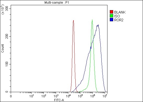
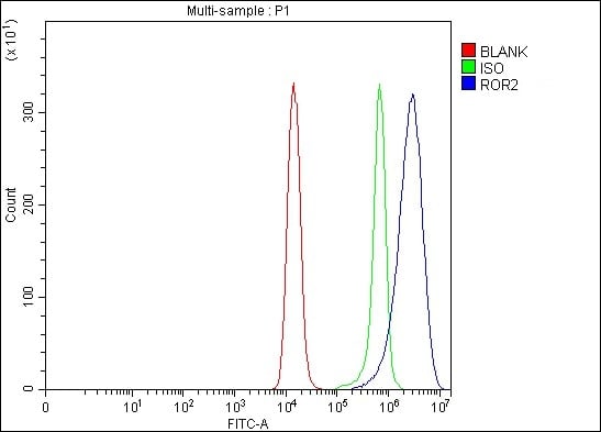
(Figure 3. Flow Cytometry analysis of Raji cells using anti-ROR2 antibody (AAA127084).Overlay histogram showing Raji cells stained with AAA127084 (Blue line). The cells were fixed with 4% paraformaldehyde and blocked with 10% normal goat serum. And then incubated with rabbit anti-ROR2 Antibody (AAA127084, 1ug/1x106 cells) for 30 min at 20 degree C. DyLight488 conjugated goat anti-rabbit IgG was used as secondary antibody for 30 minutes at 20 degree C. Isotype control antibody (Green line) was rabbit IgG (1ug/1x106) used under the same conditions. Unlabelled sample without incubation with primary antibody and secondary antibody (Red line) was used as a blank control.)
FCM/FACS (Flow Cytometry)
(Figure 3. Flow Cytometry analysis of Raji cells using anti-ROR2 antibody (AAA127084).Overlay histogram showing Raji cells stained with AAA127084 (Blue line). The cells were fixed with 4% paraformaldehyde and blocked with 10% normal goat serum. And then incubated with rabbit anti-ROR2 Antibody (AAA127084, 1ug/1x106 cells) for 30 min at 20 degree C. DyLight488 conjugated goat anti-rabbit IgG was used as secondary antibody for 30 minutes at 20 degree C. Isotype control antibody (Green line) was rabbit IgG (1ug/1x106) used under the same conditions. Unlabelled sample without incubation with primary antibody and secondary antibody (Red line) was used as a blank control.)
ROR2, Polyclonal Antibody (Cat# AAA127084)
Full Name
Anti-ROR2 Antibody Picoband
Gene Names
ROR2; BDB; BDB1; NTRKR2
Reactivity
Human, Mouse, Rat
Applications
Flow Cytometry, Western Blot
Purity
Immunogen affinity purified.
Pricing
 FCM/FACS (Flow Cytometry)
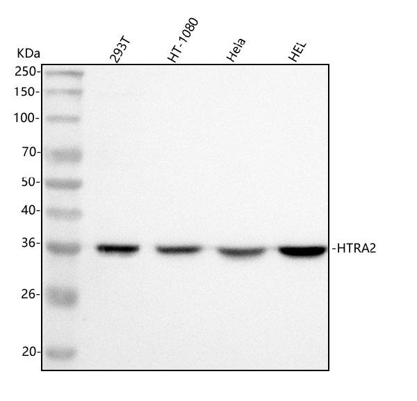
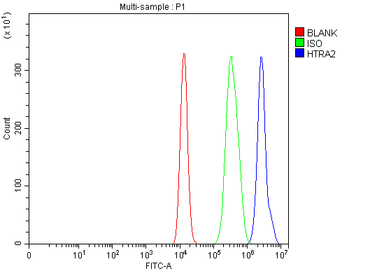
(Figure 5. Flow Cytometry analysis of HEL cells using anti-HTRA2 antibody (AAA127088).Overlay histogram showing HEL cells stained with AAA127088 (Blue line). To facilitate intracellular staining, cells were fixed with 4% paraformaldehyde and permeabilized with permeabilization buffer. The cells were blocked with 10% normal goat serum. And then incubated with rabbit anti-HTRA2 Antibody (AAA127088, 1ug/1x106 cells) for 30 min at 20 degree C. DyLight488 conjugated goat anti-rabbit IgG was used as secondary antibody for 30 minutes at 20 degree C. Isotype control antibody (Green line) was rabbit IgG (1ug/1x106) used under the same conditions. Unlabelled sample (Red line) was also used as a control.)
FCM/FACS (Flow Cytometry)
(Figure 5. Flow Cytometry analysis of HEL cells using anti-HTRA2 antibody (AAA127088).Overlay histogram showing HEL cells stained with AAA127088 (Blue line). To facilitate intracellular staining, cells were fixed with 4% paraformaldehyde and permeabilized with permeabilization buffer. The cells were blocked with 10% normal goat serum. And then incubated with rabbit anti-HTRA2 Antibody (AAA127088, 1ug/1x106 cells) for 30 min at 20 degree C. DyLight488 conjugated goat anti-rabbit IgG was used as secondary antibody for 30 minutes at 20 degree C. Isotype control antibody (Green line) was rabbit IgG (1ug/1x106) used under the same conditions. Unlabelled sample (Red line) was also used as a control.)
HTRA2, Polyclonal Antibody (Cat# AAA127088)
Full Name
Anti-HTRA2 Antibody Picoband
Gene Names
HTRA2; OMI; PARK13; PRSS25
Reactivity
Human
Applications
Flow Cytometry, Immunofluorescence, Immunocytochemistry, Immunohistochemistry, Western Blot
Purity
Immunogen affinity purified.
Pricing
 FCM/FACS (Flow Cytometry)
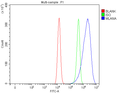
(Figure 2. Flow Cytometry analysis of HEL cells using anti-Melan-A/MLANA antibody (AAA127095).Overlay histogram showing HEL cells stained with AAA127095 (Blue line). To facilitate intracellular staining, cells were fixed with 4% paraformaldehyde and permeabilized with permeabilization buffer. The cells were blocked with 10% normal goat serum. And then incubated with rabbit anti-Melan-A/MLANA Antibody (AAA127095, 1ug/1x106 cells) for 30 min at 20 degree C. DyLight488 conjugated goat anti-rabbit IgG was used as secondary antibody for 30 minutes at 20 degree C. Isotype control antibody (Green line) was rabbit IgG (1ug/1x106) used under the same conditions. Unlabelled sample (Red line) was also used as a control.)
FCM/FACS (Flow Cytometry)
(Figure 2. Flow Cytometry analysis of HEL cells using anti-Melan-A/MLANA antibody (AAA127095).Overlay histogram showing HEL cells stained with AAA127095 (Blue line). To facilitate intracellular staining, cells were fixed with 4% paraformaldehyde and permeabilized with permeabilization buffer. The cells were blocked with 10% normal goat serum. And then incubated with rabbit anti-Melan-A/MLANA Antibody (AAA127095, 1ug/1x106 cells) for 30 min at 20 degree C. DyLight488 conjugated goat anti-rabbit IgG was used as secondary antibody for 30 minutes at 20 degree C. Isotype control antibody (Green line) was rabbit IgG (1ug/1x106) used under the same conditions. Unlabelled sample (Red line) was also used as a control.)
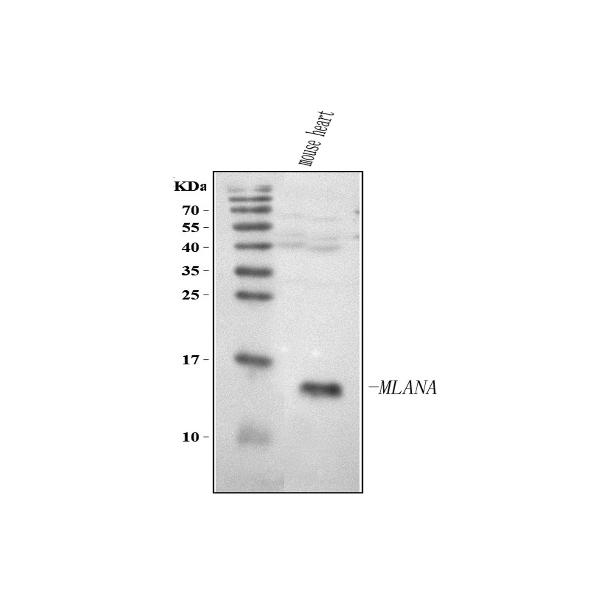
Melan-A/MLANA, Polyclonal Antibody (Cat# AAA127095)
Full Name
Anti-Melan-A/MLANA Antibody Picoband
Gene Names
MLANA; MART1; MART-1
Reactivity
Human, Mouse
Applications
Flow Cytometry, Western Blot
Purity
Immunogen affinity purified.
Pricing
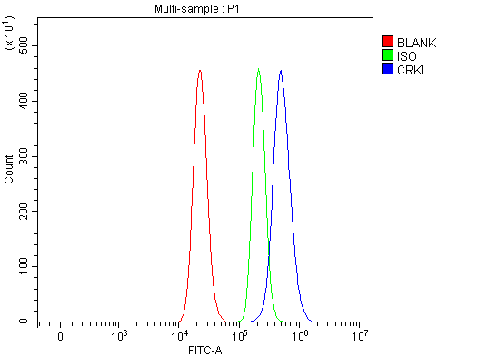 FCM/FACS (Flow Cytometry)
(Figure 3. Flow Cytometry analysis of RT4 cells using anti-CRKL antibody (AAA127100).Overlay histogram showing RT4 cells stained with AAA127100 (Blue line). To facilitate intracellular staining, cells were fixed with 4% paraformaldehyde and permeabilized with permeabilization buffer. The cells were blocked with 10% normal goat serum. And then incubated with rabbit anti-CRKL Antibody (AAA127100, 1ug/1x106 cells) for 30 min at 20 degree C. DyLight488 conjugated goat anti-rabbit IgG was used as secondary antibody for 30 minutes at 20 degree C. Isotype control antibody (Green line) was rabbit IgG (1ug/1x106) used under the same conditions. Unlabelled sample without incubation with primary antibody and secondary antibody (Red line) was used as a blank control.)
FCM/FACS (Flow Cytometry)
(Figure 3. Flow Cytometry analysis of RT4 cells using anti-CRKL antibody (AAA127100).Overlay histogram showing RT4 cells stained with AAA127100 (Blue line). To facilitate intracellular staining, cells were fixed with 4% paraformaldehyde and permeabilized with permeabilization buffer. The cells were blocked with 10% normal goat serum. And then incubated with rabbit anti-CRKL Antibody (AAA127100, 1ug/1x106 cells) for 30 min at 20 degree C. DyLight488 conjugated goat anti-rabbit IgG was used as secondary antibody for 30 minutes at 20 degree C. Isotype control antibody (Green line) was rabbit IgG (1ug/1x106) used under the same conditions. Unlabelled sample without incubation with primary antibody and secondary antibody (Red line) was used as a blank control.)
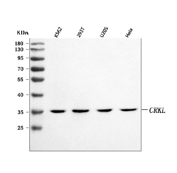
CRKL, Polyclonal Antibody (Cat# AAA127100)
Full Name
Anti-CRKL Antibody Picoband
Reactivity
Human
Applications
Flow Cytometry, Immunofluorescence, Immunocytochemistry, Western Blot
Purity
Immunogen affinity purified.
Pricing
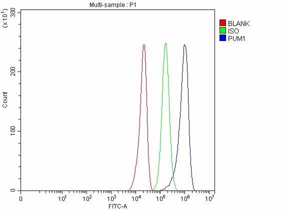 FCM/FACS (Flow Cytometry)
(Figure 3. Flow Cytometry analysis of U251 cells using anti-PUM1 antibody (AAA127102).Overlay histogram showing U251 cells stained with AAA127102 (Blue line). To facilitate intracellular staining, cells were fixed with 4% paraformaldehyde and permeabilized with permeabilization buffer. The cells were blocked with 10% normal goat serum. And then incubated with rabbit anti-PUM1 Antibody (AAA127102, 1ug/1x106 cells) for 30 min at 20 degree C. DyLight488 conjugated goat anti-rabbit IgG was used as secondary antibody for 30 minutes at 20 degree C. Isotype control antibody (Green line) was rabbit IgG (1ug/1x106) used under the same conditions. Unlabelled sample without incubation with primary antibody and secondary antibody (Red line) was used as a blank control.)
FCM/FACS (Flow Cytometry)
(Figure 3. Flow Cytometry analysis of U251 cells using anti-PUM1 antibody (AAA127102).Overlay histogram showing U251 cells stained with AAA127102 (Blue line). To facilitate intracellular staining, cells were fixed with 4% paraformaldehyde and permeabilized with permeabilization buffer. The cells were blocked with 10% normal goat serum. And then incubated with rabbit anti-PUM1 Antibody (AAA127102, 1ug/1x106 cells) for 30 min at 20 degree C. DyLight488 conjugated goat anti-rabbit IgG was used as secondary antibody for 30 minutes at 20 degree C. Isotype control antibody (Green line) was rabbit IgG (1ug/1x106) used under the same conditions. Unlabelled sample without incubation with primary antibody and secondary antibody (Red line) was used as a blank control.)
PUM1, Polyclonal Antibody (Cat# AAA127102)
Full Name
Anti-PUM1 Antibody Picoband
Gene Names
PUM1; PUMH; HSPUM; PUMH1; PUML1; SCA47
Reactivity
Human, Mouse, Rat
Applications
Flow Cytometry, Immunofluorescence, Immunocytochemistry, Western Blot
Purity
Immunogen affinity purified.
Pricing
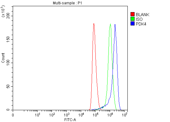 FCM/FACS (Flow Cytometry)
(Figure 5. Flow Cytometry analysis of Hela cells using anti-PDK4 antibody (AAA127103).Overlay histogram showing Hela cells stained with AAA127103 (Blue line). To facilitate intracellular staining, cells were fixed with 4% paraformaldehyde and permeabilized with permeabilization buffer. The cells were blocked with 10% normal goat serum. And then incubated with rabbit anti-PDK4 Antibody (AAA127103, 1ug/1x106 cells) for 30 min at 20 degree C. DyLight488 conjugated goat anti-rabbit IgG was used as secondary antibody for 30 minutes at 20 degree C. Isotype control antibody (Green line) was rabbit IgG (1ug/1x106) used under the same conditions. Unlabelled sample (Red line) was also used as a control.)
FCM/FACS (Flow Cytometry)
(Figure 5. Flow Cytometry analysis of Hela cells using anti-PDK4 antibody (AAA127103).Overlay histogram showing Hela cells stained with AAA127103 (Blue line). To facilitate intracellular staining, cells were fixed with 4% paraformaldehyde and permeabilized with permeabilization buffer. The cells were blocked with 10% normal goat serum. And then incubated with rabbit anti-PDK4 Antibody (AAA127103, 1ug/1x106 cells) for 30 min at 20 degree C. DyLight488 conjugated goat anti-rabbit IgG was used as secondary antibody for 30 minutes at 20 degree C. Isotype control antibody (Green line) was rabbit IgG (1ug/1x106) used under the same conditions. Unlabelled sample (Red line) was also used as a control.)
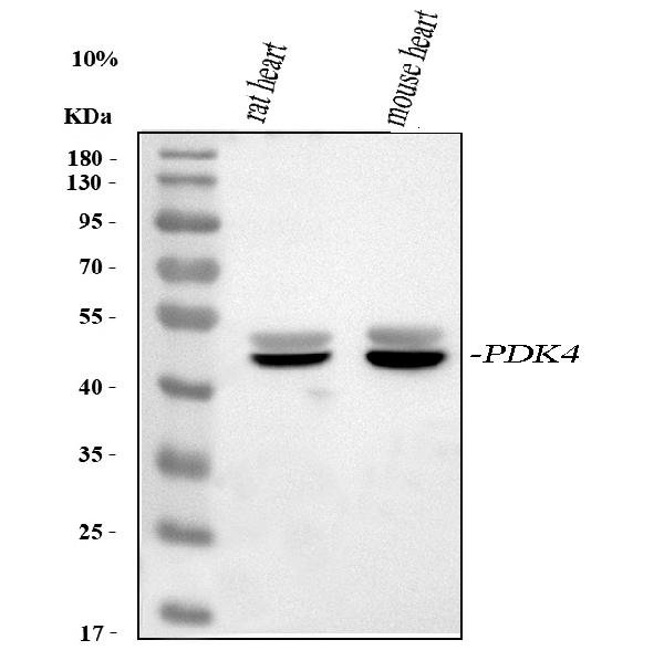
PDK4, Polyclonal Antibody (Cat# AAA127103)
Full Name
Anti-PDK4 Antibody Picoband
Reactivity
Human, Mouse, Rat
Applications
Flow Cytometry, Immunofluorescence, Immunocytochemistry, Immunohistochemistry, Western Blot
Purity
Immunogen affinity purified.
Pricing
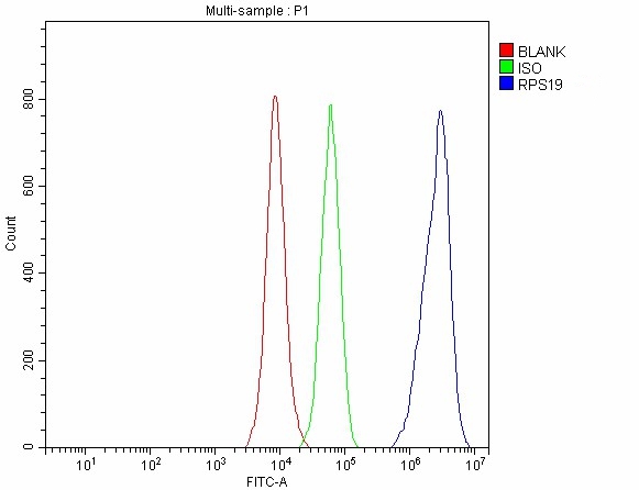 FCM/FACS (Flow Cytometry)
(Figure 3. Flow Cytometry analysis of 293T cells using anti-RPS19 antibody (AAA127107).Overlay histogram showing 293T cells stained with AAA127107 (Blue line). To facilitate intracellular staining, cells were fixed with 4% paraformaldehyde and permeabilized with permeabilization buffer. The cells were blocked with 10% normal goat serum. And then incubated with rabbit anti-RPS19 Antibody (AAA127107, 1ug/1x106 cells) for 30 min at 20 degree C. DyLight488 conjugated goat anti-rabbit IgG was used as secondary antibody for 30 minutes at 20 degree C. Isotype control antibody (Green line) was rabbit IgG (1ug/1x106) used under the same conditions. Unlabelled sample without incubation with primary antibody and secondary antibody (Red line) was used as a blank control.)
FCM/FACS (Flow Cytometry)
(Figure 3. Flow Cytometry analysis of 293T cells using anti-RPS19 antibody (AAA127107).Overlay histogram showing 293T cells stained with AAA127107 (Blue line). To facilitate intracellular staining, cells were fixed with 4% paraformaldehyde and permeabilized with permeabilization buffer. The cells were blocked with 10% normal goat serum. And then incubated with rabbit anti-RPS19 Antibody (AAA127107, 1ug/1x106 cells) for 30 min at 20 degree C. DyLight488 conjugated goat anti-rabbit IgG was used as secondary antibody for 30 minutes at 20 degree C. Isotype control antibody (Green line) was rabbit IgG (1ug/1x106) used under the same conditions. Unlabelled sample without incubation with primary antibody and secondary antibody (Red line) was used as a blank control.)
RPS19, Polyclonal Antibody (Cat# AAA127107)
Full Name
Anti-RPS19 Antibody Picoband
Gene Names
RPS19; DBA; S19; DBA1
Reactivity
Human, Mouse, Rat
Applications
Flow Cytometry, Immunofluorescence, Immunocytochemistry, Western Blot
Purity
Immunogen affinity purified.
Pricing
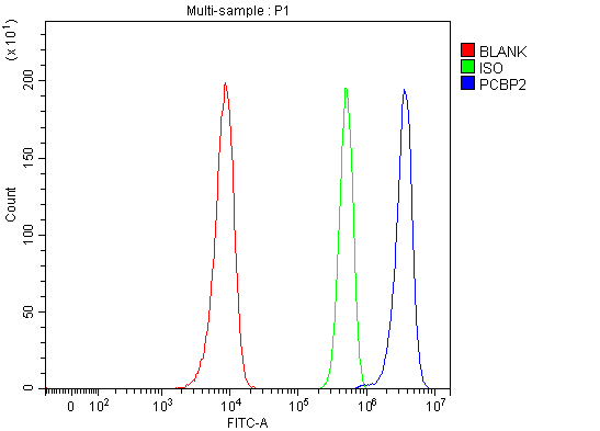 FCM/FACS (Flow Cytometry)
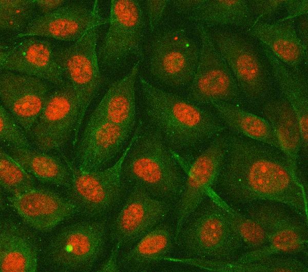
(Figure 3. Flow Cytometry analysis of Hela cells using anti-hnRNP E2/PCBP2 antibody (AAA127115).Overlay histogram showing Hela cells stained with AAA127115 (Blue line). To facilitate intracellular staining, cells were fixed with 4% paraformaldehyde and permeabilized with permeabilization buffer. The cells were blocked with 10% normal goat serum. And then incubated with rabbit anti-hnRNP E2/PCBP2 Antibody (AAA127115, 1ug/1x106 cells) for 30 min at 20 degree C. DyLight488 conjugated goat anti-rabbit IgG was used as secondary antibody for 30 minutes at 20 degree C. Isotype control antibody (Green line) was rabbit IgG (1ug/1x106) used under the same conditions. Unlabelled sample without incubation with primary antibody and secondary antibody (Red line) was used as a blank control.)
FCM/FACS (Flow Cytometry)
(Figure 3. Flow Cytometry analysis of Hela cells using anti-hnRNP E2/PCBP2 antibody (AAA127115).Overlay histogram showing Hela cells stained with AAA127115 (Blue line). To facilitate intracellular staining, cells were fixed with 4% paraformaldehyde and permeabilized with permeabilization buffer. The cells were blocked with 10% normal goat serum. And then incubated with rabbit anti-hnRNP E2/PCBP2 Antibody (AAA127115, 1ug/1x106 cells) for 30 min at 20 degree C. DyLight488 conjugated goat anti-rabbit IgG was used as secondary antibody for 30 minutes at 20 degree C. Isotype control antibody (Green line) was rabbit IgG (1ug/1x106) used under the same conditions. Unlabelled sample without incubation with primary antibody and secondary antibody (Red line) was used as a blank control.)
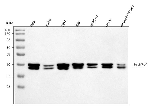
hnRNP E2/PCBP2, Polyclonal Antibody (Cat# AAA127115)
Full Name
Anti-hnRNP E2/PCBP2 Antibody Picoband
Gene Names
PCBP2; HNRPE2; HNRNPE2; hnRNP-E2
Reactivity
Human, Mouse, Rat
Applications
Western Blot, Immunofluorescence, Immunocytochemistry, Flow Cytometry
Purity
Immunogen affinity purified.
Pricing
 FCM/FACS (Flow Cytometry)
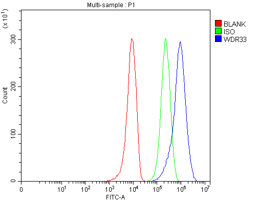
(Figure 2. Flow Cytometry analysis of 293T cells using anti-WDR33 antibody (AAA127596).Overlay histogram showing 293T cells stained with AAA127596 (Blue line). To facilitate intracellular staining, cells were fixed with 4% paraformaldehyde and permeabilized with permeabilization buffer. The cells were blocked with 10% normal goat serum. And then incubated with rabbit anti-WDR33 Antibody (AAA127596, 1ug/1x106 cells) for 30 min at 20 degree C. DyLight488 conjugated goat anti-rabbit IgG was used as secondary antibody for 30 minutes at 20 degree C. Isotype control antibody (Green line) was rabbit IgG (1ug/1x106) used under the same conditions. Unlabelled sample without incubation with primary antibody and secondary antibody (Red line) was used as a blank control.)
FCM/FACS (Flow Cytometry)
(Figure 2. Flow Cytometry analysis of 293T cells using anti-WDR33 antibody (AAA127596).Overlay histogram showing 293T cells stained with AAA127596 (Blue line). To facilitate intracellular staining, cells were fixed with 4% paraformaldehyde and permeabilized with permeabilization buffer. The cells were blocked with 10% normal goat serum. And then incubated with rabbit anti-WDR33 Antibody (AAA127596, 1ug/1x106 cells) for 30 min at 20 degree C. DyLight488 conjugated goat anti-rabbit IgG was used as secondary antibody for 30 minutes at 20 degree C. Isotype control antibody (Green line) was rabbit IgG (1ug/1x106) used under the same conditions. Unlabelled sample without incubation with primary antibody and secondary antibody (Red line) was used as a blank control.)
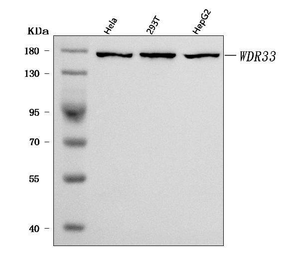
WDR33, Polyclonal Antibody (Cat# AAA127596)
Full Name
Anti-WDR33 Antibody Picoband
Gene Names
WDR33; NET14; WDC146
Reactivity
Human
Applications
Flow Cytometry, Western Blot
Purity
Immunogen affinity purified.
Pricing
 FCM/FACS (Flow Cytometry)
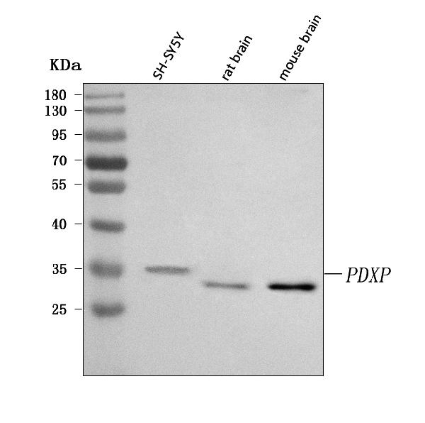
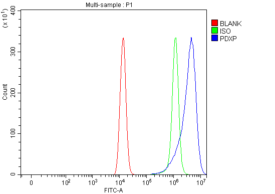
(Figure 2. Flow Cytometry analysis of HEL cells using anti-PDXP antibody (AAA127600).Overlay histogram showing HEL cells stained with AAA127600 (Blue line). To facilitate intracellular staining, cells were fixed with 4% paraformaldehyde and permeabilized with permeabilization buffer. The cells were blocked with 10% normal goat serum. And then incubated with rabbit anti-PDXP Antibody (AAA127600, 1ug/1x106 cells) for 30 min at 20 degree C. DyLight488 conjugated goat anti-rabbit IgG was used as secondary antibody for 30 minutes at 20 degree C. Isotype control antibody (Green line) was rabbit IgG (1ug/1x106) used under the same conditions. Unlabelled sample (Red line) was also used as a control.)
FCM/FACS (Flow Cytometry)
(Figure 2. Flow Cytometry analysis of HEL cells using anti-PDXP antibody (AAA127600).Overlay histogram showing HEL cells stained with AAA127600 (Blue line). To facilitate intracellular staining, cells were fixed with 4% paraformaldehyde and permeabilized with permeabilization buffer. The cells were blocked with 10% normal goat serum. And then incubated with rabbit anti-PDXP Antibody (AAA127600, 1ug/1x106 cells) for 30 min at 20 degree C. DyLight488 conjugated goat anti-rabbit IgG was used as secondary antibody for 30 minutes at 20 degree C. Isotype control antibody (Green line) was rabbit IgG (1ug/1x106) used under the same conditions. Unlabelled sample (Red line) was also used as a control.)
PDXP, Polyclonal Antibody (Cat# AAA127600)
Full Name
Anti-PDXP Antibody Picoband
Gene Names
PDXP; CIN; PLP; dJ37E16.5
Reactivity
Human, Mouse, Rat
Applications
Flow Cytometry, Western Blot
Purity
Immunogen affinity purified.
Pricing
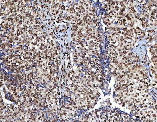
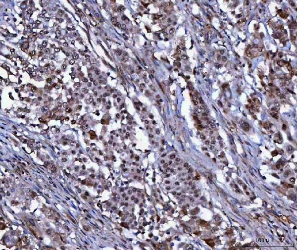
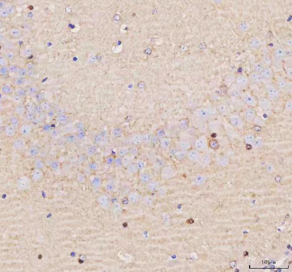
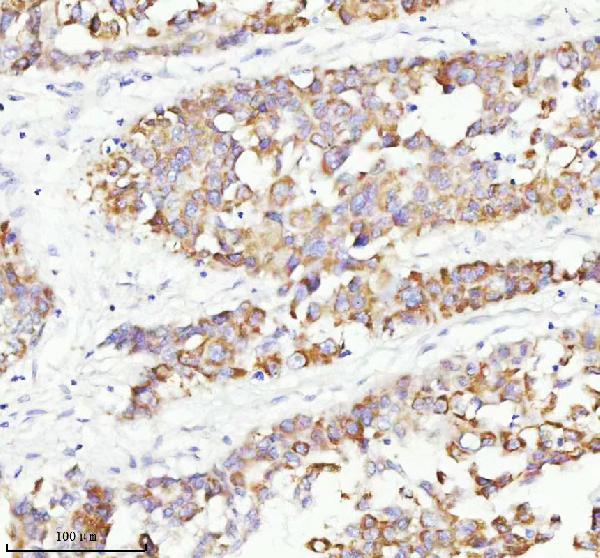
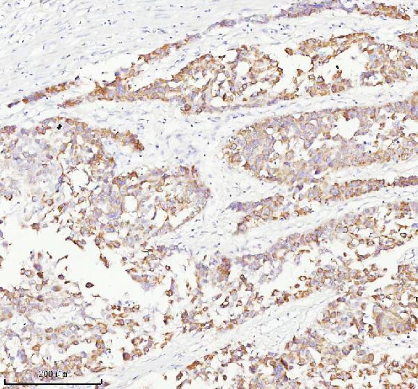
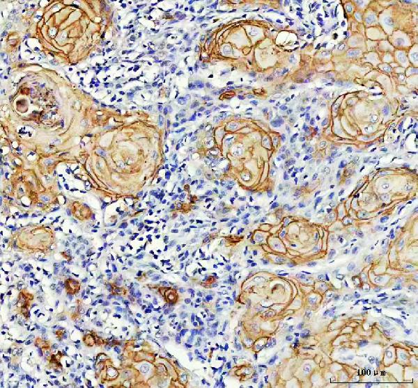 IHC (Immunohiostchemistry)
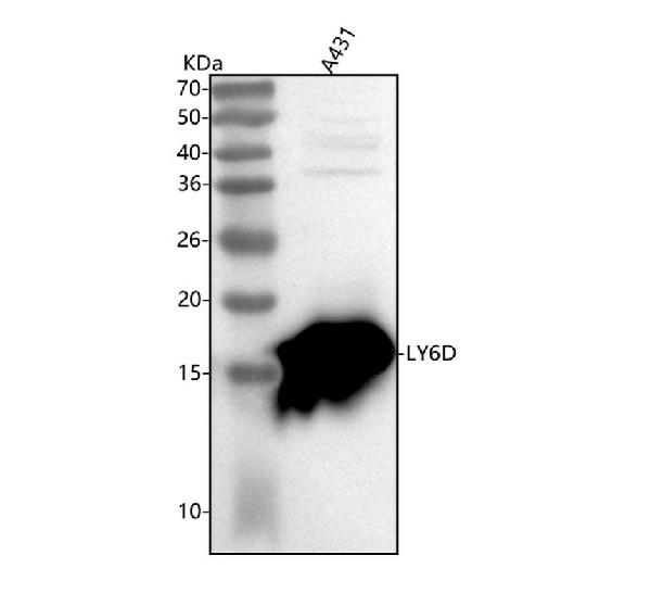
(Figure 2. IHC analysis of LY6D using anti-LY6D antibody (AAA127604).LY6D was detected in a paraffin-embedded section of human skin cancer tissue. Heat mediated antigen retrieval was performed in EDTA buffer (pH 8.0, epitope retrieval solution). The tissue section was blocked with 10% goat serum. The tissue section was then incubated with 2ug/ml rabbit anti-LY6D Antibody (AAA127604) overnight at 4 degree C. Peroxidase Conjugated Goat Anti-rabbit IgG was used as secondary antibody and incubated for 30 minutes at 37 degree C. The tissue section was developed using HRP Conjugated Rabbit IgG Super Vision Assay Kit with DAB as the chromogen.)
IHC (Immunohiostchemistry)
(Figure 2. IHC analysis of LY6D using anti-LY6D antibody (AAA127604).LY6D was detected in a paraffin-embedded section of human skin cancer tissue. Heat mediated antigen retrieval was performed in EDTA buffer (pH 8.0, epitope retrieval solution). The tissue section was blocked with 10% goat serum. The tissue section was then incubated with 2ug/ml rabbit anti-LY6D Antibody (AAA127604) overnight at 4 degree C. Peroxidase Conjugated Goat Anti-rabbit IgG was used as secondary antibody and incubated for 30 minutes at 37 degree C. The tissue section was developed using HRP Conjugated Rabbit IgG Super Vision Assay Kit with DAB as the chromogen.)
LY6D, Polyclonal Antibody (Cat# AAA127604)
Full Name
Anti-LY6D Antibody Picoband
Gene Names
LY6D; E48; Ly-6D
Reactivity
Human
Applications
Immunohistochemistry, Western Blot
Purity
Immunogen affinity purified.
Pricing

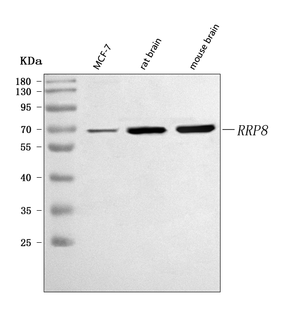
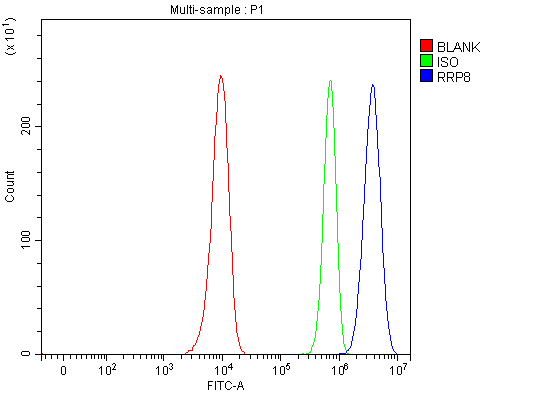 FCM/FACS (Flow Cytometry)
(Figure 3. Flow Cytometry analysis of Hela cells using anti-RRP8 antibody (AAA127621).Overlay histogram showing Hela cells stained with AAA127621 (Blue line). To facilitate intracellular staining, cells were fixed with 4% paraformaldehyde and permeabilized with permeabilization buffer. The cells were blocked with 10% normal goat serum. And then incubated with rabbit anti-RRP8 Antibody (AAA127621, 1ug/1x106 cells) for 30 min at 20 degree C. DyLight488 conjugated goat anti-rabbit IgG was used as secondary antibody for 30 minutes at 20 degree C. Isotype control antibody (Green line) was rabbit IgG (1ug/1x106) used under the same conditions. Unlabelled sample without incubation with primary antibody and secondary antibody (Red line) was used as a blank control.)
FCM/FACS (Flow Cytometry)
(Figure 3. Flow Cytometry analysis of Hela cells using anti-RRP8 antibody (AAA127621).Overlay histogram showing Hela cells stained with AAA127621 (Blue line). To facilitate intracellular staining, cells were fixed with 4% paraformaldehyde and permeabilized with permeabilization buffer. The cells were blocked with 10% normal goat serum. And then incubated with rabbit anti-RRP8 Antibody (AAA127621, 1ug/1x106 cells) for 30 min at 20 degree C. DyLight488 conjugated goat anti-rabbit IgG was used as secondary antibody for 30 minutes at 20 degree C. Isotype control antibody (Green line) was rabbit IgG (1ug/1x106) used under the same conditions. Unlabelled sample without incubation with primary antibody and secondary antibody (Red line) was used as a blank control.)
RRP8, Polyclonal Antibody (Cat# AAA127621)
Full Name
Anti-RRP8 Antibody Picoband
Gene Names
RRP8; KIAA0409
Reactivity
Human, Mouse, Rat
Applications
Flow Cytometry, Immunofluorescence, Immunocytochemistry, Western Blot
Purity
Immunogen affinity purified.
Pricing
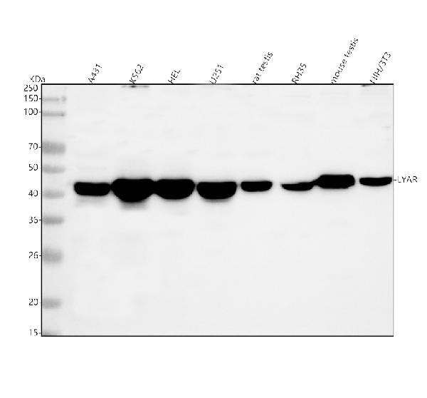
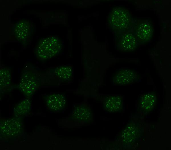
 IF (Immunofluorescence)
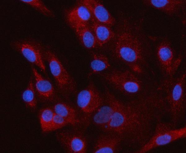
(Figure 2. IF analysis of LYAR using anti-LYAR antibody (AAA127622) and anti-Beta Tubulin antibody (M01857-3).LYAR was detected in immunocytochemical section of HELA cell. Enzyme antigen retrieval was performed using IHC enzyme antigen retrieval reagent for 15 mins. The cells were blocked with 10% goat serum. And then incubated with 5ug/mL rabbit anti-LYAR Antibody (AAA127622) and mouse anti-Beta Tubulin antibody (M01857-3) overnight at 4 degree C. DyLight488 Conjugated Goat Anti-Rabbit IgG and Cy3 Conjugated Goat Anti-Mouse IgG (BA1031) were used as secondary antibody at 1:500 dilution and incubated for 30 minutes at 37 degree C. The section was counterstained with DAPI. Visualize using a fluorescence microscope and filter sets appropriate for the label used.)
IF (Immunofluorescence)
(Figure 2. IF analysis of LYAR using anti-LYAR antibody (AAA127622) and anti-Beta Tubulin antibody (M01857-3).LYAR was detected in immunocytochemical section of HELA cell. Enzyme antigen retrieval was performed using IHC enzyme antigen retrieval reagent for 15 mins. The cells were blocked with 10% goat serum. And then incubated with 5ug/mL rabbit anti-LYAR Antibody (AAA127622) and mouse anti-Beta Tubulin antibody (M01857-3) overnight at 4 degree C. DyLight488 Conjugated Goat Anti-Rabbit IgG and Cy3 Conjugated Goat Anti-Mouse IgG (BA1031) were used as secondary antibody at 1:500 dilution and incubated for 30 minutes at 37 degree C. The section was counterstained with DAPI. Visualize using a fluorescence microscope and filter sets appropriate for the label used.)
LYAR, Polyclonal Antibody (Cat# AAA127622)
Full Name
Anti-LYAR Antibody Picoband
Gene Names
LYAR; ZLYAR; ZC2HC2
Reactivity
Human, Mouse, Rat
Applications
Immunofluorescence, Immunocytochemistry, Western Blot
Purity
Immunogen affinity purified.
Pricing

 IF (Immunofluorescence)
(Figure 2. IF analysis of HSD3B7 using anti-HSD3B7 antibody (AAA127624).HSD3B7 was detected in an immunocytochemical section of A549 cells. Enzyme antigen retrieval was performed using IHC enzyme antigen retrieval reagent for 15 mins. The cells were blocked with 10% goat serum. And then incubated with 5ug/mL rabbit anti-HSD3B7 Antibody (AAA127624) overnight at 4 degree C. Cy3 Conjugated Goat Anti-Rabbit IgG (BA1032) was used as secondary antibody at 1:500 dilution and incubated for 30 minutes at 37 degree C. The section was counterstained with DAPI. Visualize using a fluorescence microscope and filter sets appropriate for the label used.)
IF (Immunofluorescence)
(Figure 2. IF analysis of HSD3B7 using anti-HSD3B7 antibody (AAA127624).HSD3B7 was detected in an immunocytochemical section of A549 cells. Enzyme antigen retrieval was performed using IHC enzyme antigen retrieval reagent for 15 mins. The cells were blocked with 10% goat serum. And then incubated with 5ug/mL rabbit anti-HSD3B7 Antibody (AAA127624) overnight at 4 degree C. Cy3 Conjugated Goat Anti-Rabbit IgG (BA1032) was used as secondary antibody at 1:500 dilution and incubated for 30 minutes at 37 degree C. The section was counterstained with DAPI. Visualize using a fluorescence microscope and filter sets appropriate for the label used.)
HSD3B7, Polyclonal Antibody (Cat# AAA127624)
Full Name
Anti-HSD3B7 Antibody Picoband
Gene Names
HSD3B7; CBAS1; PFIC4; SDR11E3
Reactivity
Human
Applications
Immunofluorescence, Immunocytochemistry, Western Blot
Purity
Immunogen affinity purified.
Pricing
 FCM/FACS (Flow Cytometry)
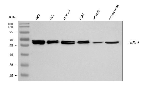
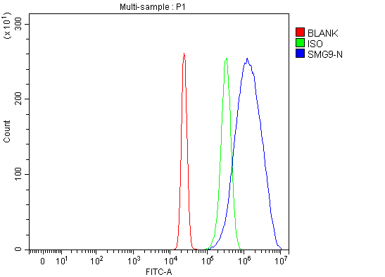
(Figure 3. Flow Cytometry analysis of HepG2 cells using anti-C19orf61/SMG9 antibody (AAA127639).Overlay histogram showing HepG2 cells stained with AAA127639 (Blue line). To facilitate intracellular staining, cells were fixed with 4% paraformaldehyde and permeabilized with permeabilization buffer. The cells were blocked with 10% normal goat serum. And then incubated with rabbit anti-C19orf61/SMG9 Antibody (AAA127639, 1ug/1x106 cells) for 30 min at 20 degree C. DyLight488 conjugated goat anti-rabbit IgG was used as secondary antibody for 30 minutes at 20 degree C. Isotype control antibody (Green line) was rabbit IgG (1ug/1x106) used under the same conditions. Unlabelled sample without incubation with primary antibody and secondary antibody (Red line) was used as a blank control.)
FCM/FACS (Flow Cytometry)
(Figure 3. Flow Cytometry analysis of HepG2 cells using anti-C19orf61/SMG9 antibody (AAA127639).Overlay histogram showing HepG2 cells stained with AAA127639 (Blue line). To facilitate intracellular staining, cells were fixed with 4% paraformaldehyde and permeabilized with permeabilization buffer. The cells were blocked with 10% normal goat serum. And then incubated with rabbit anti-C19orf61/SMG9 Antibody (AAA127639, 1ug/1x106 cells) for 30 min at 20 degree C. DyLight488 conjugated goat anti-rabbit IgG was used as secondary antibody for 30 minutes at 20 degree C. Isotype control antibody (Green line) was rabbit IgG (1ug/1x106) used under the same conditions. Unlabelled sample without incubation with primary antibody and secondary antibody (Red line) was used as a blank control.)
C19orf61/SMG9, Polyclonal Antibody (Cat# AAA127639)
Full Name
Anti-C19orf61/SMG9 Antibody Picoband
Gene Names
SMG9; C19orf61; F17127_1
Reactivity
Human, Mouse, Rat
Applications
Flow Cytometry, Immunofluorescence, Immunocytochemistry, Western Blot
Purity
Immunogen affinity purified.
Pricing
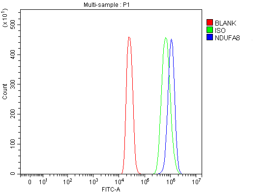
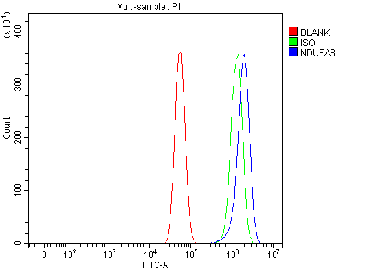 FCM/FACS (Flow Cytometry)
(Figure 3. Flow Cytometry analysis of U87 cells using anti-NDUFA8 antibody (AAA127661).Overlay histogram showing U87 cells stained with AAA127661 (Blue line). To facilitate intracellular staining, cells were fixed with 4% paraformaldehyde and permeabilized with permeabilization buffer. The cells were blocked with 10% normal goat serum. And then incubated with rabbit anti-NDUFA8 Antibody (AAA127661, 1ug/1x106 cells) for 30 min at 20 degree C. DyLight488 conjugated goat anti-rabbit IgG was used as secondary antibody for 30 minutes at 20 degree C. Isotype control antibody (Green line) was rabbit IgG (1ug/1x106) used under the same conditions. Unlabelled sample (Red line) was also used as a control.)
FCM/FACS (Flow Cytometry)
(Figure 3. Flow Cytometry analysis of U87 cells using anti-NDUFA8 antibody (AAA127661).Overlay histogram showing U87 cells stained with AAA127661 (Blue line). To facilitate intracellular staining, cells were fixed with 4% paraformaldehyde and permeabilized with permeabilization buffer. The cells were blocked with 10% normal goat serum. And then incubated with rabbit anti-NDUFA8 Antibody (AAA127661, 1ug/1x106 cells) for 30 min at 20 degree C. DyLight488 conjugated goat anti-rabbit IgG was used as secondary antibody for 30 minutes at 20 degree C. Isotype control antibody (Green line) was rabbit IgG (1ug/1x106) used under the same conditions. Unlabelled sample (Red line) was also used as a control.)
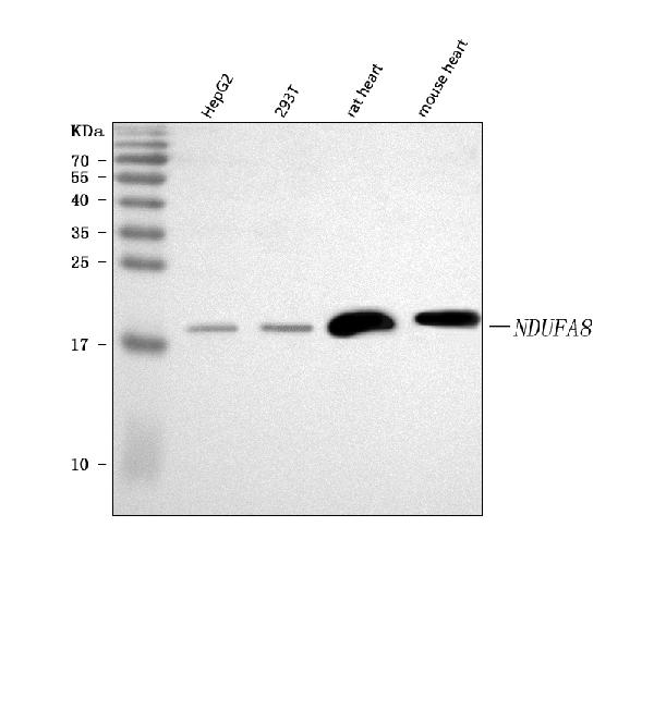
NDUFA8, Polyclonal Antibody (Cat# AAA127661)
Full Name
Anti-NDUFA8 Antibody Picoband
Gene Names
NDUFA8; PGIV; CI-19KD; CI-PGIV
Reactivity
Human, Mouse, Rat
Applications
Flow Cytometry, Western Blot
Purity
Immunogen affinity purified.
Pricing
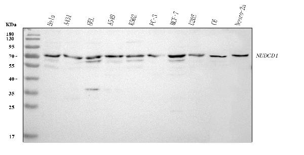
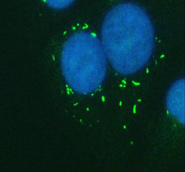
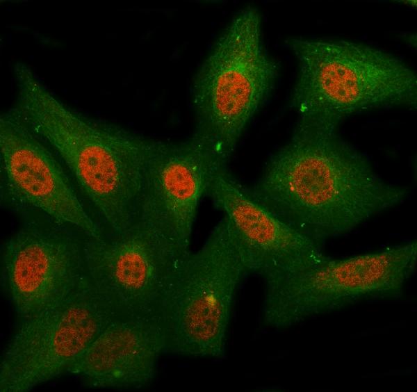 IF (Immunofluorescence)
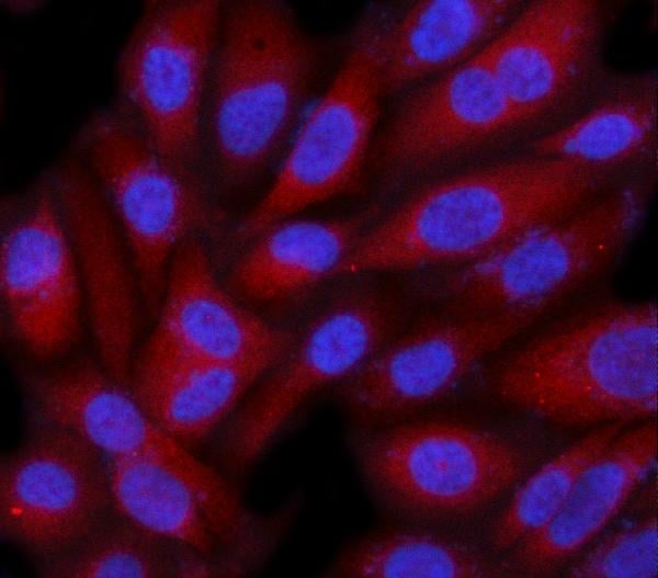
(Figure 4. IF analysis of NUDCD1 using anti-NUDCD1 antibody (AAA127674) and anti-Beta Tubulin antibody (M01857-3).NUDCD1 was detected in immunocytochemical section of HELA cell. Enzyme antigen retrieval was performed using IHC enzyme antigen retrieval reagent for 15 mins. The cells were blocked with 10% goat serum. And then incubated with 5ug/mL rabbit anti-NUDCD1 Antibody (AAA127674) and mouse anti-Beta Tubulin antibody (M01857-3) overnight at 4 degree C. Cy3 Conjugated Goat Anti-Rabbit IgG (BA1032) and DyLight488 Conjugated Goat Anti-Mouse IgG (BA1126) were used as secondary antibody at 1:500 dilution and incubated for 30 minutes at 37 degree C. Visualize using a fluorescence microscope and filter sets appropriate for the label used.)
IF (Immunofluorescence)
(Figure 4. IF analysis of NUDCD1 using anti-NUDCD1 antibody (AAA127674) and anti-Beta Tubulin antibody (M01857-3).NUDCD1 was detected in immunocytochemical section of HELA cell. Enzyme antigen retrieval was performed using IHC enzyme antigen retrieval reagent for 15 mins. The cells were blocked with 10% goat serum. And then incubated with 5ug/mL rabbit anti-NUDCD1 Antibody (AAA127674) and mouse anti-Beta Tubulin antibody (M01857-3) overnight at 4 degree C. Cy3 Conjugated Goat Anti-Rabbit IgG (BA1032) and DyLight488 Conjugated Goat Anti-Mouse IgG (BA1126) were used as secondary antibody at 1:500 dilution and incubated for 30 minutes at 37 degree C. Visualize using a fluorescence microscope and filter sets appropriate for the label used.)
NUDCD1, Polyclonal Antibody (Cat# AAA127674)
Full Name
Anti-NUDCD1 Antibody Picoband
Gene Names
NUDCD1; CML66; OVA66
Reactivity
Human, Mouse, Rat
Applications
Immunofluorescence, Immunocytochemistry, Immunohistochemistry, Western Blot
Purity
Immunogen affinity purified.
Pricing
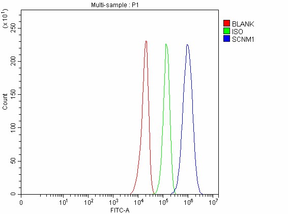 FCM/FACS (Flow Cytometry)
(Figure 5. Flow Cytometry analysis of U251 cells using anti-SCNM1 antibody (AAA127689).Overlay histogram showing U251 cells stained with AAA127689 (Blue line). To facilitate intracellular staining, cells were fixed with 4% paraformaldehyde and permeabilized with permeabilization buffer. The cells were blocked with 10% normal goat serum. And then incubated with rabbit anti-SCNM1 Antibody (AAA127689, 1ug/1x106 cells) for 30 min at 20 degree C. DyLight488 conjugated goat anti-rabbit IgG was used as secondary antibody for 30 minutes at 20 degree C. Isotype control antibody (Green line) was rabbit IgG (1ug/1x106) used under the same conditions. Unlabelled sample without incubation with primary antibody and secondary antibody (Red line) was used as a blank control.)
FCM/FACS (Flow Cytometry)
(Figure 5. Flow Cytometry analysis of U251 cells using anti-SCNM1 antibody (AAA127689).Overlay histogram showing U251 cells stained with AAA127689 (Blue line). To facilitate intracellular staining, cells were fixed with 4% paraformaldehyde and permeabilized with permeabilization buffer. The cells were blocked with 10% normal goat serum. And then incubated with rabbit anti-SCNM1 Antibody (AAA127689, 1ug/1x106 cells) for 30 min at 20 degree C. DyLight488 conjugated goat anti-rabbit IgG was used as secondary antibody for 30 minutes at 20 degree C. Isotype control antibody (Green line) was rabbit IgG (1ug/1x106) used under the same conditions. Unlabelled sample without incubation with primary antibody and secondary antibody (Red line) was used as a blank control.)
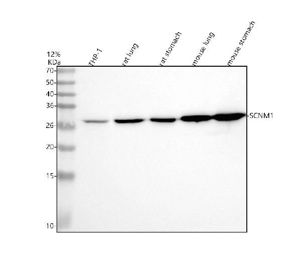
SCNM1, Polyclonal Antibody (Cat# AAA127689)
Full Name
Anti-SCNM1 Antibody Picoband
Gene Names
SCNM1; MGC3180
Reactivity
Human, Mouse, Rat
Applications
Flow Cytometry, Immunohistochemistry, Western Blot
Purity
Immunogen affinity purified.
Pricing
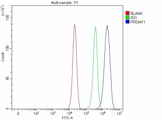 FCM/FACS (Flow Cytometry)
(Figure 3. Flow Cytometry analysis of A549 cells using anti-PRDM11 antibody (AAA127696).Overlay histogram showing A549 cells stained with AAA127696 (Blue line). To facilitate intracellular staining, cells were fixed with 4% paraformaldehyde and permeabilized with permeabilization buffer. The cells were blocked with 10% normal goat serum. And then incubated with rabbit anti-PRDM11 Antibody (AAA127696, 1ug/1x106 cells) for 30 min at 20 degree C. DyLight488 conjugated goat anti-rabbit IgG was used as secondary antibody for 30 minutes at 20 degree C. Isotype control antibody (Green line) was rabbit IgG (1ug/1x106) used under the same conditions. Unlabelled sample without incubation with primary antibody and secondary antibody (Red line) was used as a blank control.)
FCM/FACS (Flow Cytometry)
(Figure 3. Flow Cytometry analysis of A549 cells using anti-PRDM11 antibody (AAA127696).Overlay histogram showing A549 cells stained with AAA127696 (Blue line). To facilitate intracellular staining, cells were fixed with 4% paraformaldehyde and permeabilized with permeabilization buffer. The cells were blocked with 10% normal goat serum. And then incubated with rabbit anti-PRDM11 Antibody (AAA127696, 1ug/1x106 cells) for 30 min at 20 degree C. DyLight488 conjugated goat anti-rabbit IgG was used as secondary antibody for 30 minutes at 20 degree C. Isotype control antibody (Green line) was rabbit IgG (1ug/1x106) used under the same conditions. Unlabelled sample without incubation with primary antibody and secondary antibody (Red line) was used as a blank control.)
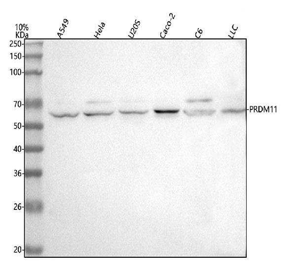
PRDM11, Polyclonal Antibody (Cat# AAA127696)
Full Name
Anti-PRDM11 Antibody Picoband
Gene Names
PRDM11; PFM8
Reactivity
Human, Mouse, Rat
Applications
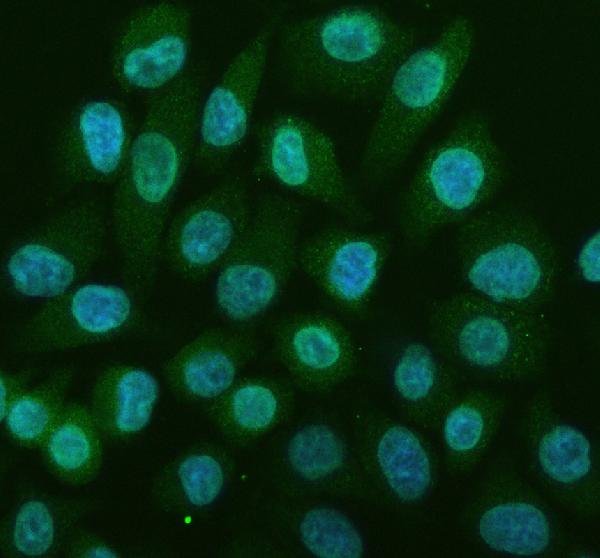
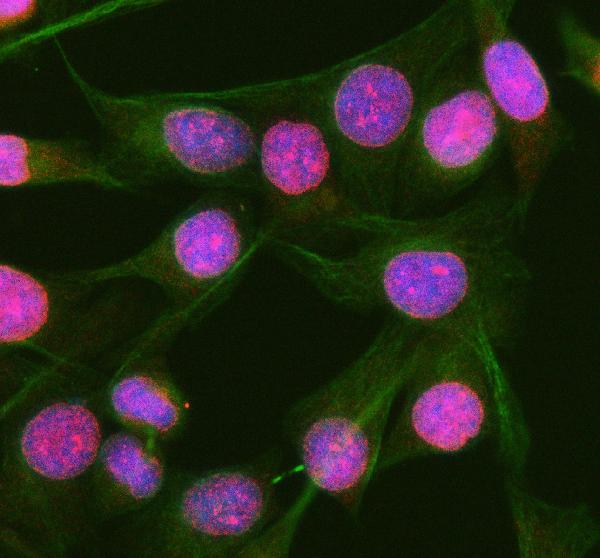
Flow Cytometry, Immunofluorescence, Immunocytochemistry, Western Blot
Purity
Immunogen affinity purified.
Pricing
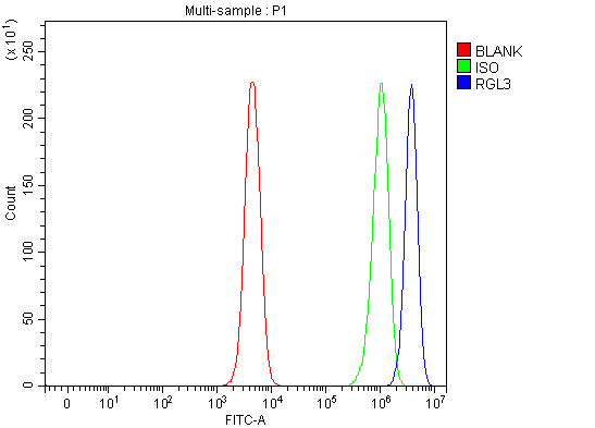 FCM/FACS (Flow Cytometry)
(Figure 5. Flow Cytometry analysis of SH-SY5Y cells using anti-RGL3 antibody (AAA127734).Overlay histogram showing SH-SY5Y cells stained with AAA127734 (Blue line). To facilitate intracellular staining, cells were fixed with 4% paraformaldehyde and permeabilized with permeabilization buffer. The cells were blocked with 10% normal goat serum. And then incubated with rabbit anti-RGL3 Antibody (AAA127734, 1ug/1x106 cells) for 30 min at 20 degree C. DyLight488 conjugated goat anti-rabbit IgG was used as secondary antibody for 30 minutes at 20 degree C. Isotype control antibody (Green line) was rabbit IgG (1ug/1x106) used under the same conditions. Unlabelled sample (Red line) was also used as a control.)
FCM/FACS (Flow Cytometry)
(Figure 5. Flow Cytometry analysis of SH-SY5Y cells using anti-RGL3 antibody (AAA127734).Overlay histogram showing SH-SY5Y cells stained with AAA127734 (Blue line). To facilitate intracellular staining, cells were fixed with 4% paraformaldehyde and permeabilized with permeabilization buffer. The cells were blocked with 10% normal goat serum. And then incubated with rabbit anti-RGL3 Antibody (AAA127734, 1ug/1x106 cells) for 30 min at 20 degree C. DyLight488 conjugated goat anti-rabbit IgG was used as secondary antibody for 30 minutes at 20 degree C. Isotype control antibody (Green line) was rabbit IgG (1ug/1x106) used under the same conditions. Unlabelled sample (Red line) was also used as a control.)
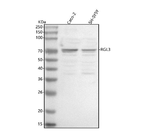
RGL3, Polyclonal Antibody (Cat# AAA127734)
Full Name
Anti-RGL3 Antibody Picoband
Reactivity
Human
Applications
Flow Cytometry, Immunofluorescence, Immunocytochemistry, Immunohistochemistry, Western Blot
Purity
Immunogen affinity purified.
Pricing
 IF (Immunofluorescence)
(Figure 2. IF analysis of PRTFDC1 using anti-PRTFDC1 antibody (AAA127736).PRTFDC1 was detected in an immunocytochemical section of A431 cells. Enzyme antigen retrieval was performed using IHC enzyme antigen retrieval reagent for 15 mins. The cells were blocked with 10% goat serum. And then incubated with 5ug/mL rabbit anti-PRTFDC1 Antibody (AAA127736) overnight at 4 degree C. Cy3 Conjugated Goat Anti-Rabbit IgG (BA1032) was used as secondary antibody at 1:500 dilution and incubated for 30 minutes at 37 degree C. The section was counterstained with DAPI. Visualize using a fluorescence microscope and filter sets appropriate for the label used.)
IF (Immunofluorescence)
(Figure 2. IF analysis of PRTFDC1 using anti-PRTFDC1 antibody (AAA127736).PRTFDC1 was detected in an immunocytochemical section of A431 cells. Enzyme antigen retrieval was performed using IHC enzyme antigen retrieval reagent for 15 mins. The cells were blocked with 10% goat serum. And then incubated with 5ug/mL rabbit anti-PRTFDC1 Antibody (AAA127736) overnight at 4 degree C. Cy3 Conjugated Goat Anti-Rabbit IgG (BA1032) was used as secondary antibody at 1:500 dilution and incubated for 30 minutes at 37 degree C. The section was counterstained with DAPI. Visualize using a fluorescence microscope and filter sets appropriate for the label used.)
PRTFDC1, Polyclonal Antibody (Cat# AAA127736)
Full Name
Anti-PRTFDC1 Antibody Picoband
Gene Names
PRTFDC1; HHGP
Reactivity
Human, Mouse, Rat
Applications
Immunofluorescence, Immunocytochemistry, Western Blot
Purity
Immunogen affinity purified.
Pricing
 IF (Immunofluorescence)
(Figure 2. IF analysis of PBX4 using anti-PBX4 antibody (AAA127742) and anti-Beta Tubulin antibody (M01857-3).PBX4 was detected in immunocytochemical section of A431 cell. Enzyme antigen retrieval was performed using IHC enzyme antigen retrieval reagent for 15 mins. The cells were blocked with 10% goat serum. And then incubated with 5ug/mL rabbit anti-PBX4 Antibody (AAA127742) and mouse anti-Beta Tubulin antibody (M01857-3) overnight at 4 degree C. Cy3 Conjugated Goat Anti-Rabbit IgG (BA1032) and FITC Conjugated Goat Anti-Mouse IgG (BA1101) were used as secondary antibody at 1:500 dilution and incubated for 30 minutes at 37 degree C. Visualize using a fluorescence microscope and filter sets appropriate for the label used.)
IF (Immunofluorescence)
(Figure 2. IF analysis of PBX4 using anti-PBX4 antibody (AAA127742) and anti-Beta Tubulin antibody (M01857-3).PBX4 was detected in immunocytochemical section of A431 cell. Enzyme antigen retrieval was performed using IHC enzyme antigen retrieval reagent for 15 mins. The cells were blocked with 10% goat serum. And then incubated with 5ug/mL rabbit anti-PBX4 Antibody (AAA127742) and mouse anti-Beta Tubulin antibody (M01857-3) overnight at 4 degree C. Cy3 Conjugated Goat Anti-Rabbit IgG (BA1032) and FITC Conjugated Goat Anti-Mouse IgG (BA1101) were used as secondary antibody at 1:500 dilution and incubated for 30 minutes at 37 degree C. Visualize using a fluorescence microscope and filter sets appropriate for the label used.)
PBX4, Polyclonal Antibody (Cat# AAA127742)
Full Name
Anti-PBX4 Antibody Picoband
Reactivity
Human, Mouse
Applications
Immunofluorescence, Immunocytochemistry, Western Blot
Purity
Immunogen affinity purified.
Pricing
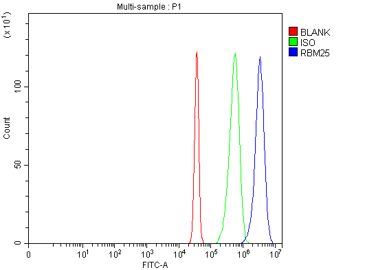 FCM/FACS (Flow Cytometry)
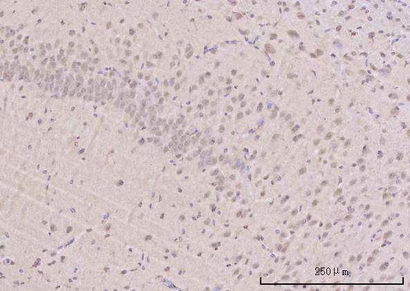
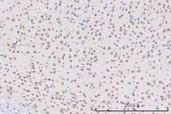
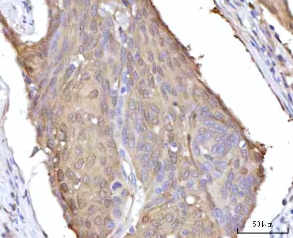
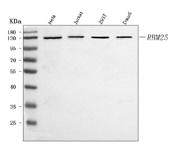
(Figure 5. Flow Cytometry analysis of Jurkat cells using anti-RBM25 antibody (AAA127449).Overlay histogram showing Jurkat cells stained with AAA127449 (Blue line). To facilitate intracellular staining, cells were fixed with 4% paraformaldehyde and permeabilized with permeabilization buffer. The cells were blocked with 10% normal goat serum. And then incubated with rabbit anti-RBM25 Antibody (AAA127449, 1ug/1x106 cells) for 30 min at 20 degree C. DyLight488 conjugated goat anti-rabbit IgG was used as secondary antibody for 30 minutes at 20 degree C. Isotype control antibody (Green line) was rabbit IgG (1ug/1x106) used under the same conditions. Unlabelled sample without incubation with primary antibody and secondary antibody (Red line) was used as a blank control.)
FCM/FACS (Flow Cytometry)
(Figure 5. Flow Cytometry analysis of Jurkat cells using anti-RBM25 antibody (AAA127449).Overlay histogram showing Jurkat cells stained with AAA127449 (Blue line). To facilitate intracellular staining, cells were fixed with 4% paraformaldehyde and permeabilized with permeabilization buffer. The cells were blocked with 10% normal goat serum. And then incubated with rabbit anti-RBM25 Antibody (AAA127449, 1ug/1x106 cells) for 30 min at 20 degree C. DyLight488 conjugated goat anti-rabbit IgG was used as secondary antibody for 30 minutes at 20 degree C. Isotype control antibody (Green line) was rabbit IgG (1ug/1x106) used under the same conditions. Unlabelled sample without incubation with primary antibody and secondary antibody (Red line) was used as a blank control.)
RBM25, Polyclonal Antibody (Cat# AAA127449)
Full Name
Anti-RBM25 Antibody Picoband
Gene Names
RBM25; S164; NET52; RNPC7; Snu71; RED120; fSAP94; MGC105088; MGC117168
Reactivity
Human, Mouse, Rat
Applications
Western Blot, Immunohistochemistry, Flow Cytometry
Purity
Immunogen affinity purified.
Pricing
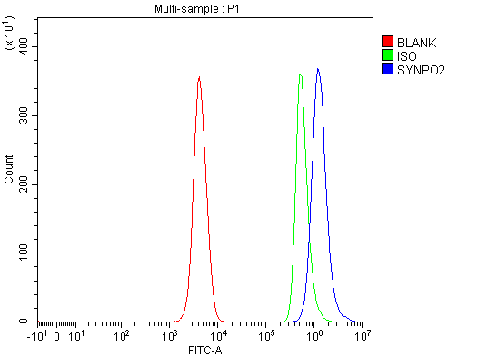 FCM/FACS (Flow Cytometry)
(Figure 2. Flow Cytometry analysis of SH-SY5Y cells using anti-SYNPO2 antibody (AAA127462).Overlay histogram showing SH-SY5Y cells stained with AAA127462 (Blue line). To facilitate intracellular staining, cells were fixed with 4% paraformaldehyde and permeabilized with permeabilization buffer. The cells were blocked with 10% normal goat serum. And then incubated with rabbit anti-SYNPO2 Antibody (AAA127462, 1ug/1x106 cells) for 30 min at 20 degree C. DyLight488 conjugated goat anti-rabbit IgG was used as secondary antibody for 30 minutes at 20 degree C. Isotype control antibody (Green line) was rabbit IgG (1ug/1x106) used under the same conditions. Unlabelled sample (Red line) was also used as a control.)
FCM/FACS (Flow Cytometry)
(Figure 2. Flow Cytometry analysis of SH-SY5Y cells using anti-SYNPO2 antibody (AAA127462).Overlay histogram showing SH-SY5Y cells stained with AAA127462 (Blue line). To facilitate intracellular staining, cells were fixed with 4% paraformaldehyde and permeabilized with permeabilization buffer. The cells were blocked with 10% normal goat serum. And then incubated with rabbit anti-SYNPO2 Antibody (AAA127462, 1ug/1x106 cells) for 30 min at 20 degree C. DyLight488 conjugated goat anti-rabbit IgG was used as secondary antibody for 30 minutes at 20 degree C. Isotype control antibody (Green line) was rabbit IgG (1ug/1x106) used under the same conditions. Unlabelled sample (Red line) was also used as a control.)
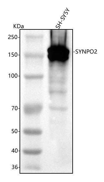
SYNPO2, Polyclonal Antibody (Cat# AAA127462)
Full Name
Anti-SYNPO2 Antibody Picoband
Reactivity
Human
Applications
Flow Cytometry, Western Blot
Purity
Immunogen affinity purified.
Pricing
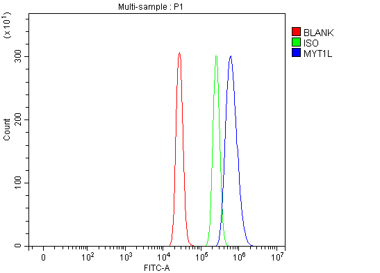 FCM/FACS (Flow Cytometry)
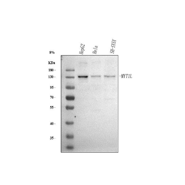
(Figure 3. Flow Cytometry analysis of HepG2 cells using anti-MYT1L antibody (AAA127482).Overlay histogram showing HepG2 cells stained with AAA127482 (Blue line). To facilitate intracellular staining, cells were fixed with 4% paraformaldehyde and permeabilized with permeabilization buffer. The cells were blocked with 10% normal goat serum. And then incubated with rabbit anti-MYT1L Antibody (AAA127482, 1ug/1x106 cells) for 30 min at 20 degree C. DyLight488 conjugated goat anti-rabbit IgG was used as secondary antibody for 30 minutes at 20 degree C. Isotype control antibody (Green line) was rabbit IgG (1ug/1x106) used under the same conditions. Unlabelled sample (Red line) was also used as a control.)
FCM/FACS (Flow Cytometry)
(Figure 3. Flow Cytometry analysis of HepG2 cells using anti-MYT1L antibody (AAA127482).Overlay histogram showing HepG2 cells stained with AAA127482 (Blue line). To facilitate intracellular staining, cells were fixed with 4% paraformaldehyde and permeabilized with permeabilization buffer. The cells were blocked with 10% normal goat serum. And then incubated with rabbit anti-MYT1L Antibody (AAA127482, 1ug/1x106 cells) for 30 min at 20 degree C. DyLight488 conjugated goat anti-rabbit IgG was used as secondary antibody for 30 minutes at 20 degree C. Isotype control antibody (Green line) was rabbit IgG (1ug/1x106) used under the same conditions. Unlabelled sample (Red line) was also used as a control.)
MYT1L, Polyclonal Antibody (Cat# AAA127482)
Full Name
Anti-MYT1L Antibody Picoband
Gene Names
MYT1L; NZF1; ZC2HC4B
Reactivity
Human
Applications
Flow Cytometry, Western Blot, Immunofluorescence, Immunocytochemistry
Purity
Immunogen affinity purified.
Pricing
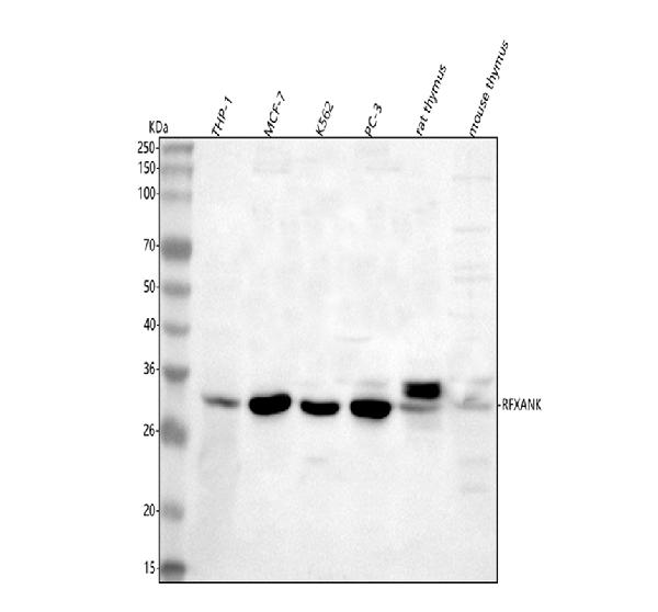
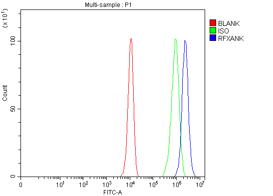 FCM/FACS (Flow Cytometry)
(Figure 3. Flow Cytometry analysis of PC-3 cells using anti-RFXANK antibody (AAA127488).Overlay histogram showing PC-3 cells stained with AAA127488 (Blue line). To facilitate intracellular staining, cells were fixed with 4% paraformaldehyde and permeabilized with permeabilization buffer. The cells were blocked with 10% normal goat serum. And then incubated with rabbit anti-RFXANK Antibody (AAA127488, 1ug/1x106 cells) for 30 min at 20 degree C. DyLight488 conjugated goat anti-rabbit IgG was used as secondary antibody for 30 minutes at 20 degree C. Isotype control antibody (Green line) was rabbit IgG (1ug/1x106) used under the same conditions. Unlabelled sample (Red line) was also used as a control.)
FCM/FACS (Flow Cytometry)
(Figure 3. Flow Cytometry analysis of PC-3 cells using anti-RFXANK antibody (AAA127488).Overlay histogram showing PC-3 cells stained with AAA127488 (Blue line). To facilitate intracellular staining, cells were fixed with 4% paraformaldehyde and permeabilized with permeabilization buffer. The cells were blocked with 10% normal goat serum. And then incubated with rabbit anti-RFXANK Antibody (AAA127488, 1ug/1x106 cells) for 30 min at 20 degree C. DyLight488 conjugated goat anti-rabbit IgG was used as secondary antibody for 30 minutes at 20 degree C. Isotype control antibody (Green line) was rabbit IgG (1ug/1x106) used under the same conditions. Unlabelled sample (Red line) was also used as a control.)
RFXANK, Polyclonal Antibody (Cat# AAA127488)
Full Name
Anti-RFXANK Antibody Picoband
Gene Names
RFXANK; BLS; RFX-B; ANKRA1; F14150_1
Reactivity
Human, Mouse, Rat
Applications
Flow Cytometry, Immunofluorescence, Immunocytochemistry, Western Blot
Purity
Immunogen affinity purified.
Pricing

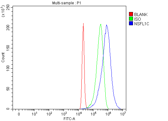 FCM/FACS (Flow Cytometry)
(Figure 3. Flow Cytometry analysis of 293T cells using anti-NSFL1C antibody (AAA127501).Overlay histogram showing 293T cells stained with AAA127501 (Blue line). To facilitate intracellular staining, cells were fixed with 4% paraformaldehyde and permeabilized with permeabilization buffer. The cells were blocked with 10% normal goat serum. And then incubated with rabbit anti-NSFL1C Antibody (AAA127501, 1ug/1x106 cells) for 30 min at 20 degree C. DyLight488 conjugated goat anti-rabbit IgG was used as secondary antibody for 30 minutes at 20 degree C. Isotype control antibody (Green line) was rabbit IgG (1ug/1x106) used under the same conditions. Unlabelled sample (Red line) was also used as a control.)
FCM/FACS (Flow Cytometry)
(Figure 3. Flow Cytometry analysis of 293T cells using anti-NSFL1C antibody (AAA127501).Overlay histogram showing 293T cells stained with AAA127501 (Blue line). To facilitate intracellular staining, cells were fixed with 4% paraformaldehyde and permeabilized with permeabilization buffer. The cells were blocked with 10% normal goat serum. And then incubated with rabbit anti-NSFL1C Antibody (AAA127501, 1ug/1x106 cells) for 30 min at 20 degree C. DyLight488 conjugated goat anti-rabbit IgG was used as secondary antibody for 30 minutes at 20 degree C. Isotype control antibody (Green line) was rabbit IgG (1ug/1x106) used under the same conditions. Unlabelled sample (Red line) was also used as a control.)
NSFL1C, Polyclonal Antibody (Cat# AAA127501)
Full Name
Anti-NSFL1C Antibody Picoband
Gene Names
NSFL1C; P47; UBX1; UBXD10; UBXN2C; dJ776F14.1
Reactivity
Human, Mouse, Rat
Applications
Flow Cytometry, Western Blot, Immunofluorescence, Immunocytochemistry
Purity
Immunogen affinity purified.
Pricing
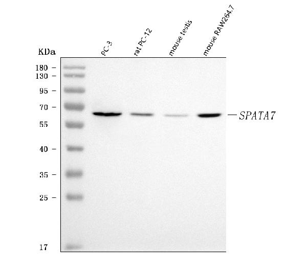
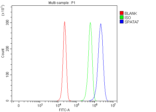 FCM/FACS (Flow Cytometry)
(Figure 2. Flow Cytometry analysis of HepG2 cells using anti-SPATA7 antibody (AAA127514).Overlay histogram showing HepG2 cells stained with AAA127514 (Blue line). To facilitate intracellular staining, cells were fixed with 4% paraformaldehyde and permeabilized with permeabilization buffer. The cells were blocked with 10% normal goat serum. And then incubated with rabbit anti-SPATA7 Antibody (AAA127514, 1ug/1x106 cells) for 30 min at 20 degree C. DyLight488 conjugated goat anti-rabbit IgG was used as secondary antibody for 30 minutes at 20 degree C. Isotype control antibody (Green line) was rabbit IgG (1ug/1x106) used under the same conditions. Unlabelled sample without incubation with primary antibody and secondary antibody (Red line) was used as a blank control.)
FCM/FACS (Flow Cytometry)
(Figure 2. Flow Cytometry analysis of HepG2 cells using anti-SPATA7 antibody (AAA127514).Overlay histogram showing HepG2 cells stained with AAA127514 (Blue line). To facilitate intracellular staining, cells were fixed with 4% paraformaldehyde and permeabilized with permeabilization buffer. The cells were blocked with 10% normal goat serum. And then incubated with rabbit anti-SPATA7 Antibody (AAA127514, 1ug/1x106 cells) for 30 min at 20 degree C. DyLight488 conjugated goat anti-rabbit IgG was used as secondary antibody for 30 minutes at 20 degree C. Isotype control antibody (Green line) was rabbit IgG (1ug/1x106) used under the same conditions. Unlabelled sample without incubation with primary antibody and secondary antibody (Red line) was used as a blank control.)
SPATA7, Polyclonal Antibody (Cat# AAA127514)
Full Name
Anti-SPATA7 Antibody Picoband
Gene Names
SPATA7; HSD3; LCA3; HSD-3.1; HEL-S-296
Reactivity
Human, Mouse, Rat
Applications
Flow Cytometry, Western Blot
Purity
Immunogen affinity purified.
Pricing
 FCM/FACS (Flow Cytometry)
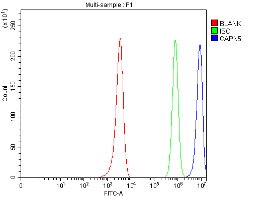
(Figure 3. Flow Cytometry analysis of U251 cells using anti-CAPN5 antibody (AAA127518).Overlay histogram showing U251 cells stained with AAA127518 (Blue line). To facilitate intracellular staining, cells were fixed with 4% paraformaldehyde and permeabilized with permeabilization buffer. The cells were blocked with 10% normal goat serum. And then incubated with rabbit anti-CAPN5 Antibody (AAA127518, 1ug/1x106 cells) for 30 min at 20 degree C. DyLight488 conjugated goat anti-rabbit IgG was used as secondary antibody for 30 minutes at 20 degree C. Isotype control antibody (Green line) was rabbit IgG (1ug/1x106) used under the same conditions. Unlabelled sample (Red line) was also used as a control.)
FCM/FACS (Flow Cytometry)
(Figure 3. Flow Cytometry analysis of U251 cells using anti-CAPN5 antibody (AAA127518).Overlay histogram showing U251 cells stained with AAA127518 (Blue line). To facilitate intracellular staining, cells were fixed with 4% paraformaldehyde and permeabilized with permeabilization buffer. The cells were blocked with 10% normal goat serum. And then incubated with rabbit anti-CAPN5 Antibody (AAA127518, 1ug/1x106 cells) for 30 min at 20 degree C. DyLight488 conjugated goat anti-rabbit IgG was used as secondary antibody for 30 minutes at 20 degree C. Isotype control antibody (Green line) was rabbit IgG (1ug/1x106) used under the same conditions. Unlabelled sample (Red line) was also used as a control.)
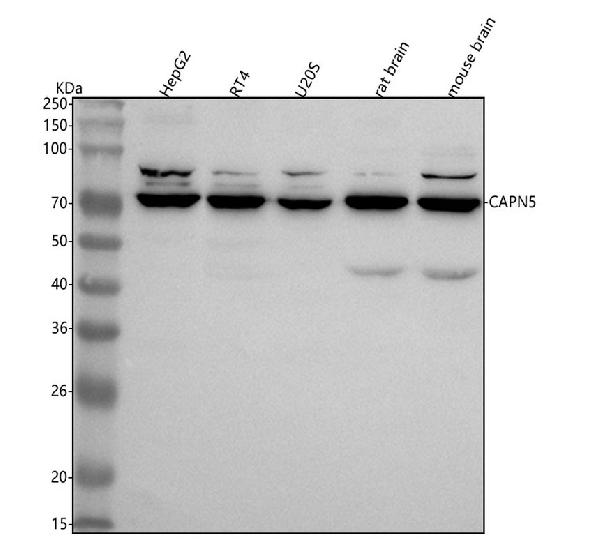
CAPN5, Polyclonal Antibody (Cat# AAA127518)
Full Name
Anti-CAPN5 Antibody Picoband
Gene Names
CAPN5; VRNI; ADNIV; HTRA3; nCL-3
Reactivity
Human, Mouse, Rat
Applications
Flow Cytometry, Immunofluorescence, Immunocytochemistry, Western Blot
Purity
Immunogen affinity purified.
Pricing

 FCM/FACS (Flow Cytometry)
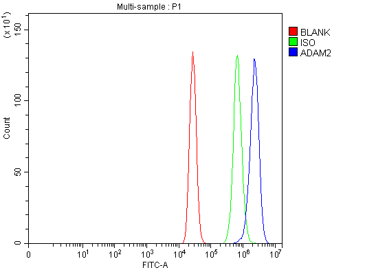
(Figure 2. Flow Cytometry analysis of MCF-7 cells using anti-ADAM2 antibody (AAA127519).Overlay histogram showing MCF-7 cells stained with AAA127519 (Blue line). The cells were fixed with 4% paraformaldehyde and blocked with 10% normal goat serum. And then incubated with rabbit anti-ADAM2 Antibody (AAA127519, 1ug/1x106 cells) for 30 min at 20 degree C. DyLight488 conjugated goat anti-rabbit IgG was used as secondary antibody for 30 minutes at 20 degree C. Isotype control antibody (Green line) was rabbit IgG (1ug/1x106) used under the same conditions. Unlabelled sample (Red line) was also used as a control.)
FCM/FACS (Flow Cytometry)
(Figure 2. Flow Cytometry analysis of MCF-7 cells using anti-ADAM2 antibody (AAA127519).Overlay histogram showing MCF-7 cells stained with AAA127519 (Blue line). The cells were fixed with 4% paraformaldehyde and blocked with 10% normal goat serum. And then incubated with rabbit anti-ADAM2 Antibody (AAA127519, 1ug/1x106 cells) for 30 min at 20 degree C. DyLight488 conjugated goat anti-rabbit IgG was used as secondary antibody for 30 minutes at 20 degree C. Isotype control antibody (Green line) was rabbit IgG (1ug/1x106) used under the same conditions. Unlabelled sample (Red line) was also used as a control.)
ADAM2, Polyclonal Antibody (Cat# AAA127519)
Full Name
Anti-ADAM2 Antibody Picoband
Gene Names
ADAM2; CT15; FTNB; PH30; CRYN1; CRYN2; PH-30b; PH30-beta
Reactivity
Human
Applications
Flow Cytometry, Western Blot
Purity
Immunogen affinity purified.
Pricing
 FCM/FACS (Flow Cytometry)
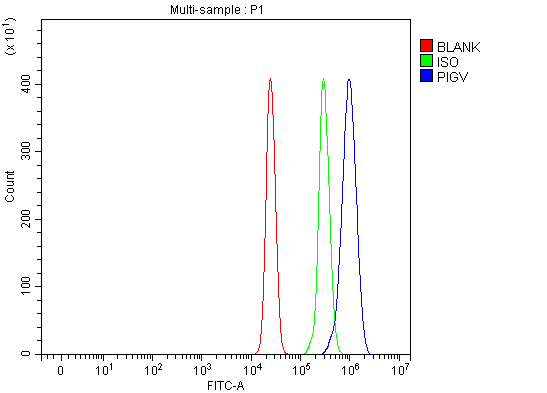
(Figure 2. Flow Cytometry analysis of U937 cells using anti-PIGV antibody (AAA127540).Overlay histogram showing U937 cells stained with AAA127540 (Blue line). To facilitate intracellular staining, cells were fixed with 4% paraformaldehyde and permeabilized with permeabilization buffer. The cells were blocked with 10% normal goat serum. And then incubated with rabbit anti-PIGV Antibody (AAA127540, 1ug/1x106 cells) for 30 min at 20 degree C. DyLight488 conjugated goat anti-rabbit IgG was used as secondary antibody for 30 minutes at 20 degree C. Isotype control antibody (Green line) was rabbit IgG (1ug/1x106) used under the same conditions. Unlabelled sample (Red line) was also used as a control.)
FCM/FACS (Flow Cytometry)
(Figure 2. Flow Cytometry analysis of U937 cells using anti-PIGV antibody (AAA127540).Overlay histogram showing U937 cells stained with AAA127540 (Blue line). To facilitate intracellular staining, cells were fixed with 4% paraformaldehyde and permeabilized with permeabilization buffer. The cells were blocked with 10% normal goat serum. And then incubated with rabbit anti-PIGV Antibody (AAA127540, 1ug/1x106 cells) for 30 min at 20 degree C. DyLight488 conjugated goat anti-rabbit IgG was used as secondary antibody for 30 minutes at 20 degree C. Isotype control antibody (Green line) was rabbit IgG (1ug/1x106) used under the same conditions. Unlabelled sample (Red line) was also used as a control.)
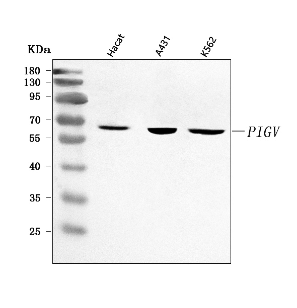
PIGV, Polyclonal Antibody (Cat# AAA127540)
Full Name
Anti-PIGV Antibody Picoband
Gene Names
PIGV; PIG-V; HPMRS1; GPI-MT-II
Reactivity
Human
Applications
Flow Cytometry, Western Blot
Purity
Immunogen affinity purified.
Pricing
 FCM/FACS (Flow Cytometry)
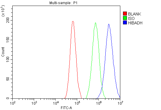
(Figure 2. Flow Cytometry analysis of A549 cells using anti-HIBADH antibody (AAA127543).Overlay histogram showing A549 cells stained with AAA127543 (Blue line). To facilitate intracellular staining, cells were fixed with 4% paraformaldehyde and permeabilized with permeabilization buffer. The cells were blocked with 10% normal goat serum. And then incubated with rabbit anti-HIBADH Antibody (AAA127543, 1ug/1x106 cells) for 30 min at 20 degree C. DyLight488 conjugated goat anti-rabbit IgG was used as secondary antibody for 30 minutes at 20 degree C. Isotype control antibody (Green line) was rabbit IgG (1ug/1x106) used under the same conditions. Unlabelled sample (Red line) was also used as a control.)
FCM/FACS (Flow Cytometry)
(Figure 2. Flow Cytometry analysis of A549 cells using anti-HIBADH antibody (AAA127543).Overlay histogram showing A549 cells stained with AAA127543 (Blue line). To facilitate intracellular staining, cells were fixed with 4% paraformaldehyde and permeabilized with permeabilization buffer. The cells were blocked with 10% normal goat serum. And then incubated with rabbit anti-HIBADH Antibody (AAA127543, 1ug/1x106 cells) for 30 min at 20 degree C. DyLight488 conjugated goat anti-rabbit IgG was used as secondary antibody for 30 minutes at 20 degree C. Isotype control antibody (Green line) was rabbit IgG (1ug/1x106) used under the same conditions. Unlabelled sample (Red line) was also used as a control.)
HIBADH, Polyclonal Antibody (Cat# AAA127543)
Full Name
Anti-HIBADH Antibody Picoband
Gene Names
HIBADH; NS5ATP1
Reactivity
Human, Mouse, Rat
Applications
Flow Cytometry, Western Blot
Purity
Immunogen affinity purified.
Pricing
 FCM/FACS (Flow Cytometry)
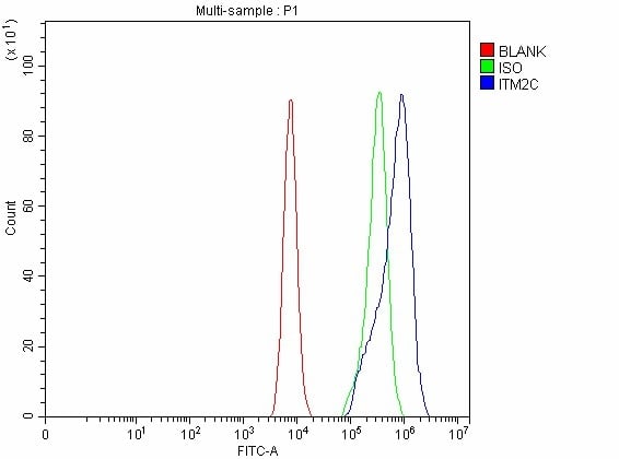
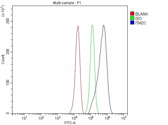
(Figure 3. Flow Cytometry analysis of PC-3 cells using anti-ITM2C antibody (AAA127549).Overlay histogram showing PC-3 cells stained with AAA127549 (Blue line). The cells were fixed with 4% paraformaldehyde and blocked with 10% normal goat serum. And then incubated with rabbit anti-ITM2C Antibody (AAA127549, 1ug/1x106 cells) for 30 min at 20 degree C. DyLight488 conjugated goat anti-rabbit IgG was used as secondary antibody for 30 minutes at 20 degree C. Isotype control antibody (Green line) was rabbit IgG (1ug/1x106) used under the same conditions. Unlabelled sample without incubation with primary antibody and secondary antibody (Red line) was used as a blank control.)
FCM/FACS (Flow Cytometry)
(Figure 3. Flow Cytometry analysis of PC-3 cells using anti-ITM2C antibody (AAA127549).Overlay histogram showing PC-3 cells stained with AAA127549 (Blue line). The cells were fixed with 4% paraformaldehyde and blocked with 10% normal goat serum. And then incubated with rabbit anti-ITM2C Antibody (AAA127549, 1ug/1x106 cells) for 30 min at 20 degree C. DyLight488 conjugated goat anti-rabbit IgG was used as secondary antibody for 30 minutes at 20 degree C. Isotype control antibody (Green line) was rabbit IgG (1ug/1x106) used under the same conditions. Unlabelled sample without incubation with primary antibody and secondary antibody (Red line) was used as a blank control.)
ITM2C, Polyclonal Antibody (Cat# AAA127549)
Full Name
Anti-ITM2C Antibody Picoband
Gene Names
ITM2C; E25; BRI3; E25C; ITM3; BRICD2C
Reactivity
Human, Mouse, Rat
Applications
Flow Cytometry, Western Blot
Purity
Immunogen affinity purified.
Pricing

 FCM/FACS (Flow Cytometry)
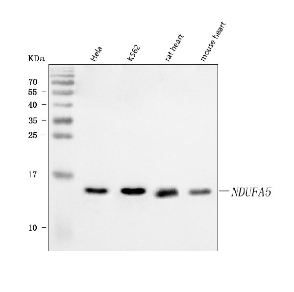
(Figure 3. Flow Cytometry analysis of U937 cells using anti-NDUFA5 antibody (AAA127555).Overlay histogram showing U937 cells stained with AAA127555 (Blue line). To facilitate intracellular staining, cells were fixed with 4% paraformaldehyde and permeabilized with permeabilization buffer. The cells were blocked with 10% normal goat serum. And then incubated with rabbit anti-NDUFA5 Antibody (AAA127555, 1ug/1x106 cells) for 30 min at 20 degree C. DyLight488 conjugated goat anti-rabbit IgG was used as secondary antibody for 30 minutes at 20 degree C. Isotype control antibody (Green line) was rabbit IgG (1ug/1x106) used under the same conditions. Unlabelled sample (Red line) was also used as a control.)
FCM/FACS (Flow Cytometry)
(Figure 3. Flow Cytometry analysis of U937 cells using anti-NDUFA5 antibody (AAA127555).Overlay histogram showing U937 cells stained with AAA127555 (Blue line). To facilitate intracellular staining, cells were fixed with 4% paraformaldehyde and permeabilized with permeabilization buffer. The cells were blocked with 10% normal goat serum. And then incubated with rabbit anti-NDUFA5 Antibody (AAA127555, 1ug/1x106 cells) for 30 min at 20 degree C. DyLight488 conjugated goat anti-rabbit IgG was used as secondary antibody for 30 minutes at 20 degree C. Isotype control antibody (Green line) was rabbit IgG (1ug/1x106) used under the same conditions. Unlabelled sample (Red line) was also used as a control.)
NDUFA5, Polyclonal Antibody (Cat# AAA127555)
Full Name
Anti-NDUFA5 Antibody Picoband
Gene Names
NDUFA5; B13; NUFM; UQOR13; CI-13kB; CI-13KD-B
Reactivity
Human, Mouse, Rat
Applications
Flow Cytometry, Immunofluorescence, Immunocytochemistry, Western Blot
Purity
Immunogen affinity purified.
Pricing
 FCM/FACS (Flow Cytometry)
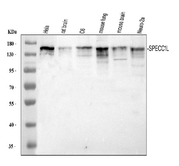
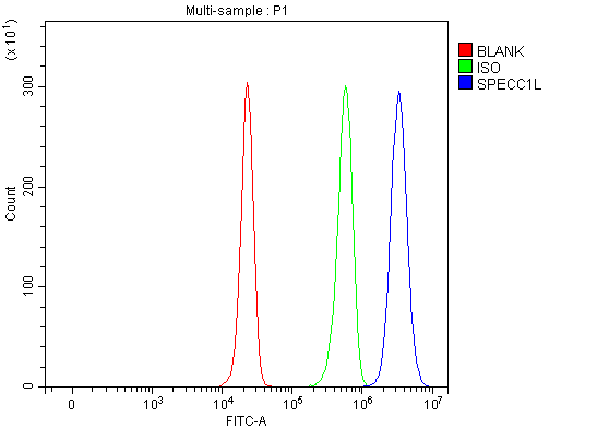
(Figure 3. Flow Cytometry analysis of HepG2 cells using anti-SPECC1L antibody (AAA127568).Overlay histogram showing HepG2 cells stained with AAA127568 (Blue line). To facilitate intracellular staining, cells were fixed with 4% paraformaldehyde and permeabilized with permeabilization buffer. The cells were blocked with 10% normal goat serum. And then incubated with rabbit anti-SPECC1L Antibody (AAA127568, 1ug/1x106 cells) for 30 min at 20 degree C. DyLight488 conjugated goat anti-rabbit IgG was used as secondary antibody for 30 minutes at 20 degree C. Isotype control antibody (Green line) was rabbit IgG (1ug/1x106) used under the same conditions. Unlabelled sample without incubation with primary antibody and secondary antibody (Red line) was used as a blank control.)
FCM/FACS (Flow Cytometry)
(Figure 3. Flow Cytometry analysis of HepG2 cells using anti-SPECC1L antibody (AAA127568).Overlay histogram showing HepG2 cells stained with AAA127568 (Blue line). To facilitate intracellular staining, cells were fixed with 4% paraformaldehyde and permeabilized with permeabilization buffer. The cells were blocked with 10% normal goat serum. And then incubated with rabbit anti-SPECC1L Antibody (AAA127568, 1ug/1x106 cells) for 30 min at 20 degree C. DyLight488 conjugated goat anti-rabbit IgG was used as secondary antibody for 30 minutes at 20 degree C. Isotype control antibody (Green line) was rabbit IgG (1ug/1x106) used under the same conditions. Unlabelled sample without incubation with primary antibody and secondary antibody (Red line) was used as a blank control.)
SPECC1L, Polyclonal Antibody (Cat# AAA127568)
Full Name
Anti-SPECC1L Antibody Picoband
Gene Names
SPECC1L; CYTSA; OBLFC1
Reactivity
Human, Mouse, Rat
Applications
Flow Cytometry, Immunofluorescence, Immunocytochemistry, Western Blot
Purity
Immunogen affinity purified.
Pricing
 FCM/FACS (Flow Cytometry)
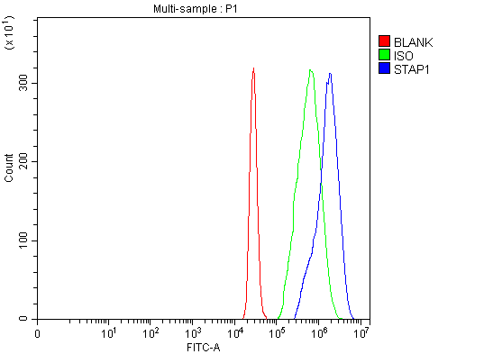
(Figure 2. Flow Cytometry analysis of U937 cells using anti-STAP-1/STAP1 antibody (AAA127569).Overlay histogram showing U937 cells stained with AAA127569 (Blue line). To facilitate intracellular staining, cells were fixed with 4% paraformaldehyde and permeabilized with permeabilization buffer. The cells were blocked with 10% normal goat serum. And then incubated with rabbit anti-STAP-1/STAP1 Antibody (AAA127569, 1ug/1x106 cells) for 30 min at 20 degree C. DyLight488 conjugated goat anti-rabbit IgG was used as secondary antibody for 30 minutes at 20 degree C. Isotype control antibody (Green line) was rabbit IgG (1ug/1x106) used under the same conditions. Unlabelled sample without incubation with primary antibody and secondary antibody (Red line) was used as a blank control.)
FCM/FACS (Flow Cytometry)
(Figure 2. Flow Cytometry analysis of U937 cells using anti-STAP-1/STAP1 antibody (AAA127569).Overlay histogram showing U937 cells stained with AAA127569 (Blue line). To facilitate intracellular staining, cells were fixed with 4% paraformaldehyde and permeabilized with permeabilization buffer. The cells were blocked with 10% normal goat serum. And then incubated with rabbit anti-STAP-1/STAP1 Antibody (AAA127569, 1ug/1x106 cells) for 30 min at 20 degree C. DyLight488 conjugated goat anti-rabbit IgG was used as secondary antibody for 30 minutes at 20 degree C. Isotype control antibody (Green line) was rabbit IgG (1ug/1x106) used under the same conditions. Unlabelled sample without incubation with primary antibody and secondary antibody (Red line) was used as a blank control.)
STAP-1/STAP1, Polyclonal Antibody (Cat# AAA127569)
Full Name
Anti-STAP-1/STAP1 Antibody Picoband
Gene Names
STAP1; BRDG1; STAP-1
Reactivity
Human
Applications
Flow Cytometry, Western Blot
Purity
Immunogen affinity purified.
Pricing
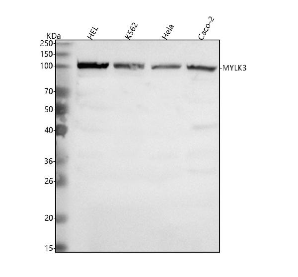
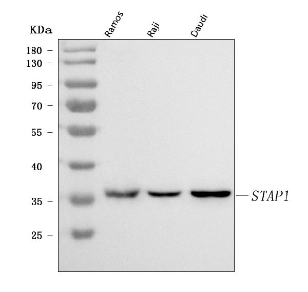
 FCM/FACS (Flow Cytometry)
(Figure 2. Flow Cytometry analysis of HEL cells using anti-MYLK3 antibody (AAA127571).Overlay histogram showing HEL cells stained with AAA127571 (Blue line). To facilitate intracellular staining, cells were fixed with 4% paraformaldehyde and permeabilized with permeabilization buffer. The cells were blocked with 10% normal goat serum. And then incubated with rabbit anti-MYLK3 Antibody (AAA127571, 1ug/1x106 cells) for 30 min at 20 degree C. DyLight488 conjugated goat anti-rabbit IgG was used as secondary antibody for 30 minutes at 20 degree C. Isotype control antibody (Green line) was rabbit IgG (1ug/1x106) used under the same conditions. Unlabelled sample (Red line) was also used as a control.)
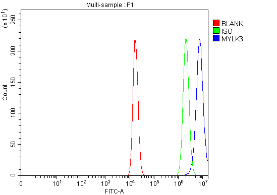
FCM/FACS (Flow Cytometry)
(Figure 2. Flow Cytometry analysis of HEL cells using anti-MYLK3 antibody (AAA127571).Overlay histogram showing HEL cells stained with AAA127571 (Blue line). To facilitate intracellular staining, cells were fixed with 4% paraformaldehyde and permeabilized with permeabilization buffer. The cells were blocked with 10% normal goat serum. And then incubated with rabbit anti-MYLK3 Antibody (AAA127571, 1ug/1x106 cells) for 30 min at 20 degree C. DyLight488 conjugated goat anti-rabbit IgG was used as secondary antibody for 30 minutes at 20 degree C. Isotype control antibody (Green line) was rabbit IgG (1ug/1x106) used under the same conditions. Unlabelled sample (Red line) was also used as a control.)
MYLK3, Polyclonal Antibody (Cat# AAA127571)
Full Name
Anti-MYLK3 Antibody Picoband
Gene Names
MYLK3; MLCK; MLCK2; caMLCK
Reactivity
Human
Applications
Flow Cytometry, Western Blot
Purity
Immunogen affinity purified.
Pricing
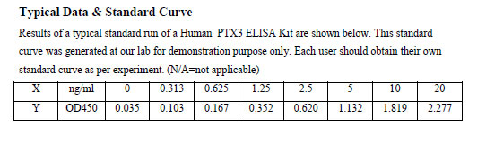
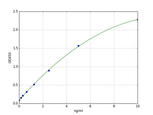
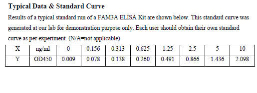
 Standard Curve (Sample)
Pentraxin-relatedh.protein PTX3, ELISA Kit (Cat# AAA99881)
Full Name
Human Pentraxin-relatedh.protein PTX3 ELISA Kit
Reactivity
Human
Pricing
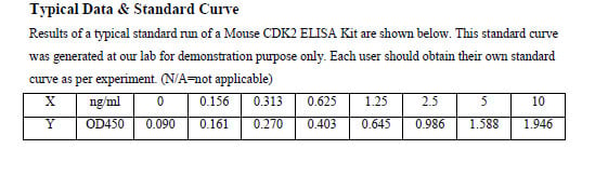
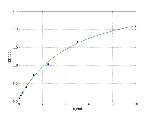
 Standard Curve (Sample)
Cyclin-dependent kinase 2, ELISA Kit (Cat# AAA99905)
Full Name
Mouse Cyclin-dependent kinase 2 ELISA Kit
Reactivity
Mouse
Pricing
 Standard Curve (Sample)
Protein FAM3A, ELISA Kit (Cat# AAA100014)
Full Name
Human Protein FAM3A ELISA Kit
Gene Names
FAM3A; DLD; 2.19; XAP-7; DXS560S
Reactivity
Human
Pricing
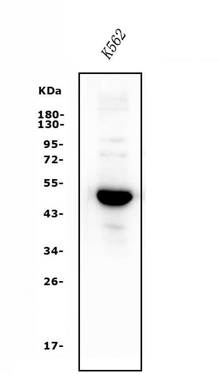
 FCM/FACS (Flow Cytometry)
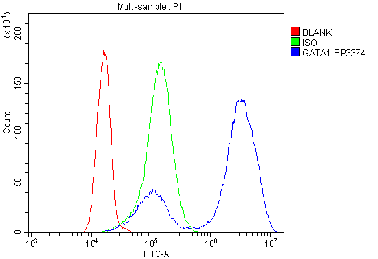
(Figure 2. Flow Cytometry analysis of K562 cells using anti- GATA1 antibody (AAA125545).Overlay histogram showing K562 cells stained with AAA125545 (Blue line). The cells were blocked with 10% normal goat serum. And then incubated with rabbit anti- GATA1 Antibody (AAA125545, 1μg/1x106 cells) for 30 min at 20 degree C. DyLight®488 conjugated goat anti-rabbit IgG (5-10μg/1x106 cells) was used as secondary antibody for 30 minutes at 20 degree C. Isotype control antibody (Green line) was rabbit IgG (1μg/1x106) used under the same conditions. Unlabelled sample (Red line) was also used as a control.)
FCM/FACS (Flow Cytometry)
(Figure 2. Flow Cytometry analysis of K562 cells using anti- GATA1 antibody (AAA125545).Overlay histogram showing K562 cells stained with AAA125545 (Blue line). The cells were blocked with 10% normal goat serum. And then incubated with rabbit anti- GATA1 Antibody (AAA125545, 1μg/1x106 cells) for 30 min at 20 degree C. DyLight®488 conjugated goat anti-rabbit IgG (5-10μg/1x106 cells) was used as secondary antibody for 30 minutes at 20 degree C. Isotype control antibody (Green line) was rabbit IgG (1μg/1x106) used under the same conditions. Unlabelled sample (Red line) was also used as a control.)
GATA1, Polyclonal Antibody (Cat# AAA125545)
Full Name
Anti-GATA1 Antibody
Gene Names
GATA1; GF1; GF-1; NFE1; XLTT; ERYF1; XLANP; XLTDA; GATA-1
Reactivity
Human
Applications
Direct ELISA, Flow Cytometry, Western Blot
Purity
Immunogen affinity purified.
Pricing
 FCM/FACS (Flow Cytometry)
(Figure 2. Flow Cytometry analysis of A549 cells using anti-TRPA1/TSA antibody (AAA125546).Overlay histogram showing A549 cells stained with AAA125546 (Blue line). The cells were blocked with 10% normal goat serum. And then incubated with rabbit anti-TRPA1/TSA Antibody (AAA125546, 1μg/1x106 cells) for 30 min at 20 degree C. DyLight®488 conjugated goat anti-rabbit IgG (5-10μg/1x106 cells) was used as secondary antibody for 30 minutes at 20 degree C. Isotype control antibody (Green line) was rabbit IgG (1μg/1x106) used under the same conditions. Unlabelled sample (Red line) was also used as a control.)
FCM/FACS (Flow Cytometry)
(Figure 2. Flow Cytometry analysis of A549 cells using anti-TRPA1/TSA antibody (AAA125546).Overlay histogram showing A549 cells stained with AAA125546 (Blue line). The cells were blocked with 10% normal goat serum. And then incubated with rabbit anti-TRPA1/TSA Antibody (AAA125546, 1μg/1x106 cells) for 30 min at 20 degree C. DyLight®488 conjugated goat anti-rabbit IgG (5-10μg/1x106 cells) was used as secondary antibody for 30 minutes at 20 degree C. Isotype control antibody (Green line) was rabbit IgG (1μg/1x106) used under the same conditions. Unlabelled sample (Red line) was also used as a control.)
TRPA1/TSA, Polyclonal Antibody (Cat# AAA125546)
Full Name
Anti-TRPA1/TSA Antibody
Gene Names
TRPA1; FEPS; ANKTM1
Reactivity
Human
Applications
Direct ELISA, Flow Cytometry, Western Blot
Purity
Immunogen affinity purified.
Pricing
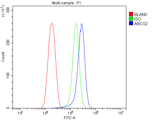 FCM/FACS (Flow Cytometry)
(Figure 4. Flow Cytometry analysis of SiHa cells using anti-BCRP/ABCG2 antibody (AAA125549).Overlay histogram showing SiHa cells stained with AAA125549 (Blue line). The cells were blocked with 10% normal goat serum. And then incubated with rabbit anti-BCRP/ABCG2 Antibody (AAA125549,1μg/1x106 cells) for 30 min at 20 degree C. DyLight®488 conjugated goat anti-rabbit IgG (5-10μg/1x106 cells) was used as secondary antibody for 30 minutes at 20 degree C. Isotype control antibody (Green line) was rabbit IgG (1μg/1x106) used under the same conditions. Unlabelled sample (Red line) was also used as a control.)
FCM/FACS (Flow Cytometry)
(Figure 4. Flow Cytometry analysis of SiHa cells using anti-BCRP/ABCG2 antibody (AAA125549).Overlay histogram showing SiHa cells stained with AAA125549 (Blue line). The cells were blocked with 10% normal goat serum. And then incubated with rabbit anti-BCRP/ABCG2 Antibody (AAA125549,1μg/1x106 cells) for 30 min at 20 degree C. DyLight®488 conjugated goat anti-rabbit IgG (5-10μg/1x106 cells) was used as secondary antibody for 30 minutes at 20 degree C. Isotype control antibody (Green line) was rabbit IgG (1μg/1x106) used under the same conditions. Unlabelled sample (Red line) was also used as a control.)
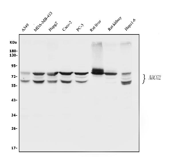
BCRP/ABCG2, Polyclonal Antibody (Cat# AAA125549)
Full Name
Anti-BCRP/ABCG2 Antibody
Gene Names
ABCG2; MRX; MXR; ABCP; BCRP; BMDP; MXR1; ABC15; BCRP1; CD338; GOUT1; CDw338; UAQTL1; EST157481
Reactivity
Human, Mouse, Rat
Applications
Direct ELISA, Flow Cytometry, Immunohistochemistry, Western Blot
Purity
Immunogen affinity purified.
Pricing
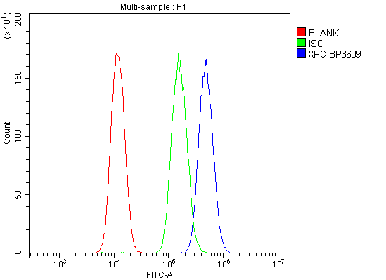
 IF (Immunofluorescence)
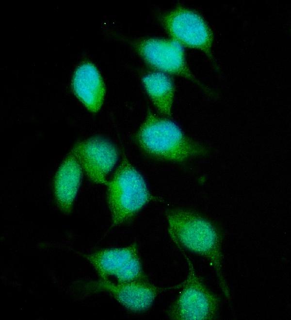
(Figure 3. IF analysis of XPC using anti- XPC antibody (AAA125551).XPC was detected in immunocytochemical section of HELA cells. Enzyme antigen retrieval was performed using IHC enzyme antigen retrieval reagent for 15 mins. The cells were blocked with 10% goat serum. And then incubated with 5μg/mL rabbit anti-XPC Antibody (AAA125551) overnight at 4 degree C. DyLight®488 Conjugated Goat Anti-Rabbit IgG was used as secondary antibody at 1:100 dilution and incubated for 30 minutes at 37 degree C. The section was counterstained with DAPI. Visualize using a fluorescence microscope and filter sets appropriate for the label used.)
IF (Immunofluorescence)
(Figure 3. IF analysis of XPC using anti- XPC antibody (AAA125551).XPC was detected in immunocytochemical section of HELA cells. Enzyme antigen retrieval was performed using IHC enzyme antigen retrieval reagent for 15 mins. The cells were blocked with 10% goat serum. And then incubated with 5μg/mL rabbit anti-XPC Antibody (AAA125551) overnight at 4 degree C. DyLight®488 Conjugated Goat Anti-Rabbit IgG was used as secondary antibody at 1:100 dilution and incubated for 30 minutes at 37 degree C. The section was counterstained with DAPI. Visualize using a fluorescence microscope and filter sets appropriate for the label used.)
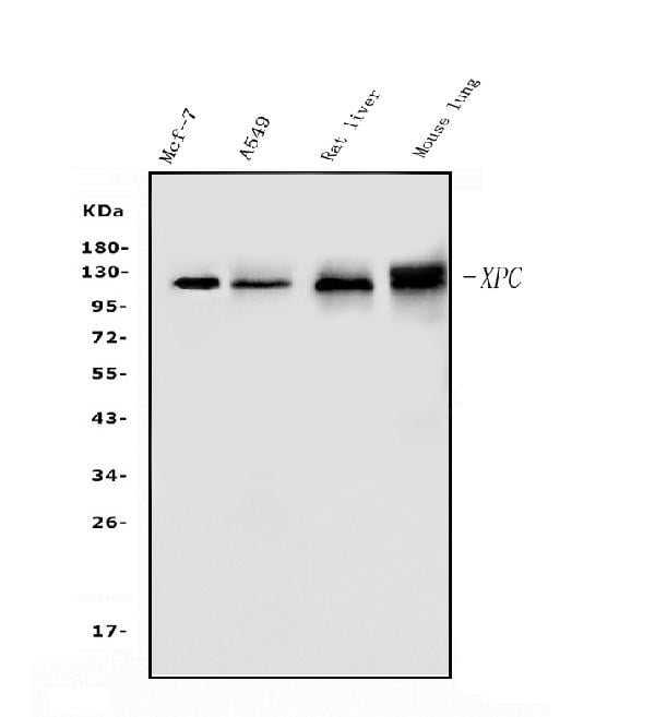
XPC, Polyclonal Antibody (Cat# AAA125551)
Full Name
Anti-XPC Antibody
Gene Names
XPC; XP3; RAD4; XPCC; p125
Reactivity
Human, Mouse, Rat
Applications
Direct ELISA, Flow Cytometry, Immunofluorescence, Immunocytochemistry, Western Blot
Purity
Immunogen affinity purified.
Pricing
 FCM/FACS (Flow Cytometry)
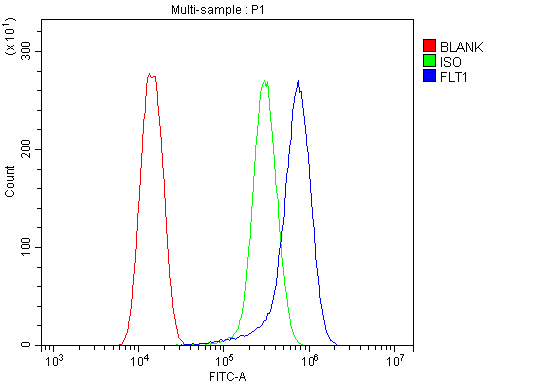
(Figure 3. Flow Cytometry analysis of U20S cells using anti-FLT1 antibody (AAA125557).Overlay histogram showing U20S cells stained with AAA125557 (Blue line). The cells were blocked with 10% normal goat serum. And then incubated with rabbit anti-FLT1 Antibody (AAA125557, 1μg/1x106 cells) for 30 min at 20 degree C. DyLight®488 conjugated goat anti-rabbit IgG (5-10μg/1x106 cells) was used as secondary antibody for 30 minutes at 20 degree C. Isotype control antibody (Green line) was rabbit IgG (1μg/1x106) used under the same conditions. Unlabelled sample (Red line) was also used as a control.)
FCM/FACS (Flow Cytometry)
(Figure 3. Flow Cytometry analysis of U20S cells using anti-FLT1 antibody (AAA125557).Overlay histogram showing U20S cells stained with AAA125557 (Blue line). The cells were blocked with 10% normal goat serum. And then incubated with rabbit anti-FLT1 Antibody (AAA125557, 1μg/1x106 cells) for 30 min at 20 degree C. DyLight®488 conjugated goat anti-rabbit IgG (5-10μg/1x106 cells) was used as secondary antibody for 30 minutes at 20 degree C. Isotype control antibody (Green line) was rabbit IgG (1μg/1x106) used under the same conditions. Unlabelled sample (Red line) was also used as a control.)
FLT1, Polyclonal Antibody (Cat# AAA125557)
Full Name
Anti-FLT1 Antibody
Gene Names
FLT1; FLT; FLT-1; VEGFR1; VEGFR-1
Reactivity
Human
Applications
Direct ELISA, Flow Cytometry, Immunofluorescence, Immunocytochemistry, Western Blot
Purity
Immunogen affinity purified.
Pricing
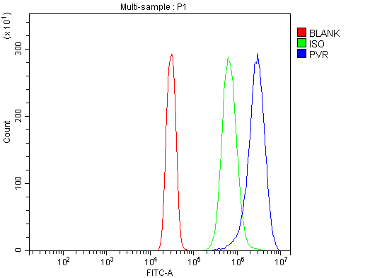 FCM/FACS (Flow Cytometry)
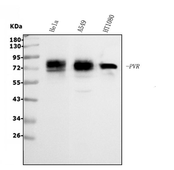
(Figure 5. Flow Cytometry analysis of SiHa cells using anti-Poliovirus Receptor/PVR antibody (AAA125567).Overlay histogram showing SiHa cells stained with AAA125567 (Blue line). The cells were blocked with 10% normal goat serum. And then incubated with rabbit anti-Poliovirus Receptor/PVR Antibody (AAA125567, 1μg/1x106 cells) for 30 min at 20 degree C. DyLight®488 conjugated goat anti-rabbit IgG (5-10μg/1x106 cells) was used as secondary antibody for 30 minutes at 20 degree C. Isotype control antibody (Green line) was rabbit IgG (1μg/1x106) used under the same conditions. Unlabelled sample (Red line) was also used as a control.)
FCM/FACS (Flow Cytometry)
(Figure 5. Flow Cytometry analysis of SiHa cells using anti-Poliovirus Receptor/PVR antibody (AAA125567).Overlay histogram showing SiHa cells stained with AAA125567 (Blue line). The cells were blocked with 10% normal goat serum. And then incubated with rabbit anti-Poliovirus Receptor/PVR Antibody (AAA125567, 1μg/1x106 cells) for 30 min at 20 degree C. DyLight®488 conjugated goat anti-rabbit IgG (5-10μg/1x106 cells) was used as secondary antibody for 30 minutes at 20 degree C. Isotype control antibody (Green line) was rabbit IgG (1μg/1x106) used under the same conditions. Unlabelled sample (Red line) was also used as a control.)
Poliovirus Receptor/PVR, Polyclonal Antibody (Cat# AAA125567)
Full Name
Anti-Poliovirus Receptor/PVR Antibody
Gene Names
PVR; PVS; HVED; CD155; NECL5; TAGE4; Necl-5
Reactivity
Human
Applications
Direct ELISA, Flow Cytometry, Immunofluorescence, Immunocytochemistry, Immunohistochemistry, Western Blot
Purity
Immunogen affinity purified.
Pricing
 FCM/FACS (Flow Cytometry)
(Figure 2. Flow Cytometry analysis of U20S cells using anti-Thrombospondin/THBS1 antibody (AAA125568).Overlay histogram showing U20S cells stained with AAA125568 (Blue line). The cells were blocked with 10% normal goat serum. And then incubated with rabbit anti-Thrombospondin/THBS1 Antibody (AAA125568, 1μg/1x106 cells) for 30 min at 20 degree C. DyLight®488 conjugated goat anti-rabbit IgG (5-10μg/1x106 cells) was used as secondary antibody for 30 minutes at 20 degree C. Isotype control antibody (Green line) was rabbit IgG (1μg/1x106) used under the same conditions. Unlabelled sample (Red line) was also used as a control.)
FCM/FACS (Flow Cytometry)
(Figure 2. Flow Cytometry analysis of U20S cells using anti-Thrombospondin/THBS1 antibody (AAA125568).Overlay histogram showing U20S cells stained with AAA125568 (Blue line). The cells were blocked with 10% normal goat serum. And then incubated with rabbit anti-Thrombospondin/THBS1 Antibody (AAA125568, 1μg/1x106 cells) for 30 min at 20 degree C. DyLight®488 conjugated goat anti-rabbit IgG (5-10μg/1x106 cells) was used as secondary antibody for 30 minutes at 20 degree C. Isotype control antibody (Green line) was rabbit IgG (1μg/1x106) used under the same conditions. Unlabelled sample (Red line) was also used as a control.)
Thrombospondin/THBS1, Polyclonal Antibody (Cat# AAA125568)
Full Name
Anti-Thrombospondin/THBS1 Antibody
Gene Names
THBS1; TSP; THBS; TSP1; TSP-1; THBS-1
Reactivity
Human
Applications
Direct ELISA, Flow Cytometry, Western Blot
Purity
Immunogen affinity purified.
Pricing
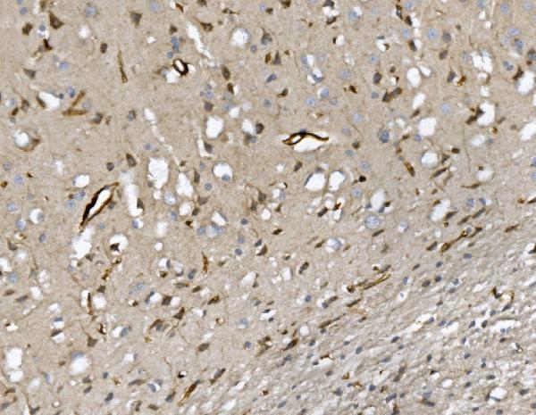
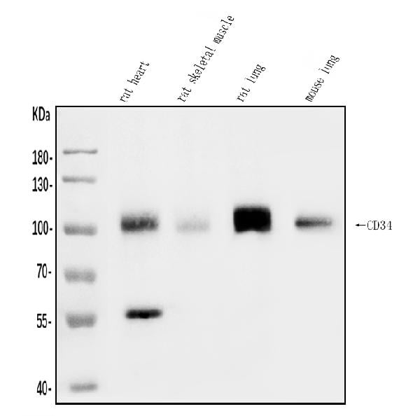
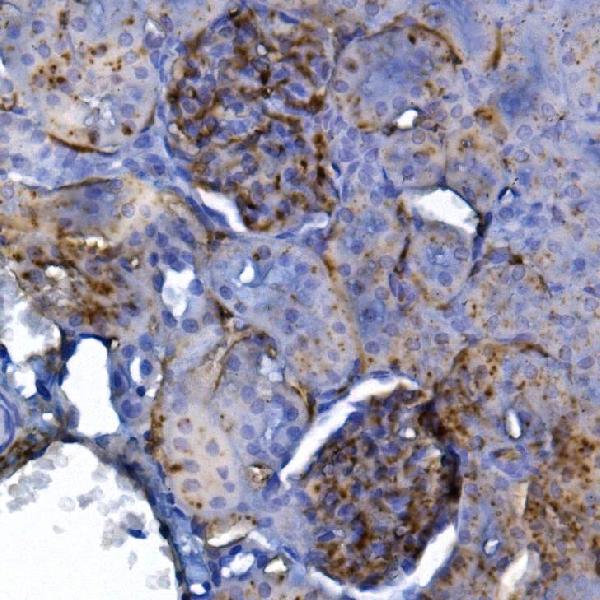 IHC (Immunohistochemisry)
(Figure 3. IHC analysis of CD34 using anti-CD34 antibody (AAA125577).CD34 was detected in paraffin-embedded section of rat kidney tissue. Heat mediated antigen retrieval was performed in EDTA buffer (pH8. 0, epitope retrieval solution). The tissue section was blocked with 10% goat serum. The tissue section was then incubated with 2μg/ml rabbit anti-CD34 Antibody (AAA125577) overnight at 4 degree C. Biotinylated goat anti-rabbit IgG was used as secondary antibody and incubated for 30 minutes at 37 degree C. The tissue section was developed using Strepavidin-Biotin-Complex (SABC) with DAB as the chromogen.)
IHC (Immunohistochemisry)
(Figure 3. IHC analysis of CD34 using anti-CD34 antibody (AAA125577).CD34 was detected in paraffin-embedded section of rat kidney tissue. Heat mediated antigen retrieval was performed in EDTA buffer (pH8. 0, epitope retrieval solution). The tissue section was blocked with 10% goat serum. The tissue section was then incubated with 2μg/ml rabbit anti-CD34 Antibody (AAA125577) overnight at 4 degree C. Biotinylated goat anti-rabbit IgG was used as secondary antibody and incubated for 30 minutes at 37 degree C. The tissue section was developed using Strepavidin-Biotin-Complex (SABC) with DAB as the chromogen.)
CD34, Polyclonal Antibody (Cat# AAA125577)
Full Name
Anti-CD34 Antibody
Reactivity
Mouse, Rat
Applications
Direct ELISA, Immunohistochemistry, Western Blot
Purity
Immunogen affinity purified.
Pricing
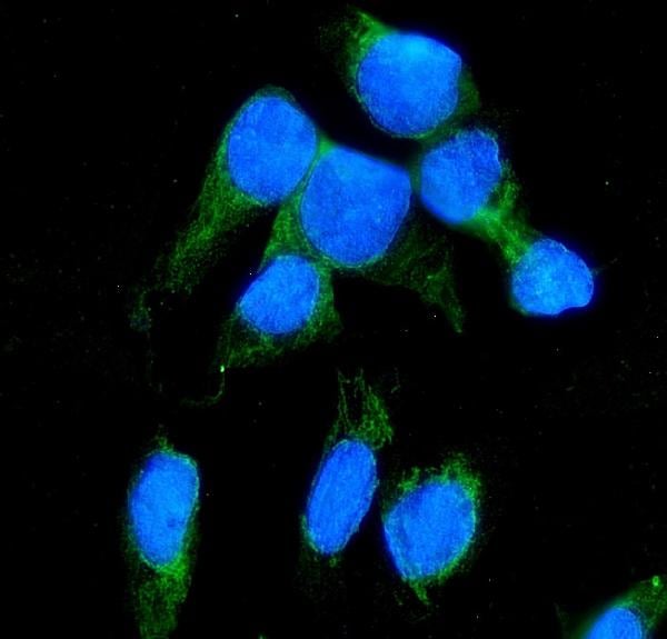
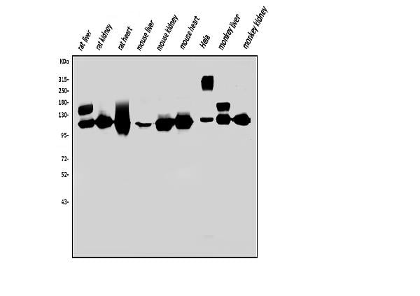
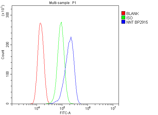 FCM/FACS (Flow Cytometry)
(Figure 3. Flow Cytometry analysis of THP-1 cells using anti-NNT antibody (AAA125578).Overlay histogram showing THP-1 cells stained with AAA125578 (Blue line). The cells were blocked with 10% normal goat serum. And then incubated with rabbit anti-NNT Antibody (AAA125578,1μg/1x106 cells) for 30 min at 20 degree C. DyLight®488 conjugated goat anti-rabbit IgG (5-10μg/1x106 cells) was used as secondary antibody for 30 minutes at 20 degree C. Isotype control antibody (Green line) was rabbit IgG (1μg/1x106) used under the same conditions. Unlabelled sample (Red line) was also used as a control.)
FCM/FACS (Flow Cytometry)
(Figure 3. Flow Cytometry analysis of THP-1 cells using anti-NNT antibody (AAA125578).Overlay histogram showing THP-1 cells stained with AAA125578 (Blue line). The cells were blocked with 10% normal goat serum. And then incubated with rabbit anti-NNT Antibody (AAA125578,1μg/1x106 cells) for 30 min at 20 degree C. DyLight®488 conjugated goat anti-rabbit IgG (5-10μg/1x106 cells) was used as secondary antibody for 30 minutes at 20 degree C. Isotype control antibody (Green line) was rabbit IgG (1μg/1x106) used under the same conditions. Unlabelled sample (Red line) was also used as a control.)
NNT, Polyclonal Antibody (Cat# AAA125578)
Full Name
Anti-NNT Antibody
Gene Names
NNT; GCCD4
Reactivity
Human, Mouse, Monkey, Rat
Applications
Direct ELISA, Flow Cytometry, Immunofluorescence, Immunocytochemistry, Western Blot
Purity
Immunogen affinity purified.
Pricing
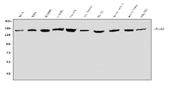
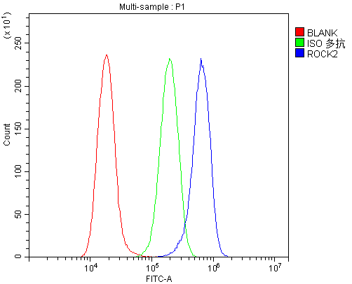
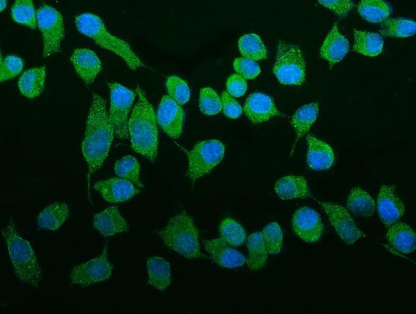 IF (Immunofluorescence)
(Figure 3. IF analysis of ROCK2 using anti- ROCK2 antibody (AAA125586).ROCK2 was detected in immunocytochemical section of Hep cells. Enzyme antigen retrieval was performed using IHC enzyme antigen retrieval reagent for 15 mins. The cells were blocked with 10% goat serum. And then incubated with 5μg/mL rabbit anti- ROCK2 Antibody (AAA125586) overnight at 4 degree C. DyLight®488 Conjugated Goat Anti-Rabbit IgG was used as secondary antibody at 1:100 dilution and incubated for 30 minutes at 37 degree C. The section was counterstained with DAPI. Visualize using a fluorescence microscope and filter sets appropriate for the label used.)
IF (Immunofluorescence)
(Figure 3. IF analysis of ROCK2 using anti- ROCK2 antibody (AAA125586).ROCK2 was detected in immunocytochemical section of Hep cells. Enzyme antigen retrieval was performed using IHC enzyme antigen retrieval reagent for 15 mins. The cells were blocked with 10% goat serum. And then incubated with 5μg/mL rabbit anti- ROCK2 Antibody (AAA125586) overnight at 4 degree C. DyLight®488 Conjugated Goat Anti-Rabbit IgG was used as secondary antibody at 1:100 dilution and incubated for 30 minutes at 37 degree C. The section was counterstained with DAPI. Visualize using a fluorescence microscope and filter sets appropriate for the label used.)
ROCK2, Polyclonal Antibody (Cat# AAA125586)
Full Name
Anti-ROCK2 Antibody
Gene Names
ROCK2; ROCK-II
Reactivity
Human, Mouse, Rat
Applications
Direct ELISA, Flow Cytometry, Immunofluorescence, Immunocytochemistry, Western Blot
Purity
Immunogen affinity purified.
Pricing