Filters
▼Clonality
▼Type
▼Reactivity
▼Gene Name
▼Isotype
▼Host
▼Application
▼Clone
▼Viewing 4150-4200 of 265407 product results
 FCM/FACS (Flow Cytometry)
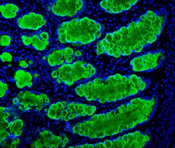
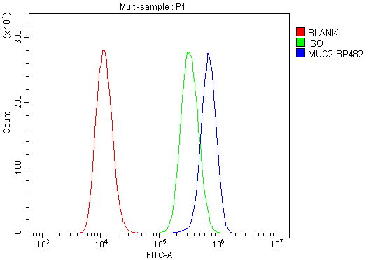
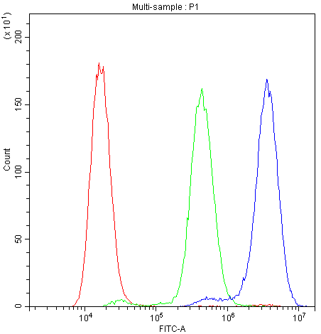
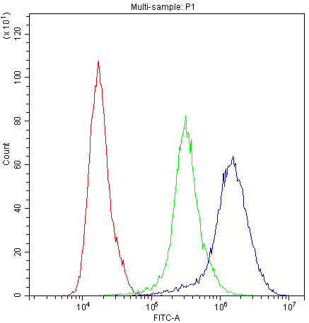
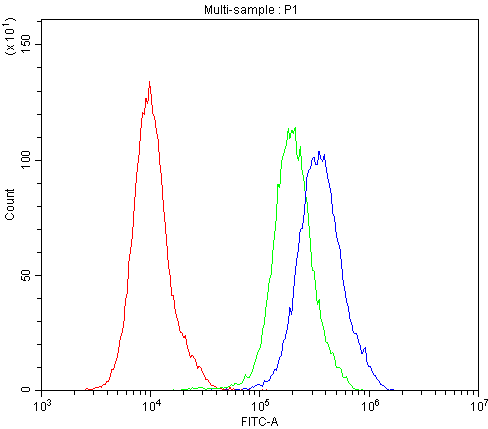
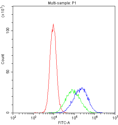
(Figure 3. Flow Cytometry analysis of U20S cells using anti-MUC2 antibody (AAA125594).Overlay histogram showing U20S cells stained with AAA125594 (Blue line). The cells were blocked with 10% normal goat serum. And then incubated with rabbit anti-MUC2 Antibody (AAA125594,1μg/1x106 cells) for 30 min at 20 degree C. DyLight®488 conjugated goat anti-rabbit IgG (5-10μg/1x106 cells) was used as secondary antibody for 30 minutes at 20 degree C. Isotype control antibody (Green line) was rabbit IgG (1μg/1x106) used under the same conditions. Unlabelled sample (Red line) was also used as a control.)
FCM/FACS (Flow Cytometry)
(Figure 3. Flow Cytometry analysis of U20S cells using anti-MUC2 antibody (AAA125594).Overlay histogram showing U20S cells stained with AAA125594 (Blue line). The cells were blocked with 10% normal goat serum. And then incubated with rabbit anti-MUC2 Antibody (AAA125594,1μg/1x106 cells) for 30 min at 20 degree C. DyLight®488 conjugated goat anti-rabbit IgG (5-10μg/1x106 cells) was used as secondary antibody for 30 minutes at 20 degree C. Isotype control antibody (Green line) was rabbit IgG (1μg/1x106) used under the same conditions. Unlabelled sample (Red line) was also used as a control.)
MUC2, Polyclonal Antibody (Cat# AAA125594)
Full Name
Anti-MUC2 Antibody
Gene Names
MUC2; MLP; SMUC; MUC-2
Reactivity
Human
Applications
Direct ELISA, Flow Cytometry, Immunohistochemistry
Purity
Immunogen affinity purified.
Pricing
 IHC (Immunohiostchemistry)
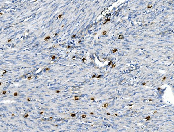
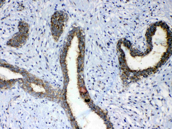
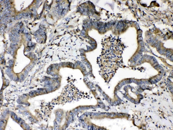
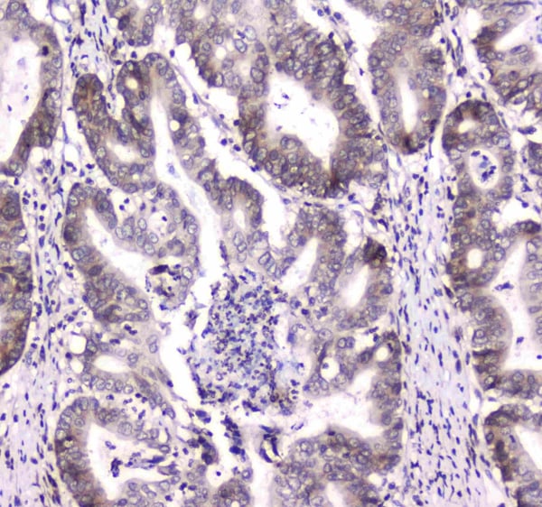
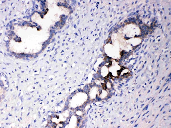
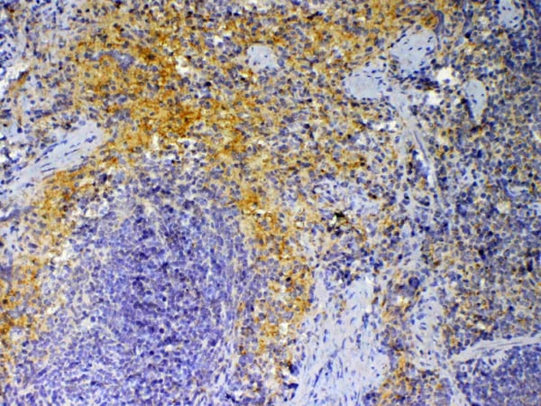
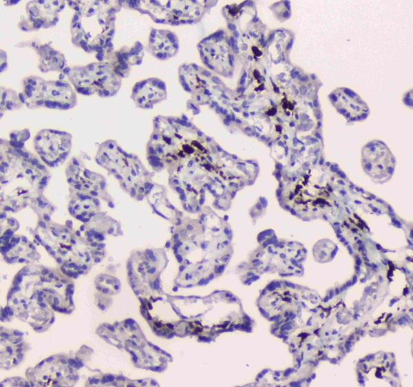
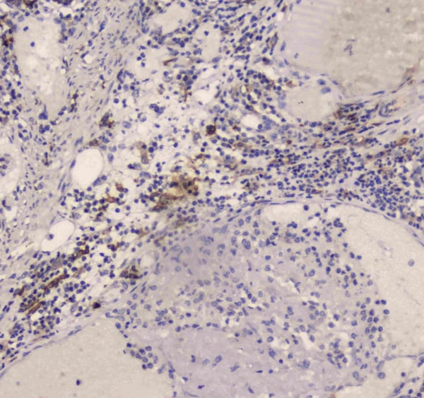
(Figure 2. IHC analysis of CD30/TNFRSF8 using anti-CD30/TNFRSF8 antibody (AAA125595).CD30/TNFRSF8 was detected in paraffin-embedded section of human endometrial carcinoma tissue. Heat mediated antigen retrieval was performed in EDTA buffer (pH8. 0, epitope retrieval solution). The tissue section was blocked with 10% goat serum. The tissue section was then incubated with 2μg/ml rabbit anti-CD30/TNFRSF8 Antibody (AAA125595) overnight at 4 degree C. Biotinylated goat anti-rabbit IgG was used as secondary antibody and incubated for 30 minutes at 37 degree C. The tissue section was developed using Strepavidin-Biotin-Complex (SABC) with DAB as the chromogen.)
IHC (Immunohiostchemistry)
(Figure 2. IHC analysis of CD30/TNFRSF8 using anti-CD30/TNFRSF8 antibody (AAA125595).CD30/TNFRSF8 was detected in paraffin-embedded section of human endometrial carcinoma tissue. Heat mediated antigen retrieval was performed in EDTA buffer (pH8. 0, epitope retrieval solution). The tissue section was blocked with 10% goat serum. The tissue section was then incubated with 2μg/ml rabbit anti-CD30/TNFRSF8 Antibody (AAA125595) overnight at 4 degree C. Biotinylated goat anti-rabbit IgG was used as secondary antibody and incubated for 30 minutes at 37 degree C. The tissue section was developed using Strepavidin-Biotin-Complex (SABC) with DAB as the chromogen.)
CD30/TNFRSF8, Polyclonal Antibody (Cat# AAA125595)
Full Name
Anti-CD30/TNFRSF8 Antibody
Gene Names
TNFRSF8; CD30; Ki-1; D1S166E
Reactivity
Human, Mouse
Applications
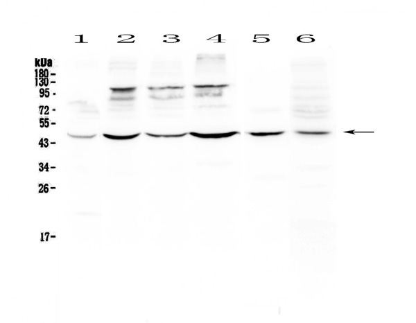
Immunohistochemistry, Western Blot
Purity
Immunogen affinity purified.
Pricing
 FCM/FACS (Flow Cytometry)
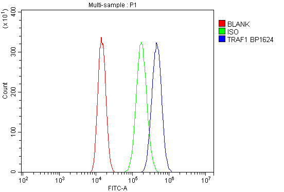
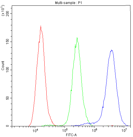
(Figure 2. Flow Cytometry analysis of A549 cells using anti-TRAF1 antibody (AAA125606).Overlay histogram showing A549 cells stained with AAA125606 (Blue line). The cells were blocked with 10% normal goat serum. And then incubated with rabbit anti-TRAF1 Antibody (AAA125606, 1μg/1x106 cells) for 30 min at 20 degree C. DyLight®488 conjugated goat anti-rabbit IgG (5-10μg/1x106 cells) was used as secondary antibody for 30 minutes at 20 degree C. Isotype control antibody (Green line) was rabbit IgG (1μg/1x106) used under the same conditions. Unlabelled sample (Red line) was also used as a control.)
FCM/FACS (Flow Cytometry)
(Figure 2. Flow Cytometry analysis of A549 cells using anti-TRAF1 antibody (AAA125606).Overlay histogram showing A549 cells stained with AAA125606 (Blue line). The cells were blocked with 10% normal goat serum. And then incubated with rabbit anti-TRAF1 Antibody (AAA125606, 1μg/1x106 cells) for 30 min at 20 degree C. DyLight®488 conjugated goat anti-rabbit IgG (5-10μg/1x106 cells) was used as secondary antibody for 30 minutes at 20 degree C. Isotype control antibody (Green line) was rabbit IgG (1μg/1x106) used under the same conditions. Unlabelled sample (Red line) was also used as a control.)
TRAF1, Polyclonal Antibody (Cat# AAA125606)
Full Name
Anti-TRAF1 Antibody
Gene Names
TRAF1; EBI6; MGC:10353
Reactivity
Human
Applications
Direct ELISA, Flow Cytometry, Western Blot
Purity
Immunogen affinity purified.
Pricing
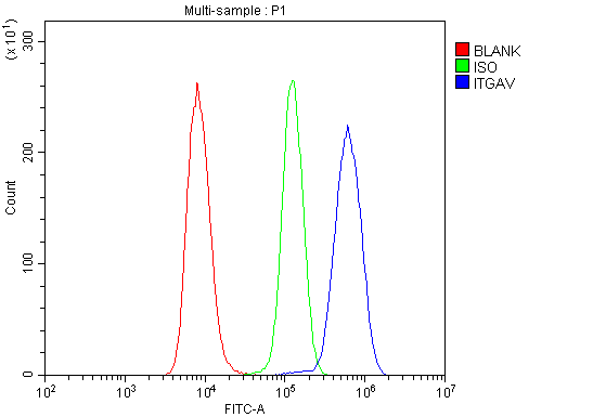 FCM/FACS (Flow Cytometry)
(Figure 2. Flow Cytometry analysis of Raji cells using anti-Integrin alpha V/ITGAV antibody (AAA125610).Overlay histogram showing Raji cells stained with AAA125610 (Blue line). The cells were blocked with 10% normal goat serum. And then incubated with rabbit anti-Integrin alpha V/ITGAV Antibody (AAA125610,1μg/1x106 cells) for 30 min at 20 degree C. DyLight®488 conjugated goat anti-rabbit IgG (5-10μg/1x106 cells) was used as secondary antibody for 30 minutes at 20 degree C. Isotype control antibody (Green line) was rabbit IgG (1μg/1x106) used under the same conditions. Unlabelled sample (Red line) was also used as a control.)
FCM/FACS (Flow Cytometry)
(Figure 2. Flow Cytometry analysis of Raji cells using anti-Integrin alpha V/ITGAV antibody (AAA125610).Overlay histogram showing Raji cells stained with AAA125610 (Blue line). The cells were blocked with 10% normal goat serum. And then incubated with rabbit anti-Integrin alpha V/ITGAV Antibody (AAA125610,1μg/1x106 cells) for 30 min at 20 degree C. DyLight®488 conjugated goat anti-rabbit IgG (5-10μg/1x106 cells) was used as secondary antibody for 30 minutes at 20 degree C. Isotype control antibody (Green line) was rabbit IgG (1μg/1x106) used under the same conditions. Unlabelled sample (Red line) was also used as a control.)
Integrin alpha V/ITGAV, Polyclonal Antibody (Cat# AAA125610)
Full Name
Anti-Integrin alpha V/ITGAV Antibody
Gene Names
ITGAV; CD51; MSK8; VNRA; VTNR
Reactivity
Human
Applications
Direct ELISA, Flow Cytometry, Western Blot
Purity
Immunogen affinity purified.
Pricing

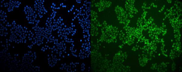
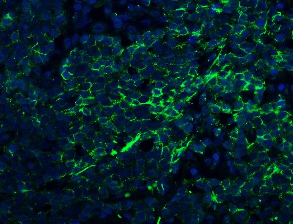 IF (Immunofluorescence)
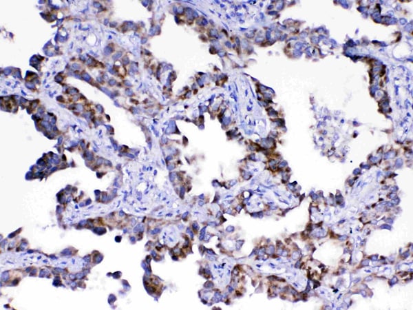
(Figure 5. IF analysis of Claudin 1/CLDN1 using anti- Claudin 1/CLDN1 antibody (AAA125612).Claudin 1/CLDN1 was detected in paraffin-embedded section of human lung cancer tissue. Heat mediated antigen retrieval was performed in EDTA buffer (pH8. 0, epitope retrieval solution). The tissue section was blocked with 10% goat serum. The tissue section was then incubated with 5μg/mL rabbit anti- Claudin 1/CLDN1 Antibody (AAA125612) overnight at 4 degree C. DyLight®488 Conjugated Goat Anti-Rabbit IgG was used as secondary antibody at 1:100 dilution and incubated for 30 minutes at 37 degree C. The section was counterstained with DAPI. Visualize using a fluorescence microscope and filter sets appropriate for the label used.)
IF (Immunofluorescence)
(Figure 5. IF analysis of Claudin 1/CLDN1 using anti- Claudin 1/CLDN1 antibody (AAA125612).Claudin 1/CLDN1 was detected in paraffin-embedded section of human lung cancer tissue. Heat mediated antigen retrieval was performed in EDTA buffer (pH8. 0, epitope retrieval solution). The tissue section was blocked with 10% goat serum. The tissue section was then incubated with 5μg/mL rabbit anti- Claudin 1/CLDN1 Antibody (AAA125612) overnight at 4 degree C. DyLight®488 Conjugated Goat Anti-Rabbit IgG was used as secondary antibody at 1:100 dilution and incubated for 30 minutes at 37 degree C. The section was counterstained with DAPI. Visualize using a fluorescence microscope and filter sets appropriate for the label used.)
Claudin 1/CLDN1, Polyclonal Antibody (Cat# AAA125612)
Full Name
Anti-Claudin 1/CLDN1 Antibody
Gene Names
CLDN1; CLD1; SEMP1; ILVASC
Reactivity
Human
Applications
Immunofluorescence, Immunocytochemistry, Immunohistochemistry
Purity
Immunogen affinity purified.
Pricing
 FCM/FACS (Flow Cytometry)
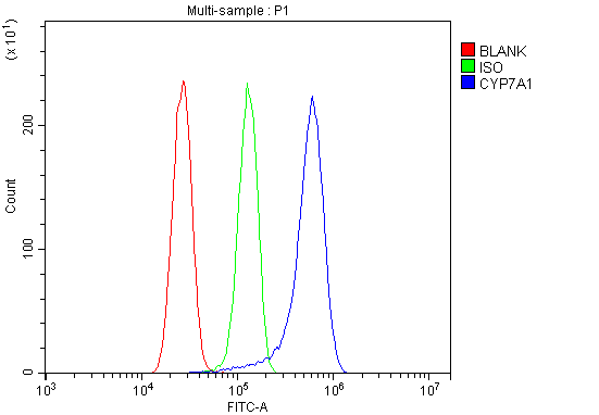
(Figure 2. Flow Cytometry analysis of U87 cells using anti-CYP7A1 antibody (AAA125614).Overlay histogram showing U87 cells stained with AAA125614 (Blue line). The cells were blocked with 10% normal goat serum. And then incubated with rabbit anti-CYP7A1 Antibody (AAA125614, 1μg/1x106 cells) for 30 min at 20 degree C. DyLight®488 conjugated goat anti-rabbit IgG (5-10μg/1x106 cells) was used as secondary antibody for 30 minutes at 20 degree C. Isotype control antibody (Green line) was rabbit IgG (1μg/1x106) used under the same conditions. Unlabelled sample (Red line) was also used as a control.)
FCM/FACS (Flow Cytometry)
(Figure 2. Flow Cytometry analysis of U87 cells using anti-CYP7A1 antibody (AAA125614).Overlay histogram showing U87 cells stained with AAA125614 (Blue line). The cells were blocked with 10% normal goat serum. And then incubated with rabbit anti-CYP7A1 Antibody (AAA125614, 1μg/1x106 cells) for 30 min at 20 degree C. DyLight®488 conjugated goat anti-rabbit IgG (5-10μg/1x106 cells) was used as secondary antibody for 30 minutes at 20 degree C. Isotype control antibody (Green line) was rabbit IgG (1μg/1x106) used under the same conditions. Unlabelled sample (Red line) was also used as a control.)
CYP7A1, Polyclonal Antibody (Cat# AAA125614)
Full Name
Anti-CYP7A1 Antibody
Gene Names
CYP7A1; CP7A; CYP7; CYPVII
Reactivity
Human, Mouse, Rat, Monkey
Applications
Direct ELISA, Flow Cytometry, Western Blot
Purity
Immunogen affinity purified.
Pricing
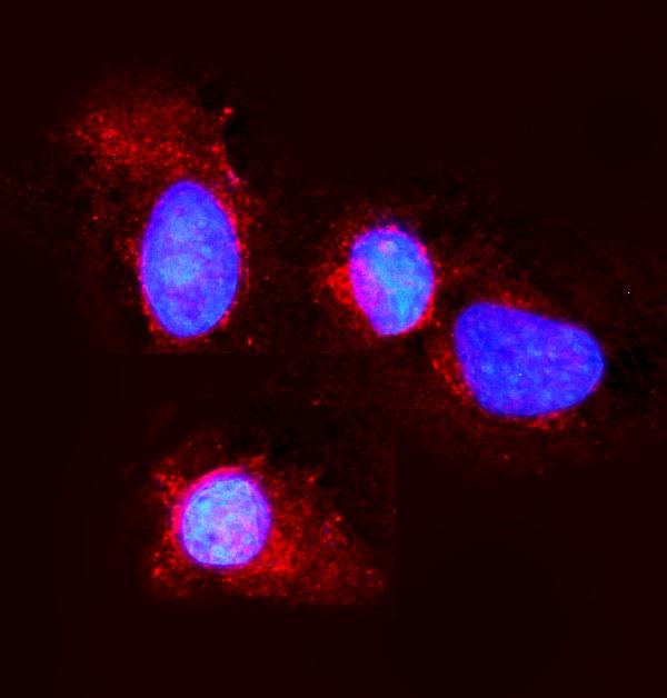
 IF (Immunofluorescence)
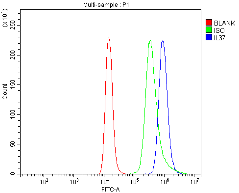
(Figure 4. IF analysis of IL37 using anti- IL37 antibody (AAA125697).IL37 was detected in immunocytochemical section of A431 cells. Enzyme antigen retrieval was performed using IHC enzyme antigen retrieval reagent for 15 mins. The cells were blocked with 10% goat serum. And then incubated with 5μg/mL rabbit anti- IL37 Antibody (AAA125697) overnight at 4 degree C. DyLight®488 Conjugated Goat Anti-Rabbit IgG was used as secondary antibody at 1:100 dilution and incubated for 30 minutes at 37 degree C. The section was counterstained with DAPI. Visualize using a fluorescence microscope and filter sets appropriate for the label used.)
IF (Immunofluorescence)
(Figure 4. IF analysis of IL37 using anti- IL37 antibody (AAA125697).IL37 was detected in immunocytochemical section of A431 cells. Enzyme antigen retrieval was performed using IHC enzyme antigen retrieval reagent for 15 mins. The cells were blocked with 10% goat serum. And then incubated with 5μg/mL rabbit anti- IL37 Antibody (AAA125697) overnight at 4 degree C. DyLight®488 Conjugated Goat Anti-Rabbit IgG was used as secondary antibody at 1:100 dilution and incubated for 30 minutes at 37 degree C. The section was counterstained with DAPI. Visualize using a fluorescence microscope and filter sets appropriate for the label used.)
IL37, Polyclonal Antibody (Cat# AAA125697)
Full Name
Anti-IL37 Antibody
Gene Names
IL37; FIL1; FIL1Z; IL-1H; IL-37; IL1F7; IL1H4; IL-1F7; IL-1H4; IL1RP1; IL-1RP1; FIL1(ZETA)
Reactivity
Human
Applications
Direct ELISA, Flow Cytometry, Immunofluorescence, Immunocytochemistry, Immunohistochemistry
Purity
Immunogen affinity purified.
Pricing
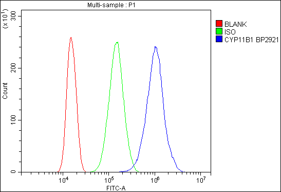 FCM/FACS (Flow Cytometry)
(Figure 2. Flow Cytometry analysis of 293T cells using anti-CYP11B1/C11B2/CYP11B2 antibody (AAA125703).Overlay histogram showing 293T cells stained with AAA125703 (Blue line). The cells were blocked with 10% normal goat serum. And then incubated with rabbit anti-CYP11B1/C11B2/CYP11B2 Antibody (AAA125703,1μg/1x106 cells) for 30 min at 20 degree C. DyLight®488 conjugated goat anti-rabbit IgG (5-10μg/1x106 cells) was used as secondary antibody for 30 minutes at 20 degree C. Isotype control antibody (Green line) was rabbit IgG (1μg/1x106) used under the same conditions. Unlabelled sample (Red line) was also used as a control.)
FCM/FACS (Flow Cytometry)
(Figure 2. Flow Cytometry analysis of 293T cells using anti-CYP11B1/C11B2/CYP11B2 antibody (AAA125703).Overlay histogram showing 293T cells stained with AAA125703 (Blue line). The cells were blocked with 10% normal goat serum. And then incubated with rabbit anti-CYP11B1/C11B2/CYP11B2 Antibody (AAA125703,1μg/1x106 cells) for 30 min at 20 degree C. DyLight®488 conjugated goat anti-rabbit IgG (5-10μg/1x106 cells) was used as secondary antibody for 30 minutes at 20 degree C. Isotype control antibody (Green line) was rabbit IgG (1μg/1x106) used under the same conditions. Unlabelled sample (Red line) was also used as a control.)
CYP11B1/C11B2/CYP11B2, Polyclonal Antibody (Cat# AAA125703)
Full Name
Anti-CYP11B1/C11B2/CYP11B2 Antibody
Gene Names
CYP11B1; FHI; CPN1; CYP11B; P450C11
Reactivity
Human, Mouse, Rat
Applications
Direct ELISA, Flow Cytometry, Western Blot
Purity
Immunogen affinity purified.
Pricing
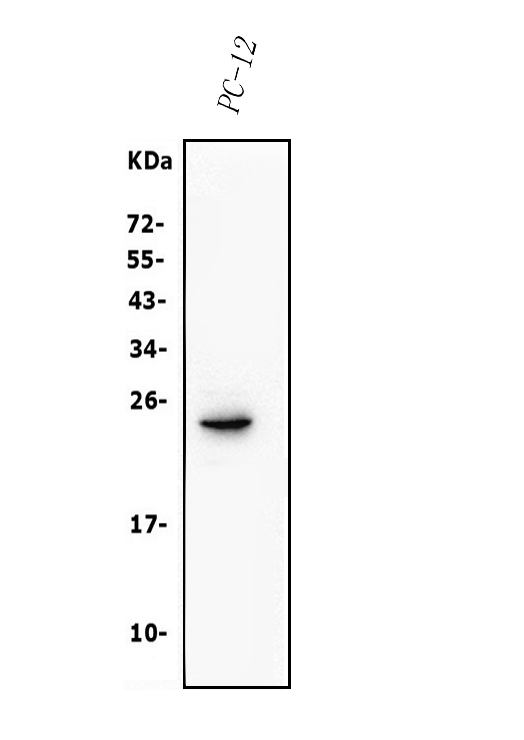 WB (Western Blot)
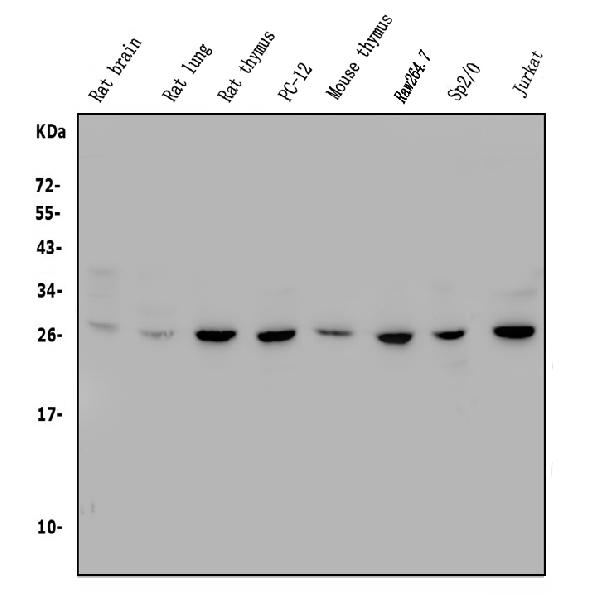
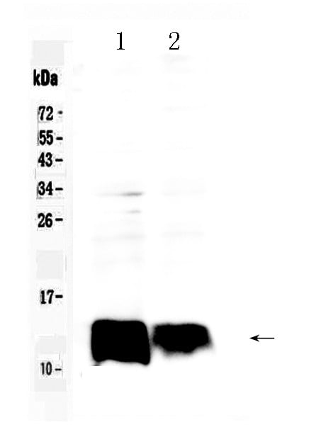
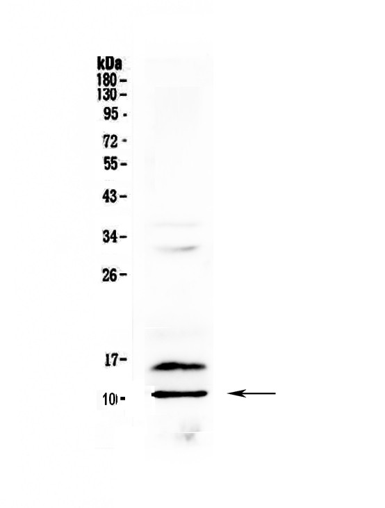
(Figure 2. Western blot analysis of Claudin 3/CLDN3 using anti-Claudin 3/CLDN3 antibody (AAA125723).Electrophoresis was performed on a 5-20% SDS-PAGE gel at 70V (Stacking gel) / 90V (Resolving gel) for 2-3 hours. The sample well of each lane was loaded with 50ug of sample under reducing conditions.Lane 1: rat PC-12 whole cell lysates.After Electrophoresis, proteins were transferred to a Nitrocellulose membrane at 150mA for 50-90 minutes. Blocked the membrane with 5% Non-fat Milk/ TBS for 1. 5 hour at RT. The membrane was incubated with rabbit anti-Claudin 3/CLDN3 antigen affinity purified polyclonal antibody (Catalog # AAA125723) at 0. 5 μg/mL overnight at 4 degree C, then washed with TBS-0. 1%Tween 3 times with 5 minutes each and probed with a goat anti-rabbit IgG-HRP secondary antibody at a dilution of 1:5000 for 1. 5 hour at RT. The signal is developed using an Enhanced Chemiluminescent detection (ECL) kit with Tanon 5200 system. A specific band was detected for Claudin 3/CLDN3 at approximately 23KD. The expected band size for Claudin 3/CLDN3 is at 23KD.)
WB (Western Blot)
(Figure 2. Western blot analysis of Claudin 3/CLDN3 using anti-Claudin 3/CLDN3 antibody (AAA125723).Electrophoresis was performed on a 5-20% SDS-PAGE gel at 70V (Stacking gel) / 90V (Resolving gel) for 2-3 hours. The sample well of each lane was loaded with 50ug of sample under reducing conditions.Lane 1: rat PC-12 whole cell lysates.After Electrophoresis, proteins were transferred to a Nitrocellulose membrane at 150mA for 50-90 minutes. Blocked the membrane with 5% Non-fat Milk/ TBS for 1. 5 hour at RT. The membrane was incubated with rabbit anti-Claudin 3/CLDN3 antigen affinity purified polyclonal antibody (Catalog # AAA125723) at 0. 5 μg/mL overnight at 4 degree C, then washed with TBS-0. 1%Tween 3 times with 5 minutes each and probed with a goat anti-rabbit IgG-HRP secondary antibody at a dilution of 1:5000 for 1. 5 hour at RT. The signal is developed using an Enhanced Chemiluminescent detection (ECL) kit with Tanon 5200 system. A specific band was detected for Claudin 3/CLDN3 at approximately 23KD. The expected band size for Claudin 3/CLDN3 is at 23KD.)
Claudin 3/CLDN3, Polyclonal Antibody (Cat# AAA125723)
Full Name
Anti-Claudin 3/CLDN3 Antibody
Gene Names
Cldn3; mRVP1; Cpetr2; AI182374
Reactivity
Mouse, Rat
Applications
Immunofluorescence, Immunocytochemistry, Western Blot
Purity
Immunogen affinity purified.
Pricing
 FCM/FACS (Flow Cytometry)
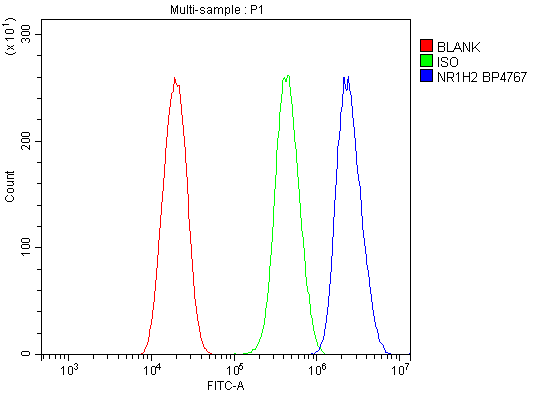
(Figure 3. Flow Cytometry analysis of U20S cells using anti-LXR beta/NER/NR1H2 antibody (AAA125726).Overlay histogram showing U20S cells stained with AAA125726 (Blue line). The cells were blocked with 10% normal goat serum. And then incubated with rabbit anti-LXR beta/NER/NR1H2 Antibody (AAA125726, 1μg/1x106 cells) for 30 min at 20 degree C. DyLight®488 conjugated goat anti-rabbit IgG (5-10μg/1x106 cells) was used as secondary antibody for 30 minutes at 20 degree C. Isotype control antibody (Green line) was rabbit IgG (1μg/1x106) used under the same conditions. Unlabelled sample (Red line) was also used as a control.)
FCM/FACS (Flow Cytometry)
(Figure 3. Flow Cytometry analysis of U20S cells using anti-LXR beta/NER/NR1H2 antibody (AAA125726).Overlay histogram showing U20S cells stained with AAA125726 (Blue line). The cells were blocked with 10% normal goat serum. And then incubated with rabbit anti-LXR beta/NER/NR1H2 Antibody (AAA125726, 1μg/1x106 cells) for 30 min at 20 degree C. DyLight®488 conjugated goat anti-rabbit IgG (5-10μg/1x106 cells) was used as secondary antibody for 30 minutes at 20 degree C. Isotype control antibody (Green line) was rabbit IgG (1μg/1x106) used under the same conditions. Unlabelled sample (Red line) was also used as a control.)
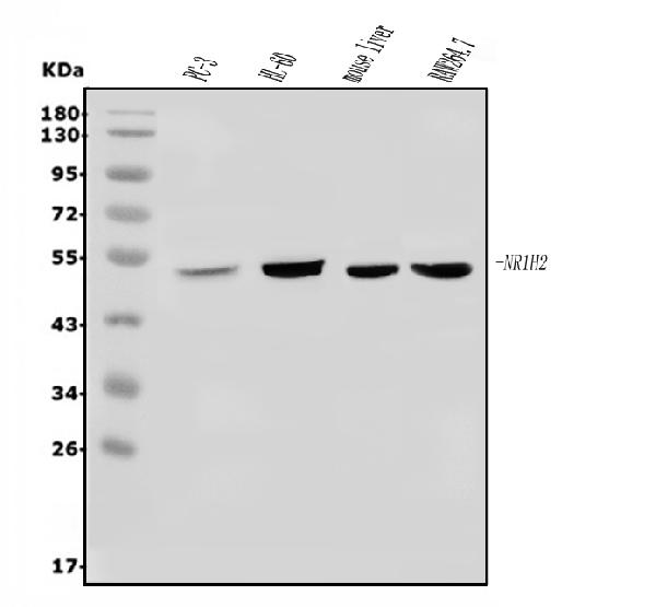
LXR beta/NER/NR1H2, Polyclonal Antibody (Cat# AAA125726)
Full Name
Anti-LXR beta/NER/NR1H2 Antibody
Gene Names
NR1H2; NER; UNR; LXRB; LXR-b; NER-I; RIP15
Reactivity
Human, Mouse
Applications
Direct ELISA, Flow Cytometry, Immunofluorescence, Immunocytochemistry, Western Blot
Purity
Immunogen affinity purified.
Pricing
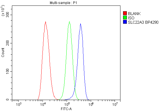 FCM/FACS (Flow Cytometry)
(Figure 2. Flow Cytometry analysis of Hela cells using anti-SLC22A3 antibody (AAA125731).Overlay histogram showing Hela cells stained with AAA125731 (Blue line). The cells were blocked with 10% normal goat serum. And then incubated with rabbit anti-SLC22A3 Antibody (AAA125731, 1μg/1x106 cells) for 30 min at 20 degree C. DyLight®488 conjugated goat anti-rabbit IgG (5-10μg/1x106 cells) was used as secondary antibody for 30 minutes at 20 degree C. Isotype control antibody (Green line) was rabbit IgG (1μg/1x106) used under the same conditions. Unlabelled sample (Red line) was also used as a control.)
FCM/FACS (Flow Cytometry)
(Figure 2. Flow Cytometry analysis of Hela cells using anti-SLC22A3 antibody (AAA125731).Overlay histogram showing Hela cells stained with AAA125731 (Blue line). The cells were blocked with 10% normal goat serum. And then incubated with rabbit anti-SLC22A3 Antibody (AAA125731, 1μg/1x106 cells) for 30 min at 20 degree C. DyLight®488 conjugated goat anti-rabbit IgG (5-10μg/1x106 cells) was used as secondary antibody for 30 minutes at 20 degree C. Isotype control antibody (Green line) was rabbit IgG (1μg/1x106) used under the same conditions. Unlabelled sample (Red line) was also used as a control.)
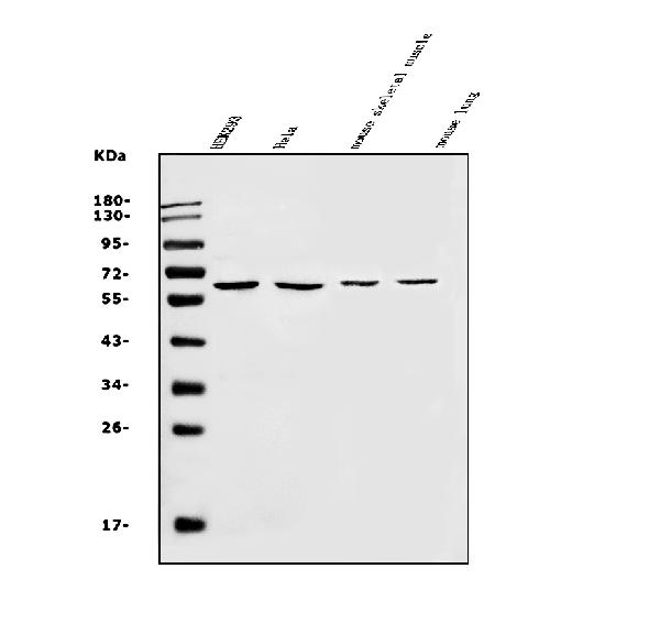
SLC22A3, Polyclonal Antibody (Cat# AAA125731)
Full Name
Anti-SLC22A3 Antibody
Gene Names
SLC22A3; EMT; EMTH; OCT3
Reactivity
Human, Mouse
Applications
Flow Cytometry, Western Blot
Purity
Immunogen affinity purified.
Pricing
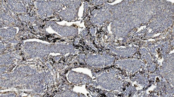
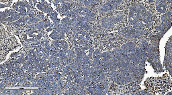
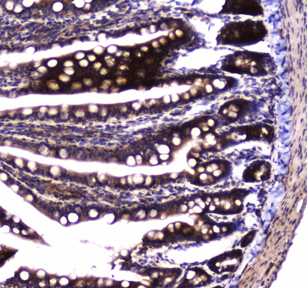
 IHC (Immunohistochemistry)
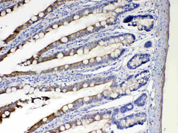
(Figure 5. IHC analysis of LOC134147/CMBL using anti-LOC134147/CMBL antibody (AAA125733).LOC134147/CMBL was detected in paraffin-embedded section of human colorectal cancer tissue. Heat mediated antigen retrieval was performed in EDTA buffer (pH8. 0, epitope retrieval solution). The tissue section was blocked with 10% goat serum. The tissue section was then incubated with 2μg/ml rabbit anti-LOC134147/CMBL Antibody (AAA125733) overnight at 4 degree C. Biotinylated goat anti-rabbit IgG was used as secondary antibody and incubated for 30 minutes at 37 degree C. The tissue section was developed using Strepavidin-Biotin-Complex (SABC) with DAB as the chromogen.)
IHC (Immunohistochemistry)
(Figure 5. IHC analysis of LOC134147/CMBL using anti-LOC134147/CMBL antibody (AAA125733).LOC134147/CMBL was detected in paraffin-embedded section of human colorectal cancer tissue. Heat mediated antigen retrieval was performed in EDTA buffer (pH8. 0, epitope retrieval solution). The tissue section was blocked with 10% goat serum. The tissue section was then incubated with 2μg/ml rabbit anti-LOC134147/CMBL Antibody (AAA125733) overnight at 4 degree C. Biotinylated goat anti-rabbit IgG was used as secondary antibody and incubated for 30 minutes at 37 degree C. The tissue section was developed using Strepavidin-Biotin-Complex (SABC) with DAB as the chromogen.)
LOC134147/CMBL, Polyclonal Antibody (Cat# AAA125733)
Full Name
Anti-LOC134147/CMBL Antibody
Gene Names
CMBL; JS-1
Reactivity
Human, Mouse, Rat
Applications
Direct ELISA, Flow Cytometry, Immunohistochemistry, Western Blot
Purity
Immunogen affinity purified.
Pricing
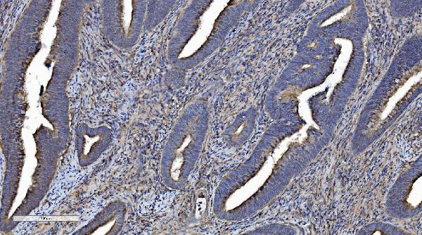
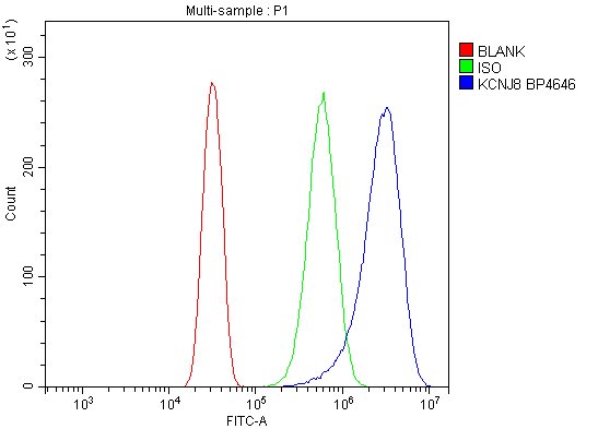 FCM/FACS (Flow Cytometry)
(Figure 3. Flow Cytometry analysis of U20S cells using anti-Kir6. 1/KCNJ8 antibody (AAA125737).Overlay histogram showing U20S cells stained with AAA125737 (Blue line). The cells were blocked with 10% normal goat serum. And then incubated with rabbit anti-Kir6. 1/KCNJ8 Antibody (AAA125737, 1μg/1x106 cells) for 30 min at 20 degree C. DyLight®488 conjugated goat anti-rabbit IgG (5-10μg/1x106 cells) was used as secondary antibody for 30 minutes at 20 degree C. Isotype control antibody (Green line) was rabbit IgG (1μg/1x106) used under the same conditions. Unlabelled sample (Red line) was also used as a control.)
FCM/FACS (Flow Cytometry)
(Figure 3. Flow Cytometry analysis of U20S cells using anti-Kir6. 1/KCNJ8 antibody (AAA125737).Overlay histogram showing U20S cells stained with AAA125737 (Blue line). The cells were blocked with 10% normal goat serum. And then incubated with rabbit anti-Kir6. 1/KCNJ8 Antibody (AAA125737, 1μg/1x106 cells) for 30 min at 20 degree C. DyLight®488 conjugated goat anti-rabbit IgG (5-10μg/1x106 cells) was used as secondary antibody for 30 minutes at 20 degree C. Isotype control antibody (Green line) was rabbit IgG (1μg/1x106) used under the same conditions. Unlabelled sample (Red line) was also used as a control.)
Kir6. 1/KCNJ8, Polyclonal Antibody (Cat# AAA125737)
Full Name
Anti-Kir6. 1/KCNJ8 Antibody
Gene Names
KCNJ8; KIR6.1; uKATP-1
Reactivity
Human, Mouse, Rat
Applications
Direct ELISA, Flow Cytometry, Immunohistochemistry, Western Blot
Purity
Immunogen affinity purified.
Pricing
 IF (Immunofluorescence)
(Figure 2. IF analysis of ACADS/SCAD using anti-ACADS/SCAD antibody (AAA125738).ACADS/SCAD was detected in immunocytochemical section of MCF-7 cells. Enzyme antigen retrieval was performed using IHC enzyme antigen retrieval reagent for 15 mins. The cells were blocked with 10% goat serum. And then incubated with 5μg/mL rabbit anti-ACADS/SCAD Antibody (AAA125738) overnight at 4 degree C. DyLight®488 Conjugated Goat Anti-Rabbit IgG was used as secondary antibody at 1:100 dilution and incubated for 30 minutes at 37 degree C. The section was counterstained with DAPI. Visualize using a fluorescence microscope and filter sets appropriate for the label used.)
IF (Immunofluorescence)
(Figure 2. IF analysis of ACADS/SCAD using anti-ACADS/SCAD antibody (AAA125738).ACADS/SCAD was detected in immunocytochemical section of MCF-7 cells. Enzyme antigen retrieval was performed using IHC enzyme antigen retrieval reagent for 15 mins. The cells were blocked with 10% goat serum. And then incubated with 5μg/mL rabbit anti-ACADS/SCAD Antibody (AAA125738) overnight at 4 degree C. DyLight®488 Conjugated Goat Anti-Rabbit IgG was used as secondary antibody at 1:100 dilution and incubated for 30 minutes at 37 degree C. The section was counterstained with DAPI. Visualize using a fluorescence microscope and filter sets appropriate for the label used.)
ACADS/SCAD, Polyclonal Antibody (Cat# AAA125738)
Full Name
Anti-ACADS/SCAD Antibody
Gene Names
ACADS; SCAD; ACAD3
Reactivity
Human, Mouse, Rat
Applications
Immunofluorescence, Immunocytochemistry, Western Blot
Purity
Immunogen affinity purified.
Pricing
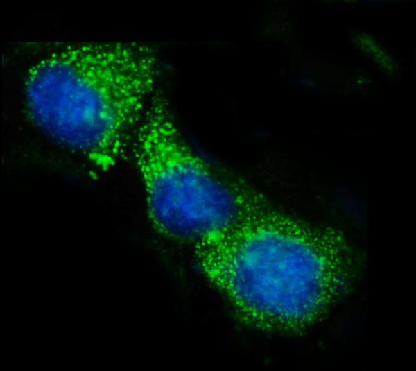
 IF (Immunofluorescence)
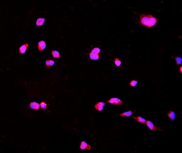
(Figure 4. IF analysis of CTHRC1 using anti-CTHRC1 antibody (AAA125741).CTHRC1 was detected in immunocytochemical section of HPEPA1-6 cells. Enzyme antigen retrieval was performed using IHC enzyme antigen retrieval reagent for 15 mins. The cells were blocked with 10% goat serum. And then incubated with 4μg/mL rabbit anti-CTHRC1 Antibody (AAA125741) overnight at 4 degree C. DyLight®550 Conjugated Goat Anti-Rabbit IgG (BA1135) was used as secondary antibody at 1:100 dilution and incubated for 30 minutes at 37 degree C. The section was counterstained with DAPI. Visualize using a fluorescence microscope and filter sets appropriate for the label used.)
IF (Immunofluorescence)
(Figure 4. IF analysis of CTHRC1 using anti-CTHRC1 antibody (AAA125741).CTHRC1 was detected in immunocytochemical section of HPEPA1-6 cells. Enzyme antigen retrieval was performed using IHC enzyme antigen retrieval reagent for 15 mins. The cells were blocked with 10% goat serum. And then incubated with 4μg/mL rabbit anti-CTHRC1 Antibody (AAA125741) overnight at 4 degree C. DyLight®550 Conjugated Goat Anti-Rabbit IgG (BA1135) was used as secondary antibody at 1:100 dilution and incubated for 30 minutes at 37 degree C. The section was counterstained with DAPI. Visualize using a fluorescence microscope and filter sets appropriate for the label used.)
CTHRC1, Polyclonal Antibody (Cat# AAA125741)
Full Name
Anti-CTHRC1 Antibody
Reactivity
Human, Mouse, Rat
Applications
Direct ELISA, Flow Cytometry, Immunofluorescence, Immunocytochemistry, Immunohistochemistry, Western Blot
Purity
Immunogen affinity purified.
Pricing

 FCM/FACS (Flow Cytometry)
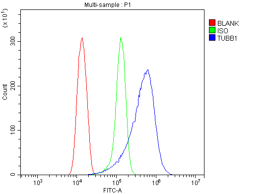
(Figure 3. Flow Cytometry analysis of Hela cells using anti-Tubulin beta antibody (AAA125748).Overlay histogram showing Hela cells stained with AAA125748 (Blue line). The cells were blocked with 10% normal goat serum. And then incubated with rabbit anti-Tubulin beta Antibody (AAA125748,1μg/1x106 cells) for 30 min at 20 degree C. DyLight®488 conjugated goat anti-rabbit IgG (5-10μg/1x106 cells) was used as secondary antibody for 30 minutes at 20 degree C. Isotype control antibody (Green line) was rabbit IgG (1μg/1x106) used under the same conditions. Unlabelled sample (Red line) was also used as a control.)
FCM/FACS (Flow Cytometry)
(Figure 3. Flow Cytometry analysis of Hela cells using anti-Tubulin beta antibody (AAA125748).Overlay histogram showing Hela cells stained with AAA125748 (Blue line). The cells were blocked with 10% normal goat serum. And then incubated with rabbit anti-Tubulin beta Antibody (AAA125748,1μg/1x106 cells) for 30 min at 20 degree C. DyLight®488 conjugated goat anti-rabbit IgG (5-10μg/1x106 cells) was used as secondary antibody for 30 minutes at 20 degree C. Isotype control antibody (Green line) was rabbit IgG (1μg/1x106) used under the same conditions. Unlabelled sample (Red line) was also used as a control.)
Tubulin beta, Polyclonal Antibody (Cat# AAA125748)
Full Name
Anti-Tubulin beta Antibody
Reactivity
Human, Mouse, Rat
Applications
Direct ELISA, Flow Cytometry, Immunofluorescence, Immunocytochemistry, Western Blot
Purity
Immunogen affinity purified.
Pricing

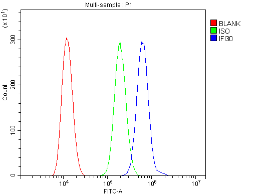 FCM/FACS (Flow Cytometry)
(Figure 5. Flow Cytometry analysis of HEPA1-6 cells using anti-GILT/IFI30 antibody (AAA125756).Overlay histogram showing HEPA1-6 cells stained with AAA125756 (Blue line). The cells were blocked with 10% normal goat serum. And then incubated with rabbit anti-GILT/IFI30 Antibody (AAA125756, 1μg/1x106 cells) for 30 min at 20 degree C. DyLight®488 conjugated goat anti-rabbit IgG (5-10μg/1x106 cells) was used as secondary antibody for 30 minutes at 20 degree C. Isotype control antibody (Green line) was rabbit IgG (1μg/1x106) used under the same conditions. Unlabelled sample (Red line) was also used as a control.)
FCM/FACS (Flow Cytometry)
(Figure 5. Flow Cytometry analysis of HEPA1-6 cells using anti-GILT/IFI30 antibody (AAA125756).Overlay histogram showing HEPA1-6 cells stained with AAA125756 (Blue line). The cells were blocked with 10% normal goat serum. And then incubated with rabbit anti-GILT/IFI30 Antibody (AAA125756, 1μg/1x106 cells) for 30 min at 20 degree C. DyLight®488 conjugated goat anti-rabbit IgG (5-10μg/1x106 cells) was used as secondary antibody for 30 minutes at 20 degree C. Isotype control antibody (Green line) was rabbit IgG (1μg/1x106) used under the same conditions. Unlabelled sample (Red line) was also used as a control.)
GILT/IFI30, Polyclonal Antibody (Cat# AAA125756)
Full Name
Anti-GILT/IFI30 Antibody
Gene Names
Ifi30; GILT; IP30
Reactivity
Mouse, Rat
Applications
Direct ELISA, Flow Cytometry, Immunohistochemistry, Western Blot
Purity
Immunogen affinity purified.
Pricing
 FCM/FACS (Flow Cytometry)
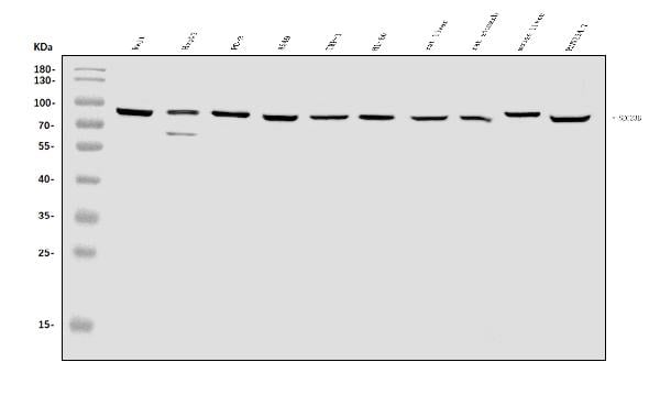
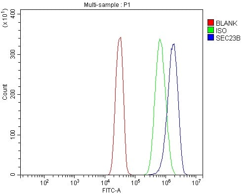
(Figure 2. Flow Cytometry analysis of SiHa cells using anti-SEC23B antibody (AAA125762).Overlay histogram showing SiHa cells stained with AAA125762 (Blue line). The cells were blocked with 10% normal goat serum. And then incubated with rabbit anti-SEC23B Antibody (AAA125762, 1μg/1x106 cells) for 30 min at 20 degree C. DyLight®488 conjugated goat anti-rabbit IgG (5-10μg/1x106 cells) was used as secondary antibody for 30 minutes at 20 degree C. Isotype control antibody (Green line) was rabbit IgG (1μg/1x106) used under the same conditions. Unlabelled sample (Red line) was also used as a control.)
FCM/FACS (Flow Cytometry)
(Figure 2. Flow Cytometry analysis of SiHa cells using anti-SEC23B antibody (AAA125762).Overlay histogram showing SiHa cells stained with AAA125762 (Blue line). The cells were blocked with 10% normal goat serum. And then incubated with rabbit anti-SEC23B Antibody (AAA125762, 1μg/1x106 cells) for 30 min at 20 degree C. DyLight®488 conjugated goat anti-rabbit IgG (5-10μg/1x106 cells) was used as secondary antibody for 30 minutes at 20 degree C. Isotype control antibody (Green line) was rabbit IgG (1μg/1x106) used under the same conditions. Unlabelled sample (Red line) was also used as a control.)
SEC23B, Polyclonal Antibody (Cat# AAA125762)
Full Name
Anti-SEC23B Antibody
Gene Names
SEC23B; CDAII; CDAN2; CDA-II; HEMPAS
Reactivity
Human, Mouse, Rat
Applications
Direct ELISA, Flow Cytometry, Western Blot
Purity
Immunogen affinity purified.
Pricing
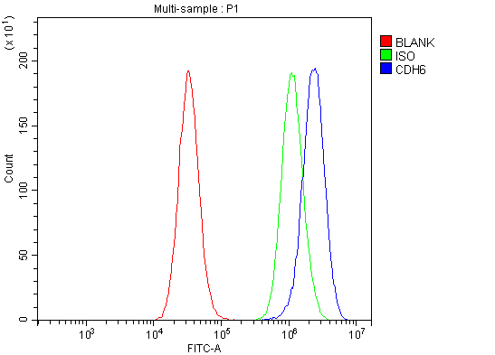 FCM/FACS (Flow Cytometry)
(Figure 3. Flow Cytometry analysis of PC-3 cells using anti-K Cadherin/CDH6 antibody (AAA125770).Overlay histogram showing PC-3 cells stained with AAA125770 (Blue line). The cells were blocked with 10% normal goat serum. And then incubated with rabbit anti-K Cadherin/CDH6 Antibody (AAA125770, 1μg/1x106 cells) for 30 min at 20 degree C. DyLight®488 conjugated goat anti-rabbit IgG (5-10μg/1x106 cells) was used as secondary antibody for 30 minutes at 20 degree C. Isotype control antibody (Green line) was rabbit IgG (1μg/1x106) used under the same conditions. Unlabelled sample (Red line) was also used as a control.)
FCM/FACS (Flow Cytometry)
(Figure 3. Flow Cytometry analysis of PC-3 cells using anti-K Cadherin/CDH6 antibody (AAA125770).Overlay histogram showing PC-3 cells stained with AAA125770 (Blue line). The cells were blocked with 10% normal goat serum. And then incubated with rabbit anti-K Cadherin/CDH6 Antibody (AAA125770, 1μg/1x106 cells) for 30 min at 20 degree C. DyLight®488 conjugated goat anti-rabbit IgG (5-10μg/1x106 cells) was used as secondary antibody for 30 minutes at 20 degree C. Isotype control antibody (Green line) was rabbit IgG (1μg/1x106) used under the same conditions. Unlabelled sample (Red line) was also used as a control.)
K Cadherin/CDH6, Polyclonal Antibody (Cat# AAA125770)
Full Name
Anti-K Cadherin/CDH6 Antibody
Gene Names
CDH6; CAD6; KCAD
Reactivity
Human, Mouse, Rat
Applications
Direct ELISA, Flow Cytometry, Immunohistochemistry, Western Blot
Purity
Immunogen affinity purified.
Pricing
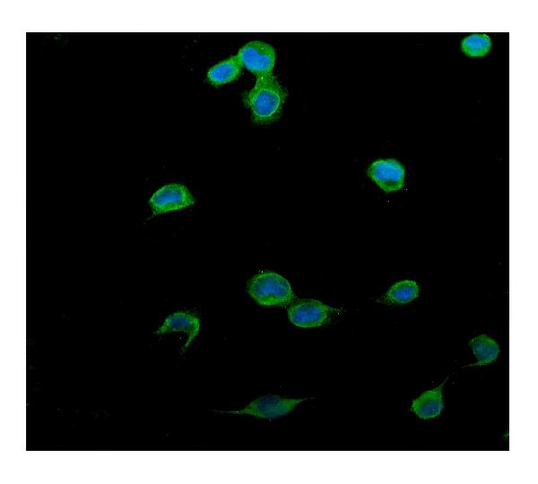 IF (Immunofluorescence)
(Figure 4. IF analysis of PTCH2 using anti- PTCH2 antibody (AAA125771).PTCH2 was detected in immunocytochemical section of U20S cells. Enzyme antigen retrieval was performed using IHC enzyme antigen retrieval reagent for 15 mins. The cells were blocked with 10% goat serum. And then incubated with 5μg/mL rabbit anti-PTCH2 Antibody (AAA125771) overnight at 4 degree C. DyLight®488 Conjugated Goat Anti-Rabbit IgG was used as secondary antibody at 1:100 dilution and incubated for 30 minutes at 37 degree C. The section was counterstained with DAPI. Visualize using a fluorescence microscope and filter sets appropriate for the label used.)
IF (Immunofluorescence)
(Figure 4. IF analysis of PTCH2 using anti- PTCH2 antibody (AAA125771).PTCH2 was detected in immunocytochemical section of U20S cells. Enzyme antigen retrieval was performed using IHC enzyme antigen retrieval reagent for 15 mins. The cells were blocked with 10% goat serum. And then incubated with 5μg/mL rabbit anti-PTCH2 Antibody (AAA125771) overnight at 4 degree C. DyLight®488 Conjugated Goat Anti-Rabbit IgG was used as secondary antibody at 1:100 dilution and incubated for 30 minutes at 37 degree C. The section was counterstained with DAPI. Visualize using a fluorescence microscope and filter sets appropriate for the label used.)
PTCH2, Polyclonal Antibody (Cat# AAA125771)
Full Name
Anti-PTCH2 Antibody
Gene Names
PTCH2; PTC2
Reactivity
Human
Applications
Immunofluorescence, Immunocytochemistry, Immunohistochemistry, Western Blot
Purity
Immunogen affinity purified.
Pricing
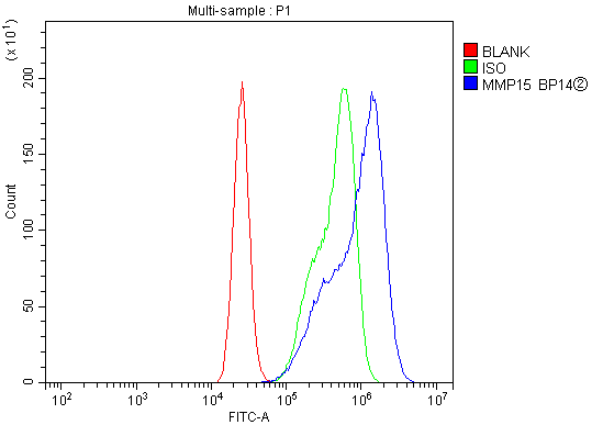 FCM/FACS (Flow Cytometry)
(Figure 3. Flow Cytometry analysis of CACO-2 cells using anti-MT2-MMP/MMP15 antibody (AAA125772).Overlay histogram showing CACO-2 cells stained with AAA125772 (Blue line). The cells were blocked with 10% normal goat serum. And then incubated with rabbit anti-MT2-MMP/MMP15 Antibody (AAA125772, 1μg/1x106 cells) for 30 min at 20 degree C. DyLight®488 conjugated goat anti-rabbit IgG (5-10μg/1x106 cells) was used as secondary antibody for 30 minutes at 20 degree C. Isotype control antibody (Green line) was rabbit IgG (1μg/1x106) used under the same conditions. Unlabelled sample (Red line) was also used as a control.)
FCM/FACS (Flow Cytometry)
(Figure 3. Flow Cytometry analysis of CACO-2 cells using anti-MT2-MMP/MMP15 antibody (AAA125772).Overlay histogram showing CACO-2 cells stained with AAA125772 (Blue line). The cells were blocked with 10% normal goat serum. And then incubated with rabbit anti-MT2-MMP/MMP15 Antibody (AAA125772, 1μg/1x106 cells) for 30 min at 20 degree C. DyLight®488 conjugated goat anti-rabbit IgG (5-10μg/1x106 cells) was used as secondary antibody for 30 minutes at 20 degree C. Isotype control antibody (Green line) was rabbit IgG (1μg/1x106) used under the same conditions. Unlabelled sample (Red line) was also used as a control.)
MT2-MMP/MMP15, Polyclonal Antibody (Cat# AAA125772)
Full Name
Anti-MT2-MMP/MMP15 Antibody
Gene Names
MMP15; MTMMP2; SMCP-2; MT2-MMP
Reactivity
Human, Mouse, Rat
Applications
Direct ELISA, Flow Cytometry, Immunofluorescence, Immunocytochemistry, Western Blot
Purity
Immunogen affinity purified.
Pricing
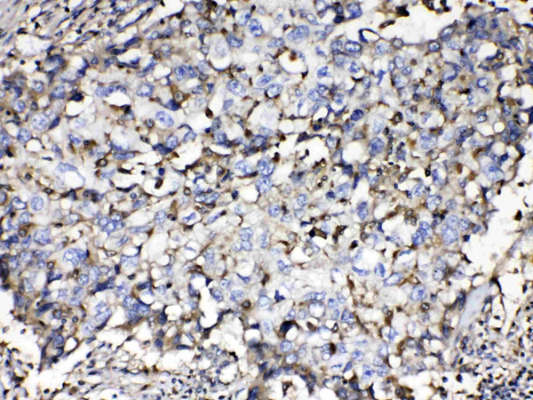
 IHC (Immunohistochemistry)
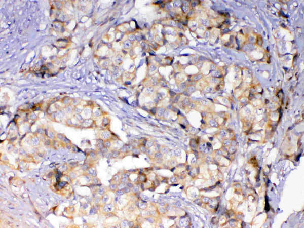
(Figure 5. IHC analysis of ErbB 4 using anti-ErbB 4 antibody (AAA124563).ErbB 4 was detected in paraffin-embedded section of human mammary cancer tissue. Heat mediated antigen retrieval was performed in citrate buffer (pH6, epitope retrieval solution) for 20 mins. The tissue section was blocked with 10% goat serum. The tissue section was then incubated with 1ug/ml rabbit anti-ErbB 4 Antibody (AAA124563) overnight at 4 degree C. Biotinylated goat anti-rabbit IgG was used as secondary antibody and incubated for 30 minutes at 37 degree C. The tissue section was developed using Strepavidin-Biotin-Complex (SABC) with DAB as the chromogen.)
IHC (Immunohistochemistry)
(Figure 5. IHC analysis of ErbB 4 using anti-ErbB 4 antibody (AAA124563).ErbB 4 was detected in paraffin-embedded section of human mammary cancer tissue. Heat mediated antigen retrieval was performed in citrate buffer (pH6, epitope retrieval solution) for 20 mins. The tissue section was blocked with 10% goat serum. The tissue section was then incubated with 1ug/ml rabbit anti-ErbB 4 Antibody (AAA124563) overnight at 4 degree C. Biotinylated goat anti-rabbit IgG was used as secondary antibody and incubated for 30 minutes at 37 degree C. The tissue section was developed using Strepavidin-Biotin-Complex (SABC) with DAB as the chromogen.)
ErbB 4, Polyclonal Antibody (Cat# AAA124563)
Full Name
Anti-ErbB 4 Picoband antibody
Gene Names
ERBB4; HER4; ALS19; p180erbB4
Reactivity
Human, Mouse, Rat
No cross reactivity with other proteins.
No cross reactivity with other proteins.
Applications
Western Blot, Immunohistochemistry
Pricing

 FCM/FACS (Flow Cytometry)
(Figure 5. Flow Cytometry analysis of Jurkat cells using anti-NFATC1 antibody (AAA124564).Overlay histogram showing Jurkat cells stained with AAA124564 (Blue line).The cells were blocked with 10% normal goat serum. And then incubated with rabbit anti-NFATC1 Antibody (AAA124564,1ug/1x10^6 cells) for 30 min at 20 degree C. DyLight®488 conjugated goat anti-rabbit IgG (5-10ug/1x10^6 cells) was used as secondary antibody for 30 minutes at 20 degree C. Isotype control antibody (Green line) was rabbit IgG (1ug/1x106) used under the same conditions. Unlabelled sample (Red line) was also used as a control.)
FCM/FACS (Flow Cytometry)
(Figure 5. Flow Cytometry analysis of Jurkat cells using anti-NFATC1 antibody (AAA124564).Overlay histogram showing Jurkat cells stained with AAA124564 (Blue line).The cells were blocked with 10% normal goat serum. And then incubated with rabbit anti-NFATC1 Antibody (AAA124564,1ug/1x10^6 cells) for 30 min at 20 degree C. DyLight®488 conjugated goat anti-rabbit IgG (5-10ug/1x10^6 cells) was used as secondary antibody for 30 minutes at 20 degree C. Isotype control antibody (Green line) was rabbit IgG (1ug/1x106) used under the same conditions. Unlabelled sample (Red line) was also used as a control.)
NFAT2, Polyclonal Antibody (Cat# AAA124564)
Full Name
Anti-NFAT2 Picoband Antibody
Gene Names
NFATC1; NFAT2; NFATc; NF-ATC; NF-ATc1.2
Reactivity
Human, Mouse, Rat
No cross reactivity with other proteins.
No cross reactivity with other proteins.
Applications
Western Blot, Immunocytochemistry, Immunohistochemistry, Flow Cytometry
Purity
Immunogen affinity purified
Pricing
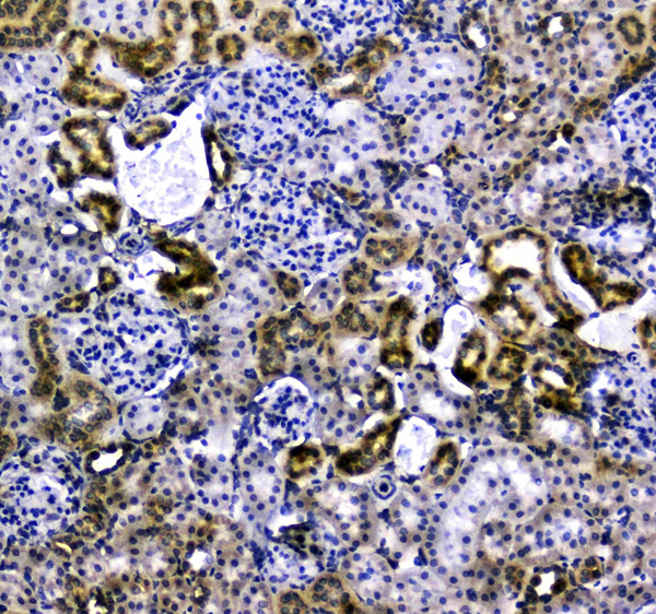
 IHC (Immunohistochemisry)
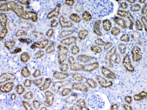
(Figure 3. IHC analysis of NADPH oxidase 4 using anti-NADPH oxidase 4 antibody (AAA124570).NADPH oxidase 4 was detected in paraffin-embedded section of human renal cancer tissue. Heat mediated antigen retrieval was performed in citrate buffer (pH6, epitope retrieval solution) for 20 mins. The tissue section was blocked with 10% goat serum. The tissue section was then incubated with 1ug/ml rabbit anti-NADPH oxidase 4 Antibody (AAA124570) overnight at 4 degree C. Biotinylated goat anti-rabbit IgG was used as secondary antibody and incubated for 30 minutes at 37 degree C. The tissue section was developed using Strepavidin-Biotin-Complex (SABC) with DAB as the chromogen.)
IHC (Immunohistochemisry)
(Figure 3. IHC analysis of NADPH oxidase 4 using anti-NADPH oxidase 4 antibody (AAA124570).NADPH oxidase 4 was detected in paraffin-embedded section of human renal cancer tissue. Heat mediated antigen retrieval was performed in citrate buffer (pH6, epitope retrieval solution) for 20 mins. The tissue section was blocked with 10% goat serum. The tissue section was then incubated with 1ug/ml rabbit anti-NADPH oxidase 4 Antibody (AAA124570) overnight at 4 degree C. Biotinylated goat anti-rabbit IgG was used as secondary antibody and incubated for 30 minutes at 37 degree C. The tissue section was developed using Strepavidin-Biotin-Complex (SABC) with DAB as the chromogen.)
NADPH oxidase 4, Polyclonal Antibody (Cat# AAA124570)
Full Name
Anti-NADPH oxidase 4 Picoband antibody
Gene Names
NOX4; KOX; KOX-1; RENOX
Reactivity
Human, Mouse, Rat
No cross reactivity with other proteins.
No cross reactivity with other proteins.
Applications
Western Blot, Immunohistochemistry
Pricing
 IHC (Immunohistochemisry)
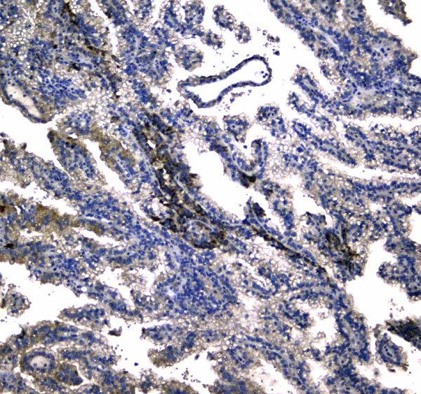
(Figure 3. IHC analysis of Beta 2 Microglobulin using anti- Beta 2 Microglobulin antibody (AAA124573).Beta 2 Microglobulin was detected in paraffin-embedded section of human lung cancer tissues. Heat mediated antigen retrieval was performed in citrate buffer (pH6, epitope retrieval solution) for 20 mins. The tissue section was blocked with 10% goat serum. The tissue section was then incubated with 1ug/ml rabbit anti- Beta 2 Microglobulin Antibody (AAA124573) overnight at 4 degree C. Biotinylated goat anti-rabbit IgG was used as secondary antibody and incubated for 30 minutes at 37 degree C. The tissue section was developed using Strepavidin-Biotin-Complex (SABC) with DAB as the chromogen.)
IHC (Immunohistochemisry)
(Figure 3. IHC analysis of Beta 2 Microglobulin using anti- Beta 2 Microglobulin antibody (AAA124573).Beta 2 Microglobulin was detected in paraffin-embedded section of human lung cancer tissues. Heat mediated antigen retrieval was performed in citrate buffer (pH6, epitope retrieval solution) for 20 mins. The tissue section was blocked with 10% goat serum. The tissue section was then incubated with 1ug/ml rabbit anti- Beta 2 Microglobulin Antibody (AAA124573) overnight at 4 degree C. Biotinylated goat anti-rabbit IgG was used as secondary antibody and incubated for 30 minutes at 37 degree C. The tissue section was developed using Strepavidin-Biotin-Complex (SABC) with DAB as the chromogen.)
Beta 2 Microglobulin, Polyclonal Antibody (Cat# AAA124573)
Full Name
Anti-Beta 2 Microglobulin Picoband Antibody
Gene Names
B2M; IMD43
Reactivity
Human
No cross reactivity with other proteins
No cross reactivity with other proteins
Applications
Western Blot, Immunohistochemistry
Purity
Immunogen affinity purified
Pricing
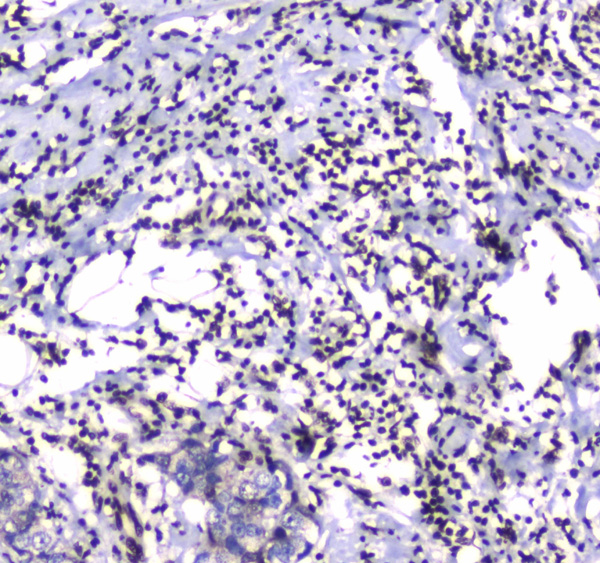
 IHC (Immunohiostchemistry)
(Figure 2. IHC analysis of DGCR8 using anti-DGCR8 antibody (AAA124576).DGCR8 was detected in paraffin-embedded section of human rectal cancer tissue. Heat mediated antigen retrieval was performed in citrate buffer (pH6, epitope retrieval solution) for 20 mins. The tissue section was blocked with 10% goat serum. The tissue section was then incubated with 1ug/ml rabbit anti-DGCR8 Antibody (AAA124576) overnight at 4 degree C. Biotinylated goat anti-rabbit IgG was used as secondary antibody and incubated for 30 minutes at 37 degree C. The tissue section was developed using Strepavidin-Biotin-Complex (SABC) with DAB as the chromogen.)
IHC (Immunohiostchemistry)
(Figure 2. IHC analysis of DGCR8 using anti-DGCR8 antibody (AAA124576).DGCR8 was detected in paraffin-embedded section of human rectal cancer tissue. Heat mediated antigen retrieval was performed in citrate buffer (pH6, epitope retrieval solution) for 20 mins. The tissue section was blocked with 10% goat serum. The tissue section was then incubated with 1ug/ml rabbit anti-DGCR8 Antibody (AAA124576) overnight at 4 degree C. Biotinylated goat anti-rabbit IgG was used as secondary antibody and incubated for 30 minutes at 37 degree C. The tissue section was developed using Strepavidin-Biotin-Complex (SABC) with DAB as the chromogen.)
DGCR8, Polyclonal Antibody (Cat# AAA124576)
Full Name
Anti-DGCR8 Picoband antibody
Gene Names
DGCR8; Gy1; pasha; DGCRK6; C22orf12
Reactivity
Human, Mouse, Rat
No cross reactivity with other proteins.
No cross reactivity with other proteins.
Applications
Western Blot, Immunohistochemistry
Pricing
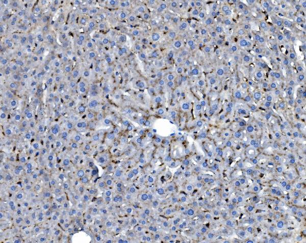
 IHC (Immunohistochemistry)
(Figure 5. IHC analysis of Cytochrome P450 2D6 using anti- Cytochrome P450 2D6 antibody (AAA124578).Cytochrome P450 2D6 was detected in paraffin-embedded section of human liver cancer tissues. Heat mediated antigen retrieval was performed in citrate buffer (pH6, epitope retrieval solution) for 20 mins. The tissue section was blocked with 10% goat serum. The tissue section was then incubated with 1ug/ml rabbit anti- Cytochrome P450 2D6 Antibody (AAA124578) overnight at 4 degree C. Biotinylated goat anti-rabbit IgG was used as secondary antibody and incubated for 30 minutes at 37 degree C. The tissue section was developed using Strepavidin-Biotin-Complex (SABC) with DAB as the chromogen.)
IHC (Immunohistochemistry)
(Figure 5. IHC analysis of Cytochrome P450 2D6 using anti- Cytochrome P450 2D6 antibody (AAA124578).Cytochrome P450 2D6 was detected in paraffin-embedded section of human liver cancer tissues. Heat mediated antigen retrieval was performed in citrate buffer (pH6, epitope retrieval solution) for 20 mins. The tissue section was blocked with 10% goat serum. The tissue section was then incubated with 1ug/ml rabbit anti- Cytochrome P450 2D6 Antibody (AAA124578) overnight at 4 degree C. Biotinylated goat anti-rabbit IgG was used as secondary antibody and incubated for 30 minutes at 37 degree C. The tissue section was developed using Strepavidin-Biotin-Complex (SABC) with DAB as the chromogen.)
Cytochrome P450 2D6, Polyclonal Antibody (Cat# AAA124578)
Full Name
Anti-Cytochrome P450 2D6 Picoband Antibody
Gene Names
CYP2D6; CPD6; CYP2D; CYP2DL1; CYPIID6; P450C2D; P450DB1; CYP2D7AP; CYP2D7BP; CYP2D7P2; CYP2D8P2; P450-DB1
Reactivity
Human, Mouse, Rat
No cross reactivity with other proteins
No cross reactivity with other proteins
Applications
Western Blot, Immunohistochemistry
Purity
Immunogen affinity purified
Pricing
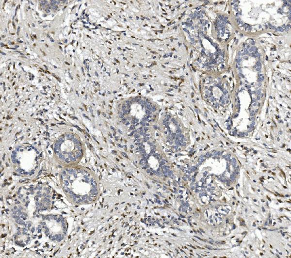
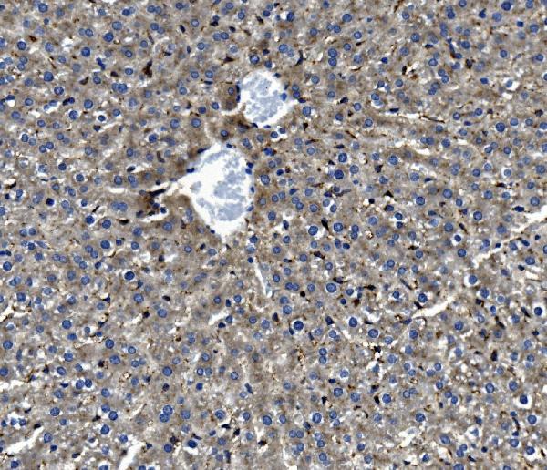
 IHC (Immunohistochemistry)
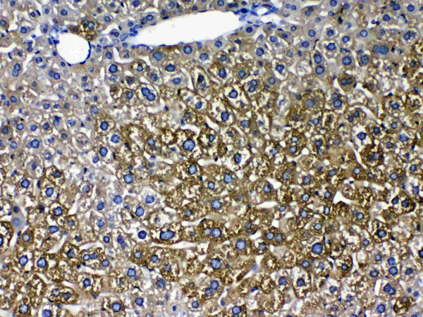
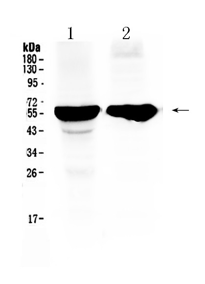
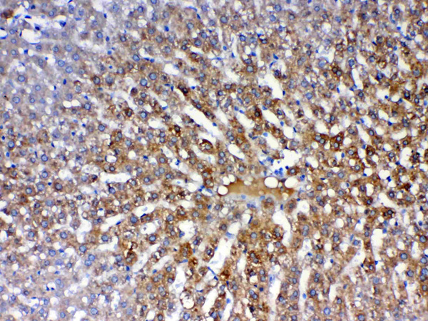
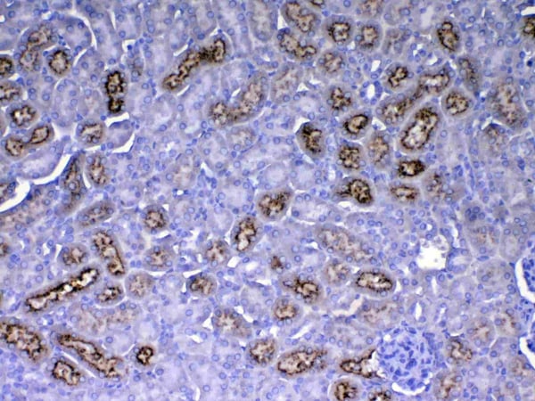
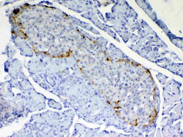
(IHC analysis of PON1 using anti-PON1 antibody (AAA124579). PON1 was detected in paraffin-embedded section of rat liver tissue. Heat mediated antigen retrieval was performed in citrate buffer (pH6, epitope retrieval solution) for 20 mins. The tissue section was blocked with 10% goat serum. The tissue section was then incubated with 1ug/ml rabbit anti-PON1 Antibody (AAA124579) overnight at 4 degree C. Biotinylated goat anti-rabbit IgG was used as secondary antibody and incubated for 30 minutes at 37 degree C. The tissue section was developed using Strepavidin-Biotin-Complex (SABC) with DAB as the chromogen.)
IHC (Immunohistochemistry)
(IHC analysis of PON1 using anti-PON1 antibody (AAA124579). PON1 was detected in paraffin-embedded section of rat liver tissue. Heat mediated antigen retrieval was performed in citrate buffer (pH6, epitope retrieval solution) for 20 mins. The tissue section was blocked with 10% goat serum. The tissue section was then incubated with 1ug/ml rabbit anti-PON1 Antibody (AAA124579) overnight at 4 degree C. Biotinylated goat anti-rabbit IgG was used as secondary antibody and incubated for 30 minutes at 37 degree C. The tissue section was developed using Strepavidin-Biotin-Complex (SABC) with DAB as the chromogen.)
PON1/Paraoxonase 1, Polyclonal Antibody (Cat# AAA124579)
Full Name
Anti-PON1/Paraoxonase 1 Picoband Antibody
Gene Names
Pon1; Pon
Reactivity
Mouse
No cross reactivity with other proteins.
No cross reactivity with other proteins.
Applications
Western Blot, Immunohistochemistry
Purity
Immunogen affinity purified
Pricing
 IHC (Immunohistochemisry)
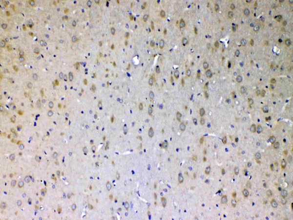
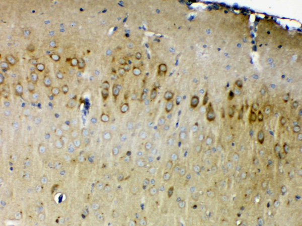
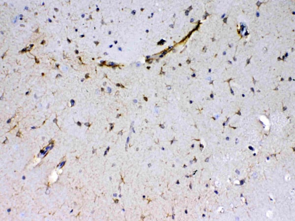
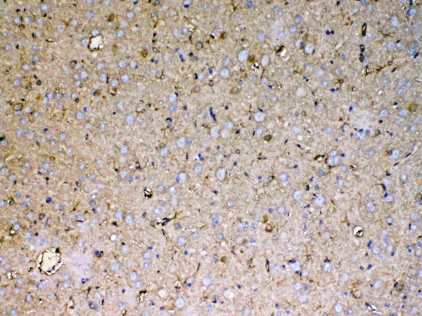
(Figure 3. IHC analysis of Sacsin using anti-Sacsin antibody (AAA124580).Sacsin was detected in paraffin-embedded section of rat brain tissue. Heat mediated antigen retrieval was performed in citrate buffer (pH6, epitope retrieval solution) for 20 mins. The tissue section was blocked with 10% goat serum. The tissue section was then incubated with 1ug/ml rabbit anti-Sacsin Antibody (AAA124580) overnight at 4 degree C. Biotinylated goat anti-rabbit IgG was used as secondary antibody and incubated for 30 minutes at 37 degree C. The tissue section was developed using Strepavidin-Biotin-Complex (SABC) with DAB as the chromogen.)
IHC (Immunohistochemisry)
(Figure 3. IHC analysis of Sacsin using anti-Sacsin antibody (AAA124580).Sacsin was detected in paraffin-embedded section of rat brain tissue. Heat mediated antigen retrieval was performed in citrate buffer (pH6, epitope retrieval solution) for 20 mins. The tissue section was blocked with 10% goat serum. The tissue section was then incubated with 1ug/ml rabbit anti-Sacsin Antibody (AAA124580) overnight at 4 degree C. Biotinylated goat anti-rabbit IgG was used as secondary antibody and incubated for 30 minutes at 37 degree C. The tissue section was developed using Strepavidin-Biotin-Complex (SABC) with DAB as the chromogen.)
Sacsin, Polyclonal Antibody (Cat# AAA124580)
Full Name
Anti-Sacsin Picoband antibody
Gene Names
SACS; SPAX6; ARSACS; DNAJC29; PPP1R138
Reactivity
Human, Mouse, Rat
No cross reactivity with other proteins.
No cross reactivity with other proteins.
Applications
Western Blot, Immunohistochemistry
Purity
Immunogen affinity purified
Pricing
 WB (Western Blot)
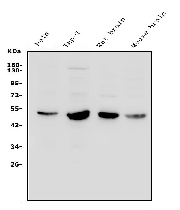
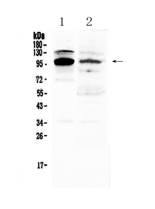
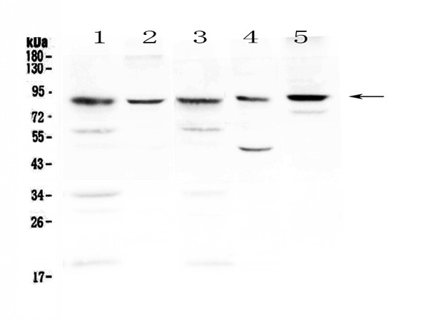
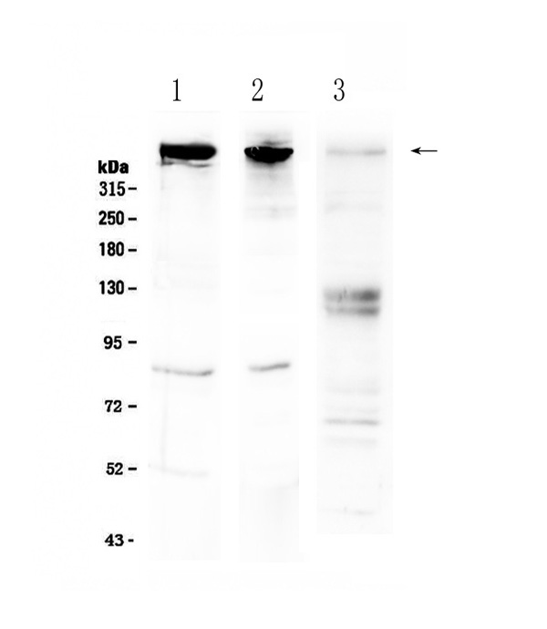
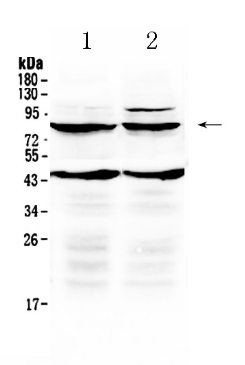
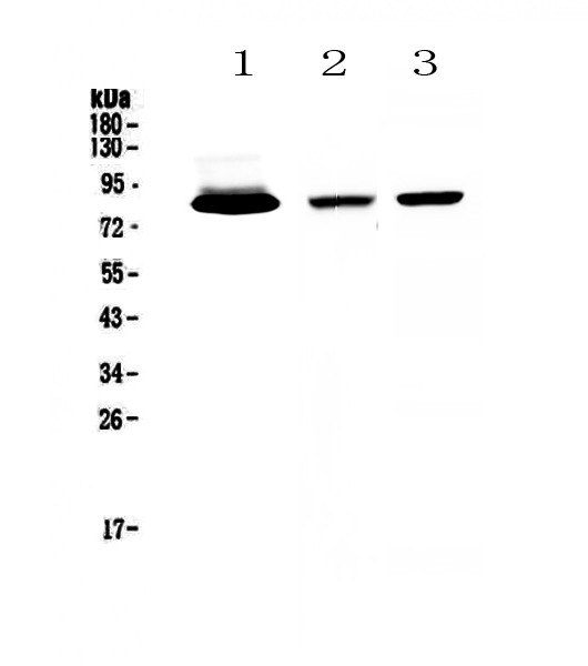
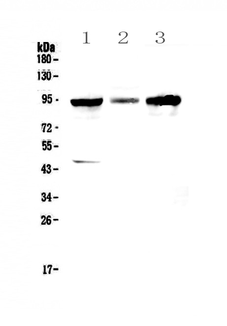
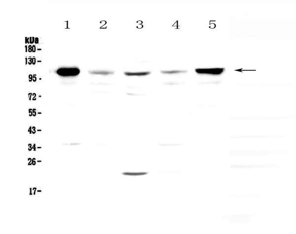
(Figure 1. Western blot analysis of TSH Receptor using anti-TSH Receptor antibody (AAA124582).Electrophoresis was performed on a 5-20% SDS-PAGE gel at 70V (Stacking gel) / 90V (Resolving gel) for 2-3 hours. The sample well of each lane was loaded with 50ug of sample under reducing conditions.Lane 1: rat brain tissue lysates,Lane 2: mouse brain tissue lysates.After Electrophoresis, proteins were transferred to a Nitrocellulose membrane at 150mA for 50-90 minutes. Blocked the membrane with 5% Non-fat Milk/ TBS for 1.5 hour at RT. The membrane was incubated with rabbit anti-TSH Receptor antigen affinity purified polyclonal antibody at 0.5ug/mL overnight at 4 degree C, then washed with TBS-0.1%Tween 3 times with 5 minutes each and probed with a goat anti-rabbit IgG-HRP secondary antibody at a dilution of 1:10000 for 1.5 hour at RT. The signal is developed using an Enhanced Chemiluminescent detection (ECL) kit with Tanon 5200 system. A specific band was detected for TSH Receptor at approximately 86KD. The expected band size for TSH Receptor is at 86KD.)
WB (Western Blot)
(Figure 1. Western blot analysis of TSH Receptor using anti-TSH Receptor antibody (AAA124582).Electrophoresis was performed on a 5-20% SDS-PAGE gel at 70V (Stacking gel) / 90V (Resolving gel) for 2-3 hours. The sample well of each lane was loaded with 50ug of sample under reducing conditions.Lane 1: rat brain tissue lysates,Lane 2: mouse brain tissue lysates.After Electrophoresis, proteins were transferred to a Nitrocellulose membrane at 150mA for 50-90 minutes. Blocked the membrane with 5% Non-fat Milk/ TBS for 1.5 hour at RT. The membrane was incubated with rabbit anti-TSH Receptor antigen affinity purified polyclonal antibody at 0.5ug/mL overnight at 4 degree C, then washed with TBS-0.1%Tween 3 times with 5 minutes each and probed with a goat anti-rabbit IgG-HRP secondary antibody at a dilution of 1:10000 for 1.5 hour at RT. The signal is developed using an Enhanced Chemiluminescent detection (ECL) kit with Tanon 5200 system. A specific band was detected for TSH Receptor at approximately 86KD. The expected band size for TSH Receptor is at 86KD.)
TSH Receptor, Polyclonal Antibody (Cat# AAA124582)
Full Name
Anti-TSH Receptor Picoband Antibody
Gene Names
TSHR; LGR3; CHNG1; hTSHR-I
Reactivity
Reacts with: Mouse, Rat
Predicted to work with: Human
Predicted to work with: Human
Applications
Western Blot
Purity
Immunogen affinity purified
Pricing
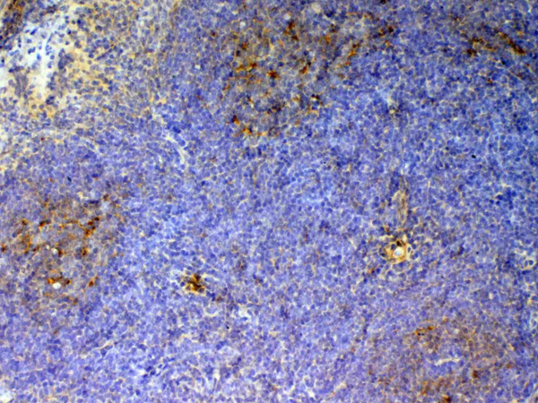
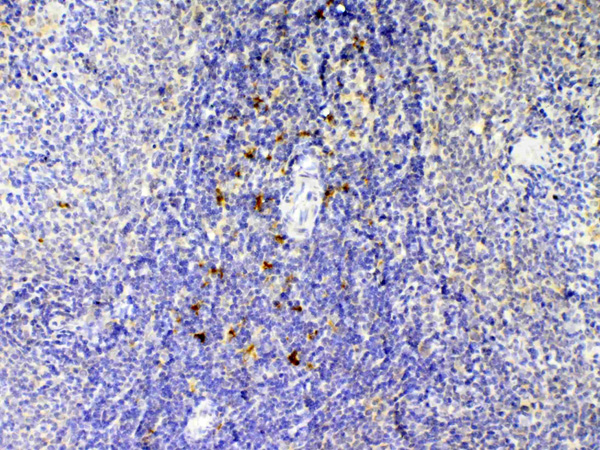
 IHC (Immunohistochemistry)
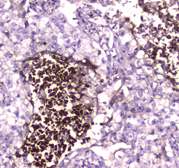
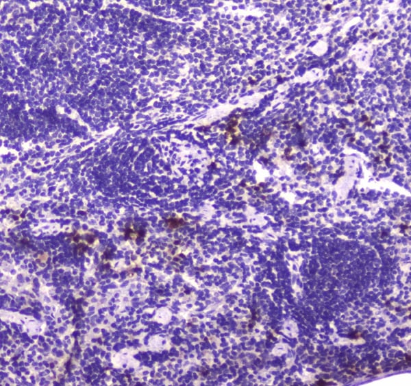
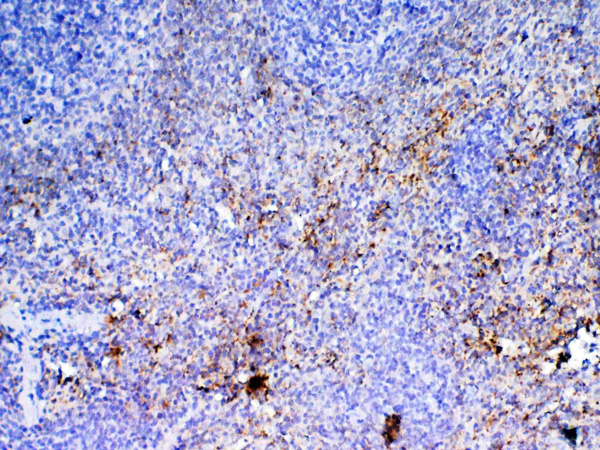
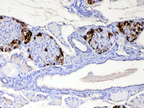
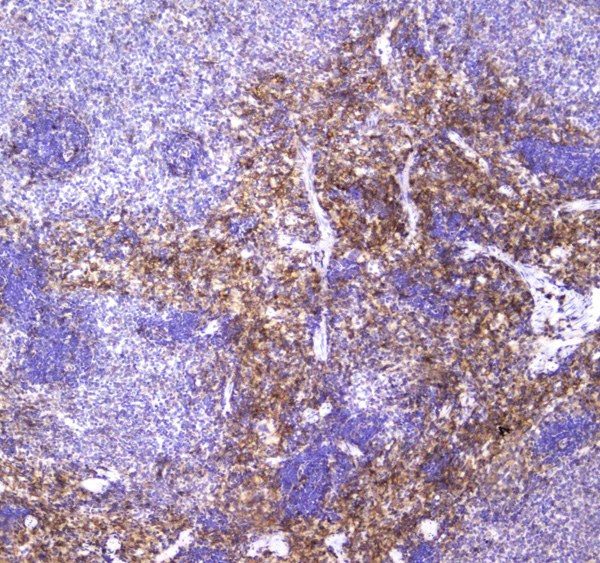
(Figure 4. IHC analysis of Lactoferrin using anti-Lactoferrin antibody (AAA124585).Lactoferrin was detected in paraffin-embedded section of mouse spleen tissue. Heat mediated antigen retrieval was performed in citrate buffer (pH6, epitope retrieval solution) for 20 mins. The tissue section was blocked with 10% goat serum. The tissue section was then incubated with 2ug/ml rabbit anti-Lactoferrin Antibody (AAA124585) overnight at 4 degree C. Biotinylated goat anti-rabbit IgG was used as secondary antibody and incubated for 30 minutes at 37 degree C. The tissue section was developed using Strepavidin-Biotin-Complex (SABC) with DAB as the chromogen.)
IHC (Immunohistochemistry)
(Figure 4. IHC analysis of Lactoferrin using anti-Lactoferrin antibody (AAA124585).Lactoferrin was detected in paraffin-embedded section of mouse spleen tissue. Heat mediated antigen retrieval was performed in citrate buffer (pH6, epitope retrieval solution) for 20 mins. The tissue section was blocked with 10% goat serum. The tissue section was then incubated with 2ug/ml rabbit anti-Lactoferrin Antibody (AAA124585) overnight at 4 degree C. Biotinylated goat anti-rabbit IgG was used as secondary antibody and incubated for 30 minutes at 37 degree C. The tissue section was developed using Strepavidin-Biotin-Complex (SABC) with DAB as the chromogen.)
Lactoferrin, Polyclonal Antibody (Cat# AAA124585)
Full Name
Anti-Lactoferrin Picoband antibody
Gene Names
LTF; LF; HLF2; GIG12; HEL110
Reactivity
Human, Mouse, Rat
No cross reactivity with other proteins.
No cross reactivity with other proteins.
Applications
Western Blot, Immunohistochemistry
Pricing
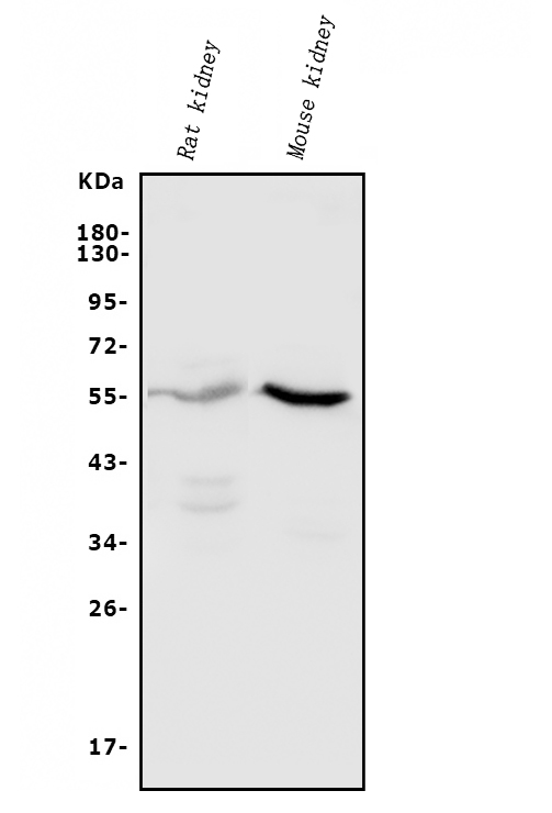
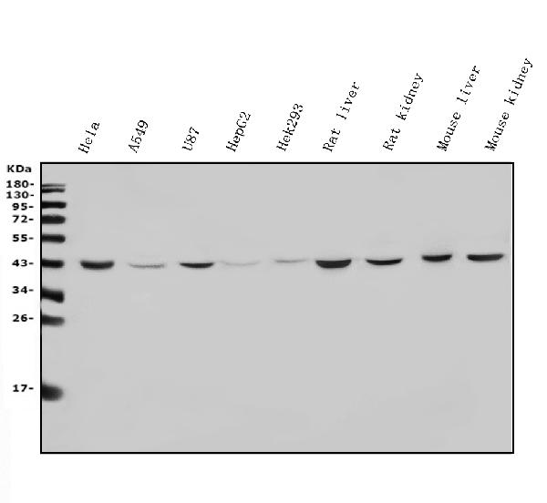
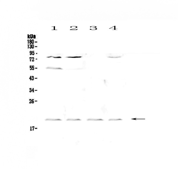
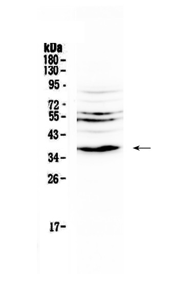
 WB (Western Blot)
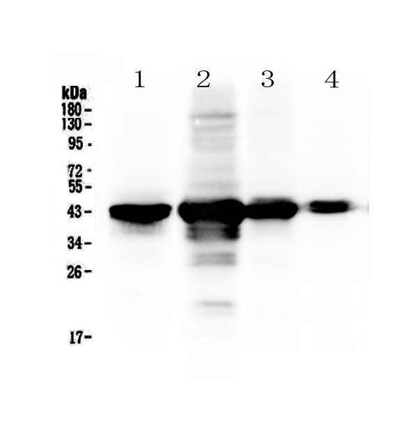
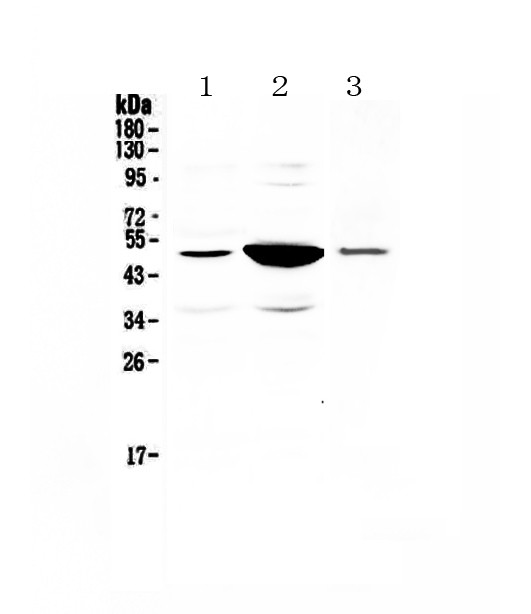
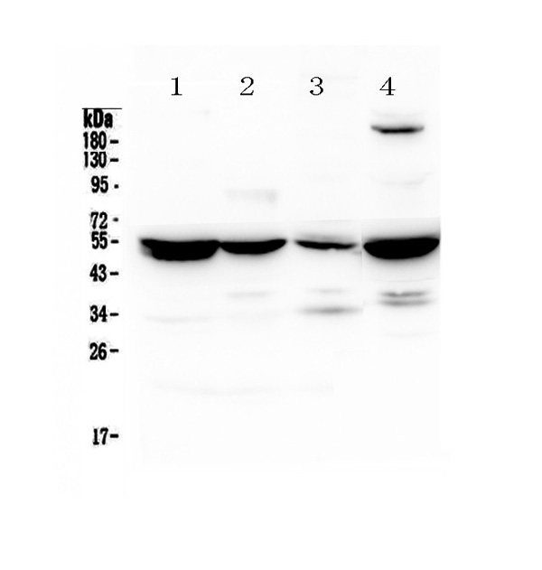
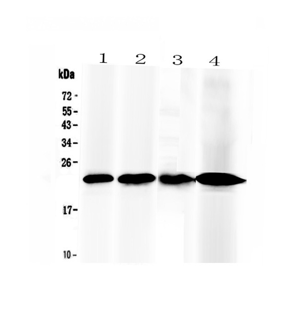
(Figure 1. Western blot analysis of PAI1 using anti-PAI1 antibody (AAA124586).Electrophoresis was performed on a 5-20% SDS-PAGE gel at 70V (Stacking gel) / 90V (Resolving gel) for 2-3 hours. The sample well of each lane was loaded with 50ug of sample under reducing conditions.Lane 1: rat small intestine tissue lysates,Lane 2: rat kidney tissue lysates,Lane 3: mouse kidney tissue lysates.After Electrophoresis, proteins were transferred to a Nitrocellulose membrane at 150mA for 50-90 minutes. Blocked the membrane with 5% Non-fat Milk/ TBS for 1.5 hour at RT. The membrane was incubated with rabbit anti-PAI1 antigen affinity purified polyclonal antibody at 0.5ug/mL overnight at 4 degree C, then washed with TBS-0.1%Tween 3 times with 5 minutes each and probed with a goat anti-rabbit IgG-HRP secondary antibody at a dilution of 1:10000 for 1.5 hour at RT. The signal is developed using an Enhanced Chemiluminescent detection (ECL) kit with Tanon 5200 system. A specific band was detected for PAI1 at approximately 45-50KD. The expected band size for PAI1 is at 45KD.)
WB (Western Blot)
(Figure 1. Western blot analysis of PAI1 using anti-PAI1 antibody (AAA124586).Electrophoresis was performed on a 5-20% SDS-PAGE gel at 70V (Stacking gel) / 90V (Resolving gel) for 2-3 hours. The sample well of each lane was loaded with 50ug of sample under reducing conditions.Lane 1: rat small intestine tissue lysates,Lane 2: rat kidney tissue lysates,Lane 3: mouse kidney tissue lysates.After Electrophoresis, proteins were transferred to a Nitrocellulose membrane at 150mA for 50-90 minutes. Blocked the membrane with 5% Non-fat Milk/ TBS for 1.5 hour at RT. The membrane was incubated with rabbit anti-PAI1 antigen affinity purified polyclonal antibody at 0.5ug/mL overnight at 4 degree C, then washed with TBS-0.1%Tween 3 times with 5 minutes each and probed with a goat anti-rabbit IgG-HRP secondary antibody at a dilution of 1:10000 for 1.5 hour at RT. The signal is developed using an Enhanced Chemiluminescent detection (ECL) kit with Tanon 5200 system. A specific band was detected for PAI1 at approximately 45-50KD. The expected band size for PAI1 is at 45KD.)
PAI1, Polyclonal Antibody (Cat# AAA124586)
Full Name
Anti-PAI1 Picoband antibody
Gene Names
Serpine1; Pai1; PAI1A; Planh; Pai1aa; RATPAI1A
Reactivity
Mouse, Rat
Applications
Direct ELISA, Western Blot
Purity
Immunogen affinity purified.
Pricing
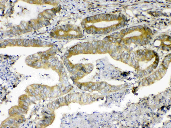
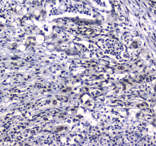
 IHC (Immunohiostchemistry)
(Figure 2. IHC analysis of ATF6 using anti- ATF6 antibody (AAA124587).ATF6 was detected in paraffin-embedded section of human mammary cancer tissues. Heat mediated antigen retrieval was performed in citrate buffer (pH6, epitope retrieval solution) for 20 mins. The tissue section was blocked with 10% goat serum. The tissue section was then incubated with 1ug/ml rabbit anti- ATF6 Antibody (AAA124587) overnight at 4 degree C. Biotinylated goat anti-rabbit IgG was used as secondary antibody and incubated for 30 minutes at 37 degree C. The tissue section was developed using Strepavidin-Biotin-Complex (SABC) with DAB as the chromogen.)
IHC (Immunohiostchemistry)
(Figure 2. IHC analysis of ATF6 using anti- ATF6 antibody (AAA124587).ATF6 was detected in paraffin-embedded section of human mammary cancer tissues. Heat mediated antigen retrieval was performed in citrate buffer (pH6, epitope retrieval solution) for 20 mins. The tissue section was blocked with 10% goat serum. The tissue section was then incubated with 1ug/ml rabbit anti- ATF6 Antibody (AAA124587) overnight at 4 degree C. Biotinylated goat anti-rabbit IgG was used as secondary antibody and incubated for 30 minutes at 37 degree C. The tissue section was developed using Strepavidin-Biotin-Complex (SABC) with DAB as the chromogen.)
ATF6, Polyclonal Antibody (Cat# AAA124587)
Full Name
Anti-ATF6 Picoband Antibody
Gene Names
ATF6; ACHM7; ATF6A
Reactivity
Human, Mouse, Rat
No cross reactivity with other proteins
No cross reactivity with other proteins
Applications
Western Blot, Immunohistochemistry
Purity
Immunogen affinity purified
Pricing
 IHC (Immunohistochemisry)
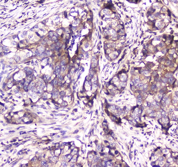
(Figure 3. IHC analysis of Adenylosuccinate Lyase using anti-Adenylosuccinate Lyase antibody (AAA124591).Adenylosuccinate Lyase was detected in paraffin-embedded section of human mammary cancer tissue. Heat mediated antigen retrieval was performed in citrate buffer (pH6, epitope retrieval solution) for 20 mins. The tissue section was blocked with 10% goat serum. The tissue section was then incubated with 1ug/ml rabbit anti-Adenylosuccinate Lyase Antibody (AAA124591) overnight at 4 degree C. Biotinylated goat anti-rabbit IgG was used as secondary antibody and incubated for 30 minutes at 37 degree C. The tissue section was developed using Strepavidin-Biotin-Complex (SABC) with DAB as the chromogen.)
IHC (Immunohistochemisry)
(Figure 3. IHC analysis of Adenylosuccinate Lyase using anti-Adenylosuccinate Lyase antibody (AAA124591).Adenylosuccinate Lyase was detected in paraffin-embedded section of human mammary cancer tissue. Heat mediated antigen retrieval was performed in citrate buffer (pH6, epitope retrieval solution) for 20 mins. The tissue section was blocked with 10% goat serum. The tissue section was then incubated with 1ug/ml rabbit anti-Adenylosuccinate Lyase Antibody (AAA124591) overnight at 4 degree C. Biotinylated goat anti-rabbit IgG was used as secondary antibody and incubated for 30 minutes at 37 degree C. The tissue section was developed using Strepavidin-Biotin-Complex (SABC) with DAB as the chromogen.)
Adenylosuccinate Lyase, Polyclonal Antibody (Cat# AAA124591)
Full Name
Anti-Adenylosuccinate Lyase Picoband Antibody
Gene Names
ASL; ASAL
Reactivity
Human, Mouse, Rat
No cross reactivity with other proteins.
No cross reactivity with other proteins.
Applications
Western Blot, Immunohistochemistry
Purity
Immunogen affinity purified
Pricing
 FCM/FACS (Flow Cytometry)
(Figure 5. Flow Cytometry analysis of THP-1 cells using anti-CD163 antibody (AAA124594).Overlay histogram showing THP-1 cells stained with AAA124594 (Blue line).The cells were blocked with 10% normal goat serum. And then incubated with rabbit anti-CD163 Antibody (AAA124594,1ug/1x10^6 cells) for 30 min at 20 degree C. DyLight®488 conjugated goat anti-rabbit IgG (5-10ug/1x10^6 cells) was used as secondary antibody for 30 minutes at 20 degree C. Isotype control antibody (Green line) was rabbit IgG (1ug/1x106) used under the same conditions. Unlabelled sample (Red line) was also used as a control.)
FCM/FACS (Flow Cytometry)
(Figure 5. Flow Cytometry analysis of THP-1 cells using anti-CD163 antibody (AAA124594).Overlay histogram showing THP-1 cells stained with AAA124594 (Blue line).The cells were blocked with 10% normal goat serum. And then incubated with rabbit anti-CD163 Antibody (AAA124594,1ug/1x10^6 cells) for 30 min at 20 degree C. DyLight®488 conjugated goat anti-rabbit IgG (5-10ug/1x10^6 cells) was used as secondary antibody for 30 minutes at 20 degree C. Isotype control antibody (Green line) was rabbit IgG (1ug/1x106) used under the same conditions. Unlabelled sample (Red line) was also used as a control.)
CD163, Antibody (Cat# AAA124594)
Full Name
Anti-CD163 Picoband antibody
Gene Names
CD163; M130; MM130; SCARI1
Reactivity
Human, Mouse, Rat
Applications
Flow Cytometry, Immunocytochemistry, Immunohistochemistry, Immunohistochemistry, Western Blot
Purity
Immunogen affinity purified
Pricing
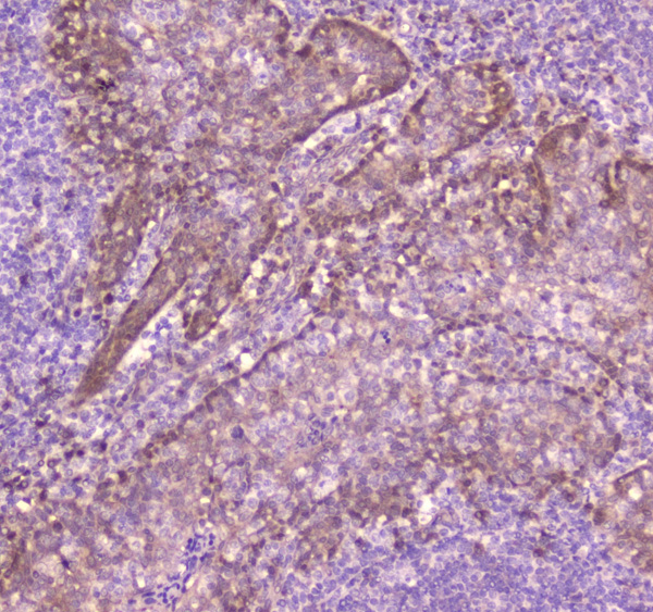
 IHC (Immunohistochemisry)
(Figure 3. IHC analysis of PF4 using anti-PF4 antibody (AAA124596).PF4 was detected in paraffin-embedded section of rat spleen tissue. Heat mediated antigen retrieval was performed in citrate buffer (pH6, epitope retrieval solution) for 20 mins. The tissue section was blocked with 10% goat serum. The tissue section was then incubated with 1ug/ml rabbit anti-PF4 Antibody (AAA124596) overnight at 4 degree C. Biotinylated goat anti-rabbit IgG was used as secondary antibody and incubated for 30 minutes at 37 degree C. The tissue section was developed using Strepavidin-Biotin-Complex (SABC) with DAB as the chromogen.)
IHC (Immunohistochemisry)
(Figure 3. IHC analysis of PF4 using anti-PF4 antibody (AAA124596).PF4 was detected in paraffin-embedded section of rat spleen tissue. Heat mediated antigen retrieval was performed in citrate buffer (pH6, epitope retrieval solution) for 20 mins. The tissue section was blocked with 10% goat serum. The tissue section was then incubated with 1ug/ml rabbit anti-PF4 Antibody (AAA124596) overnight at 4 degree C. Biotinylated goat anti-rabbit IgG was used as secondary antibody and incubated for 30 minutes at 37 degree C. The tissue section was developed using Strepavidin-Biotin-Complex (SABC) with DAB as the chromogen.)
PF4/Cxcl4, Polyclonal Antibody (Cat# AAA124596)
Full Name
Anti-PF4/Cxcl4 Picoband antibody
Gene Names
Pf4; PF-4; Pf4a; Cxcl4; RATPF4A
Reactivity
Mouse, Rat
No cross reactivity with other proteins.
No cross reactivity with other proteins.
Applications
Western Blot, Immunohistochemistry
Pricing
 IHC (Immunohiostchemistry)
(Figure 2. IHC analysis of GFI1 using anti-GFI1 antibody (AAA124598).GFI1 was detected in paraffin-embedded section of human rectal cancer tissue. Heat mediated antigen retrieval was performed in citrate buffer (pH6, epitope retrieval solution) for 20 mins. The tissue section was blocked with 10% goat serum. The tissue section was then incubated with 1ug/ml rabbit anti-GFI1 Antibody (AAA124598) overnight at 4 degree C. Biotinylated goat anti-rabbit IgG was used as secondary antibody and incubated for 30 minutes at 37 degree C. The tissue section was developed using Strepavidin-Biotin-Complex (SABC) with DAB as the chromogen.)
IHC (Immunohiostchemistry)
(Figure 2. IHC analysis of GFI1 using anti-GFI1 antibody (AAA124598).GFI1 was detected in paraffin-embedded section of human rectal cancer tissue. Heat mediated antigen retrieval was performed in citrate buffer (pH6, epitope retrieval solution) for 20 mins. The tissue section was blocked with 10% goat serum. The tissue section was then incubated with 1ug/ml rabbit anti-GFI1 Antibody (AAA124598) overnight at 4 degree C. Biotinylated goat anti-rabbit IgG was used as secondary antibody and incubated for 30 minutes at 37 degree C. The tissue section was developed using Strepavidin-Biotin-Complex (SABC) with DAB as the chromogen.)
GFI1, Polyclonal Antibody (Cat# AAA124598)
Full Name
Anti-GFI1 Picoband antibody
Gene Names
GFI1; SCN2; GFI-1; GFI1A; ZNF163
Reactivity
Human, Mouse, Rat
No cross reactivity with other proteins.
No cross reactivity with other proteins.
Applications
Immunohistochemistry
Pricing
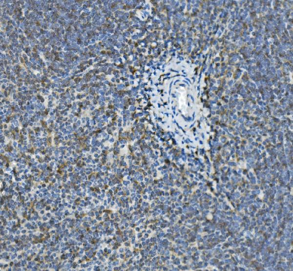
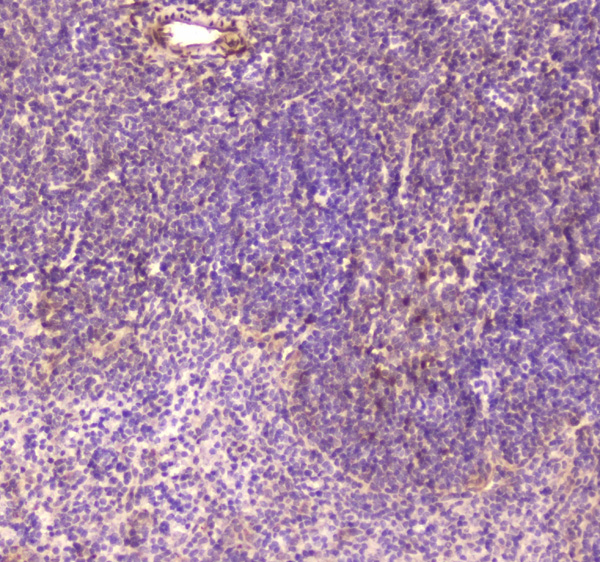
 IHC (Immunohistochemisry)
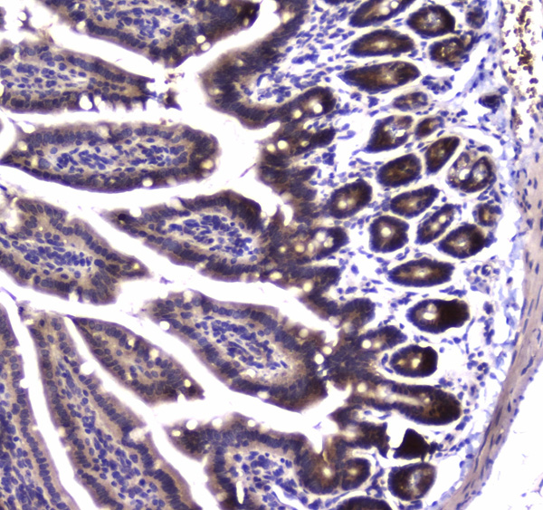
(Figure 3. IHC analysis of CD59 using anti-CD59 antibody (AAA124602).CD59 was detected in paraffin-embedded section of mouse thymus tissue. Heat mediated antigen retrieval was performed in citrate buffer (pH6, epitope retrieval solution) for 20 mins. The tissue section was blocked with 10% goat serum. The tissue section was then incubated with 1ug/ml rabbit anti-CD59 Antibody (AAA124602) overnight at 4 degree C. Biotinylated goat anti-rabbit IgG was used as secondary antibody and incubated for 30 minutes at 37 degree C. The tissue section was developed using Strepavidin-Biotin-Complex (SABC) with DAB as the chromogen.)
IHC (Immunohistochemisry)
(Figure 3. IHC analysis of CD59 using anti-CD59 antibody (AAA124602).CD59 was detected in paraffin-embedded section of mouse thymus tissue. Heat mediated antigen retrieval was performed in citrate buffer (pH6, epitope retrieval solution) for 20 mins. The tissue section was blocked with 10% goat serum. The tissue section was then incubated with 1ug/ml rabbit anti-CD59 Antibody (AAA124602) overnight at 4 degree C. Biotinylated goat anti-rabbit IgG was used as secondary antibody and incubated for 30 minutes at 37 degree C. The tissue section was developed using Strepavidin-Biotin-Complex (SABC) with DAB as the chromogen.)
CD59, Polyclonal Antibody (Cat# AAA124602)
Full Name
Anti-CD59 Picoband antibody
Gene Names
Cd59a; Cd59; AA987121; protectin
Reactivity
Mouse, Rat
No cross reactivity with other proteins.
No cross reactivity with other proteins.
Applications
Western Blot, Immunohistochemistry
Pricing
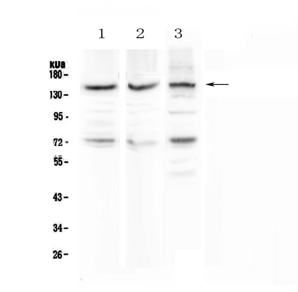
 WB (Western Blot)
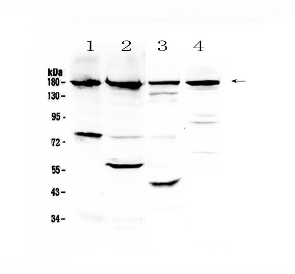
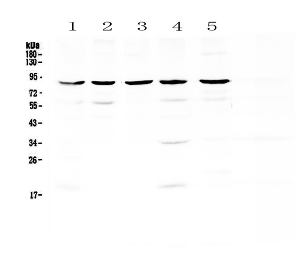
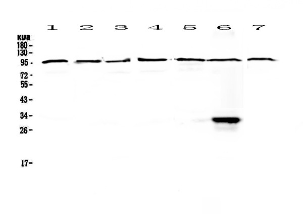
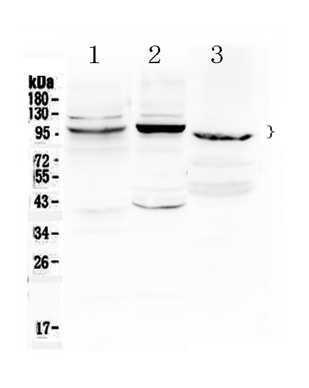
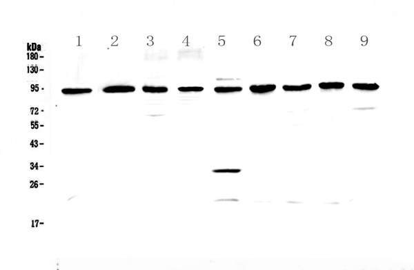
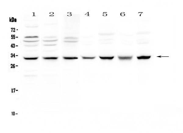
(Figure 2. Western blot analysis of GNS using anti-GNS antibody (AAA124609).Electrophoresis was performed on a 5-20% SDS-PAGE gel at 70V (Stacking gel) / 90V (Resolving gel) for 2-3 hours. The sample well of each lane was loaded with 50ug of sample under reducing conditions.Lane 1: rat lung tissue lysates,Lane 2: rat kidney tissue lysates,Lane 3: rat testis tissue lysates,Lane 4: rat PC-12 whole cell lysates,Lane 5: mouse lung tissue lysates,Lane 6: mouse kidney tissue lysates,Lane 7: mouse testis tissue lysates,Lane 8: mouse spleen tissue lysates,Lane 9: mouse thymus tissue lysates.After Electrophoresis, proteins were transferred to a Nitrocellulose membrane at 150mA for 50-90 minutes. Blocked the membrane with 5% Non-fat Milk/ TBS for 1.5 hour at RT. The membrane was incubated with rabbit anti-GNS antigen affinity purified polyclonal antibody at 0.5ug/mL overnight at 4 degree C, then washed with TBS-0.1%Tween 3 times with 5 minutes each and probed with a goat anti-rabbit IgG-HRP secondary antibody at a dilution of 1:10000 for 1.5 hour at RT. The signal is developed using an Enhanced Chemiluminescent detection (ECL) kit with Tanon 5200 system. A specific band was detected for GNS at approximately 90KD. The expected band size for GNS is at 62KD.)
WB (Western Blot)
(Figure 2. Western blot analysis of GNS using anti-GNS antibody (AAA124609).Electrophoresis was performed on a 5-20% SDS-PAGE gel at 70V (Stacking gel) / 90V (Resolving gel) for 2-3 hours. The sample well of each lane was loaded with 50ug of sample under reducing conditions.Lane 1: rat lung tissue lysates,Lane 2: rat kidney tissue lysates,Lane 3: rat testis tissue lysates,Lane 4: rat PC-12 whole cell lysates,Lane 5: mouse lung tissue lysates,Lane 6: mouse kidney tissue lysates,Lane 7: mouse testis tissue lysates,Lane 8: mouse spleen tissue lysates,Lane 9: mouse thymus tissue lysates.After Electrophoresis, proteins were transferred to a Nitrocellulose membrane at 150mA for 50-90 minutes. Blocked the membrane with 5% Non-fat Milk/ TBS for 1.5 hour at RT. The membrane was incubated with rabbit anti-GNS antigen affinity purified polyclonal antibody at 0.5ug/mL overnight at 4 degree C, then washed with TBS-0.1%Tween 3 times with 5 minutes each and probed with a goat anti-rabbit IgG-HRP secondary antibody at a dilution of 1:10000 for 1.5 hour at RT. The signal is developed using an Enhanced Chemiluminescent detection (ECL) kit with Tanon 5200 system. A specific band was detected for GNS at approximately 90KD. The expected band size for GNS is at 62KD.)
GNS, Polyclonal Antibody (Cat# AAA124609)
Full Name
Anti-GNS Picoband antibody
Gene Names
GNS; G6S
Reactivity
Human, Mouse, Rat
No cross reactivity with other proteins.
No cross reactivity with other proteins.
Applications
Western Blot
Pricing

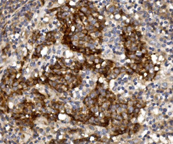
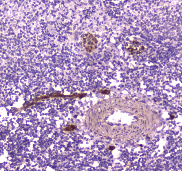
 IHC (Immunohiostchemistry)
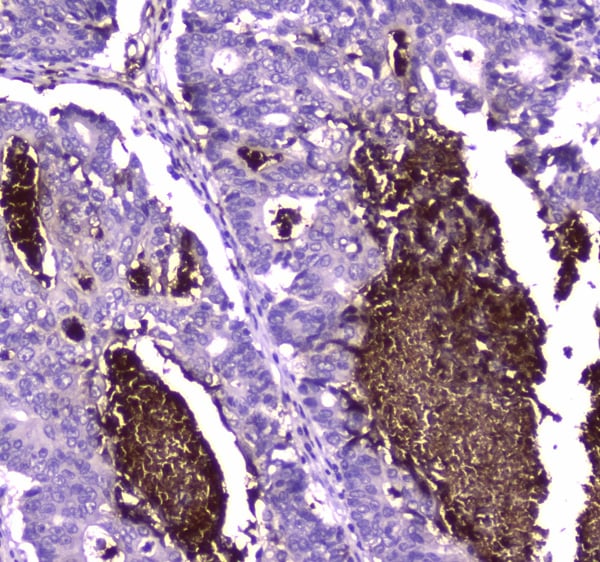
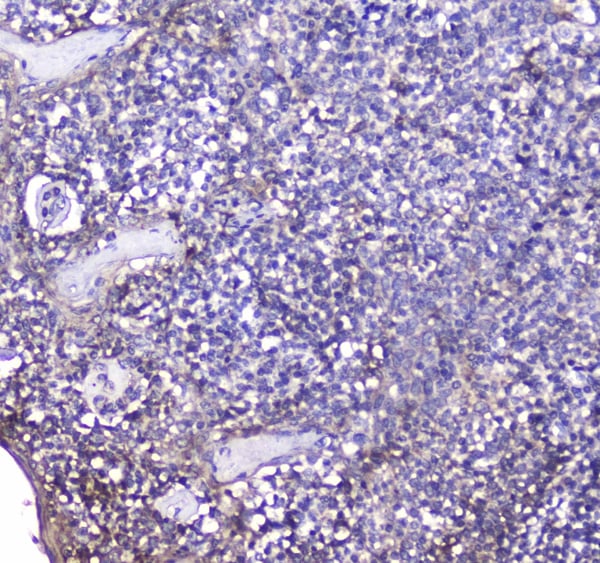
(Figure 2. IHC analysis of BCMA using anti-BCMA antibody (AAA124612).BCMA was detected in paraffin-embedded section of human tonsil tissue. Heat mediated antigen retrieval was performed in citrate buffer (pH6, epitope retrieval solution) for 20 mins. The tissue section was blocked with 10% goat serum. The tissue section was then incubated with 2ug/ml rabbit anti-BCMA Antibody (AAA124612) overnight at 4 degree C. Biotinylated goat anti-rabbit IgG was used as secondary antibody and incubated for 30 minutes at 37 degree C. The tissue section was developed using Strepavidin-Biotin-Complex (SABC) with DAB as the chromogen.)
IHC (Immunohiostchemistry)
(Figure 2. IHC analysis of BCMA using anti-BCMA antibody (AAA124612).BCMA was detected in paraffin-embedded section of human tonsil tissue. Heat mediated antigen retrieval was performed in citrate buffer (pH6, epitope retrieval solution) for 20 mins. The tissue section was blocked with 10% goat serum. The tissue section was then incubated with 2ug/ml rabbit anti-BCMA Antibody (AAA124612) overnight at 4 degree C. Biotinylated goat anti-rabbit IgG was used as secondary antibody and incubated for 30 minutes at 37 degree C. The tissue section was developed using Strepavidin-Biotin-Complex (SABC) with DAB as the chromogen.)
BCMA, Polyclonal Antibody (Cat# AAA124612)
Full Name
Anti-BCMA Picoband antibody
Gene Names
TNFRSF17; BCM; BCMA; CD269; TNFRSF13A
Reactivity
Human
No cross reactivity with other proteins.
No cross reactivity with other proteins.
Applications
Western Blot, Immunohistochemistry
Pricing
 FCM/FACS (Flow Cytometry)
(Figure 4. Flow Cytometry analysis of THP-1 cells using anti-DC-SIGN antibody (AAA124613).Overlay histogram showing THP-1 cells stained with AAA124613 (Blue line).The cells were blocked with 10% normal goat serum. And then incubated with rabbit anti-DC-SIGN Antibody (AAA124613,1ug/1x10^6 cells) for 30 min at 20 degree C. DyLight®488 conjugated goat anti-rabbit IgG (5-10ug/1x10^6 cells) was used as secondary antibody for 30 minutes at 20 degree C. Isotype control antibody (Green line) was rabbit IgG (1ug/1x106) used under the same conditions. Unlabelled sample (Red line) was also used as a control.)
FCM/FACS (Flow Cytometry)
(Figure 4. Flow Cytometry analysis of THP-1 cells using anti-DC-SIGN antibody (AAA124613).Overlay histogram showing THP-1 cells stained with AAA124613 (Blue line).The cells were blocked with 10% normal goat serum. And then incubated with rabbit anti-DC-SIGN Antibody (AAA124613,1ug/1x10^6 cells) for 30 min at 20 degree C. DyLight®488 conjugated goat anti-rabbit IgG (5-10ug/1x10^6 cells) was used as secondary antibody for 30 minutes at 20 degree C. Isotype control antibody (Green line) was rabbit IgG (1ug/1x106) used under the same conditions. Unlabelled sample (Red line) was also used as a control.)
DC-SIGN, Polyclonal Antibody (Cat# AAA124613)
Full Name
Anti-DC-SIGN Picoband antibody
Gene Names
CD209; CDSIGN; CLEC4L; DC-SIGN; DC-SIGN1
Reactivity
Human, Mouse, Rat
No cross reactivity with other proteins.
No cross reactivity with other proteins.
Applications
Western Blot, Immunohistochemistry
Pricing
 IHC (Immunohistochemistry)
(Figure 5. IHC analysis of RBP4 using anti-RBP4 antibody (AAA124617).RBP4 was detected in paraffin-embedded section of rat pancreas tissue. Heat mediated antigen retrieval was performed in citrate buffer (pH6, epitope retrieval solution) for 20 mins. The tissue section was blocked with 10% goat serum. The tissue section was then incubated with 1ug/ml rabbit anti-RBP4 Antibody (AAA124617) overnight at 4 degree C. Biotinylated goat anti-rabbit IgG was used as secondary antibody and incubated for 30 minutes at 37 degree C. The tissue section was developed using Strepavidin-Biotin-Complex (SABC) with DAB as the chromogen.)
IHC (Immunohistochemistry)
(Figure 5. IHC analysis of RBP4 using anti-RBP4 antibody (AAA124617).RBP4 was detected in paraffin-embedded section of rat pancreas tissue. Heat mediated antigen retrieval was performed in citrate buffer (pH6, epitope retrieval solution) for 20 mins. The tissue section was blocked with 10% goat serum. The tissue section was then incubated with 1ug/ml rabbit anti-RBP4 Antibody (AAA124617) overnight at 4 degree C. Biotinylated goat anti-rabbit IgG was used as secondary antibody and incubated for 30 minutes at 37 degree C. The tissue section was developed using Strepavidin-Biotin-Complex (SABC) with DAB as the chromogen.)
RBP4/Retinol Binding Protein 4, Polyclonal Antibody (Cat# AAA124617)
Full Name
Anti-RBP4/Retinol Binding Protein 4 Picoband Antibody
Gene Names
Rbp4; Rbp-4
Reactivity
Human, Mouse, Rat
No cross reactivity with other proteins.
No cross reactivity with other proteins.
Applications
Western Blot, Immunohistochemistry
Purity
Immunogen affinity purified
Pricing
 IHC (Immunohistochemistry)
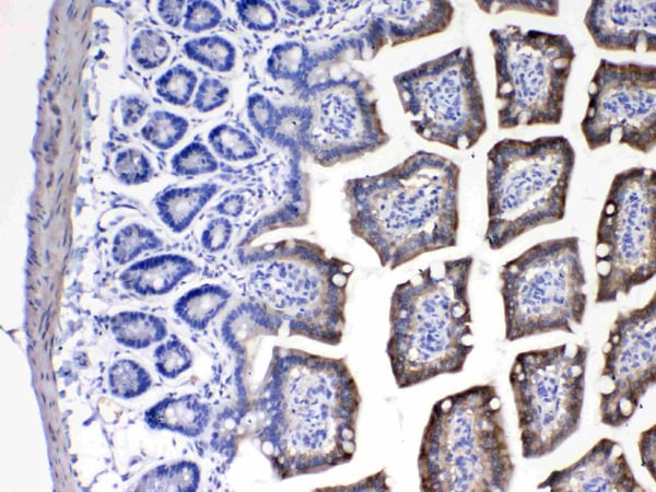
(Figure 4. IHC analysis of SDHB using anti-SDHB antibody (AAA124618).SDHB was detected in paraffin-embedded section of rat small intestine tissue. Heat mediated antigen retrieval was performed in citrate buffer (pH6, epitope retrieval solution) for 20 mins. The tissue section was blocked with 10% goat serum. The tissue section was then incubated with 1ug/ml rabbit anti-SDHB Antibody (AAA124618) overnight at 4 degree C. Biotinylated goat anti-rabbit IgG was used as secondary antibody and incubated for 30 minutes at 37 degree C. The tissue section was developed using Strepavidin-Biotin-Complex (SABC) with DAB as the chromogen.)
IHC (Immunohistochemistry)
(Figure 4. IHC analysis of SDHB using anti-SDHB antibody (AAA124618).SDHB was detected in paraffin-embedded section of rat small intestine tissue. Heat mediated antigen retrieval was performed in citrate buffer (pH6, epitope retrieval solution) for 20 mins. The tissue section was blocked with 10% goat serum. The tissue section was then incubated with 1ug/ml rabbit anti-SDHB Antibody (AAA124618) overnight at 4 degree C. Biotinylated goat anti-rabbit IgG was used as secondary antibody and incubated for 30 minutes at 37 degree C. The tissue section was developed using Strepavidin-Biotin-Complex (SABC) with DAB as the chromogen.)
SDHB, Polyclonal Antibody (Cat# AAA124618)
Full Name
Anti-SDHB Picoband antibody
Gene Names
SDHB; IP; SDH; CWS2; PGL4; SDH1; SDH2; SDHIP
Reactivity
Human, Mouse, Rat
No cross reactivity with other proteins.
No cross reactivity with other proteins.
Applications
Western Blot, Immunohistochemistry
Pricing
 IHC (Immunohiostchemistry)
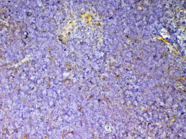
(Figure 2. IHC analysis of IL12B using anti-IL12B antibody (AAA124625).IL12B was detected in paraffin-embedded section of rat spleen tissue. Heat mediated antigen retrieval was performed in citrate buffer (pH6, epitope retrieval solution) for 20 mins. The tissue section was blocked with 10% goat serum. The tissue section was then incubated with 1ug/ml rabbit anti-IL12B Antibody (AAA124625) overnight at 4 degree C. Biotinylated goat anti-rabbit IgG was used as secondary antibody and incubated for 30 minutes at 37 degree C. The tissue section was developed using Strepavidin-Biotin-Complex (SABC) with DAB as the chromogen.)
IHC (Immunohiostchemistry)
(Figure 2. IHC analysis of IL12B using anti-IL12B antibody (AAA124625).IL12B was detected in paraffin-embedded section of rat spleen tissue. Heat mediated antigen retrieval was performed in citrate buffer (pH6, epitope retrieval solution) for 20 mins. The tissue section was blocked with 10% goat serum. The tissue section was then incubated with 1ug/ml rabbit anti-IL12B Antibody (AAA124625) overnight at 4 degree C. Biotinylated goat anti-rabbit IgG was used as secondary antibody and incubated for 30 minutes at 37 degree C. The tissue section was developed using Strepavidin-Biotin-Complex (SABC) with DAB as the chromogen.)
IL12B/Il 12, Polyclonal Antibody (Cat# AAA124625)
Full Name
Anti-IL12B/Il 12 Picoband Antibody
Gene Names
Il12b; Il12
Reactivity
Mouse, Rat
No cross reactivity with other proteins.
No cross reactivity with other proteins.
Applications
Western Blot, Immunohistochemistry
Purity
Immunogen affinity purified
Pricing
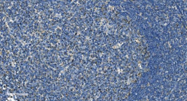
 IHC (Immunohistochemisry)
(Figure 3. IHC analysis of VCAM1 using anti-VCAM1 antibody (AAA124628).VCAM1 was detected in paraffin-embedded section of rat spleen tissue. Heat mediated antigen retrieval was performed in citrate buffer (pH6, epitope retrieval solution) for 20 mins. The tissue section was blocked with 10% goat serum. The tissue section was then incubated with 1ug/ml rabbit anti-VCAM1 Antibody (AAA124628) overnight at 4 degree C. Biotinylated goat anti-rabbit IgG was used as secondary antibody and incubated for 30 minutes at 37 degree C. The tissue section was developed using Strepavidin-Biotin-Complex (SABC) with DAB as the chromogen.)
IHC (Immunohistochemisry)
(Figure 3. IHC analysis of VCAM1 using anti-VCAM1 antibody (AAA124628).VCAM1 was detected in paraffin-embedded section of rat spleen tissue. Heat mediated antigen retrieval was performed in citrate buffer (pH6, epitope retrieval solution) for 20 mins. The tissue section was blocked with 10% goat serum. The tissue section was then incubated with 1ug/ml rabbit anti-VCAM1 Antibody (AAA124628) overnight at 4 degree C. Biotinylated goat anti-rabbit IgG was used as secondary antibody and incubated for 30 minutes at 37 degree C. The tissue section was developed using Strepavidin-Biotin-Complex (SABC) with DAB as the chromogen.)
VCAM1/Cd106, Polyclonal Antibody (Cat# AAA124628)
Full Name
Anti-VCAM1/Cd106 Picoband antibody
Gene Names
Vcam1; VCAM1B
Reactivity
Mouse, Rat
No cross reactivity with other proteins.
No cross reactivity with other proteins.
Applications
Western Blot, Immunohistochemistry
Pricing
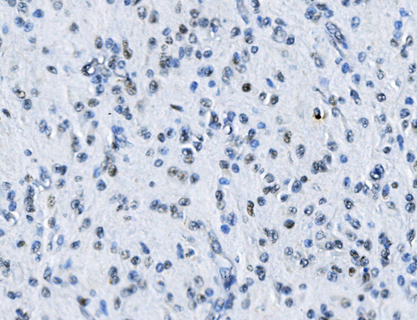
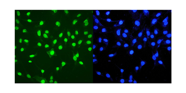
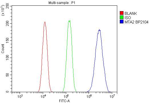
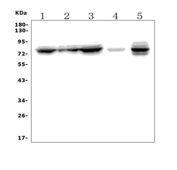 WB (Western Blot)
(Figure 1. Western blot analysis of MTA2 using anti-MTA2 antibody.)
WB (Western Blot)
(Figure 1. Western blot analysis of MTA2 using anti-MTA2 antibody.)
MTA2/PID, Polyclonal Antibody (Cat# AAA125463)
Full Name
Anti-MTA2/PID Antibody
Gene Names
MTA2; PID; MTA1L1
Reactivity
Human, Mouse, Rat
Applications
Direct ELISA, Flow Cytometry, Immunofluorescence, Immunocytochemistry, Immunohistochemistry, Western Blot
Purity
Immunogen affinity purified.
Pricing
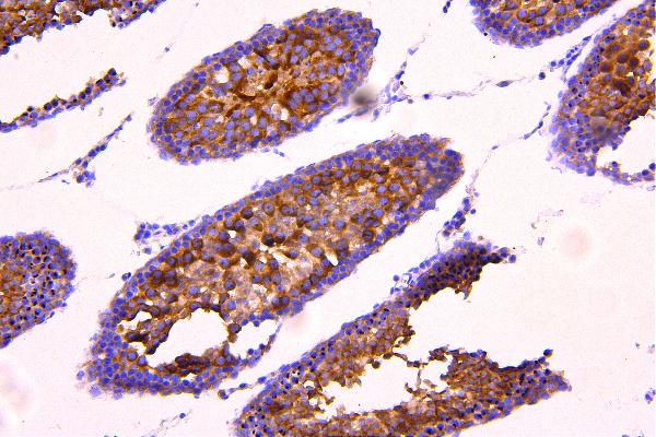
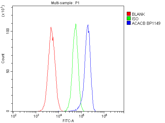
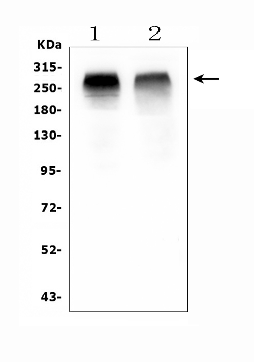 WB (Western Blot)
Acetyl Coenzyme A Carboxylase/ACACB, Polyclonal Antibody (Cat# AAA125469)
Full Name
Anti-Acetyl Coenzyme A Carboxylase/ACACB Antibody
Gene Names
ACACB; ACC2; ACCB; HACC275
Reactivity
Human, Mouse, Rat
Applications
Flow Cytometry, Immunohistochemistry, Western Blot
Purity
Immunogen affinity purified.
Pricing
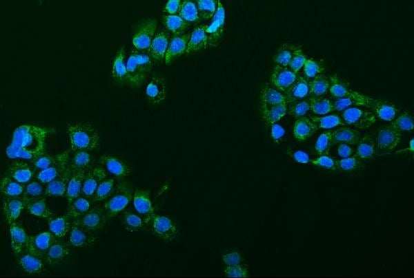
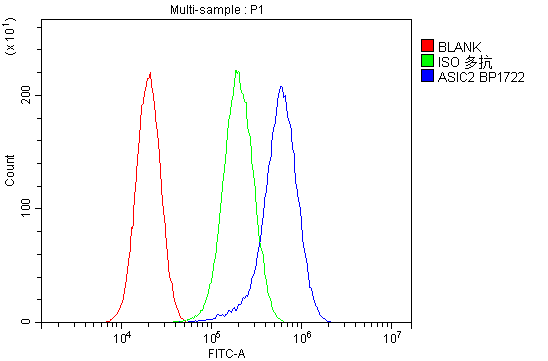
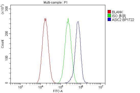
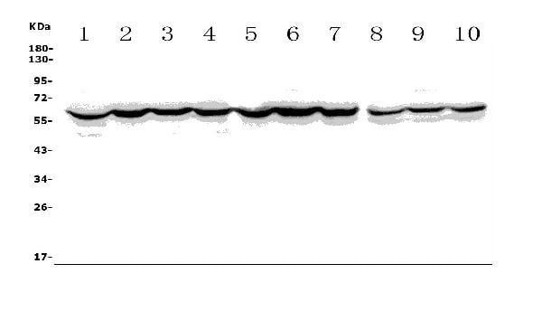 WB (Western Blot)
(Figure 1. Western blot analysis of ASIC2 using anti-ASIC2 antibody.)
WB (Western Blot)
(Figure 1. Western blot analysis of ASIC2 using anti-ASIC2 antibody.)
ACCN1/ASIC2, Polyclonal Antibody (Cat# AAA125471)
Full Name
Anti-ACCN1/ASIC2 Antibody
Gene Names
ASIC2; ACCN; BNC1; MDEG; ACCN1; BNaC1; ASIC2a; hBNaC1
Reactivity
Human, Mouse, Rat
Applications
Direct ELISA, Flow Cytometry, Immunofluorescence, Immunocytochemistry, Western Blot
Purity
Immunogen affinity purified.
Pricing
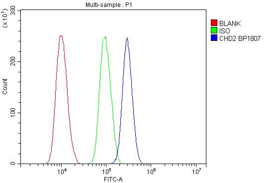
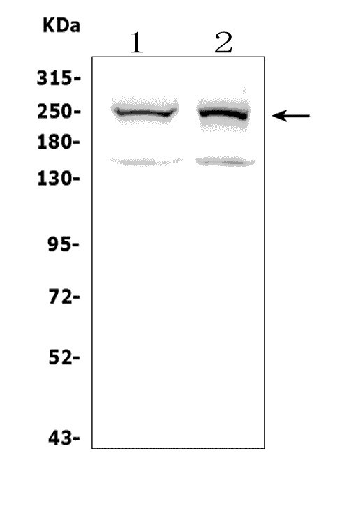 WB (Western Blot)
(Figure 1. Western blot analysis of CHD2 using anti-CHD2 antibody.)
WB (Western Blot)
(Figure 1. Western blot analysis of CHD2 using anti-CHD2 antibody.)
CHD2, Polyclonal Antibody (Cat# AAA125472)
Full Name
Anti-CHD2 Antibody
Gene Names
CHD2; EEOC
Reactivity
Human
Applications
Flow Cytometry, Western Blot
Purity
Immunogen affinity purified.
Pricing
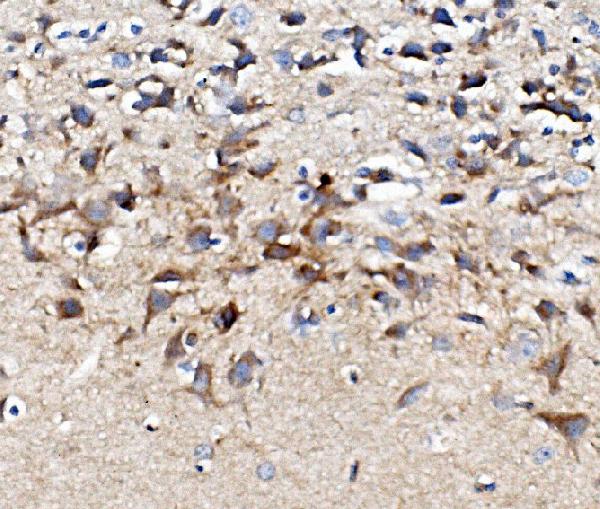
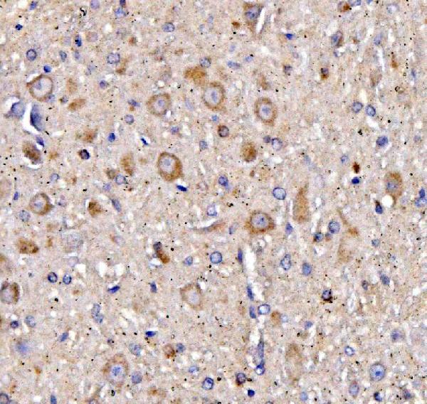
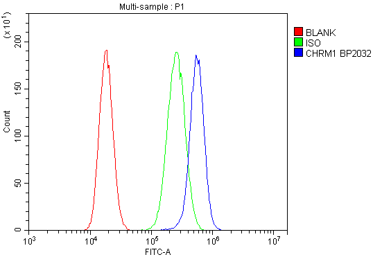
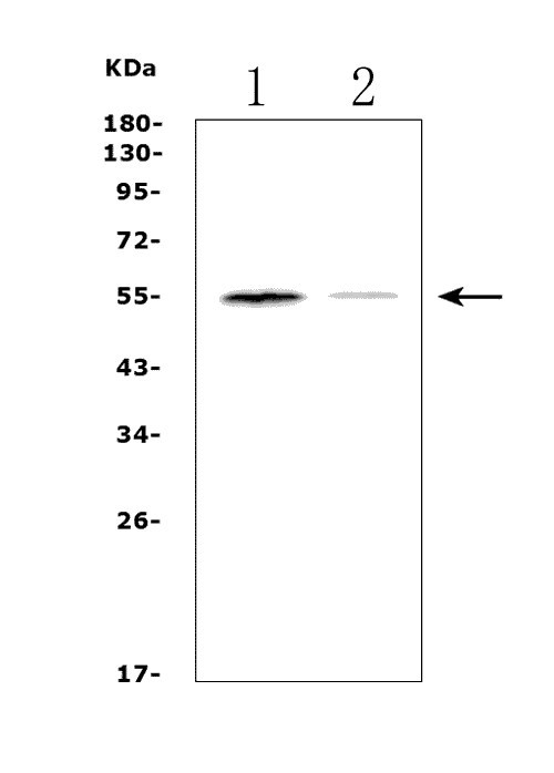 WB (Western Blot)
(Figure 1. Western blot analysis of CHRM1 using anti-CHRM1 antibody.)
WB (Western Blot)
(Figure 1. Western blot analysis of CHRM1 using anti-CHRM1 antibody.)
Muscarinic Acetylcholine Receptor 1/CHRM1, Polyclonal Antibody (Cat# AAA125473)
Full Name
Anti-Muscarinic Acetylcholine Receptor 1/CHRM1 Antibody
Gene Names
CHRM1; M1; HM1; M1R
Reactivity
Human, Mouse, Rat
Applications
Flow Cytometry, Immunohistochemistry, Western Blot
Purity
Immunogen affinity purified.
Pricing