A Beginner's Guide to Immunohistochemistry
 Cynthia
11 min read
Cynthia
11 min read
In this Article
- What Is Immunohistochemistry and How Does It Work?
- What Is Immunohistochemistry Used For?
- What Are the Main Steps in an IHC Workflow?
- Factors to Consider When Choosing an Antibody for IHC
- Why Is Blocking Important in IHC?
- Which IHC Detection Method Should You Use?
- Which Controls Are Important for Reliable IHC Results?
- How Do You Troubleshoot Common IHC Problems?


All of the products listed in AAA Biotech’s catalog are strictly for research-use only (RUO).
Key Takeaways
- Immunohistochemistry (IHC) uses antibodies to detect and locate specific proteins within intact tissue sections, revealing both presence and precise location.
- The core workflow includes tissue fixation, sectioning, antigen retrieval, blocking, antibody incubation, detection, and counterstaining.
- Choosing a validated, IHC-specific antibody is one of the most important factors in achieving reliable results.
- Positive, negative, and no-primary controls confirm whether observed staining reflects true target detection.
- Common issues include weak staining, high background, and nonspecific signal, each traceable to a specific workflow variable.
- Modern IHC increasingly incorporates multiplexing, digital pathology, and spatial biomarker analysis for deeper tissue insight.
Immunohistochemistry (IHC) is a technique that uses antibodies to detect and visualize specific proteins directly within tissue sections. By combining antigen-antibody binding with a detectable label, IHC reveals not just whether a protein is present, but precisely where it sits within a cell or tissue; information that's central to both research and pathology.
If you're new to the bench, immunohistochemistry basics can feel like a lot to absorb at once: fixation chemistry, retrieval buffers, antibody dilutions, detection chemistries. But the underlying logic is straightforward. IHC lets you see where a protein is expressed, not only confirm that it exists. That distinction matters. A receptor expressed on the cell surface tells a different biological story than the same receptor trapped in the cytoplasm, and only a tissue-based, antibody-driven method can capture that difference while preserving the surrounding architecture.
This guide walks through how IHC works, where it's applied, the full staining workflow, and the antibody-selection and troubleshooting knowledge every beginner needs before running their first experiment.
What Is Immunohistochemistry and How Does It Work?
Immunohistochemistry combines principles of immunology and histology. It uses the specificity of antibodies to identify particular antigens while retaining the structural context of the tissue.
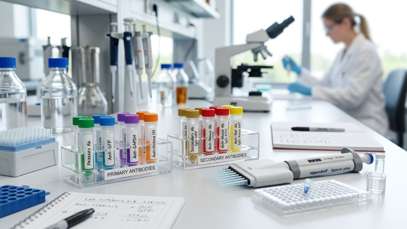
The basic principle is straightforward. A primary antibody recognizes and binds to a specific target protein, or antigen, in a tissue section. That antibody can either carry a detectable label itself or be recognized by a labeled secondary antibody. The resulting signal shows where the target is located.
This is why IHC provides information beyond simply determining whether a protein is present. It can show which cells contain the protein, where it is located within those cells, and how its distribution changes across a tissue.
In practical terms, the immunohistochemistry explanation comes down to four stages:
Target antigen → antibody binding → signal generation → microscopic visualization
The objective is to make protein detection more specific while maintaining the familiar tissue morphology seen through light microscopy. In this way, immunohistology brought together immunology and histology and helped make tissue-based diagnosis more objective while retaining conventional morphological assessment.
What Is Immunohistochemistry Used For?
IHC is used when researchers need to identify specific proteins within their natural tissue environment. Unlike techniques that analyze extracted material, IHC retains information about tissue architecture and cellular location.
Common applications include:
- Protein localization and expression studies
- Biomarker research
- Tissue characterization
- Cancer and disease research
- Cell and tissue biology
- Pathology investigations
- Clinical diagnostic applications
In immunohistochemistry pathology, staining patterns can provide information that contributes to tissue classification and disease assessment. However, clinical interpretation requires appropriately validated assays, controls, and established diagnostic criteria. Research staining should not automatically be treated as a clinical diagnostic result.
What Are the Main Steps in an IHC Workflow?
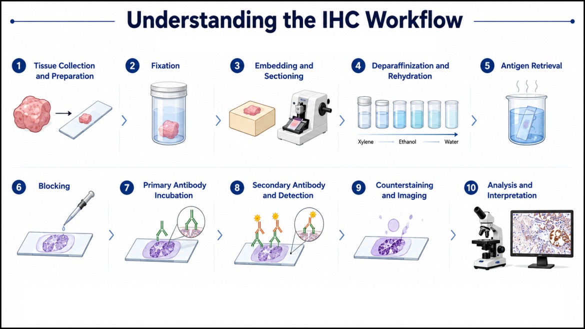
The immunohistochemistry process involves several connected stages. A problem at an early stage can affect everything that follows.
- Tissue Collection and Preparation:
Everything starts with tissue selection and careful sample handling. Delays between collection and fixation allow autolysis to begin degrading both morphology and antigenicity, so preserving tissue integrity from the moment of collection is foundational to reliable results downstream.
- Fixation:
Fixation halts tissue degradation and locks cellular architecture in place, typically using formaldehyde-based fixatives that create protein cross-links. This preservation comes at a cost: those same cross-links can mask the very epitopes your antibody needs to bind, and over-fixation makes antigen accessibility measurably worse.
- Embedding and Sectioning:
Tissue is either embedded in paraffin (FFPE) or frozen in OCT compound, each with trade-offs. Paraffin-embedded tissue offers superior long-term morphology and storage stability; frozen sections often preserve antigen conformation more faithfully but are less durable for archival use. Consistent section thickness across your slides is essential for reproducible comparisons.
- Deparaffinization and Rehydration:
FFPE sections must be cleared of paraffin (typically via xylene) and rehydrated through a graded alcohol series before staining can proceed; skipping or rushing this step compromises everything that follows.
- Antigen Retrieval:
Because fixation masks epitopes, antigen retrieval is usually required to unmask them before staining. Heat-induced epitope retrieval (HIER) and enzymatic (proteolytic) retrieval are the two standard approaches, and the right choice depends heavily on the specific target and antibody in use; there's no universal setting that works for every protein.
- Blocking:
Before primary antibody incubation, a blocking step reduces nonspecific binding sites across the tissue.
- Primary Antibody Incubation:
This is target-specific binding in action, with antibody concentration, incubation time, and temperature directly shaping signal strength and specificity.
- Secondary Antibody and Detection:
In indirect detection, a secondary antibody targets the primary, carrying either an enzymatic (chromogenic) or fluorescent label that produces the final visible signal.
- Counterstaining and Imaging:
A counterstain, commonly hematoxylin for chromogenic work or DAPI for fluorescence, provides structural context around your target signal. Consistent imaging conditions across a sample set are what make comparisons between slides meaningful rather than misleading.
Important Note! These immunohistochemistry steps should be viewed as an interconnected workflow rather than isolated tasks. The IHC staining procedure may require optimization depending on the tissue and target.
Further Reading: For a peer-reviewed deep dive into IHC methodology, reagents, and standardization, see "Principles and Methods of Immunohistochemistry" by Ramos-Vara, published in Methods in Molecular Biology and indexed on PubMed.
Factors to Consider When Choosing an Antibody for IHC
More IHC experiments go wrong at the antibody-selection stage than at any other point. Pick the right one, and the rest of the workflow has a real shot at working. Get it wrong, and no amount of optimization downstream will fully make up for it. Here's what to check before you commit tissue and reagents to a new antibody:
| Factor | What to Check |
|---|---|
| Target and epitope specificity | Does it recognize the correct protein and epitope, without cross-reacting with related proteins? |
| IHC-specific validation | An antibody that works well in Western blot won't necessarily work in IHC; WB detects a denatured, linearized protein on a membrane, while IHC needs the antibody to find its target inside intact, fixed tissue. Look for validation data run specifically for IHC, not borrowed from another application. |
| Species and tissue compatibility | The host species of your primary antibody dictates which secondaries and detection systems you can pair it with, so lock this in early. |
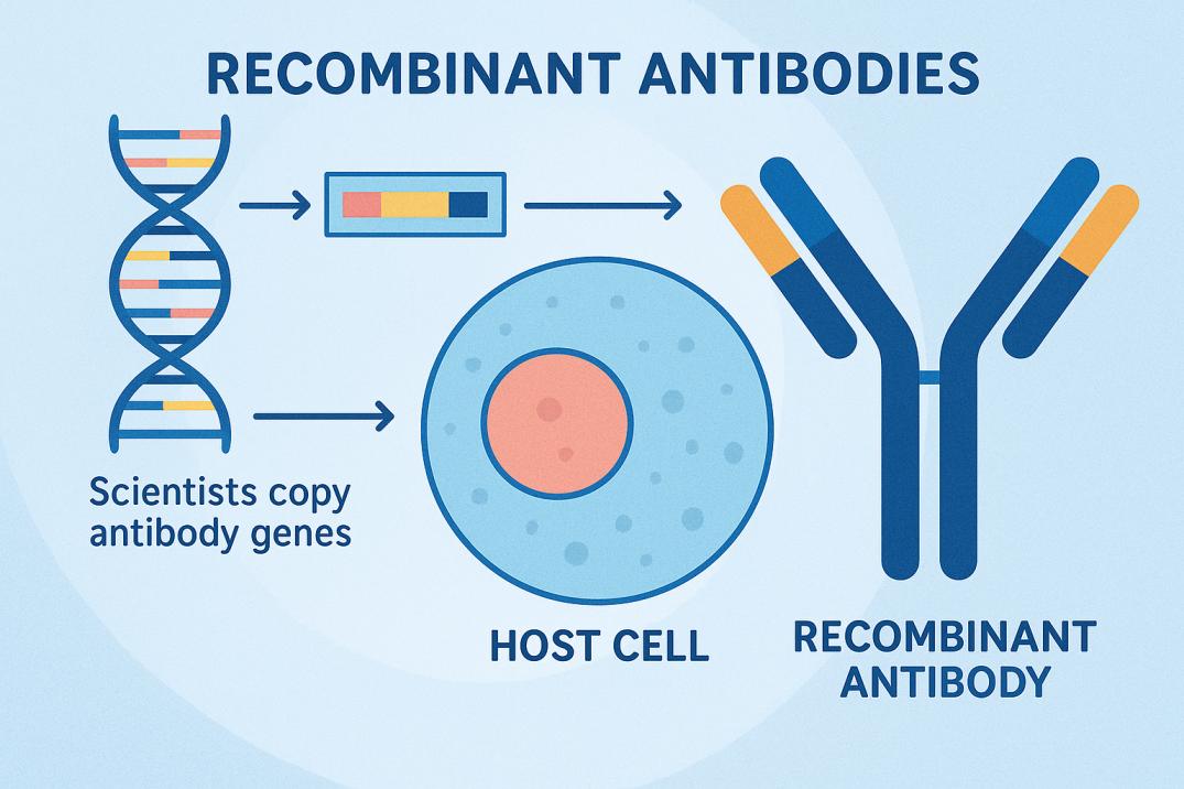
| Clonality | Monoclonals bind a single epitope; precise, and good for discriminating between closely related targets. Polyclonals bind multiple epitopes on the same target, often giving a stronger signal, which helps with low-abundance proteins. Recombinant antibodies bring an added layer of batch-to-batch consistency, since they come from a defined genetic sequence rather than an animal-derived source. |
| Expected subcellular localization | Should your target show up nuclear, cytoplasmic, or membranous? Knowing this ahead of time helps you tell real signal from an artifact. |
| Recommended dilution range | Start from the manufacturer's or published range, then optimize from there. |
| Available controls | Make sure appropriate positive and negative controls exist for your target before you start. |
Looking for IHC-Validated Antibodies?
Our extensive range of highly validated and characterized monoclonal antibodies and purified polyclonal antibodies covers both approaches, alongside high-affinity recombinant antibodies validated for IHC and related applications.
Why Is Blocking Important in IHC?
Tissue contains abundant nonspecific binding sites: charged surfaces, Fc receptors, and endogenous proteins that antibodies can bind indiscriminately, independent of true antigen recognition. A blocking buffer in immunohistochemistry, typically bovine serum albumin (BSA) or serum matched to the host species of the secondary antibody, occupies these nonspecific sites before the primary antibody is applied.
Blocking directly interacts with antibody concentration and background: too little blocking and background rises; too much and true signal can be dampened. Blocking conditions frequently require optimization for different tissue types and detection systems, so treat your first attempt as a starting point rather than a fixed setting.
Which IHC Detection Method Should You Use?
Detection strategy is one of the core immunohistochemistry techniques beginners need to understand early, since it shapes both workflow complexity and signal strength.
- Direct vs. Indirect Detection:
In direct detection, the primary antibody itself carries the label, producing a shorter workflow with fewer incubation steps. Indirect detection uses an unlabeled primary antibody paired with a labeled secondary, which introduces signal amplification and greater flexibility, at the cost of an additional step and a slightly higher risk of cross-reactivity.
Feature Direct Detection Indirect Detection Primary antibody Labeled Unlabeled Secondary antibody Not required Required Signal amplification Lower Higher Workflow Fewer steps More steps Flexibility Lower Higher - Chromogenic vs. Fluorescent Detection:
Chromogenic detection (commonly using DAB) produces a permanent, brightfield-visible signal well suited to standard microscopy and long-term slide storage; a dependable choice for most diagnostic and routine research workflows.
Fluorescent detection offers stronger multiplexing potential, allowing several targets to be visualized simultaneously, but requires fluorescence microscopy and careful management of spectral overlap and photobleaching.
Which Controls Are Important for Reliable IHC Results?
Controls are what separate genuine antigen detection from an artifact. Appropriate tissue controls tell you whether observed staining represents genuine target detection or a false signal generated somewhere else in the workflow, and skipping them is one of the fastest ways to misinterpret an otherwise well-run experiment.
- Positive control: Tissue known to express your target, confirming your protocol is capable of producing signal at all; so a blank result in your experimental tissue is meaningful rather than ambiguous.
Example: tonsil tissue is a common positive control for Ki-67, since it reliably contains a high proportion of proliferating cells.
- Negative control: Typically tissue lacking the target, confirming the antibody isn't binding nonspecifically.
Example: staining a tissue type known not to express HER2 alongside a HER2-positive breast tumor sample helps confirm the antibody isn't producing a false-positive signal.
- No-primary control: The primary antibody is omitted entirely, revealing whether your secondary antibody or detection system is generating background signal on its own.
Example: running a serial section through the full protocol minus the primary antibody, then checking it for any signal at all.
- Secondary-only control: Serves a similar purpose, isolating the detection chemistry from antibody specificity questions.
Example: applying only the labeled secondary antibody to a section to confirm it isn't binding tissue components independent of the primary.
How Do You Troubleshoot Common IHC Problems?
Even a well-designed protocol runs into snags. Immunohistochemistry troubleshooting is largely a process of isolating which variable is responsible.
- Weak or No Staining: Check antibody concentration first, then antigen retrieval conditions, fixation quality, target accessibility, and the sensitivity of your detection reagents. Any one of these, if underperforming, can suppress signal to the point of invisibility.
- High Background: Nonspecific binding, insufficient blocking, excess antibody concentration, and inadequate washing are the usual suspects. High background often masquerades as "the antibody doesn't work" when the real issue is a protocol variable upstream of antibody binding.
- Unexpected or Nonspecific Staining: Revisit antibody specificity and potential cross-reactivity, consider endogenous tissue activity (such as endogenous peroxidase in chromogenic detection), and confirm your controls were adequate to catch the issue in the first place.
- Uneven or Inconsistent Staining: Section quality, uneven reagent coverage across the slide, tissue preparation differences, and batch-to-batch processing variation are common culprits.
The single most useful troubleshooting principle: Change and evaluate one variable at a time. Adjusting the entire workflow simultaneously makes it impossible to know which change actually fixed or broke your staining.- Chromogenic vs. Fluorescent Detection:
IHC Checklist: What to Confirm Before You Begin
Before you start your next experiment, confirm the following:
- Confirm the target and tissue type
- Use an antibody validated for IHC specifically
- Check fixation and retrieval requirements for your tissue and target
- Select compatible detection reagents
- Include appropriate positive and negative controls
- Optimize antibody concentration and blocking conditions
- Check staining quality before drawing conclusions
- Document experimental conditions thoroughly for reproducibility
Final Thought
Successful IHC depends on far more than antibody staining alone. Tissue preparation, fixation, antigen retrieval, antibody selection, blocking, detection chemistry, controls, and imaging all shape your result, and a weakness at any stage can undermine an otherwise well-run experiment. Understanding each stage builds a stronger foundation for confident optimization and interpretation.
Ready to start your next IHC experiment on solid footing? Explore AAA Biotech's validated antibody range, built for dependable, reproducible tissue staining, or reach out to our team with questions on antibody selection.
Faq's
How Is IHC Different From Immunocytochemistry?
IHC is performed on intact tissue sections, preserving surrounding architecture and cell-to-cell context. Immunocytochemistry (ICC) is performed on isolated cells, such as cultured cells on a slide, without that tissue structure. The antibody-antigen chemistry is the same; the sample type differs.
Why Is Antigen Retrieval Used in Immunohistochemistry?
Fixation cross-links proteins, which can mask the epitopes antibodies need to bind. Antigen retrieval using heat or enzymes reverses this masking, unfolding proteins to expose hidden epitopes. Without it, many targets would stain weakly or not at all.
How Long Does an Immunohistochemistry Test Take?
Timing depends on tissue type, fixation method, and detection system. A typical FFPE workflow from deparaffinization through counterstaining commonly spans one to two working days, factoring in overnight primary antibody incubation and multiple wash steps.
What Does It Mean If an Immunohistochemistry Test Is Positive?
A positive result means the target antigen was present and recognized by the antibody. It doesn't automatically confirm specificity, though that depends on appropriate positive and negative controls and a staining pattern matching the target's expected localization.
What Supplies Are Needed for IHC?
Core supplies include fixed or frozen tissue sections, antigen retrieval buffer, blocking solution, a validated primary antibody, a compatible secondary antibody or detection kit, chromogenic or fluorescent reagents, a counterstain, mounting medium, slides, and staining trays.
How Should I Interpret IHC Staining?
Interpretation should weigh staining intensity, the proportion of positive cells, and whether the localization pattern — nuclear, cytoplasmic, or membranous — matches expectations for your target. Always read staining alongside your positive and negative controls, not in isolation.
Can the Same Antibody Be Used for IHC and Western Blot?
Sometimes, but it shouldn't be assumed. IHC requires an antibody to recognize its target within fixed, three-dimensional tissue, while Western blot detects a denatured, linearized protein. An antibody validated for one application may perform poorly in the other.
Lead Clinical Research Coordinator (LCRC)
Cynthia Lee is the President of AAA Biotech and specializes in understanding highly validated and characterized monoclonal/polyclonal antibodies, recombinant proteins, and ELISA kits.
Related Posts