Custom Antibody Production: Process, Timeline & When You Need It
 Cynthia
12 min read
Cynthia
12 min read
In this Article


All of the products listed in AAA Biotech’s catalog are strictly for research-use only (RUO).
Key Takeaways
- Custom antibodies are essential when catalog options lack target specificity, cannot distinguish between isoforms with more than 90% homology, target novel post-translational modifications (PTMs), or require long-term supply security for clinical and diagnostic projects.
- Development timelines vary by platform: Polyclonal antibodies take 10–14 weeks, monoclonal hybridoma production takes 16–24 weeks, and recombinant or single B-cell platforms take 6–10 weeks.
- Polyclonal antibodies offer rapid, multi-epitope binding for general screening; monoclonal antibodies provide single-epitope specificity through frozen cell lines; and recombinant antibodies deliver sequence-defined consistency from lot to lot.
Custom antibody production is used to create tailor-made antibodies designed to bind to a specific target molecule, or antigen, when standard commercial antibodies are not available. Off-the-shelf catalog antibodies fail to meet the strict specificity, target affinity, or application-specific validation required for complex research and diagnostic assays.
While commercial, off-the-shelf catalog antibodies are the backbone of biological research, industry studies show that up to 30% fail in application-specific testing.
Custom antibody development can take from 6 to 10 weeks for recombinant platforms, 10 to 14 weeks for polyclonal antibodies, and 16 to 24 weeks for traditional hybridoma monoclonals. Custom antibodies ensure sequence-level IP control, batch-to-batch reproducibility, and application-specific assay performance.
When Do You Need a Custom Antibody?
Transitioning to custom antibody production becomes necessary when experimental success depends on target-specific design, stringent selectivity, or long-term reagent stability. Custom antibody production services are required to address the following four primary technical and commercial challenges:
Transitioning to custom antibody production becomes necessary when experimental success depends on target-specific design, stringent selectivity, or long-term reagent stability. Custom antibody production services are required to address the following four primary technical and commercial challenges:
Novel Targets, Neo-Epitopes, and PTMs
Researching newly discovered proteins requires specific cleavage fragments, or post-translational modifications (e.g., phosphorylation, ubiquitination, or site-specific methylation), and commercial options are often non-existent. With custom development, researchers can design synthetic peptide immunogens tailored to exact 10–20 amino acid sequences, generating reagents that discriminate between modified and unmodified protein states with high fidelity.
High Homology and Isoform Discrimination
Protein families often share over 90% sequence identity. Off-the-shelf antibodies that frequently bind conserved domain regions can lead to severe off-target cross-reactivity.
Production of custom antibodies incorporates targeted negative selection and counter-screening protocols. This ensures that the resulting antibody isolates the target protein down to single-point mutations without binding closely related family members.
Application-Specific Conformation Demands
The performance of an antibody depends heavily on the physical state of the target antigen. Catalog antibodies validated solely under denaturing conditions (such as SDS-PAGE Western blotting) often fail in native functional assays such as Immunoprecipitation (IP), Flow Cytometry, or live-cell neutralization. Conformational epitopes must remain intact in these assays. Custom production aligns the immunization and screening conditions directly with the target assay's physical state (native, denatured, or cross-linked).
Diagnostic Scaling, Clinical Supply, and IP Control
Commercial catalog antibodies carry severe supply chain risks for in vitro diagnostic (IVD) manufacturers, therapeutic developers, or multi-year clinical trials. Catalog reagents can be discontinued, re-formulated, or suffer from significant lot-to-lot batch variation. Custom monoclonal antibody or recombinant production provides complete sequence-level intellectual property (IP) ownership. It guarantees lot-to-lot reproducibility and an indefinite supply.
Format Selection & Molecular Mechanics
You need to select the optimal antibody format, which requires balancing initial development timelines against long-term reproducibility and requirements of the target application.
Polyclonal vs. Monoclonal vs. Recombinant Antibodies
Polyclonal Antibodies (pAbs)
Polyclonal antibodies originate from multiple B-cell lineages and recognize multiple epitopes per target. Custom polyclonal antibody development can take 10–14 weeks. Batch consistency varies between host animals, and supply is limited to the animal's lifetime. These antibodies are best suited for quick screening, WB, IP, and low-budget detection.
Monoclonal Antibodies (mAbs)
Monoclonal antibodies originate from a single B-cell clone and recognize a single specific epitope. Production of custom monoclonal antibodies takes 16–24 weeks.
Best suited for sandwich ELISA, IVD assays, and long-term studies, mAbs offer high lot-to-lot consistency and indefinite supply.
Recombinant Antibodies (rAbs)
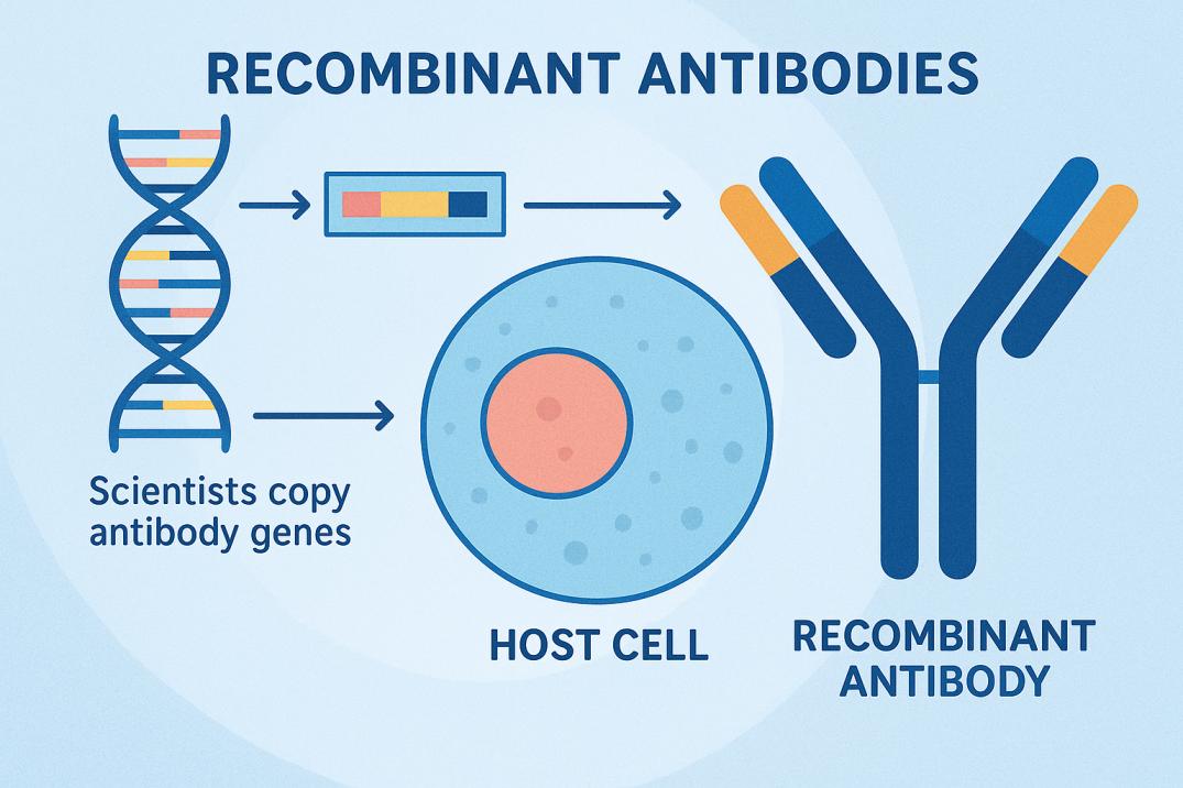
Recombinant antibodies originate from a genetically engineered sequence and target a single specific epitope. Production of custom recombinant antibodies takes 6–10 weeks (Single B-cell/Display). Custom recombinant antibodies offer exceptional lot-to-lot consistency, and supply is indefinite. These antibodies are best for therapeutic leads, ADCs, and high-scale manufacturing.
Upstream Antigen Design & Purification Mechanics
Immunogen design strictly governs the target affinity (KD) and specificity of an antibody. Poor antigen design accounts for over 40% of custom antibody project failures. This typically results in high off-target background or failure to recognize native proteins.
Synthetic Peptides (10–20 Amino Acids)
Synthetic peptides allow researchers to target specific linear regions, such as post-translational modifications (PTMs), point mutations, or site-specific cleavage fragments.
To maximize binding accessibility, target regions should come from flexible loops exposed on the protein surface.
Peptides must be conjugated to immunogenic carrier proteins to trigger an immune response. This is achieved using directional maleimide chemistry or EDC/NHS chemistry.
Recombinant Proteins
Recombinant proteins or isolated domains are required when generating antibodies against complex, discontinuous 3D conformational epitopes. Recombinant protein expression in E. coli is cost-effective. However, it lacks eukaryotic post-translational modifications and often requires refolding from inclusion bodies. Expression in mammalian systems (HEK293/CHO) ensures native folding and glycosylation. As a result, mammalian systems produce superior antibodies for functional, native-state assays (IP, flow cytometry). Large fusion tags (GST, MBP) must be cleaved prior to immunization to avoid tag-dominant immune responses.
DNA / Genetic Immunization
Plasmids or mRNA delivered in vivo via electroporation allow host cells to synthesize the antigen natively. This is the gold standard for multi-pass transmembrane targets (GPCRs, ion channels) where detergent extraction alters native tertiary structure.
Downstream Purification Methodologies
Raw antiserum, cell culture supernatant, or ascites fluid contains host proteins, albumin, lipid debris, and non-specific immunoglobulins. These contaminants must be cleared to eliminate background noise and preserve target binding affinity.
Physicochemical Separation (Clarification)
Before chromatographic loading, crude biological fluids undergo preliminary clarification to remove particulate matter and coarse protein contaminants:
Centrifugation & Depth Filtration
Harvest fluids are centrifuged (> 10,000 X g at 4℃) and passed through 0.22 µm polyethersulfone (PES) membranes. This removes insoluble cell debris, lipids, and aggregation nuclei that would otherwise clog chromatography columns.
Ammonium Sulfate Precipitation
Salting out using 35%–45% saturated ammonium sulfate (NH4)2SO4 selectively precipitates total immunoglobulins out of solution. This leaves host serum albumin and low-molecular-weight contaminants soluble. The resulting protein pellet is resuspended and dialyzed against 1X PBS before column loading.
Class-Specific Affinity Chromatography
Class-specific capture isolates total immunoglobulin (IgG) from host cell proteins based on Fc-region binding dynamics:
Protein A Resin
Protein A resin exhibits high affinity for human IgG1, IgG2, IgG4, and rabbit IgG. It binds target immunoglobulins via hydrophobic interactions at the CH2CH3 interdomain interface of the antibody heavy chain.
Protein G Resin
Protein G resin offers superior binding affinity for mouse IgG1, mouse IgG3, and rat immunoglobulins where Protein A binding is weak or non-existent.
Protein L Resin
Protein L targets kappa light chains, not the Fc heavy chain. This allows it to capture antibodies that are missed by other resins. It is used to purify the following:
- Single-chain variable fragments (scFv),
- Fab fragments,
- IgA or IgM isotypes.
Antigen-Specific Affinity Purification
Class-specific resins capture all IgG present in the sample. However, target-specific antibodies account for just 1% to 5% of total serum. Antigen-specific affinity purification is critical for eliminating non-specific host IgG:
Matrix Coupling
Synthetic peptide or recombinant protein immunogens are covalently immobilized onto NHS-activated Sepharose or CNBr-activated agarose matrices.
Adsorption & Washing
Total IgG is passed through the antigen column. Target-specific immunoglobulins bind tightly to the immobilized antigen, while non-binding host antibodies pass through in the flow-through fraction. Washing the column with a high-salt buffer eliminates non-specific ionic interactions.
Elution and pH Neutralization
Bound antibodies are eluted using an acidic buffer (0.1 M Glycine-HCl, pH 2.5–2.8). This temporarily disrupts electrostatic and hydrogen bonds at the antigen-antibody interface. Eluted fractions are collected directly into tubes containing 1 M Tris-HCl (pH 8.5–9.0) at a 1:10 volume ratio to immediately restore a neutral pH. This prevents acid-induced denaturation or irreversible aggregation.
End-to-End Timelines & Accelerated Workflows
Antibody production timelines vary significantly based on host biological responses, platform selection, and the complexity of downstream screening assays. The selection of the appropriate expression platform depends on balancing development speed against long-term reagent stability and IP requirements.
Comparative Development Schedule
| Stage | Polyclonal Timeline | Monoclonal Timeline | Recombinant Timeline |
|---|---|---|---|
| 1. Antigen Design & Preparation | Weeks 1–3 | Weeks 1–3 | Weeks 1–2 |
| 2. Host Immunization Schedule | Weeks 4–9 | Weeks 4–10 | Weeks 3–5 |
| 3. Fusion / Single B-Cell Isolation | N/A | Weeks 11–15 | Weeks 6–7 |
| 4. Subcloning & High-Throughput Screening | Weeks 10–12 (Serum Titration) | Weeks 16–20 | Weeks 7–8 |
| 5. Purification & Application QC Validation | Weeks 13–14 | Weeks 21–24 | Weeks 8–9 |
| Total Project Lead Time | 10–14 Weeks | 16–24 Weeks | 6–10 Weeks |
Polyclonal Antibodies
Antigen synthesis is followed by a multi-boost immunization protocol (typically in rabbits or goats) over 6–8 weeks to drive somatic hypermutation. Serum titers are monitored via ELISA before full bleed harvest and antigen-affinity purification. Biological variation between host animals can lead to batch-to-batch affinity shifts, which require re-validation for every production lot.
Traditional Monoclonal Hybridomas
Splenocytes harvested from immunized mice or rats are fused with immortalized myeloma cells (e.g., SP2/0) using polyethylene glycol (PEG) or electrofusion. Hybridomas undergo HAT selection, followed by limiting dilution subcloning to ensure monoclonality and stable antibody expression. Hybridoma instability or chromosome loss during early subcloning stages can result in the loss of high-performing clones, which makes cryopreservation of early parental stocks critical.
Recombinant & Single B-Cell Discovery
Single B-cell isolation platforms (and recombinant antibody workflows) bypass long hybridoma fusion cycles by directly isolating antigen-positive B cells from host blood or lymphoid tissue using microfluidic sorting or fluorophore-conjugated antigen baiting.
Actionable Timeline Optimization Strategies
Standard antibody development lead times range from 10 to 24 weeks. However, pipeline constraints and project milestones often require an accelerated timeline. Incorporating strategic technical interventions during project kickoff can reduce total lead time by 30% to 50% without compromising affinity or target specificity.
Provide Pre-Validated Antigens
Time Saved: 2 to 4 Weeks
Synthesizing, folding, and running quality control (QC) on custom peptide or recombinant protein antigens internally accounts for the first month of standard vendor workflows.
Supplying a pre-validated antigen enables immediate host animal immunization upon project kickoff. This requires providing HPLC purity profiles, mass spectrometry verification, and functional activity assays to ensure antigen quality.
Parallel Multi-Animal Immunization
Time Saved: 2 to 3 Weeks
Individual animal immune responses are inherently variable. Immunizing a single animal risks low antibody titers or non-specific binding. This requires a complete protocol restart. Running parallel immunizations across multiple host animals with varied adjuvant combinations maximizes host immune activation. This ensures at least one high-responder candidate without sequential retry cycles.
Single B-Cell Screening Platforms
Time Saved: 6 to 10 Weeks
Traditional monoclonal development relies on immortalizing splenocytes via hybridoma fusion. This is followed by lengthy HAT media selection and limiting dilution subcloning to achieve stable monoclonality.
Single B-cell isolation platforms bypass traditional hybridoma fusion entirely. Instead, they harvest antigen-positive B cells directly from peripheral blood mononuclear cells (PBMCs) or lymphoid tissue using microfluidic sorting or fluorophore-conjugated antigen baiting. Heavy (VH) and light (VL) variable genes are then sequenced, cloned, and expressed recombinantly.
Application-Specific Functional Validation
Protein spatial architecture varies drastically across experimental conditions. An antibody validated solely under denaturing conditions may fail completely in live-cell or native-state assays. Custom antibody validation must match the exact target protein conformation of the final downstream assay.
| Assay Format | Target Protein Conformation | Essential Quality Control Metrics | Validation Strategy & Controls |
|---|---|---|---|
| Western Blot (WB) | Denatured / Linearized |
|
Validate specificity using knockout/knockdown (CRISPR/siRNA) lysates or target-overexpressing cell lines under reducing conditions. |
| Immunoprecipitation (IP / Co-IP) | Native / Folded (Solubilized) |
|
|
| Immunohistochemistry (IHC / IF) | Fixed / Partially Cross-Linked |
|
|
| Flow Cytometry (FACS) | Native (Cell Surface) |
|
|
| ChIP-seq | Cross-Linked Chromatin |
|
|
Assay-Specific QC Guidelines
Denaturing Assays (Western Blot)
Denaturing buffers (SDS, 𝛽‐mercaptoethanol) unfold target proteins, which exposes linear peptide sequences. Antibodies raised against short synthetic peptides perform exceptionally well here, provided they demonstrate no cross-reactivity with off-target host proteins of similar molecular weight.
Native & Conformational Assays (IP, Flow Cytometry)
Native cell lysates and live cell surfaces preserve tertiary and quaternary protein structures. Antibodies must recognize surface-accessible, discontinuous conformational epitopes. Small-molecule tag-mediated purification (e.g., FLAG, HA) or recombinant single-chain variable fragments (scFv) often yield superior signal-to-noise ratios in these formats.
Fixed & Cross-Linked Assays (IHC, ChIP-seq)
Formaldehyde fixation creates methylene bridges that can mask target epitopes or alter local surface charge. Validation requires optimizing heat-induced epitope retrieval (HIER) protocols to unmask target sites without causing tissue damage or chromatin fragmentation.
Risk Management & CRO Selection Guide
Selecting a Contract Research Organization (CRO) for custom antibody production requires balancing technical capability, intellectual property security, and financial protection. A rigorous risk management framework is essential.
CRO Evaluation Checklist
Intellectual Property (IP) Ownership
Make sure that the contract grants full ownership of all host immunization outputs, sequence files (VH/VL chains), and derived cell lines upon final payment. For recombinant projects, demand complete plasmid sequence access to prevent vendor lock-in during bioprocess scaling.
Traceability & Animal Welfare
Verify active AAALAC accreditation, OLAW assurance, and USDA Animal Welfare Act compliance. Require individual host animal ID tracking, complete bleed logs, and a certified Certificate of Origin confirming TSE/BSE safety profiles for domestic and international material transport.
Analytical Quality Control
Require complete analytical validation packages for every production lot, including SDS-PAGE for structural integrity, SEC-HPLC demonstrating >95% monomeric purity, and mass spectrometry sequence verification for recombinant proteins.
For cell-based or in vivo applications, require endotoxin testing certified below 1 EU/mg via LAL assay along with verified sterile filtration (0.22 µm).
AAA Biotech provides antibodies, proteins, and ELISA kits for scientific research and discovery. Contact us today for validated, publication-backed antibodies.
Faq's
How long does custom polyclonal antibody production take?
Custom polyclonal antibody production typically takes 10 to 14 weeks from host immunization through final serum harvest and affinity purification.
What is the difference between polyclonal and monoclonal antibodies?
Polyclonal antibodies are a heterogeneous mixture produced by multiple B-cell clones that bind different epitopes on the same target. Monoclonal antibodies originate from a single B-cell clone and recognize a single specific epitope to provide high specificity and batch-to-batch consistency.
How can you accelerate custom antibody production timelines?
Timelines can be compressed by 30% to 50% by supplying pre-validated antigens, running parallel multi-animal immunizations, or utilizing single B-cell screening platforms, thereby bypassing 6- to 10-week hybridoma fusion and subcloning steps.
What is the difference between peptide and full-length protein immunogens?
Peptide immunogens consist of short synthetic amino acid sequences (10–20 residues) designed to target specific linear regions or post-translational modifications. Full-length protein immunogens preserve native tertiary/quaternary structures. This makes them better suited to generating antibodies that recognize complex conformational epitopes.
Lead Clinical Research Coordinator (LCRC)
Cynthia Lee is the President of AAA Biotech and specializes in understanding highly validated and characterized monoclonal/polyclonal antibodies, recombinant proteins, and ELISA kits.
Related Posts