How To Read an Antibody Datasheet
 Cynthia
19 min read
Cynthia
19 min read
In this Article


All of the products listed in AAA Biotech’s catalog are strictly for research-use only (RUO).

Key Takeaways
- Check application validation first. Make sure your exact application, such as WB, IHC, IF, ELISA, or flow cytometry, is supported by experimental data.
- Match the antibody to your sample. Check species reactivity, tissue or cell type, target form, and immunogen information before ordering.
- Use the datasheet to set your starting conditions. Review the recommended dilution, protocol, formulation, and storage conditions before your first experiment.
- Evaluate the evidence, not just the claims. Look for validation images, controls, molecular-weight data, KO/KD or blocking experiments, and application-specific results.
- Compare products using the same criteria. A useful datasheet comparison includes validation depth, clonality, immunogen, purification, formulation, citations, and available product documentation.
The right choice of antibody is not as simple as finding the right target and placing an order. Two antibodies against the same protein can perform very differently depending on the:
- Host species
- Clonality
- Target region
- Application validation
- Sample type
- Recommended protocol
These details are easier to overlook. This is where the antibody datasheet comes in. A datasheet can tell you whether an antibody has been validated for Western blot, immunohistochemistry, immunofluorescence, flow cytometry, or other applications. It can also tell you more about whether the antibody is compatible with your experiment, including the host species, isotype, reactivity, immunogen, formulation, concentration, storage conditions, and known limitations.
This guide breaks down every section of an antibody technical datasheet.
What Is an Antibody Datasheet?
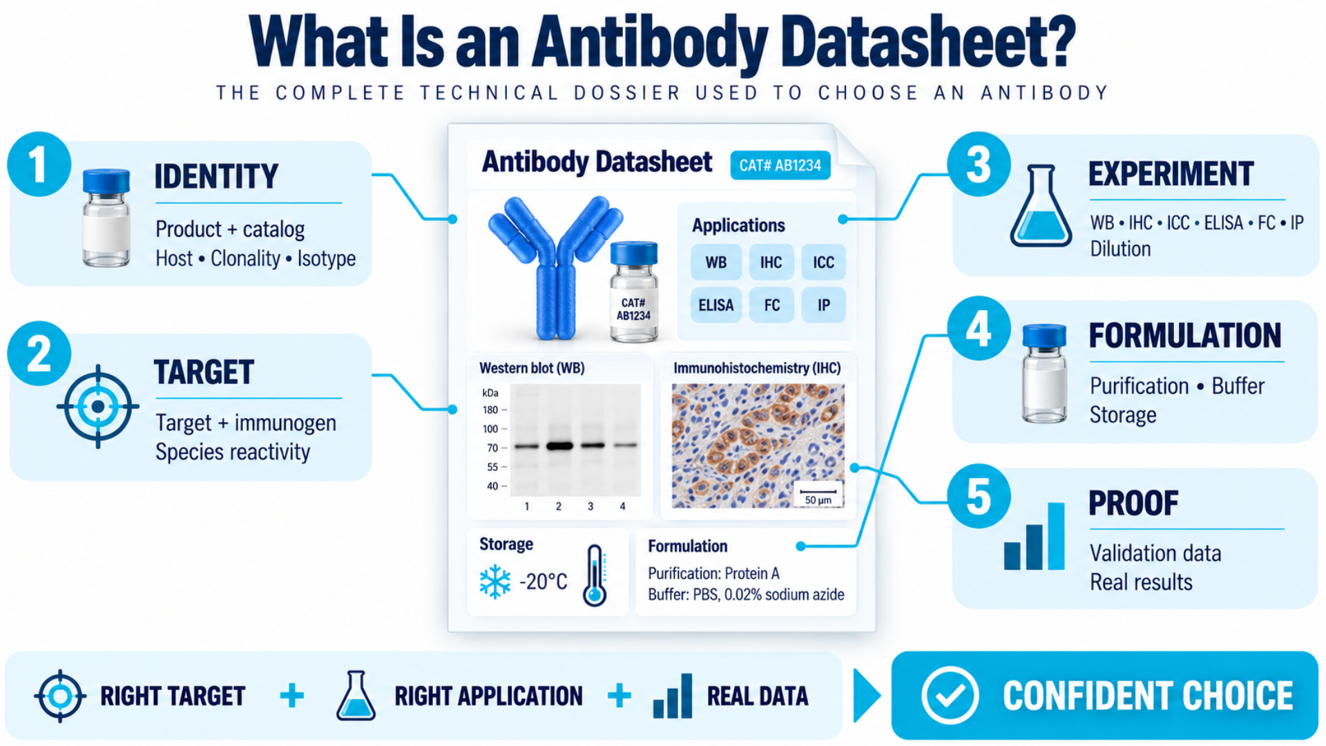
An antibody datasheet (also called an antibody specification sheet, antibody technical datasheet, or product data page) is a document provided by the antibody manufacturer or supplier. It consolidates every piece of technical information about a specific antibody product.
You can think of it as the antibody’s complete dossier. It tells you:
- What the antibody targets
- How it was made
- Which experimental applications it has been validated for
- Which species it reacts with
- How to store it
- What buffer it ships in
- If the supplier is thorough, how it actually performed in real experiments
What is Included in an Antibody Datasheet?
A typical antibody datasheet includes the following sections:
| Product Information | Description |
|---|---|
| Product name and catalog number | The antibody identity and reorder reference. |
| Host species | The animal in which the antibody was produced. |
| Clonality | Whether the antibody is monoclonal, polyclonal, or recombinant. |
| Isotype | The immunoglobulin class and subclass (e.g., IgG1, IgG2a). |
| Target and immunogen | The protein or epitope the antibody recognizes, and what was used to generate the immune response. |
| Reactivity/target species | Which organisms the antibody has been tested against or predicted to react with. |
| Validated applications | This section shows which experimental techniques the antibody has been tested and validated for, including WB, IHC, ICC, ELISA, FC, and IP. |
| Dilution recommendations | Suggested concentration ranges for each application. |
| Purification method | How the antibody was isolated (e.g., Protein A/G affinity, antigen-affinity). |
| Buffer and formulation | The storage solution the antibody ships in, including additives. |
| Storage conditions | Temperature requirements and freeze-thaw guidance. |
| Validation data | Images, blots, staining photos, and performance documentation. |
Antibody datasheets can vary from one supplier to another. Some provide detailed validation data and published studies, while others provide only basic product information. It is important to know what to check before you choose an antibody for your experiment.
Section-by-Section Breakdown: How to Read an Antibody Datasheet.
1. Product Name and Catalog Number
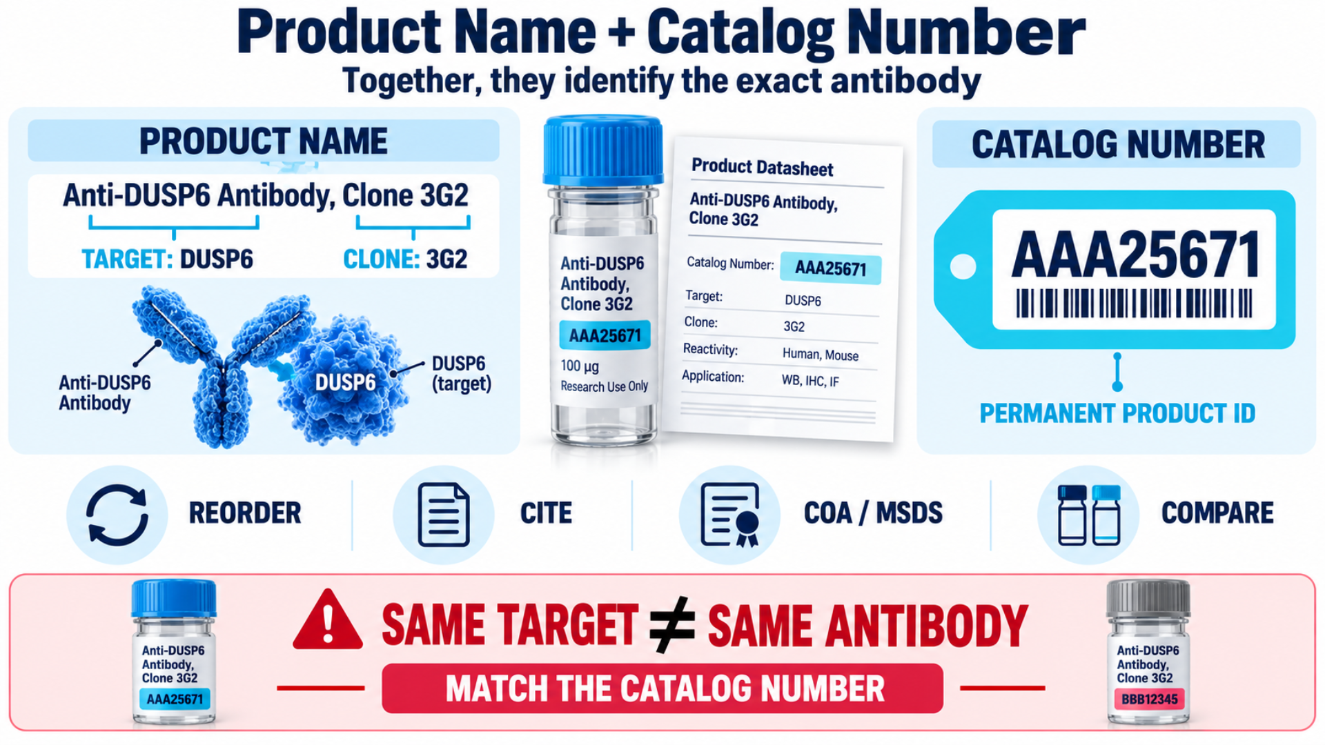
Every antibody datasheet starts with a product name and a unique catalog number. These fields help you identify, reference, and reorder the same antibody product.
What the Product Name Tells You
The name usually identifies the target protein or antigen. For example, "Anti-DUSP6 Antibody, Clone 3G2" tells you that the antibody targets Dual Specificity Protein Phosphatase 6. It also indicates that 3G2 is the specific monoclonal antibody clone.
What the Catalog Number Tells You
Every product is assigned a unique catalog number that helps identify the exact antibody. For example:
- AAA25671 for the DUSP6 monoclonal antibody
- AAA255710 for the SAE1 polyclonal antibody
This number serves as your permanent product reference. Use it to:
- Reorder the same antibody to keep your results consistent.
- Include the antibody details in your methods section and publications.
- Contact the supplier for batch-specific information, certificates of analysis (COA), or safety data sheets (MSDS).
- Compare antibody products from different suppliers.
What Researchers Miss
Researchers often reorder antibodies using only the target name. Always check the catalog number to make sure you are buying the same antibody you used before.
2. Host Species
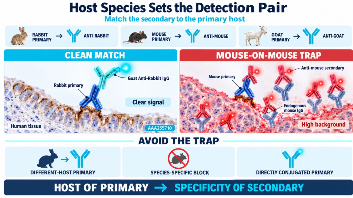
The host species is the animal used to produce the antibody. Common host species include rabbits, mice, and goats. The host species determines which secondary antibody you should use for detection.
How it Works
If you purchase a rabbit-derived primary antibody, you need an anti-rabbit secondary antibody, such as Goat Anti-Rabbit IgG, for detection. The species match between the primary and secondary antibodies is essential for accurate results.
The Same-Species Trap
This is a common problem in immunohistochemistry. When a mouse-derived monoclonal antibody is used on mouse tissue, the secondary antibody can bind to the primary antibody. It can also bind to the mouse immunoglobulins naturally present in the tissue. This creates high nonspecific background staining, which can make your results difficult to interpret.
Solutions include:
- Use a primary antibody from a different host species (for example, rabbit anti-target antibody on mouse tissue).
- Apply species-specific blocking reagents before antibody incubation.
- Use a directly conjugated primary antibody to remove the need for a secondary antibody.
Example
The Anti-SAE1 Polyclonal Antibody (Catalog #AAA255710) is produced in rabbit. When using this antibody with human tissue, you can pair it with a Goat Anti-Rabbit IgG (H+L) secondary antibody for detection.
Using a rabbit primary antibody on human tissue also helps avoid detection of endogenous human IgG by the anti-rabbit secondary antibody.
3. Clonality: Monoclonal vs. Polyclonal vs. Recombinant
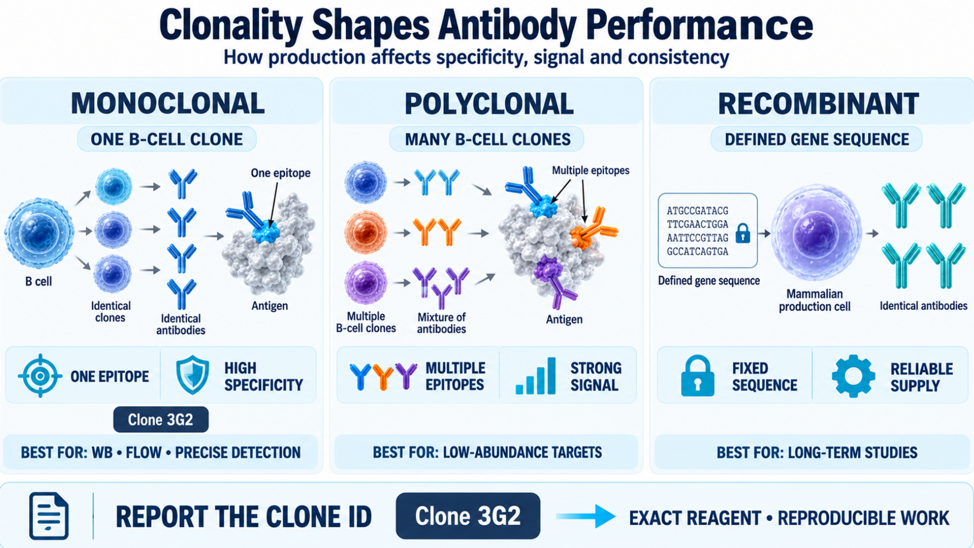
Clonality refers to whether an antibody is produced from one or multiple B-cell clones. If an antibody is made from:
- One single B-cell clone– Monoclonal (Recognizes one specific target region on the antigen).
- Many B-cell clones– Polyclonal (Recognize multiple regions on the same antigen)
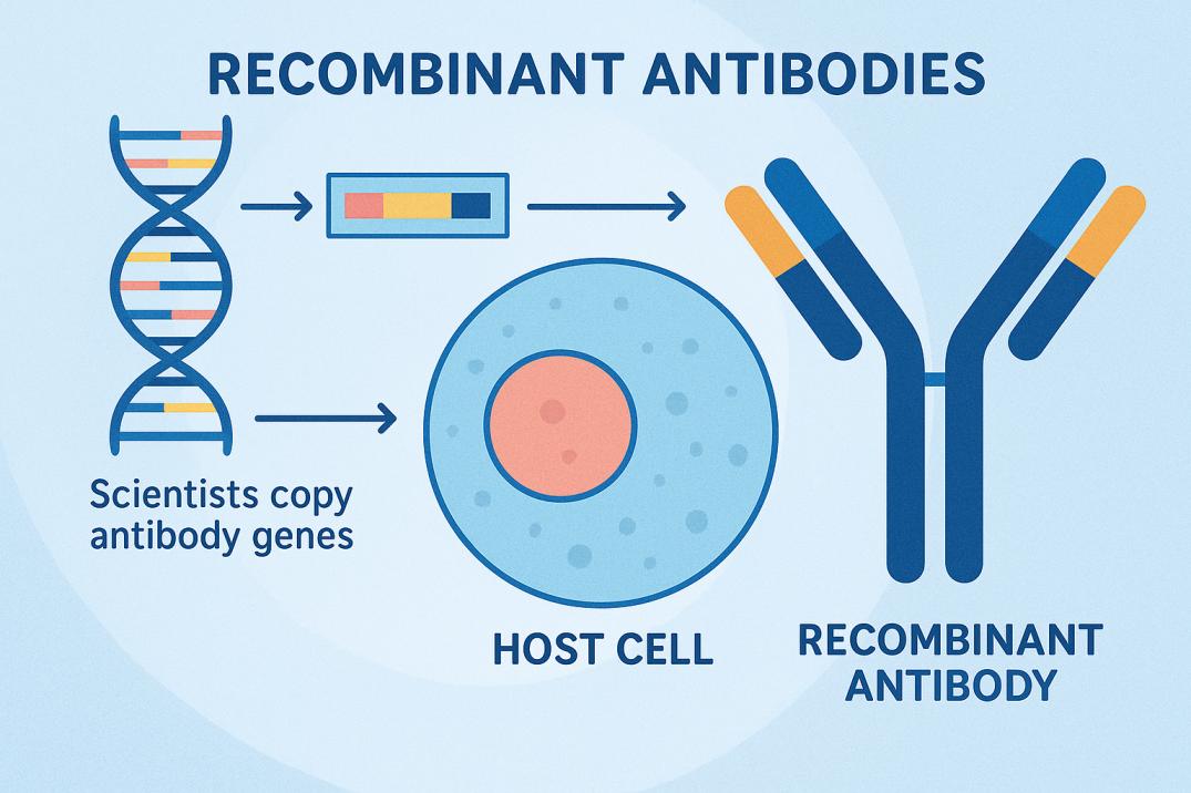
- Defined antibody gene sequence–Recombinant (Allows researchers to produce the same antibody consistently)
What Researchers Miss
A monoclonal antibody datasheet includes a clone ID, such as "Clone 3G2." Always include this clone ID when citing the antibody in your publications. It allows other researchers to identify and use the exact same reagent for experimental replication.
4. Isotype
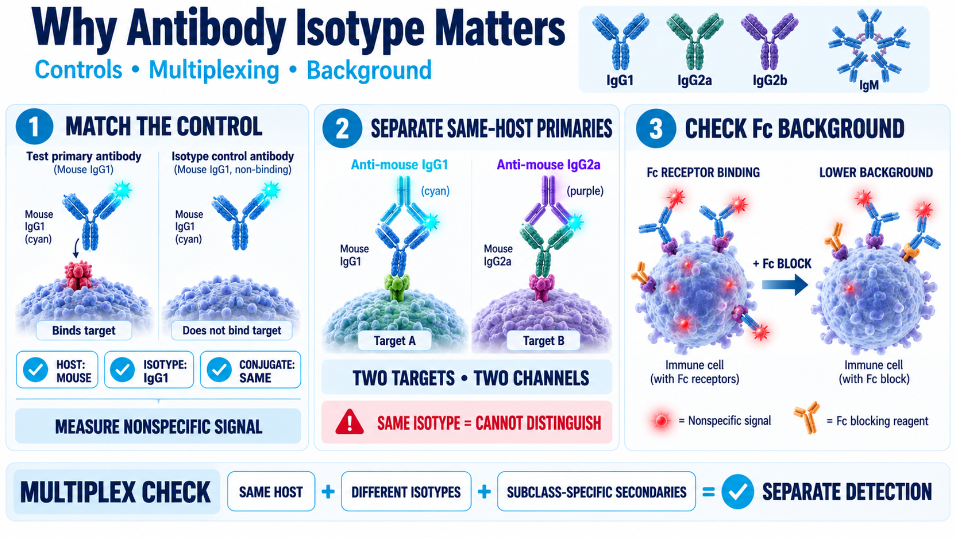
Isotype refers to the class or subclass of an antibody, such as IgG1, IgG2a, IgG2b, or IgM. Isotype selection is important for choosing controls and secondary antibodies.
What to Check
- Isotype control: Use an isotype control that matches the primary antibody's host species and isotype. It should not recognize your target. For example, if your primary antibody is mouse IgG1, use a mouse IgG1 isotype control.
- Secondary antibody: Check that the secondary antibody can detect your primary antibody's isotype. This is particularly useful when using two primary antibodies from the same host species. For example, mouse IgG1 and mouse IgG2a can be detected separately using isotype-specific secondary antibodies.
- Fc receptor binding: Some antibodies can bind to Fc receptors on certain cells and produce background staining. If this occurs, an Fc receptor blocking reagent may help reduce the background.
What Researchers Miss
Isotype selection is important in multiplex experiments when using two antibodies from the same host species. If both antibodies have the same isotype, their secondary antibodies may not distinguish between them. This can make it difficult to detect each target separately.
5. Target and Immunogen Information
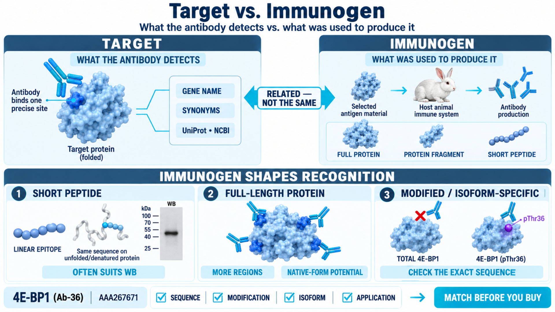
This section explains:
- What the antibody detects (the target)
- What was used to produce the antibody (the immunogen)
The target is the protein, peptide, or epitope that the antibody is designed to recognize. On an antibody datasheet, the target information is listed with gene names and known synonyms. These details help researchers cross-reference the target with databases such as UniProt and NCBI.
On the other hand, the immunogen is the material used to generate the antibody. It may be a full protein, a protein fragment, or a short peptide. This material is injected into the host animal to stimulate an immune response and produce antibodies.
Why the Immunogen Matters More Than Most Researchers Realize
- Short peptide immunogen: They are often useful for detecting denatured proteins, such as in the Western blot, but may not recognize the protein’s natural structure effectively.
- Full-length protein immunogen: They may produce antibodies that recognize multiple regions of the protein. They can be useful for applications involving proteins in their native form.
- Post-translational modifications and isoforms: If you are studying a specific modification or isoform, check whether the immunogen contains the relevant sequence or modification. This can help you assess whether the antibody is likely to recognize the form you are studying.
Example
The 4E-BP1 (Ab-36) Antibody (Catalog #AAA267671) is purified using affinity chromatography with an epitope-specific immunogen. The datasheet also lists a phospho-specific antibody against 4E-BP1 phospho-Thr36. The immunogen determines which part or form of the protein the antibody can recognize. For example, a phospho-specific antibody is designed to detect the phosphorylated form of a protein, not the total amount of 4E-BP1.
6. Species Reactivity / Target Species
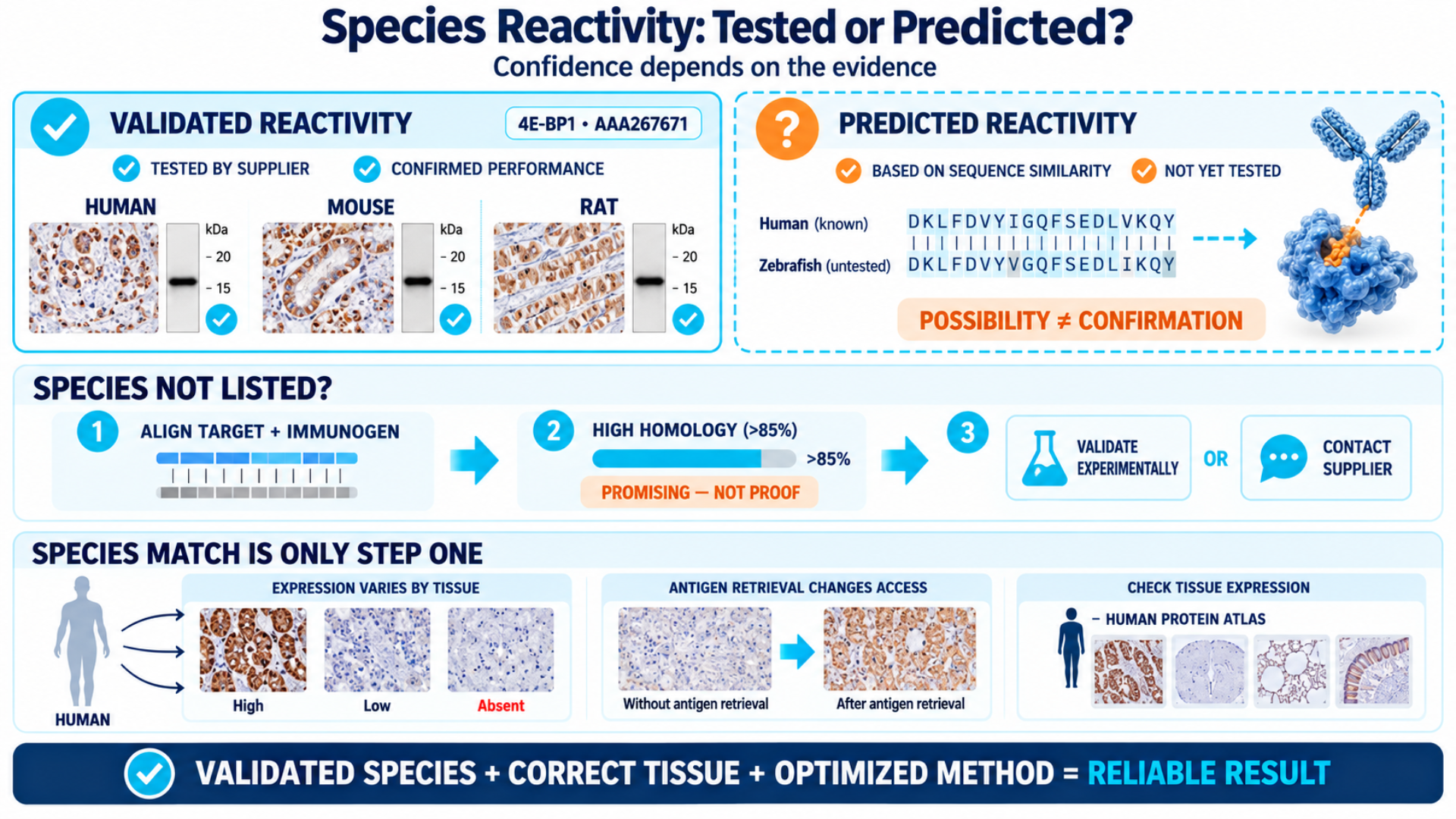
This section shows which species the antibody has been tested to recognize, such as human, mouse, or rat. Some datasheets also include species where the antibody may work based on protein sequence similarity.
There are two types of species reactivity you may see:
- Validated reactivity: The supplier has tested the antibody in that species and confirmed that it works. This is the most reliable information.
- Predicted reactivity: The antibody may work because the target protein is similar to the protein used to generate the antibody. However, the antibody has not been tested in that species. Treat predicted reactivity as a possibility, not a confirmation.
Example
The 4E-BP1 Antibody (Catalog #AAA267671) lists validated reactivity with Human, Mouse, and Rat. The product page also states that it "may cross-react with other species as described in the data sheet."
If your model organism is not listed, you have two options:
1. Check the protein sequence alignment between your species and the immunogen sequence. High sequence homology (>85%) suggests that the antibody may work, but you should validate it experimentally.
2. Contact the supplier directly.
What Researchers Miss
"Reactive with Human" does not mean the antibody will work in every human tissue type. Protein expression can vary between tissues. In IHC, antigen retrieval methods can also affect epitope accessibility.
Always compare the datasheet information with protein expression databases, such as the Human Protein Atlas, for your specific tissue of interest.
7. Validated Applications
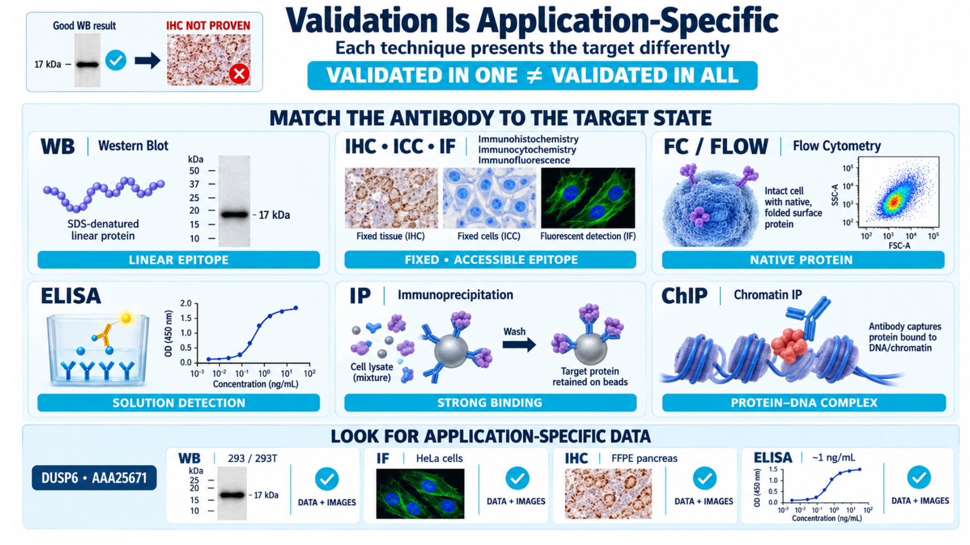
This is one of the most important sections in an antibody datasheet. It shows which experimental techniques the antibody has been tested and confirmed to work in.
Common application abbreviations include:
| Abbreviation | Full Name | What It Tests |
|---|---|---|
| WB | Western Blot | Protein size and expression in denatured lysates |
| IHC | Immunohistochemistry | Protein localization in fixed tissue sections |
| ICC | Immunohistochemistry | Protein localization in fixed cultured cells |
| IF | Immunohistochemistry | Protein visualization using fluorescent detection |
| ELISA | Enzyme-Linked Immunosorbent Assay | Quantitative protein detection in solution |
| FC/Flow | Flow Cytometry | Single-cell protein expression analysis |
| IP | Immunoprecipitation | Protein isolation from complex mixtures |
| ChIP | Chromatin Immunoprecipitation | Protein-DNA interaction analysis |
The critical rule: An antibody validated for one application is not automatically validated for another. Each technique requires the antibody to meet different conditions.
- WB: The antibody must recognize the denatured (linear) form of the protein.
- IHC and ICC: The antibody must recognize proteins that may be altered or hidden by fixatives, which can affect epitope accessibility.
- Flow cytometry: The antibody often needs to bind the native (folded) protein on live or lightly fixed cells.
- IP: The antibody must bind the target strongly enough to remain attached during washing steps.
An antibody that produces a clear 17 kDa band in Western blot may produce only background staining in IHC if it has not been validated for that application.
Example
The DUSP6 Monoclonal Antibody (Catalog #AAA25671) is validated for IF, IHC, WB, and ELISA. Its datasheet includes application-specific images and experimental data.
The Western blot data show DUSP6 detection in overexpressed or transfected 293/293T cells. The immunofluorescence data show detection in HeLa cells. The IHC data show staining in formalin-fixed paraffin-embedded (FFPE) human pancreas tissue. The ELISA data report a detection limit of approximately 1 ng/mL for recombinant GST-tagged DUSP6.
These application-specific results demonstrate that the listed techniques are supported by validation data and images, rather than relying solely on general claims.
The Difference Between "Validated" and "Suggested" Applications
Datasheets may label applications as validated or suggested. These labels do not mean the same thing.
| Validated Application | Suggested Application |
|---|---|
| Experimentally tested by the supplier | Not experimentally confirmed by the supplier |
| Supporting data or images may be available | Usually has limited or no supporting data |
| Gives you more confidence in performance | Performance is uncertain |
| Lower validation risk | You may need to optimize and validate it yourself |
What Should You Consider?
If your experiment requires reliable and reproducible results, prioritize a validated application. If only a suggested application is available, test the antibody on a small scale first and optimize the conditions before running your full experiment.
8. Dilution Recommendations
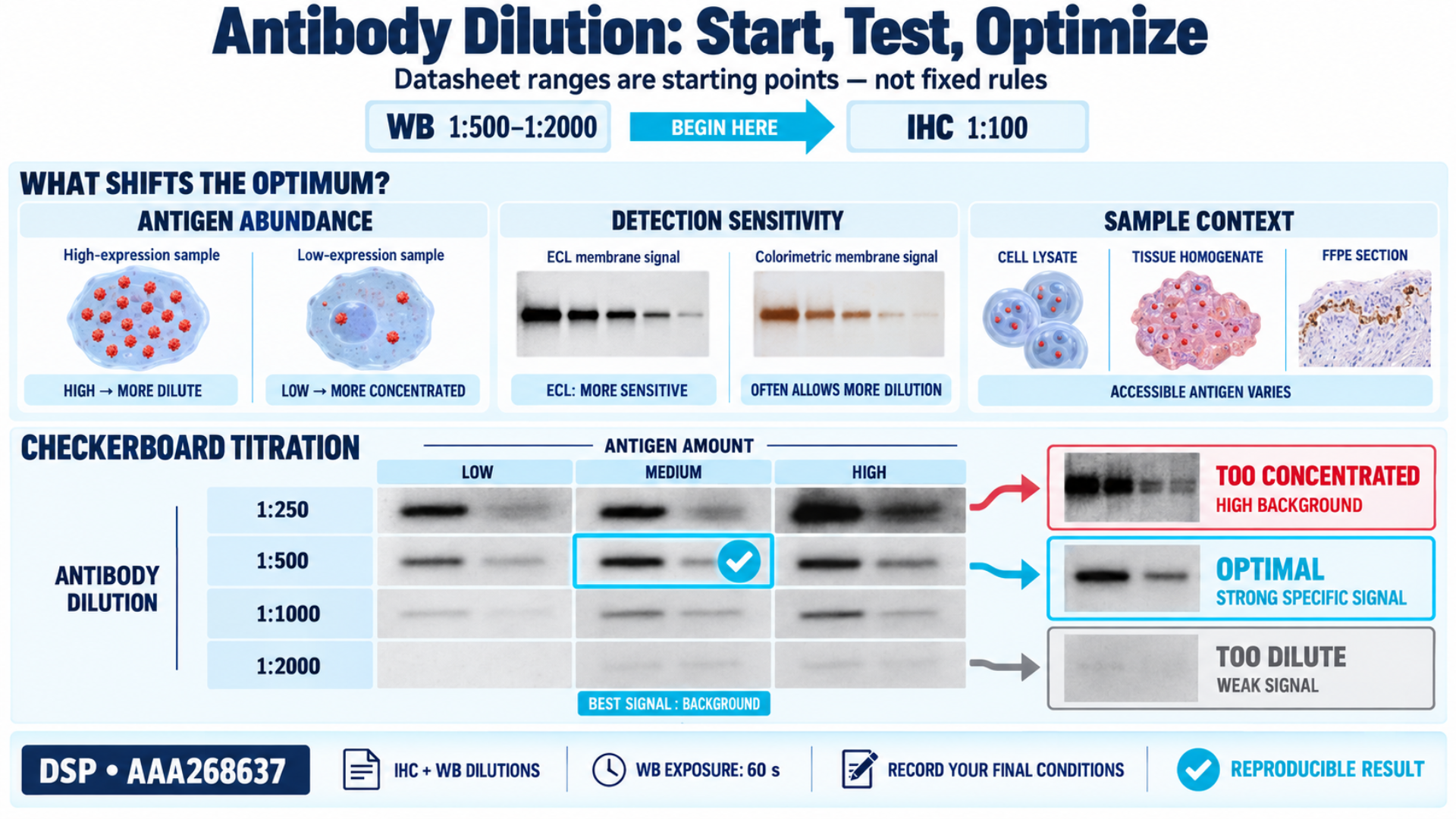
Most antibody datasheets provide a recommended dilution range for each validated application. For example, an antibody may be recommended at 1:500 to 1:2000 for Western blot or 1:100 for IHC.
These values are starting points, not strict rules. The best dilution depends on several factors:
- Antigen abundance: Highly expressed proteins may work well with more diluted antibodies. Low-abundance targets may require a higher antibody concentration.
- Detection system sensitivity: Chemiluminescent detection (ECL) is usually more sensitive than colorimetric detection. This often allows the use of more diluted antibodies.
- Sample type: Cell lysates, tissue homogenates, and FFPE sections contain different levels of accessible antigen. This can affect the optimal dilution.
How to Optimize
When using an antibody for the first time, perform a checkerboard titration. Test different antibody dilutions to find the condition that gives the strongest specific signal with the lowest background. You can then identify the dilution that provides the strongest specific signal with the lowest background.
Example
The DSP Polyclonal Antibody (Catalog #AAA268637) datasheet provides detailed dilution information for IHC and Western blot applications. The datasheet also lists a 60-second WB exposure time, giving researchers a useful starting point for their experiments.
9. Purification Method
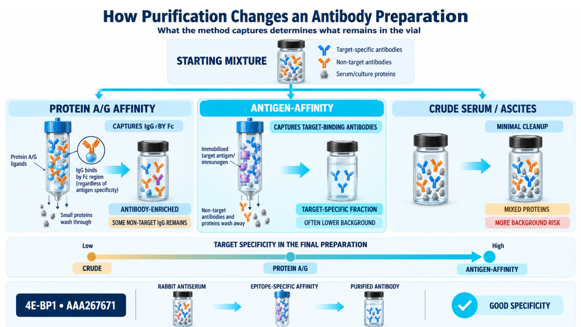
The purification method explains how the antibody was separated and cleaned from serum or cell culture medium. It can affect the antibody’s purity and performance in different applications.
- Protein A/G affinity purification: This method uses the antibody’s Fc region to separate it from other proteins. Other antibodies may also be present.
- Antigen-affinity purification: This method uses the immunogen to collect antibodies that recognize the target. It produces a purer antibody and can reduce background.
- Crude serum or ascites fluid: This contains the antibody along with other proteins. It may cause more background and nonspecific binding, especially in sensitive applications.
Example
The 4E-BP1 Antibody (Catalog #AAA267671) is affinity-purified from rabbit antiserum using an epitope-specific immunogen. This method produces a highly purified antibody with good specificity.
10. Buffer, Formulation, and Additives
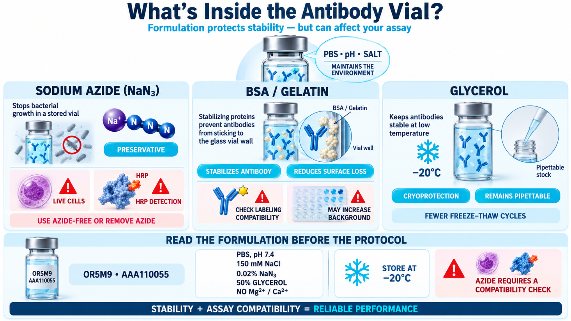
This section tells you where the antibody is stored. It can affect the antibody’s stability and performance in your experiments.
| Component | Purpose | What to Consider |
|---|---|---|
| Sodium azide (NaN₃) | Prevents bacterial growth during storage. | Can harm living cells and affect HRP-based detection. For live-cell experiments or HRP assays, use an azide-free antibody or remove the azide before use. |
| BSA or gelatin | Helps keep the antibody stable and prevents it from sticking to the vial. | BSA may affect some labeling reactions or increase background in certain experiments. |
| Glycerol | Protects the antibody during freezing and storage. | Antibodies with glycerol can usually be stored at –20°C and used directly from the stock without thawing completely. |
Example
The OR5M9 Antibody (Catalog #AAA110055) is supplied in phosphate-buffered saline (without Mg²⁺ and Ca²⁺), pH 7.4, containing 150 mM NaCl, 0.02% sodium azide, and 50% glycerol.
The 50% glycerol allows storage at –20°C without freeze-thaw damage. You can pipette the required amount and return the antibody to the freezer.
The 0.02% sodium azide means this antibody should not be used directly in live-cell assays without dilution or dialysis.
11. Storage and Handling Conditions
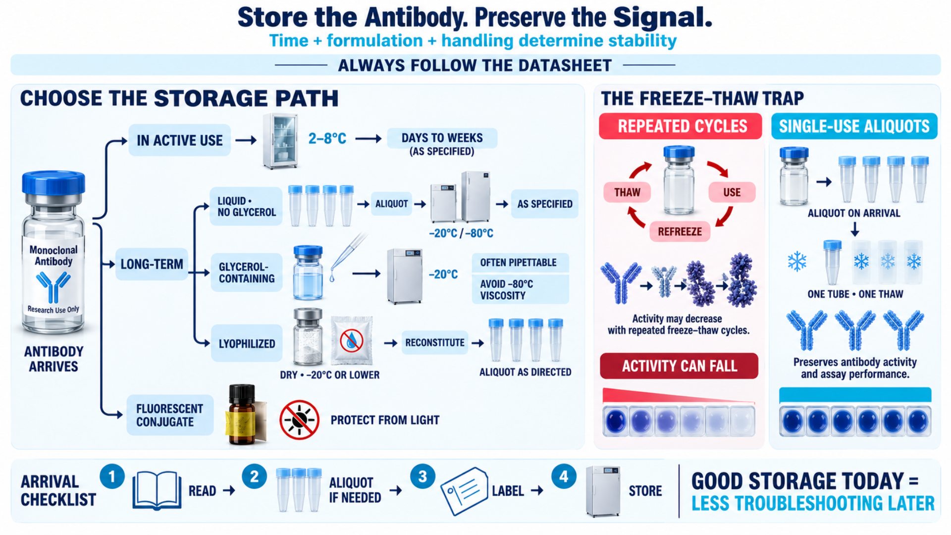
Poor storage conditions can quickly damage an otherwise effective antibody. Always follow the storage instructions provided in the datasheet.
General Principles
- Short-term storage (days to a few weeks): Store at 2–8°C (standard refrigerator). This is suitable for antibodies that are currently in use.
- Long-term storage (months to years): Store at –20°C or –80°C, depending on the antibody formulation.
- Glycerol-containing buffers: Store at –20°C. Glycerol prevents ice crystal formation, so the antibody does not need to be aliquoted. Avoid –80°C storage because glycerol can become highly viscous at this temperature.
- Lyophilized antibodies: Store in a dry environment at –20°C or lower. Reconstitute according to the datasheet instructions and prepare aliquots for storage.
- Avoid repeated freeze-thaw cycles: Each freeze-thaw cycle can damage some antibody molecules and gradually reduce binding activity. If the antibody does not contain glycerol, divide it into single-use aliquots when you first receive it.
- Light-sensitive conjugates (fluorophore-labeled antibodies): Protect them from light during storage to prevent photobleaching.
What Researchers Miss
Many laboratories store all antibodies in the same –20°C freezer box and repeatedly remove the same vial. Each time the vial is warmed, handled, and refrozen, the antibody can lose activity. Preparing aliquots when the antibody arrives can help prevent loss of activity and troubleshooting later.
12. Validation Data and Datasheet Images
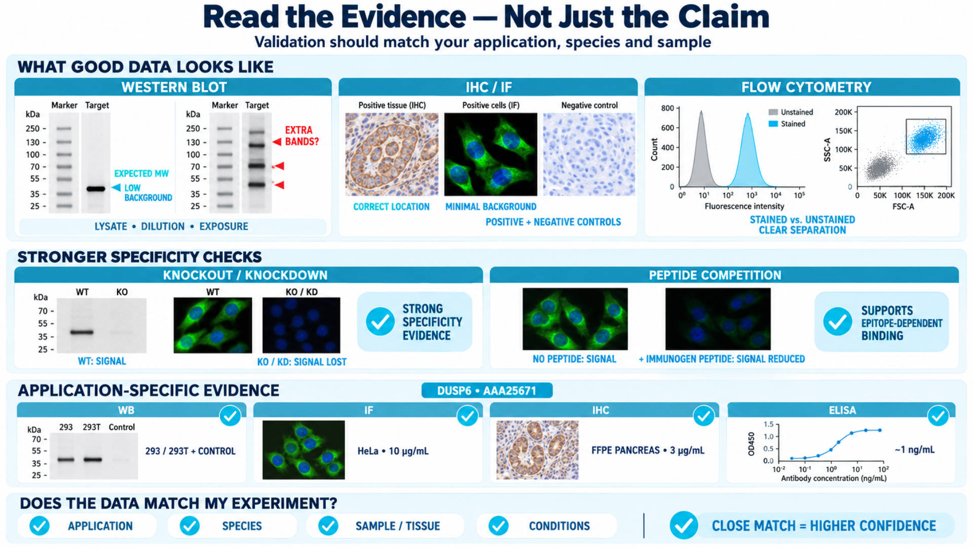
A reliable antibody datasheet includes experimental evidence showing how the antibody performs. This helps distinguish antibodies with strong validation evidence from those with limited documentation.
What to Look for in Validation Data
- Western blot images: Look for a clear band at the expected molecular weight. The datasheet should mention the sample type, antibody dilution, secondary antibody, and detection method. Multiple bands or high background may indicate poor specificity.
- IHC images: Check for specific staining in the correct subcellular location (nucleus, cytoplasm, or membrane) with minimal background. Positive and negative tissue controls improve confidence.
- Flow cytometry data: Look for histograms or dot plots showing clear separation between stained and unstained populations.
- Knockout or knockdown validation: A strong validation method where the antibody is tested in samples with reduced or removed target gene expression. Loss of the signal confirms target specificity.
- Peptide competition / blocking: The antibody is pre-incubated with the immunogen peptide. Loss of staining confirms that the antibody recognizes the intended target epitope.
Example
DUSP6 Monoclonal Antibody (Catalog #AAA25671) includes validation data for multiple applications. The Western blot data show DUSP6 detection in transfected and overexpressed 293/293T cells, along with non-transfected controls. The antibody was also tested by immunofluorescence in HeLa cells at 10 µg/mL and by immunohistochemistry in formalin-fixed paraffin-embedded human pancreas tissue at 3 µg/mL. In ELISA, the antibody showed a detection limit of approximately 1 ng/mL for recombinant GST-tagged DUSP6 when used as a capture antibody.
What Researchers Miss
Always check whether the validation data matches your application and species. A Western blot image from human lysates does not confirm that the antibody will work for IHC in mouse tissue. Choose validation data that closely matches your own experimental conditions.
13. Reviews, Citations, and Publication References
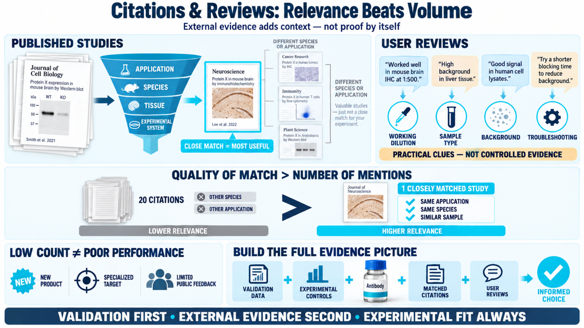
User reviews and publication references can provide extra information when evaluating an antibody.
Published studies show how researchers have used an antibody in real experiments. When reviewing citations, check whether the antibody was tested in an application, species, tissue, or experimental system that matches your research. A publication can provide useful details about antibody performance under specific conditions.
User reviews, when available, can also provide practical insights that may not appear in the datasheet. These may include working dilutions, sample types, background levels, or troubleshooting experiences.
What Researchers Miss
A low number of citations or user reviews does not always mean an antibody performs poorly. The antibody may be new, highly specialized, or have limited public feedback.
Consider citations and reviews together with validation data, experimental controls, and application-specific evidence before making a decision.
Checklist: Your Antibody Datasheet Evaluation Framework
Before you order any antibody, run through this checklist against the datasheet:
Step 1: Define Your Experiment
- What is your target protein?
- Which application will you use (WB, IHC, ELISA, FC)?
- What species is your sample from?
Step 2: Check Validated Applications
Is your specific application listed with supporting validation data not just a "suggested" label?
Step 3: Confirm Species Reactivity
Is your model organism listed under validated (not just predicted) reactivity?
Step 4: Review the Host Species
- Will the host species conflict with your sample species?
- Do you have the correct secondary antibody?
Step 5: Evaluate Clonality
- Do you need monoclonal specificity and reproducibility, or polyclonal sensitivity?
- Is a recombinant option available for long-term consistency?
Step 6: Inspect the Immunogen
Does the immunogen match the protein form you are trying to detect (denatured vs. native, phosphorylated vs. total)?
Step 7: Review Dilution and Protocol Recommendations
Does the datasheet provide application-specific dilutions, or only a generic range?
Step 8: Check the Buffer
Does the formulation contain sodium azide (problematic for live-cell work)? Does it contain glycerol (affects storage protocol)?
Step 9: Assess the Validation Data
Are there application-matched images? Are controls shown? Is there KO/KD validation?
Step 10: Look for Citations and Reviews
Has anyone published with this antibody in your application and species?
Conclusion
Researchers who get consistent and reproducible results usually begin by carefully evaluating antibody information before ordering. They are the ones who read the datasheet completely, match every specification to their experimental design, and refuse to skip the fine print.
At AAA Biotech, every antibody product page is built to give you the information you need to make an informed decision. Our datasheets include validated application data with supporting images, full buffer and formulation details, purification methods, and direct access to certificates of analysis and technical support.
Faq's
What are the key features of an antibody datasheet?
The key features are validated applications (which techniques the antibody has been confirmed to work in), species reactivity (which organisms it reacts with), clonality (which affects reproducibility and specificity), immunogen/epitope information (which determines what the antibody recognizes), and validation data (experimental evidence of performance). These features determine whether the antibody will work in your specific experiment.
What is the difference between a monoclonal antibody datasheet and a polyclonal antibody datasheet?
A monoclonal antibody datasheet will include a clone ID (e.g., Clone 3G2) and specify a single defined epitope. A polyclonal antibody datasheet will describe the immunogen (often a peptide or recombinant fragment) but will note that the antibody recognizes multiple epitopes. Monoclonal datasheets emphasize specificity and batch consistency; polyclonal datasheets often highlight sensitivity and broader epitope coverage.
How do I know if an antibody is validated for Western blot or IHC?
Look for application-specific validation data in the datasheet and actual images showing the antibody's performance in that technique. A checkmark or "suggested" label without supporting images does not constitute validation. Reputable suppliers include WB images with band sizes, IHC staining photos with tissue types and dilution conditions, and ELISA detection limit data for each validated application.
How do I read my antibody test results?
Compare your experimental results against the information provided in the antibody datasheet. For Western blot, check whether your band appears at the expected molecular weight listed in the datasheet. For IHC, compare your staining pattern (nuclear, cytoplasmic, membrane) against the datasheet images. For ELISA, verify that your signal falls within the detection range specified. The datasheet is your benchmark for interpreting whether your results are on-target or anomalous.
How do I interpret an antibody titer result?
Antibody titer is the highest dilution that still produces a measurable signal. Compare your working titer with the datasheet's recommended dilution range. For example, if the datasheet recommends 1:1000 for WB but you need 1:200 to see a signal, the antibody may have degraded due to improper storage. However, your sample may also have very low target expression. Conversely, if you get a strong signal at 1:5000, you can use less antibody per experiment. This can help extend the life of the vial.
How do you interpret an antibody panel result?
An antibody panel uses multiple antibodies to identify different markers on a cell population. Start by checking whether each antibody produces the expected staining pattern and intensity.
If a marker shows unexpected positivity or negativity, check the datasheet. Review the antibody's species reactivity and validated applications, especially flow cytometry. Also check the antibody's isotype. Isotype cross-reactivity between panel members can cause unexpected staining or artifacts.
What should I do if my antibody gives nonspecific results?
First, verify that the application is listed as validated (not just suggested) in the datasheet. Then confirm the dilution matches the datasheet recommendation. Increase blocking time or switch the blocking agent (e.g., use BSA instead of milk for phospho-specific antibodies). Confirm that the host species does not match your sample species. And finally, check whether the antibody has been stored according to the datasheet's specifications; improper storage is a leading cause of antibody degradation and nonspecific binding.
Are antibody datasheets standardized?
No. There is no universal standard or regulatory body that controls the format, content, or level of detail in antibody datasheets. Because of this, datasheet quality can vary greatly between suppliers. Some suppliers provided detailed validation data. Others may only provide basic information, such as target name, host species, etc.
Lead Clinical Research Coordinator (LCRC)
Cynthia Lee is the President of AAA Biotech and specializes in understanding highly validated and characterized monoclonal/polyclonal antibodies, recombinant proteins, and ELISA kits.
Related Posts