Secondary Antibodies: How to Choose the Right One and Avoid Cross-Reactivity
In this Article
All of the products listed in AAA Biotech’s catalog are strictly for research-use only (RUO).

Key Takeaways
- A secondary antibody binds to the primary antibody, not directly to the antigen, and carries the detectable signal in your assay.
- Matching host species and target species correctly is the single most important step in choosing a secondary antibody.
- The right conjugate (HRP, fluorescent dye, biotin) depends entirely on the detection platform you are using.
- Pre-adsorbed secondary antibodies are the most reliable way to eliminate cross-reactivity in multiplex or tissue-based experiments.
- Always run a secondary-only control. It is the fastest way to identify background caused by the secondary antibody itself.
Every experienced researcher has had this moment: the Western blot is done, the bands look right, but the background is catastrophic. Or the immunofluorescence image is a blur of non-specific signal with no clear staining. In many of these cases, the culprit is not the primary antibody. It is a poorly chosen secondary antibody.
The secondary antibody is one of the most overlooked variables in immunoassay design, yet it is one of the most consequential. Choose it correctly, and your signal is clean, bright, and specific. Choose it carelessly, and no amount of blocking or washing will fully rescue your experiment.
This guide breaks down everything you need to know about what a secondary antibody is, the exact factors that matter when choosing a secondary antibody, how cross-reactivity happens, and how to prevent it entirely.
What Is a Secondary Antibody?
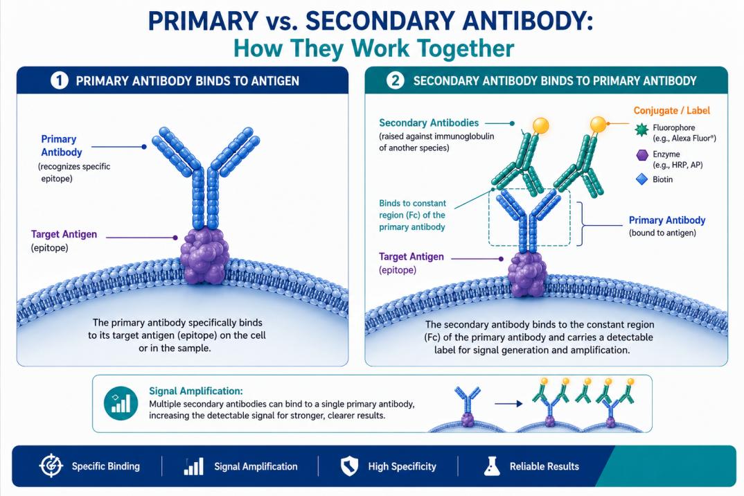
To define a secondary antibody accurately: a secondary antibody is an antibody that is raised against the immunoglobulin of another species and is used to detect a primary antibody that is already bound to a target antigen.
Rather than binding to your protein of interest directly, the secondary antibody binds to the Fc region (the constant region) of your primary antibody and carries the detectable label, whether that is an enzyme, fluorescent dye, or biotin.
Think of it this way: the primary antibody is the scout that finds the target. The secondary antibody is the signal amplifier that makes the scout visible to your detection instrument. Without a matched and validated secondary antibody, even the most specific primary antibody cannot produce a readable result.
Quick Reference: Secondary Antibody Terminology
Host Species: The animal in which the secondary antibody was raised (e.g., goat, donkey, rabbit).
Target Species: The animal in which the primary antibody was raised (e.g., mouse, rabbit, human).
Isotype: The class of immunoglobulin being recognized (e.g., IgG, IgM, IgA).
Conjugate: The detectable label attached to the secondary antibody (e.g., HRP, Alexa Fluor, biotin).
Pre-adsorbed: A secondary antibody that has been treated to remove cross-reactive antibodies targeting other species.
Explore AAA Biotech’s secondary antibody catalog for species-specific, cross-adsorbed, HRP-conjugated, fluorescent, and F(ab')2 secondary antibodies for IF, WB, ELISA, and flow cytometry workflows.
The 5 Key Factors for Choosing a Secondary Antibody
There is no single universal secondary antibody that works for every experiment. Every selection decision involves five distinct variables, and getting even one of them wrong can compromise your entire assay. Here is what to evaluate before you order.
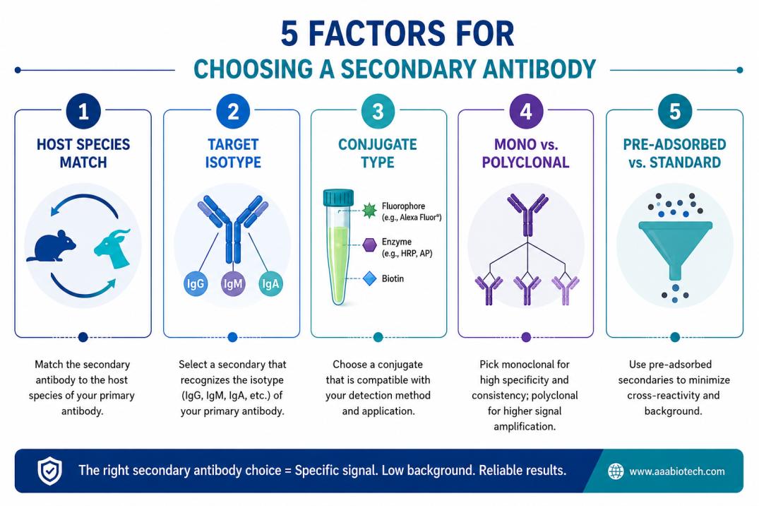
1. Match the Host Species to Your Primary Antibody
The most fundamental rule in choosing a secondary antibody is this: the secondary's target species must match the host species of your primary antibody. If your primary antibody was raised in a mouse, you need an anti-mouse secondary antibody.
If it were raised in a rabbit, you need an anti-rabbit secondary. Using the wrong species pairing means the secondary antibody has nothing to bind to, and you will see no signal at all.
| Primary Antibody Host | You Need This Secondary | Common Example |
|---|---|---|
| Mouse | Anti-Mouse IgG | Goat Anti-Mouse IgG |
| Rabbit | Anti-Rabbit IgG | Donkey Anti-Rabbit IgG |
| Goat | Anti-Goat IgG | Donkey Anti-Goat IgG |
| Human | Anti-Human IgG | Goat Anti-Human IgG |
| Rat | Anti-Rat IgG | Goat Anti-Rat IgG |
AAA Biotech offers species-specific anti-mouse secondary antibodies and anti-rabbit secondary antibodies optimized for Western blotting, immunofluorescence, ELISA, and IHC applications.
2. Know Your Target Isotype (IgG, IgM, IgA, and Others)
Most researchers use IgG-class primary antibodies, and most secondary antibodies are designed to recognize IgG by default. However, if your primary antibody is a different isotype, such as IgM or IgA, you will need an isotype-specific secondary antibody to ensure proper recognition. Using an anti-IgG secondary antibody against an IgM primary will produce no signal, regardless of how well the primary itself is working.
Isotype-specific secondary antibodies are also useful in multiplex experiments where you want to use two primary antibodies from the same species but different isotypes.
For example, using an anti-mouse IgG1 and an anti-mouse IgG2a secondary antibody together allows you to detect two different targets simultaneously within the same mouse-derived primary antibody panel.
3. Choose the Right Conjugate for Your Application
A secondary antibody's conjugate is the signal-generating component, and it must match your detection system. Choosing the wrong conjugate is one of the most common reasons researchers waste time repeating experiments that should have worked the first time.
| Application | Recommended Conjugate | Detection Method | Key Consideration |
|---|---|---|---|
| Western Blot | HRP | Chemiluminescence | High sensitivity, low background |
| ELISA | HRP or AP | Colorimetric / Fluorescent | AP preferred for alkaline conditions |
| Immunofluorescence (IF) | Alexa Fluor / Cy dyes | Fluorescence microscopy | Match fluorophore to filter set |
| Flow Cytometry | Fluorescent dye | Laser excitation | Match to cytometer laser lines |
| IHC (tissue sections) | Biotin or HRP | Chromogenic | Consider endogenous biotin signal |
| Signal Amplification | Biotin | Streptavidin detection | Best for low-abundance targets |
4. Polyclonal vs. Monoclonal Secondary Antibodies
Both types of secondary antibodies have a place in research, and the choice depends on what you are optimizing for.
- Polyclonal secondary antibodies recognize multiple epitopes on the primary antibody's Fc region. This means more secondary antibody molecules can bind per primary antibody, resulting in stronger signal amplification. They are the preferred choice for most routine applications where sensitivity is more important than ultimate specificity.
- Monoclonal secondary antibodies recognize a single defined epitope. They offer tighter lot-to-lot consistency and are better suited to applications where reproducibility and specificity are paramount, such as quantitative imaging or high-stakes multiplex panels.
Think of it this way: A polyclonal secondary antibody is like casting a wide net. It captures more signal and can improve sensitivity, but it may occasionally pull in unwanted background. A monoclonal secondary antibody is a targeted hook. It captures less overall signal, but with greater specificity for exactly the target you intended to detect.
5. Pre-Adsorbed vs. Standard: When It Actually Matters
Pre-adsorption is a manufacturing process in which the secondary antibody is exposed to immunoglobulins from other species before purification. Any antibodies that cross-react with those species are removed, leaving behind only highly specific binders.
You should reach for a pre-adsorbed secondary antibody in several situations. These include experiments where your tissue or cell sample contains endogenous immunoglobulins from a species that overlaps with your secondary host, multiplex panels where you are using primary antibodies from more than one species, and any immunohistochemistry work on tissues that are known to produce high background with standard secondaries.
Standard (non-pre-adsorbed) secondary antibodies are perfectly appropriate for straightforward single-label experiments where cross-reactivity is not a concern. They are also generally less expensive and are available in a wider range of conjugates, making them the default starting point for most researchers.
What Is Cross-Reactivity in Secondary Antibodies, and How Do You Avoid It?
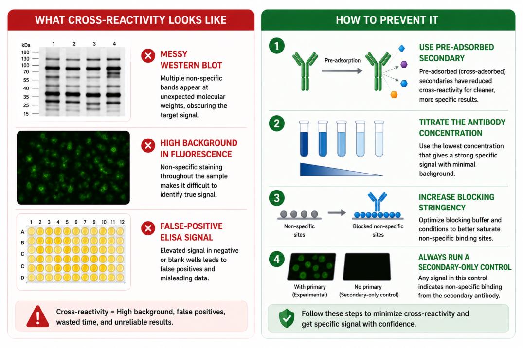
Cross-reactivity in secondary antibodies occurs when the secondary binds to something other than the intended primary antibody. This can be another antibody in your sample, an endogenous immunoglobulin in the tissue, or a structurally similar protein that shares epitopes with the intended target. The result is elevated background, false-positive signal, or mislocalized staining that can derail an entire experiment.
The Most Common Causes of Cross-Reactivity
- Endogenous immunoglobulins in tissue samples: Tissues from mammals contain their own IgG molecules. If your secondary antibody was raised in a host that is immunologically similar to the tissue species, cross-reactivity is likely without pre-adsorption.
- Shared epitopes between species: Immunoglobulins from closely related species (for example, rat and mouse) share structural similarities. A secondary antibody raised against mouse IgG may also recognize rat IgG to some degree, producing non-specific signal.
- Excessive secondary antibody concentration: Using too much secondary antibody increases the probability of low-affinity, non-specific binding events. Titration is one of the most effective and underused controls in antibody-based assays.
- Insufficient blocking: Incomplete saturation of non-specific binding sites before the secondary antibody step allows the secondary to stick to the membrane, slide, or plate surface rather than to the primary antibody.
Four Practical Steps to Eliminate Cross-Reactivity
- Use a pre-adsorbed secondary antibody. This is the single most effective intervention. Pre-adsorbed secondary antibodies have already had cross-reactive clones removed and will perform more cleanly in complex samples.
- Titrate your secondary antibody concentration. Do not assume the manufacturer's recommended dilution is optimal for your specific assay conditions. Run a dilution series and select the concentration that gives maximum specific signal with minimum background.
- Optimize your blocking conditions. The blocking buffer composition matters. BSA-based blocks tend to work better for fluorescence applications, while milk-based blocks are standard for Western blotting. Match your block to your detection system.
- Always include a secondary-only control. Remove the primary antibody from one replicate and run the secondary antibody alone. Any signal in this control is coming from the secondary itself and represents your true background baseline.
Important: A secondary-only control is non-negotiable. It is the fastest way to determine whether background signal is coming from the secondary antibody or from another component of your assay. This simple control can save hours of troubleshooting and help ensure accurate interpretation of your results.
Secondary Antibody Selection by Application
Different applications need different secondary antibodies. A secondary technique that works well in one setting may perform poorly in another.
- Western Blot: HRP-conjugated secondary antibodies are commonly used for chemiluminescent Western blot detection because of their high sensitivity and compatibility with ECL substrates. Anti-mouse and anti-rabbit HRP secondaries cover most experiments. Typical dilutions range from 1:1000 to 1:5000.
- Immunofluorescence (IF) and Immunohistochemistry (IHC): Fluorescent secondaries, especially Alexa Fluor dyes, are commonly used in IF due to their brightness and photostability. Choose fluorophores compatible with your microscope lasers. In IHC, HRP-based detection is often preferred because biotin systems can create tissue background.
- ELISA: HRP-conjugated secondaries are widely used for colorimetric ELISA detection, while AP conjugates work better in alkaline conditions. Typical dilutions are around 1:5000 to 1:10000.
- Flow Cytometry: Flow cytometry relies on fluorescent secondary antibodies matched to the cytometer’s laser lines. For complex multicolor panels, directly conjugated primary antibodies are usually preferred to reduce spectral overlap.
- Co-Immunoprecipitation (Co-IP): In Co-IP, heavy and light chains from the primary antibody can interfere with detection. Light-chain-specific secondary antibodies help reduce this background and produce cleaner blots.
Source Your Secondary Antibodies from AAA Biotech
At AAA Biotech, we understand that a reliable secondary antibody is not just a reagent. It is the foundation of reproducible data. Our catalog includes a wide range of validated secondary antibodies covering the most commonly used host-target species pairings, conjugated to HRP, Alexa Fluor dyes, FITC, biotin, and more.
Every product in our secondary antibody portfolio is quality-controlled for titer, specificity, and lot-to-lot consistency before it reaches your lab.
Browse our full catalog or contact the AAA Biotech team directly to discuss your experimental needs.
Faq's
What is the difference between a primary and a secondary antibody?
A primary antibody binds directly to the antigen you want to detect. A secondary antibody binds to the primary antibody itself, typically at its Fc region, and carries the detectable label such as an enzyme or fluorescent dye. The primary antibody provides the specificity, while the secondary antibody provides the signal.
What does pre-adsorbed mean in a secondary antibody?
Pre-adsorption is a process in which the secondary antibody is incubated with immunoglobulins from other species to remove any clones that cross-react with those species. The result is a highly specific secondary antibody with reduced background in complex or tissue-based assays. Pre-adsorbed secondary antibodies are particularly important for multiplex experiments or any work involving samples that contain endogenous immunoglobulins.
What causes non-specific binding in secondary antibodies?
Non-specific binding from secondary antibodies usually results from four factors: insufficient blocking before applying the secondary, an antibody concentration that is too high, cross-reactivity with endogenous immunoglobulins in the sample, or a secondary antibody that has not been pre-adsorbed for the relevant species. Each of these can be addressed systematically through a secondary-only control experiment followed by targeted troubleshooting.
How do I know if my secondary antibody is causing cross-reactivity?
The most direct way to identify secondary antibody cross-reactivity is to run a secondary-only control: perform the experiment exactly as normal but omit the primary antibody. If signal appears in this control, it is coming from the secondary antibody binding non-specifically. Common signs of cross-reactivity include high background across the entire membrane or slide, unexpected bands at non-target molecular weights, or signal in tissues where the target protein should not be expressed.
What is the best secondary antibody for multiplex experiments?
For multiplexing, the best approach combines isotype-specific secondary antibodies and pre-adsorbed reagents. When your primary antibodies come from different species (for example, one mouse and one rabbit primary), use species-specific secondaries conjugated to spectrally distinct fluorophores. When both primaries come from the same species, use isotype-specific secondary antibodies (for example, anti-mouse IgG1 and anti-mouse IgG2a) to distinguish the signals without cross-talk between the two detection channels.
Do secondary antibodies require special storage conditions?
Most secondary antibodies should be stored at 4 degrees Celsius for short-term use and at minus 20 degrees Celsius for long-term storage. Repeated freeze-thaw cycles degrade antibody performance significantly, so it is best practice to aliquot the stock into single-use volumes when you receive a new vial.
Lead Clinical Research Coordinator (LCRC)
Cynthia Lee is the President of AAA Biotech and specializes in understanding highly validated and characterized monoclonal/polyclonal antibodies, recombinant proteins, and ELISA kits.
Related Posts