Get accurate results in your research with our Monoclonal Antibodies, which are specially made to target exactly what you require for your research, and will produce consistent, reliable performance in lab tests.
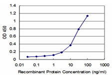
Application Data (Detection limit for recombinant GST tagged PGGT1B is ~1ng/ml as a capture antibody.)
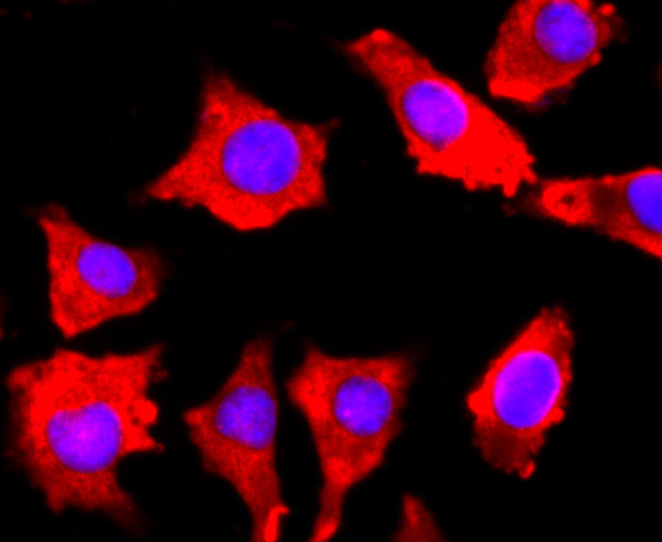
IF (Immunofluorescence) (Immunofluorescence of monoclonal antibody to PGGT1B on HeLa cell. [antibody concentration 10ug/ml])
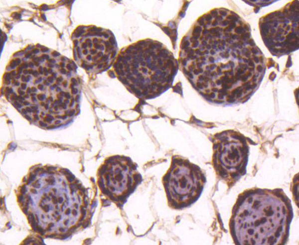
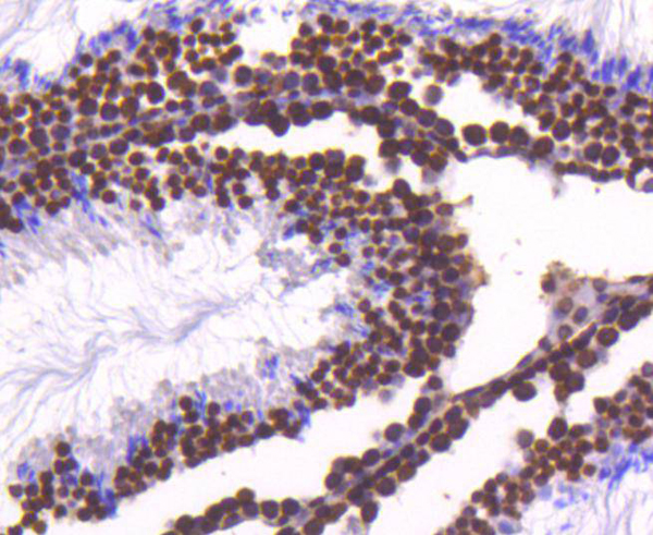
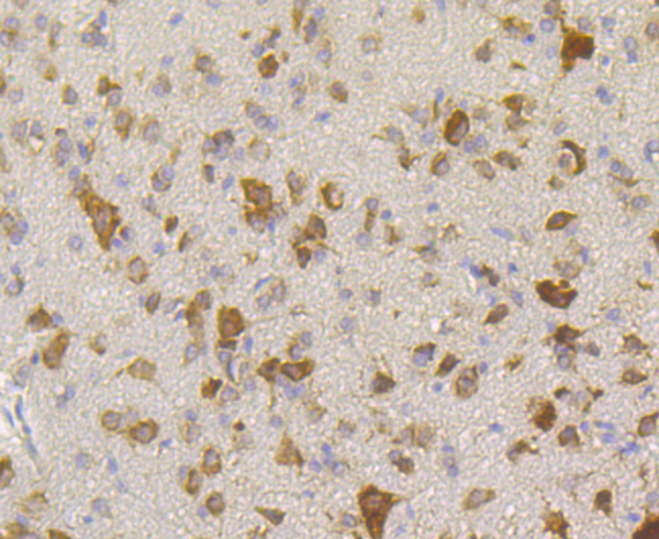
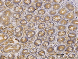
IHC (Immunohistochemistry) (Immunoperoxidase of monoclonal antibody to PGGT1B on formalin-fixed paraffin-embedded human kidney. [antibody concentration 1.2ug/ml])
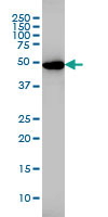
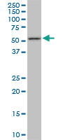
WB (Western Blot) (PGGT1B monoclonal antibody Western Blot analysis of PGGT1B expression in PC-12.)
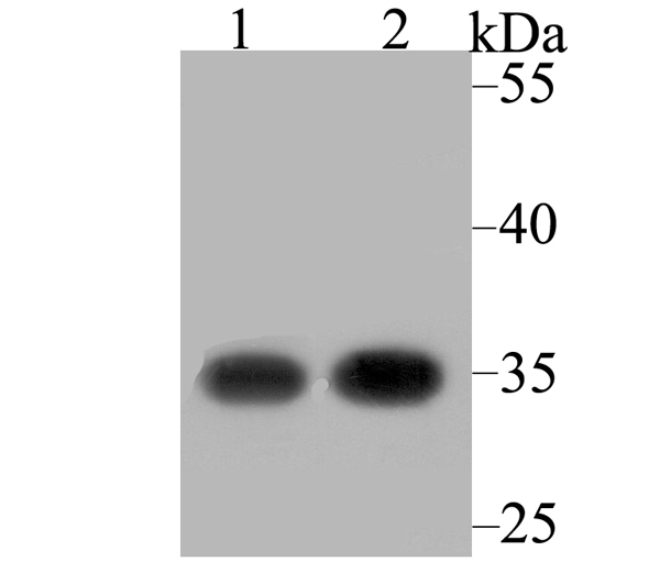
WB (Western Blot) (PGGT1B monoclonal antibody Western Blot analysis of PGGT1B expression in HeLa.)
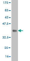
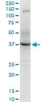
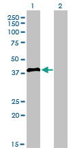
WB (Western Blot) (Western Blot detection against Immunogen (37.29kD).)
PGGT1B (Geranylgeranyl Transferase Type-1 Subunit beta, Geranylgeranyl Transferase Type I Subunit beta, GGTase-I-beta, Type I Protein Geranyl-Geranyltransferase Subunit beta) (PE)
Reactivity
Human, Rat
Applications
ELISA (EIA), Immunofluorescence (IF), Immunohistochemistry (IHC), Western Blot (WB)
Application Data (Detection limit for recombinant GST tagged NFKB1 is approximately 3ng/ml as a capture antibody.)
IF (Immunofluorescence) (Immunofluorescence of monoclonal antibody to NFKB1 on HeLa cell. [antibody concentration 10 ug/ml])
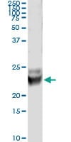
WB (Western Blot) (NFKB1 monoclonal antibody (M03), clone 3F6 Western Blot analysis of NFKB1 expression in Hela S3 NE (Cat # L013V3).)
IHC (Immunohistochemistry) (Immunoperoxidase of monoclonal antibody to NFKB1 on formalin-fixed paraffin-embedded human salivary gland. [antibody concentration 3 ug/ml])
Application Data (Immunoperoxidase of monoclonal antibody to NFKB1 on formalin-fixed paraffin-embedded human salivary gland. [antibody concentration 3 ug/ml])
ICC (Immunocytochemistry) (ICC staining Hemopexin in SK-Br-3 cells (green). The nuclear counter stain is DAPI (blue). Cells were fixed in paraformaldehyde, permeabilised with 0.25% Triton X100/PBS.)
ICC (Immunocytochemistry) (ICC staining Hemopexin in LOVO cells (green). The nuclear counter stain is DAPI (blue). Cells were fixed in paraformaldehyde, permeabilised with 0.25% Triton X100/PBS.)
ICC (Immunocytochemistry) (ICC staining Hemopexin in LO2 cells (green). The nuclear counter stain is DAPI (blue). Cells were fixed in paraformaldehyde, permeabilised with 0.25% Triton X100/PBS.)
ICC (Immunocytochemistry) (ICC staining Hemopexin in HepG2 cells (green). The nuclear counter stain is DAPI (blue). Cells were fixed in paraformaldehyde, permeabilised with 0.25% Triton X100/PBS.)
IHC (Immunohistochemistry) (Immunohistochemical analysis of paraffin-embedded human placenta tissue using anti-Hemopexin antibody. Counter stained with hematoxylin.)
IHC (Immunohistochemistry) (Immunohistochemical analysis of paraffin-embedded human liver tissue using anti-Hemopexin antibody. Counter stained with hematoxylin.)
WB (Western Blot) (Western blot analysis of Hemopexin on human plasma lysate using anti-Hemopexin antibody at 1/500 dilution.)
IP (Immunoprecipitation) (Immunoprecipitation analysis of Hela cell lysates using Bip/GRP78 (6H7) Mouse mAb.)
ICC (Immunocytochemistry) (Immunocytochemistry staining of HeLa cells using Bip/GRP78 (6H7) Mouse mAb(dilution 1:100).)
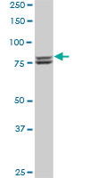
WB (Western Blot) (Western blot detection of Bip/GRP78 in Jurkat,MCF7,SKBR-3,Hela cell lysates using Bip/GRP78 (6H7) Mouse mAb(1:2000 diluted). Predicted band size:78KDa. Observed band size:78KDa.)
IHC (Immunohistochemistry) (Immunohistochemical analysis of paraffin-embedded Human Breast Caricnoma using GRP78/BipMouse mAb diluted at 1:200.)
IHC (Immunohistochemistry) (Immunohistochemical analysis of paraffin-embedded Human Colon Caricnoma using GRP78/BipMouse mAb diluted at 1:200.)
WB (Western Blot) (Western blot analysis of 1)Hela, 2) Rat LiverTissue with GRP78/Bip Mouse mAb diluted at 1:2,000.)
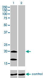
WB (Western Blot) (Western Blot analysis of FAM3B expression in transfected 293T cell line by FAM3B monoclonal antibody. Lane 1: FAM3B transfected lysate (26kD). Lane 2: Non-transfected lysate.)
WB (Western Blot) (Western blot analysis of TAF7 over-expressed 293 cell line, cotransfected with TAF7 Validated Chimera RNAi (Lane 2) or non-transfected control (Lane 1). Blot probed with TAF7 monoclonal antibody. GAPDH (36.1kD) used as specificity and loading control.)
Application Data (Detection limit for recombinant GST tagged TAF7 is ~0.3ng/ml as a capture antibody.)
IF (Immunofluorescence) (Immunofluorescence of monoclonal antibody to TAF7 on HeLa cell. [antibody concentration 10ug/ml])
IHC (Immunohistochemistry) (Immunoperoxidase of monoclonal antibody to TAF7 on formalin-fixed paraffin-embedded human placenta. [antibody concentration 1ug/ml])
IHC (Immunohistochemistry) (Immunoperoxidase of monoclonal antibody to TAF7 on formalin-fixed paraffin-embedded human lymph node. [antibody concentration 3ug/ml])
WB (Western Blot) (Western Blot analysis of TAF7 expression in transfected 293T cell line by TAF7 monoclonal antibody. Lane 1: TAF7 transfected lysate (40.3kD). Lane 2: Non-transfected lysate.)
WB (Western Blot) (TAF7 monoclonal antibody, Western Blot analysis of TAF7 expression in MCF-7.)
CD28 (CD28 Antigen, CD28 Molecule, MGC138290, T cell Antigen CD28, T cell Specific Surface Glycoprotein, T cell Specific Surface Glycoprotein CD28, Tp44) (MaxLight 550)
SDS-PAGE (Whole cell lysates were separated by 10% SDS-PAGE, and the membrane was blotted with anti-Mesothelin (ABT-MSLN) antibody. The HRP-conjugated Goat anti-M)
WB (Western Blot) (Western blot analysis of 1) Mouse Liver Tissue, 2) Rat Liver Tissue using HAO1 Monoclonal Antibody.)
IF (Immunofluorescence) (Immunofluorescence analysis of Mouse-spleen tissue. 1,HAO1 Monoclonal Antibody(Mix)(red) was diluted at 1:200(4 degree C,overnight). 2, Cy3 labled Secondary antibody was diluted at 1:300(room temperature, 50min).3, Picture B: DAPI(blue) 10min. Picture A:Target. Picture B: DAPI. Picture C: merge of A+B)
IF (Immunofluorescence) (Immunofluorescence analysis of Human-appendix tissue. 1,HAO1 Monoclonal Antibody(Mix)(red) was diluted at 1:200(4 degree C,overnight). 2, Cy3 labled Secondary antibody was diluted at 1:300(room temperature, 50min).3, Picture B: DAPI(blue) 10min. Picture A:Target. Picture B: DAPI. Picture C: merge of A+B)
IHC (Immunohistochemistry) (Immunohistochemical analysis of paraffin-embedded Mouse-heart tissue. 1,HAO1 Monoclonal Antibody(Mix) was diluted at 1:200(4 degree C,overnight). 2, Sodium citrate pH 6.0 was used for antibody retrieval(>98 degree C,20min). 3,Secondary antibody was diluted at 1:200(room tempeRature, 30min). Negative control was used by secondary antibody only.)
IHC (Immunohistochemistry) (Immunohistochemical analysis of paraffin-embedded Rat-liver tissue. 1,HAO1 Monoclonal Antibody(Mix) was diluted at 1:200(4 degree C,overnight). 2, Sodium citrate pH 6.0 was used for antibody retrieval(>98 degree C,20min). 3,Secondary antibody was diluted at 1:200(room tempeRature, 30min). Negative control was used by secondary antibody only.)
IHC (Immunohistochemistry) (Immunohistochemical analysis of paraffin-embedded Human-liver tissue. 1,HAO1 Monoclonal Antibody(Mix) was diluted at 1:200(4 degree C,overnight). 2, Sodium citrate pH 6.0 was used for antibody retrieval(>98 degree C,20min). 3,Secondary antibody was diluted at 1:200(room tempeRature, 30min). Negative control was used by secondary antibody only.)
WB (Western Blot) (AKAP8 monoclonal antibody (Western Blot analysis of AKAP8 expression in HeLa NE)
IP (Immunoprecipitation) (Immunoprecipitation of AKAP8 transfected lysate using AKAP8 monoclonal antibody and Protein A Magnetic Bead, and immunoblotted with AKAP8 monoclonal antibody.)
IF (Immunofluorescence) (Immunofluorescence of monoclonal antibody to AKAP8 on HeLa cell. [antibody concentration 10ug/ml])
IHC (Immunohistochemistry) (Immunoperoxidase of monoclonal antibody to AKAP8 on formalin-fixed paraffin-embedded human pancreas. [antibody concentration 3ug/ml])
WB (Western Blot) (Western Blot analysis of AKAP8 expression in transfected 293T cell line by AKAP8 monoclonal antibody Lane 1: AKAP8 transfected lysate (76.2kD). Lane 2: Non-transfected lysate.)
WB (Western Blot) (Western Blot detection against Immunogen (38.43kD).)
Application Data (Detection limit for recombinant GST tagged YY1 is approximately 0.03ng/ml as a capture antibody.)
IF (Immunofluorescence) (Immunofluorescence of monoclonal antibody to YY1 on HeLa cell. [antibody concentration 10 ug/ml])
WB (Western Blot) (YY1 monoclonal antibody (M03), clone 4D2 Western Blot analysis of YY1 expression in Hela S3 NE.)
IHC (Immunohistochemistry) (Immunoperoxidase of monoclonal antibody to YY1 on formalin-fixed paraffin-embedded human endometrium. [antibody concentration 3 ug/ml])
Application Data (Immunoperoxidase of monoclonal antibody to YY1 on formalin-fixed paraffin-embedded human endometrium. [antibody concentration 3 ug/ml])
Application Data (Staining of mouse peritoneal macrophages with Rat anti Mouse CD11b)
Application Data (Staining of mouse peritoneal macrophages cells with Rat anti Mouse CD11b:Alexa Fluor 488)
Application Data (Staining of total mouse peritoneal exudate cells demonstrating labelling of macrophages with Rat anti Mouse CD11b:RPE)
Application Data (Published customer image: Immunocytochemical characterisation of cultures of mouse microglia. Cultures of microglia were immunostained with anti-CD11b (a) and anti-GFAP (c). Cultures of astrocytes were immunostained with anti-CD11b (c) and anti-GFAP (d). All cells were also counterstained with DAPI (blue) to identify the cells' nuclei.From: Ferger et al. Journal of Neuroinflammation 2010 7:45)
Application Data (Staining of mouse peritoneal macrophages with Rat anti Mouse CD11b:RPE)
Application Data (Staining of mouse peritoneal macrophages cells with Rat anti Mouse CD11b:FITC)
Application Data (Immunoperoxidase staining of mouse lymph node cryosection using Rat anti Mouse CD11b antibody followed by horseradish peroxidase conjugated Goat anti Rat IgG . High power)
Application Data (Staining of mouse peritoneal macrophages with Rat anti Mouse CD11b: Alexa Fluor 647)
Application Data (Immunoperoxidase staining of mouse lymph node cryosection using Rat anti Mouse CD11b antibody followed by horseradish peroxidase conjugated Goat anti Rat IgG . Medium power)
Application Data (Immunoperoxidase staining of rat lymph node cryosection with Mouse anti Rat CD25 followed by horseradish peroxidase conjugated Goat anti Mouse IgG1 as a detection reagent. High power)
Application Data (Immunofluorescence staining of rat lymph node cryosection with Mouse anti Rat CD25 antibody , red in A and Mouse anti Rat CD8 , green in B. C is the merged image with nuclei counterstained blue using DAPI. Medium power)
Application Data (Published customer image: Analysis of graft-infiltrating T cells. Activated CD25+ T cells and CD4+ and CD8+ T cell subsets were stained at the time points of corneal allograft rejection and calculated within the graft. A, C, and E show representative histological staining for CD25, CD4, and CD8 in grafts of treated and control animals, respectively. CD25+ (B) and CD4+ (D) cells infiltrated to a statistically significantly stronger extent in 3.2.3-treated animals when compared to control treated animals (*p)
Application Data (Staining of rat spleen cells with Mouse anti Rat CD25: FITC)
Application Data (Published clone specific image: The percentage of Ly49s3+ NK cells increases in the spleen and decreases in the bone marrow following L. monocytogenes infection. A) Mononuclear leukocytes were harvested from spleen, blood, and bone marrow from uninfected and infected animals 24 and 48 hrs p.i. Cells were stained with antibodies to NKR-P1A, CD3, and Ly49s3. NK cells were gated as NKR-P1A+CD3- cells, and the percentage of Ly49s3+ NK cells was analysed by flow cytometry. The data represent the mean +/- SEM from three independent experiments, with 2 animals at each time point. Data were analyzed with the one-way ANOVA with Tukey's multiple comparisons test. B) The activation status of NK cells from uninfected (filled histogram) or infected animals 24 (grey line) and 48 hrs p.i. (black line) was tested by flow cytometry. An antibody towards the activation marker CD25 was employed.From: Shegarfi H, Naper C, Rolstad B, Inngjerdingen M (2010) Listeria monocytogenes Infection Affects a Subset of Ly49-Expressing NK Cells in the Rat. PLoS ONE 5(12): e15579.)
Application Data (Staining of stimulated rat spleen cells with Mouse anti Rat CD25:FITC)
Application Data (Immunoperoxidase staining of rat lymph node cryosection with Mouse anti Rat CD25 followed by horseradish peroxidase conjugated Goat anti Mouse IgG1 as a detection reagent. Medium power)
Application Data (Immunofluorescence staining of rat lymph node cryosection with Mouse anti Rat CD25 antibody , red in A and Mouse anti Rat CD8 , green in B. C is the merged image with nuclei counterstained blue using DAPI. High power)
Application Data (Immunofluorescence staining of rat lymph node cryosection with Mouse anti Rat CD25 antibody , red in A and Mouse anti Rat CD8 , green in B. C is the merged image with nuclei counterstained blue using DAPI. Low power)
Application Data (Staining of Con A activated rat spleen cells with Mouse anti Rat CD25: Biotin)
Application Data (Immunoperoxidase staining of rat lymph node cryosection with Mouse anti Rat CD86 antibody, clone 42F followed by horseradish peroxidase conjugated Goat anti Mouse IgG . Low power)
Application Data (Immunoperoxidase staining of rat lymph node cryosection with Mouse anti Rat CD86 antibody, clone 42F followed by horseradish peroxidase conjugated Goat anti Mouse IgG . Medium power)
Application Data (Staining of rat splenocytes with Mouse anti Rat CD86:RPE)
Application Data (Staining of rat splenocytes with Mouse anti Rat CD86:Alexa Fluor ?647)
Application Data (Staining of rat splenocytes with Mouse anti Rat CD86:Low Endotoxin)
Application Data (Staining of rat splenocytes with Mouse anti Rat CD86:FITC)
Application Data (Staining of rat splenocytes with Mouse anti Rat CD86:Alexa Fluor ?488)
Application Data (Immunoperoxidase staining of rat lymph node cryosection with Mouse anti Rat CD86 antibody, clone 42F followed by horseradish peroxidase conjugated Goat anti Mouse IgG . High power)
WB (Western Blot) (FLJ23356 monoclonal antibody. Western Blot analysis of FLJ23356 expression in HeLa.)
WB (Western Blot) (FLJ23356 monoclonal antibody. Western Blot analysis of FLJ23356 expression in HepG2.)
Application Data (Detection limit for recombinant GST tagged FLJ23356 is ~0.1ng/ml as a capture antibody.)
IF (Immunofluorescence) (Immunofluorescence of monoclonal antibody to FLJ23356 on HeLa cell. [antibody concentration 10ug/ml].)
WB (Western Blot) (Western Blot analysis of FLJ23356 expression in transfected 293T cell line by FLJ23356 monoclonal antibody. Lane 1: FLJ23356 transfected lysate (38.5kD). Lane 2: Non-transfected lysate.)
WB (Western Blot) (Western blot analysis of FLJ23356 over-expressed 293 cell line, cotransfected with FLJ23356 Validated Chimera RNAi (Lane 2) or non-transfected control (Lane 1). Blot probed with FLJ23356 monoclonal antibody. GAPDH (36.1kD) used as specificity and loading control.)
WB (Western Blot) (Western Blot detection against Immunogen (37.11kD).)
WB (Western Blot) (FGR monoclonal antibody (M02), clone 3B11. Western Blot analysis of FGR expression in Raw 264.7 (Cat # L024V1).)
WB (Western Blot) (FGR monoclonal antibody (M02), clone 3B11. Western Blot analysis of FGR expression in PC-12 (Cat # L012V1).)
Application Data (Detection limit for recombinant GST tagged FGR is approximately 0.1ng/ml as a capture antibody.)
WB (Western Blot) (FGR monoclonal antibody (M02), clone 3B11 Western Blot analysis of FGR expression in HeLa (Cat # L013V1).)
IHC (Immunohistochemistry) (Immunoperoxidase of monoclonal antibody to FGR on formalin-fixed paraffin-embedded human lymph node. [antibody concentration 3 ug/ml])
Application Data (Immunoperoxidase of monoclonal antibody to FGR on formalin-fixed paraffin-embedded human lymph node. [antibody concentration 3 ug/ml])
WB (Western Blot) (LZTFL1 monoclonal antibody, Western Blot analysis of LZTFL1 expression in HeLa.)
Application Data (Detection limit for recombinant GST tagged LZTFL1 is ~0.1ng/ml as a capture antibody.)
IF (Immunofluorescence) (Immunofluorescence of monoDetection limit for recombinant GST tagged LZTFL1 is ~0.1ng/ml as a capture antibody.clonal antibody to LZTFL1 on HeLa cell. [antibody concentration 25ug/ml].)
IHC (Immunohistochemistry) (Immunoperoxidase of monoclonal antibody to LZTFL1 on formalin-fixed paraffin-embedded human colon. [antibody concentration 3ug/ml].)
WB (Western Blot) (Western Blot analysis of LZTFL1 expression in transfected 293T cell line by LZTFL1 monoclonal antibody. Lane 1: LZTFL1 transfected lysate (34.6kD). Lane 2: Non-transfected lysate.)
WB (Western Blot) (Western Blot detection against Immunogen (37.11kD).)
WB (Western Blot) (ASB9 monoclonal antibody, Western Blot analysis of ASB9 expression in HepG2.)
Application Data (Detection limit for recombinant GST tagged ASB9 is ~0.1ng/ml as a capture antibody.)
IP (Immunoprecipitation) (Immunoprecipitation of ASB9 transfected lysate using ASB9 monoclonal antibody and Protein A Magnetic Bead and immunoblotted with ASB9 rabbit polyclonal antibody.)
IHC (Immunohistochemistry) (Immunoperoxidase of monoclonal antibody to ASB9 on formalin-fixed paraffin-embedded human lymphoma tissue. [antibody concentration 3ug/ml])
WB (Western Blot) (Western Blot analysis of ASB9 expression in transfected 293T cell line by ASB9 monoclonal antibody. Lane 1: ASB9 transfected lysate (31.9kD). Lane 2: Non-transfected lysate.)
WB (Western Blot) (Western Blot detection against Immunogen (53.83kD).)
CD28 (CD28 Antigen, CD28 Molecule, MGC138290, T cell Antigen CD28, T cell Specific Surface Glycoprotein, T cell Specific Surface Glycoprotein CD28, Tp44) (MaxLight 650)
FCM (Flow Cytometry) (Flow cytometric analysis of Hela cells with LOX antibody at 1/100 dilution (red) compared with an unlabelled control (cells without incubation with primary antibody; black). Alexa Fluor 488-conjugated goat anti rabbit IgG was used as the secondary antibody.)
ICC (Immunocytochemistry) (ICC staining LOX in SH-SY5Y cells (green). The nuclear counter stain is DAPI (blue). Cells were fixed in paraformaldehyde, permeabilised with 0.25% Triton X100/PBS.)
ICC (Immunocytochemistry) (ICC staining LOX in HUVEC cells (green). The nuclear counter stain is DAPI (blue). Cells were fixed in paraformaldehyde, permeabilised with 0.25% Triton X100/PBS.)
ICC (Immunocytochemistry) (ICC staining LOX in Hela cells (green). The nuclear counter stain is DAPI (blue). Cells were fixed in paraformaldehyde, permeabilised with 0.25% Triton X100/PBS.)
IHC (Immunohistchemistry) (Immunohistochemical analysis of paraffin-embedded mouse heart tissue using anti-LOX antibody. Counter stained with hematoxylin.)
IHC (Immunohistochemistry) (Immunohistochemical analysis of paraffin-embedded human placenta tissue using anti-LOX antibody. Counter stained with hematoxylin.)
IHC (Immunohistochemistry) (Immunohistochemical analysis of paraffin-embedded human kidney tissue using anti-LOX antibody. Counter stained with hematoxylin.)
IHC (Immunohistochemistry) (Immunohistochemical analysis of paraffin-embedded rat skeletal muscle tissue using anti-LOX antibody. Counter stained with hematoxylin.)
WB (Western Blot) (Western blot analysis of LOX on Zebrafish tissue lysates using anti-LOX antibody at 1/1, 000 dilution.)
WB (Western Blot) (Western blot analysis of LOX on different lysates using anti-LOX antibody at 1/1, 000 dilution. Positive control: Lane 1: Jurkat Lane 2: Mouse brain Lane 3: Human fetal brain)
Application Data (Immunoperoxidase staining of human tonsil cryosection using Mouse anti Human CD3 antibody, clone UCHT1 followed by horseradish peroxidase Goat anti Mouse IgG2a antibody as a detection reagent. Medium power)
Application Data (Staining of human peripheral blood lymphocytes with Mouse anti Human CD3:RPE)
Application Data (Immunoperoxidase staining of human tonsil cryosection using Mouse anti Human CD3 antibody, clone UCHT1 followed by horseradish peroxidase Goat anti Mouse IgG2a antibody as a detection reagent. Low power)
Application Data (ELISA analysis of human CD3 expression using Rat anti Human CD3ε, clone CD3-12 as a capture reagent and biotinylated Mouse anti Human CD3, clone UCHT1 as a detection reagent with recombinant human CD3 as antigen to produce the standard curve. Detection is by HRP conjugated streptavidin and substrate. Microtitre plate is read at O.D. 450 nm on the iMark Microplate Absorbance Reader . A serum (green) sample is included undiluted)
Application Data (Immunoperoxidase staining of human tonsil cryosection using Mouse anti Human CD3 antibody, clone UCHT1 followed by horseradish peroxidase Goat anti Mouse IgG2a antibody as a detection reagent. High power)
Application Data (Staining of human peripheral blood lymphocytes with Mouse anti Human CD3:Alexa Fluor 405)
Application Data (Staining of human peripheral blood lymphocytes with Mouse anti Human CD3)
Application Data (Immunoperoxidase staining of human tonsil cryosection using Mouse anti Human CD3 antibody, clone UCHT1 followed by horseradish peroxidase Goat anti Mouse IgG2a antibody as a detection reagent. High power)
Application Data (Staining of human peripheral blood lymphocytes with Mouse anti Human CD3: RPE- Alexa Fluor 750)
Application Data (Staining of human peripheral blood lymphocytes with Mouse anti Human CD3: FITC)
Application Data (Staining of mouse spleen with Rat anti Mouse CD4:RPE)
Application Data (Staining of mouse spleen with Rat anti Mouse CD4:Alexa Fluor ?488)
Application Data (Staining of mouse peripheral blood lymphocytes with Rat anti Mouse CD4:Low Endotoxin)
Application Data (Staining of mouse peripheral blood lymphocytes with Rat anti Mouse CD4:FITC)
Application Data (Staining of mouse peripheral blood lymphocytes with Rat anti Mouse CD4 Alexa Fluor ?647)
Application Data (Published customer image: Intrafollicular location of MZB cells in B6.TC spleens. A. Representative spleen sections from 3 mo old B6.TC (left) and B6 (right) mice stained with Moma-1-FITC, CD1d-PE and B220-PB. The B220+ CD1d+ MZB cells show as bright pink while the other B cells show as blue. The ring of Moma-1+ metallophillic macrophages delineates the MZ inner edge. B. Percentage of CD1d+ B220+ B cells relative to total B220+ B cells outside (MZ) and inside (FO) the Moma-1+ ring in 3 mo and 10 mo old B6 and B6.TC mice. The data show means and standard errors of the mean (SEM) calculated of 4 MZ and FO areas for each mouse. ***: p < 0.001 for t tests. C. Representative spleen sections from 10 mo old B6.TC (left) and B6 (right) mice stained with CD4-FITC, CD1d-PE and B220-PB. Boxed areas show multiple contacts between green B6.TC CD4 T cells and pink MZB cells, but not in the B6 spleen. Original magnification: 200X.From: Zhou et al. BMC Immunology 2011 12:7.)
Application Data (Immunoperoxidase staining of a mouse lymph node cryosection with Rat anti Mouse CD4 antibody, clone GK1.5 followed by horseradish peroxidase conjugated Goat anti Rat IgG . Low power)
Application Data (Immunoperoxidase staining of a mouse lymph node cryosection with Rat anti Mouse CD4 antibody, clone GK1.5 followed by horseradish peroxidase conjugated Goat anti Rat IgG . High power)
Application Data (Immunoperoxidase staining of a mouse lymph node cryosection with Rat anti Mouse CD4 antibody, clone GK1.5 followed by horseradish peroxidase conjugated Goat anti Rat IgG . Medium power)
Application Data (Staining of mouse spleen with Rat anti Mouse CD4)
WB (Western Blot) (Western blot analysis of ARID3A over-expressed 293 cell line, cotransfected with ARID3A Validated Chimera RNAi (Lane 2) or non-transfected control (Lane 1). Blot probed with ARID3A monoclonal antibody. GAPDH (36.1kD) used as specificity and loading control.)
Application Data (Detection limit for recombinant GST tagged ARID3A is ~0.03ng/ml as a capture antibody.)
IF (Immunofluorescence) (Immunofluorescence of monoclonal antibody to ARID3A on HeLa cell. [antibody concentration 10ug/ml].)
IHC (Immunohistochemistry) (Immunoperoxidase of monoclonal antibody to ARID3A on formalin-fixed paraffin-embedded human placenta. [antibody concentration 3ug/ml].)
WB (Western Blot) (ARID3A monoclonal antibody, Western Blot analysis of ARID3A expression in K-562.)
WB (Western Blot) (Western Blot detection against Immunogen (36.74kD).)
ARID3A (AT-rich Interactive Domain-containing Protein 3A, ARID Domain-containing Protein 3A, B Cell Regulator of IgH Transcription, Bright, Dead Ringer-like Protein 1, E2F-binding Protein 1, DRIL1, DRIL3, DRX, E2FBP1) (FITC)
Gene Names
ARID3A; DRIL1; DRIL3; BRIGHT; E2FBP1
Reactivity
Human
Applications
ELISA (EIA), Immunofluorescence (IF), Immunohistochemistry (IHC), Western Blot (WB)
Application Data (Proximity Ligation Analysis of protein-protein interactions between CRKL and PIK3R1. Mahlavu cells were stained with anti-CRKL rabbit purified polyclonal 1:1200 and anti-PIK3R1 mouse monoclonal antibody 1:50. Each red dot represents the detection of protein-protein interaction complex, and nuclei were counterstained with DAPI (blue).)
Application Data (Proximity Ligation Analysis of protein-protein interactions between FLT1 and PIK3R1 Huh7 cells were stained with anti-FLT1 rabbit purified polyclonal 1:1200 and anti-PIK3R1 mouse monoclonal antibody 1:50. Each red dot represents the detection of protein-protein interaction complex, and nuclei were counterstained with DAPI (blue).)
Application Data (Proximity Ligation Analysis of protein-protein interactions between EGFR and PIK3R1. HeLa cells were stained with EGFR rabbit purified polyclonal 1:1200 and PIK3R1 mouse monoclonal antibody 1:50. Each red dot represents the detection of protein-protein interaction complex, and nuclei were counterstained with DAPI (blue))
Application Data (Detection limit for recombinant GST tagged PIK3R1 is ~0.1ng/ml as a capture antibody.)
WB (Western Blot) (PIK3R1 monoclonal antibody Western Blot analysis of PIK3R1 expression in PC-12.)
WB (Western Blot) (PIK3R1 monoclonal antibody Western Blot analysis of PIK3R1 expression in HeLa.)
WB (Western Blot) (Western Blot detection against Immunogen (75.68kD).)
Application Data (Detection limit for recombinant GST tagged TP53 is approximately 1ng/ml as a capture antibody.)
WB (Western Blot) (TP53 monoclonal antibody (M01), clone 2C3 Western Blot analysis of TP53 expression in A-431 (Cat # L015V1).)
IF (Immunofluorescence) (Immunofluorescence of monoclonal antibody to TP53 on A-431 cell. [antibody concentration 10 ug/ml])
IHC (Immunohistochemistry) (Immunoperoxidase of monoclonal antibody to TP53 on formalin-fixed paraffin-embedded human salivary gland. [antibody concentration 3 ug/ml])
Application Data (Immunoperoxidase of monoclonal antibody to TP53 on formalin-fixed paraffin-embedded human salivary gland. [antibody concentration 3 ug/ml])
Application Data (Published customer image: Representative images of the inflammatory changes in the facial nucleus during axonal regeneration, one week following facial nerve transaction. a, b: CD11b immunoreactivity for microglia is increased in the axotomized facial nucleus, and microglia enwrap the facial motor neurons, e.g. at arrows. The regenerating neurons were retrogradely labelled with fluorogold. c, d: CD6- positive T-cells accumulated in the injured motor nucleus (arrows). They had little cytoplasm but dense nuclei (c) and were sometimes clustered around neurons retrogradely labelled with fluorogold (d). The scale bar in (a) also applies to (b) and that in (c) also applies to (d).From: Shokouhi et al. BMC Neuroscience 2010 11:13.)
Application Data (Published customer image: CD11b immunoreactive (red) microglia in the motor cortex following cervical corticospinal tract injury. Representative images of motor cortex - a, b: one week (deconvolved images); c, d: two weeks following corticospinal tract injury. The corticospinal neurons were retrogradely labelled with fluorogold applied to the spinal cord at the time of injury. There is little sign of microglial activation. The inset in (c) shows a CD6-positive T-cell in the motor cortex, but no accumulation of such cells was detected.From: Shokouhi BN, Wong BZ, Siddiqui S, Lieberman AR, Campbell G, Tohyama K, Anderson PN. Microglial responses around intrinsic CNS neurons are correlated with axonal regeneration. BMC Neurosci. 2010 Feb 5;11:13.)
Application Data (Staining of rat peritoneal macrophages with Mouse anti Rat CD11b)
Application Data (Published customer image: Microglia in the red nucleus (a-d and inset) and motor cortex (e, f) 4 weeks after nerve graft implantation into the rubrospinal tract and cervical dorsal columns respectively. Neurons with regenerating axons were retrogradely labelled with fluorogold. Microglia established close contacts with regenerating rubrospinal neurons (arrows), which also expressed ATF3 (inset). No retrogradely labelled corticospinal neurons were detected (e and f) and there was no microglial activation in the motor cortex. (a-c) are non-deconvolved and (d-f plus inset) are deconvolved images. The scale bar in (a) also applies to (b).From: Shokouhi BN, Wong BZ, Siddiqui S, Lieberman AR, Campbell G, Tohyama K, Anderson PN. Microglial responses around intrinsic CNS neurons are correlated with axonal regeneration. BMC Neurosci. 2010 Feb 5;11:13.)
Application Data (Immunoperoxidase staining of rat lymph node cryosection with Mouse anti Rat CD11b antibody followed by horseradish peroxidase conjugated Goat anti Mouse IgG2a as a detection reagent. High power)
Application Data (Staining of rat peritoneal macrophages cells with Mouse anti Rat CD11b:Alexa Fluor 488)
Application Data (Published customer image: Rubrospinal injury produces little microglial activation and no accumulation of T-cells in the red nucleus. a, b: Representative images show there is little difference in CD11b immunoreactivity for microglia in the red nucleus (*) one week following axotomy. c, d: confocal images of beta thymosin immunoreactive microglia in the red nucleus 3 weeks after injury. e: a rare CD6-positive T lymphocyte in the red nucleus of an unoperated rat. f: one week after injury no T lymphocytes can be identified in the red nucleus around the axotomized rubrospinal neurons, which can be recognised by their expression of ATF3 (green). Neuronal cytoplasm has been visualised by high gain in the red signal; this does not represent CD6 signal.From: Shokouhi BN, Wong BZ, Siddiqui S, Lieberman AR, Campbell G, Tohyama K, Anderson PN. Microglial responses around intrinsic CNS neurons are correlated with axonal regeneration. BMC Neurosci. 2010 Feb 5;11:13.)
Application Data (Staining of rat peritoneal macrophages with Mouse anti Rat CD11b:RPE-Alexa Fluor 647)
Application Data (Immunofluoescence staining of rat lymph node cryosection with Mouse anti Rat CD11b antibody , red in A and Mouse anti Rat CD8 , green in B. C is the merged image with nuclei counter-stained in blue using DAPI. High power)
Application Data (Immunofluoescence staining of rat lymph node cryosection with Mouse anti Rat CD11b antibody , red in A and Mouse anti Rat CD8 , green in B. C is the merged image with nuclei counter-stained in blue using DAPI. Low power)
Application Data (Staining of mouse spleen with Rat anti Mouse CD4:RPE)
Application Data (Staining of mouse spleen with Rat anti Mouse CD4:Alexa Fluor ?488)
Application Data (Staining of mouse peripheral blood lymphocytes with Rat anti Mouse CD4:Low Endotoxin)
Application Data (Staining of mouse peripheral blood lymphocytes with Rat anti Mouse CD4:FITC)
Application Data (Staining of mouse peripheral blood lymphocytes with Rat anti Mouse CD4 Alexa Fluor ?647)
Application Data (Published customer image: Intrafollicular location of MZB cells in B6.TC spleens. A. Representative spleen sections from 3 mo old B6.TC (left) and B6 (right) mice stained with Moma-1-FITC, CD1d-PE and B220-PB. The B220+ CD1d+ MZB cells show as bright pink while the other B cells show as blue. The ring of Moma-1+ metallophillic macrophages delineates the MZ inner edge. B. Percentage of CD1d+ B220+ B cells relative to total B220+ B cells outside (MZ) and inside (FO) the Moma-1+ ring in 3 mo and 10 mo old B6 and B6.TC mice. The data show means and standard errors of the mean (SEM) calculated of 4 MZ and FO areas for each mouse. ***: p < 0.001 for t tests. C. Representative spleen sections from 10 mo old B6.TC (left) and B6 (right) mice stained with CD4-FITC, CD1d-PE and B220-PB. Boxed areas show multiple contacts between green B6.TC CD4 T cells and pink MZB cells, but not in the B6 spleen. Original magnification: 200X.From: Zhou et al. BMC Immunology 2011 12:7.)
Application Data (Immunoperoxidase staining of a mouse lymph node cryosection with Rat anti Mouse CD4 antibody, clone GK1.5 followed by horseradish peroxidase conjugated Goat anti Rat IgG . Low power)
Application Data (Immunoperoxidase staining of a mouse lymph node cryosection with Rat anti Mouse CD4 antibody, clone GK1.5 followed by horseradish peroxidase conjugated Goat anti Rat IgG . High power)
Application Data (Immunoperoxidase staining of a mouse lymph node cryosection with Rat anti Mouse CD4 antibody, clone GK1.5 followed by horseradish peroxidase conjugated Goat anti Rat IgG . Medium power)
Application Data (Staining of mouse spleen with Rat anti Mouse CD4)
WB (Western Blot) (Western blot analysis of SGK over-expressed 293 cell line, cotransfected with SGK Validated Chimera RNAi (Lane 2) or non-transfected control (Lane 1). Blot probed with SGK monoclonal antibody. GAPDH (36.1kD) used as specificity and loading control.)
Application Data (Detection limit for recombinant GST tagged SGK is ~1ng/ml as a capture antibody.)
IF (Immunofluorescence) (Immunofluorescence of monoclonal antibody to SGK on HeLa cell. [antibody concentration 10ug/ml].)
IHC (Immunohistochemistry) (Immunoperoxidase of monoclonal antibody to SGK on formalin-fixed paraffin-embedded human stomach tissue. [antibody concentration 5ug/ml].)
IHC (Immunohistochemistry) (Immunoperoxidase of monoclonal antibody to SGK on formalin-fixed paraffin-embedded human kidney. [antibody concentration 3ug/ml].)
WB (Western Blot) (Western Blot analysis of SGK expression in transfected 293T cell line by SGK monoclonal antibody. Lane 1: SGK transfected lysate (48.9kD). Lane 2: Non-transfected lysate.)
WB (Western Blot) (Western Blot detection against Immunogen (73.52kD).)
Application Data (Detection limit for recombinant GST tagged YY1 is approximately 0.03ng/ml as a capture antibody.)
WB (Western Blot) (YY1 monoclonal antibody (M02), clone 4A5 Western Blot analysis of YY1 expression in Hela S3 NE (Cat # L013V3).)
IF (Immunofluorescence) (Immunofluorescence of monoclonal antibody to YY1 on HeLa cell. [antibody concentration 10 ug/ml])
IHC (Immunohistochemistry) (Immunoperoxidase of monoclonal antibody to YY1 on formalin-fixed paraffin-embedded human small Intestine. [antibody concentration 3 ug/ml])
Application Data (Immunoperoxidase of monoclonal antibody to YY1 on formalin-fixed paraffin-embedded human small Intestine. [antibody concentration 3 ug/ml])
IHC (Immunohistchemistry) (Immunohistochemistry analysis of paraffin-embedded Human breast cancer using Phospho-Smad3-S423/S425 Rabbit mAb (AAA28611) at dilution of 1:200 (40x lens). High pressure antigen retrieval performed with 0.01M Citrate Bufferr (pH 6.0) prior to IHC staining.)
IHC (Immunohistochemistry) (Immunohistochemistry analysis of paraffin-embedded Rat lung using Phospho-Smad3-S423/S425 Rabbit mAb (AAA28611) at dilution of 1:200 (40x lens). High pressure antigen retrieval performed with 0.01M Citrate Bufferr (pH 6.0) prior to IHC staining.)
IHC (Immunohistochemistry) (Immunohistochemistry analysis of paraffin-embedded Human colon carcinoma using Phospho-Smad3-S423/S425 Rabbit mAb (AAA28611) at dilution of 1:200 (40x lens). High pressure antigen retrieval performed with 0.01M Citrate Bufferr (pH 6.0) prior to IHC staining.)
IHC (Immunohistochemistry) (Immunohistochemistry analysis of paraffin-embedded Rat ovary using Phospho-Smad3-S423/S425 Rabbit mAb (AAA28611) at dilution of 1:200 (40x lens). High pressure antigen retrieval performed with 0.01M Citrate Bufferr (pH 6.0) prior to IHC staining.)
IHC (Immunohistochemistry) (Immunohistochemistry analysis of paraffin-embedded Mouse kidney using Phospho-Smad3-S423/S425 Rabbit mAb (AAA28611) at dilution of 1:200 (40x lens). High pressure antigen retrieval performed with 0.01M Citrate Bufferr (pH 6.0) prior to IHC staining.)
WB (Western Blot) (Western blot analysis of lysates from C6 cells, using Phospho-Smad3-S423/S425 Rabbit mAb (AAA28611) at 1:1000 dilution. C6 cells were treated by PMA/TPA (200 nM) at 37? for 10 minutes.Secondary antibody: HRP-conjugated Goat anti-Rabbit IgG (H+L) (AS014) at 1:10000 dilution.Lysates/proteins: 25ug per lane.Blocking buffer: 3% nonfat dry milk in TBST.Detection: ECL Basic Kit (RM00020).Exposure time: 60s.)
ICC (Immunocytochemistry) (ICC staining EpCAM in SW1990 cells (green). The nuclear counter stain is DAPI (blue). Cells were fixed in paraformaldehyde, permeabilised with 0.25% Triton X100/PBS.)
ICC (Immunocytochemistry) (ICC staining EpCAM in A431 cells (green). The nuclear counter stain is DAPI (blue). Cells were fixed in paraformaldehyde, permeabilised with 0.25% Triton X100/PBS.)
ICC (Immunocytochemistry) (ICC staining EpCAM in D3 cells (red). Cells were fixed in paraformaldehyde, permeabilised with 0.25% Triton X100/PBS.)
IHC (Immunohistochemistry) (Immunohistochemical analysis of paraffin-embedded mouse kidney tissue using EpCAM antibody. Counter stained with)
IHC (Immunohistochemistry) (Immunohistochemical analysis of paraffin-embedded human breast carcinoma tissue using EpCAM antibody. Counter stained with hematoxylin.)
IHC (Immunohistochemistry) (Immunohistochemical analysis of paraffin-embedded human kidney tissue using EpCAM antibody. Counter stained with hematoxylin.)
IHC (Immunohistochemistry) (Immunohistochemical analysis of paraffin-embedded human colon carcinoma tissue using EpCAM antibody. Counter stained with hematoxylin.)
WB (Western Blot) (Western blot analysis of EpCAM on different cell lysates using anti- EpCAM antibody at 1/1000 dilution. Positive control: Lane1: Hela Lane2: NIH/3T3 Lane3: MCF-7)
WB (Western Blot) (Western blot analysis of ARID3A over-expressed 293 cell line, cotransfected with ARID3A Validated Chimera RNAi (Lane 2) or non-transfected control (Lane 1). Blot probed with ARID3A monoclonal antibody. GAPDH (36.1kD) used as specificity and loading control.)
Application Data (Detection limit for recombinant GST tagged ARID3A is ~0.03ng/ml as a capture antibody.)
IF (Immunofluorescence) (Immunofluorescence of monoclonal antibody to ARID3A on HeLa cell. [antibody concentration 10ug/ml].)
IHC (Immunohistochemistry) (Immunoperoxidase of monoclonal antibody to ARID3A on formalin-fixed paraffin-embedded human placenta. [antibody concentration 3ug/ml].)
WB (Western Blot) (ARID3A monoclonal antibody, Western Blot analysis of ARID3A expression in K-562.)
WB (Western Blot) (Western Blot detection against Immunogen (36.74kD).)
ARID3A (AT-rich Interactive Domain-containing Protein 3A, ARID Domain-containing Protein 3A, B-cell Regulator of IgH Transcription, Bright, Dead Ringer-like Protein 1, E2F-binding Protein 1, DRIL1, DRIL3, DRX, E2FBP1)
Gene Names
ARID3A; DRIL1; DRIL3; BRIGHT; E2FBP1
Reactivity
Human
Applications
ELISA (EL/EIA), Western Blot (WB), Immunohistochemistry (IHC), Immunofluorescence (IF)
Purity
Affinity Purified Purified by Protein A affinity chromatography.
ICC (Immunocytochemistry) (ICC staining macroH2A.1 in PC-3M cells (green). The nuclear counter stain is DAPI (blue). Cells were fixed in paraformaldehyde, permeabilised with 0.25% Triton X100/PBS.)
ICC (Immunocytochemistry) (ICC staining macroH2A.1 in HepG2 cells (green). The nuclear counter stain is DAPI (blue). Cells were fixed in paraformaldehyde, permeabilised with 0.25% Triton X100/PBS.)
ICC (Immunocytochemistry) (ICC staining macroH2A.1 in Hela cells (green). The nuclear counter stain is DAPI (blue). Cells were fixed in paraformaldehyde, permeabilised with 0.25% Triton X100/PBS.)
IHC (Immunohistochemistry) (Immunohistochemical analysis of paraffin-embedded mouse fallopian tube tissue using anti-macroH2A.1 antibody. Counter stained with hematoxylin.)
IHC (Immunohistochemistry) (Immunohistochemical analysis of paraffin-embedded human placenta tissue using anti-macroH2A.1 antibody. Counter stained with hematoxylin.)
IHC (Immunohistochemistry) (Immunohistochemical analysis of paraffin-embedded human thyroid gland tissue using anti-macroH2A.1 antibody. Counter stained with hematoxylin.)
IHC (Immunohistochemistry) (Immunohistochemical analysis of paraffin-embedded human lung cancer tissue using anti-macroH2A.1 antibody. Counter stained with hematoxylin.)
WB (Western Blot) (Western blot analysis of macroH2A.1 on different lysates using anti-macroH2A.1 antibody at 1/500 dilution. Positive control: Lane 1: Hela Lane 2: Mouse placenta tissue Lane 3: MCF-7)
Application Data (N.B. Appropriate control samples should always be included in any labeling studies.* For optimal results in various applications, it is recommended that each investigator determine dilutions appropriate for individual use.)
WB (Western Blot) (LDB3 monoclonal antibody Western Blot analysis of LDB3 expression in A-431.)
WB (Western Blot) (Western blot analysis of LDB3 over-expressed 293 cell line, cotransfected with LDB3 Validated Chimera RNAi ((Lane 2) or non-transfected control (Lane 1). Blot probed with LDB3 monoclonal antibody GAPDH (36.1kD) used as specificity and loading control.)
Application Data (Detection limit for recombinant GST tagged LDB3 is ~0.03ng/ml as a capture antibody.)
IF (Immunofluorescence) (Immunofluorescence of monoclonal antibody to LDB3 on A-431 cell. [antibody concentration 10ug/ml])
WB (Western Blot) (Western Blot analysis of LDB3 expression in transfected 293T cell line by LDB3 monoclonal antibody. Lane 1: LDB3 transfected lysate (31kD). Lane 2: Non-transfected lysate.)
WB (Western Blot) (Western Blot detection against Immunogen (57.24kD).)
Application Data (Detection limit for recombinant GST tagged PHB is ~0.03ng/ml as a capture antibody.)
IF (Immunofluorescence) (Immunofluorescence of monoclonal antibody to PHB on HeLa cell. [antibody concentration 1 ~ 10ug/ml])
IHC (Immunohistochemistry) (Immunoperoxidase of monoclonal antibody to PHB on formalin-fixed paraffin-embedded human tonsil tissue.[antibody concentration 5ug/ml])
IHC (Immunohistochemistry) (Immunoperoxidase of monoclonal antibody to PHB on formalin-fixed paraffin-embedded human colon adenocarcinoma. [antibody concentration 5ug/ml])
WB (Western Blot) (PHB monoclonal antibody. Western Blot analysis of PHB expression in Hela.)
WB (Western Blot) (Western Blot detection against Immunogen (55.66kD).)
IF (Immunofluorescence) (Immunofluorescence analysis of U-2 OS cells using Syntenin Rabbit mAb (AAA28607) at dilution of 1:100 (40x lens). Secondary antibody: Cy3-conjugated Goat anti-Rabbit IgG (H+L) (AS007) at 1:500 dilution. Blue: DAPI for nuclear staining.)
IHC (Immunohistochemistry) (Immunohistochemistry analysis of paraffin-embedded Human tonsil tissue using Syntenin Rabbit mAb (AAA28607) at a dilution of 1:200 (40x lens). High pressure antigen retrieval performed with 0.01M Tris-EDTA Buffer (pH 9.0) prior to IHC staining.)
IHC (Immunohistochemistry) (Immunohistochemistry analysis of paraffin-embedded Mouse brain using Syntenin Rabbit mAb (AAA28607) at dilution of 1:100 (40x lens). Microwave antigen retrieval performed with 0.01M Tris/EDTA Buffer (pH 9.0) prior to IHC staining.)
IHC (Immunohistochemistry) (Immunohistochemistry analysis of paraffin-embedded Human colon using Syntenin Rabbit mAb (AAA28607) at dilution of 1:100 (40x lens). Microwave antigen retrieval performed with 0.01M Tris/EDTA Buffer (pH 9.0) prior to IHC staining.)
IHC (Immunohistochemistry) (Immunohistochemistry analysis of paraffin-embedded Rat brain using Syntenin Rabbit mAb (AAA28607) at dilution of 1:100 (40x lens). Microwave antigen retrieval performed with 0.01M Tris/EDTA Buffer (pH 9.0) prior to IHC staining.)
WB (Western Blot) (Western blot analysis of various lysates using Syntenin Rabbit mAb (AAA28607) at 1:1000 dilution.Secondary antibody: HRP-conjugated Goat anti-Rabbit IgG (H+L) (AS014) at 1:10000 dilution.Lysates/proteins: 25ug per lane.Blocking buffer: 3% nonfat dry milk in TBST.Detection: ECL Basic Kit (RM00020).Exposure time: 3min.)
FCM (Flow Cytometry) (Flow cytometric analysis of Hela cells with Zyxin antibody at 1/100 dilution (red) compared with an unlabelled control (cells without incubation with primary antibody; black). Alexa Fluor 488-conjugated goat anti rabbit IgG was used as the secondary antibody.)
ICC (Immunocytochemistry) (ICC staining Zyxin in PC-3M cells (green). The nuclear counter stain is DAPI (blue). Cells were fixed in paraformaldehyde, permeabilised with 0.25% Triton X100/PBS.)
ICC (Immunocytochemistry) (ICC staining Zyxin in LOVO cells (green). The nuclear counter stain is DAPI (blue). Cells were fixed in paraformaldehyde, permeabilised with 0.25% Triton X100/PBS.)
IHC (Immunohistochemistry) (Immunohistochemical analysis of paraffin-embedded mouse smooth muscle tissue using anti-Zyxin antibody. Counter stained with hematoxylin.)
IHC (Immunohistochemistry) (Immunohistochemical analysis of paraffin-embedded human kidney tissue using anti-Zyxin antibody. Counter stained with hematoxylin.)
WB (Western Blot) (Western blot analysis of Zyxin on MCF-7 cell using anti-Zyxin antibody at 1/500 dilution.)
SDS-PAGE (K562 whole cell lysates were separated by 10% SDS-PAGE, and the membrane was blotted with anti-PIK3CA H1047R(ABT453) antibody. The HRP-conjugated Goat anti-M)
WB (Western Blot) (CRYM monoclonal antibody, Western Blot analysis of CRYM expression in Jurkat.)
Application Data (Detection limit for recombinant GST tagged CRYM is ~0.03ng/ml as a capture antibody.)
IP (Immunoprecipitation) (Immunoprecipitation of CRYM transfected lysate using CRYM monoclonal antibody and Protein A Magnetic Bead and immunoblotted with CRYM rabbit polyclonal antibody.)
IHC (Immunohistochemistry) (Immunoperoxidase of monoclonal antibody to CRYM on formalin-fixed paraffin-embedded human skeletal muscle. [antibody concentration 3ug/ml].)
WB (Western Blot) (Western Blot analysis of CRYM expression in transfected 293T cell line by CRYM monoclonal antibody. Lane 1: CRYM transfected lysate (33.8kD). Lane 2: Non-transfected lysate.)
WB (Western Blot) (Western Blot detection against Immunogen (36.74kD).)
Monoclonal antibodies are specialized laboratory-produced proteins developed for binding to specific biological antigens or other molecular targets. Since they come from a single cell (or clone), they are especially consistent and accurate in the data they are involved in producing.
This type of antibody material has been shown to be a powerful tool in finding and subsequently destroying harmful cells in an organism, such as those found in cancers or various autoimmune diseases. This makes them excellent aids in medical testing and research, which is why they are so widely used.
AAA Biotech offers a comprehensive range of high-quality monoclonal antibodies that perform effectively in various laboratory tests, including (amongst others) ELISA, western blotting, immunohistochemistry, and flow cytometry. All of the products in our catalog are thoroughly quality tested to make sure that they are reliable and will consistently perform well in your research.
What Are The Uses of Monoclonal Antibodies
Monoclonal antibodies are used in many lab tests, including (amongst others) ELISA, western blotting, immunohistochemistry, and flow cytometry.
ELISA is a test that helps detect a specific substance/analyte in a sample. It uses antibodies (often monoclonal) bound to a solid surface (such as the well of a microplate) to “capture” the substance/analyte in the sample and immobilize it so that the detection antibody component can then bind to it and produce a signal, which can then be measured.
Western blotting identifies specific proteins in a sample. The sample is first separated on a gel, and then antibodies are applied that will typically bind to the target, which will all be localized to a single band in a lane.
Immunohistochemistry helps locate specific proteins in cells or tissue samples using antibodies.
Flow cytometry looks at and sorts cells. It uses antibodies that are conjugated to reporter molecules called “fluorophores”, which, under special lights, emit light themselves, which can then be measured by a detector instrument.
How Monoclonal Antibodies Are Used as Medicine?
Please note that all of the products listed in AAA Biotech’s catalog are strictly for research-use only (RUO).
Monoclonal antibodies can also be used as therapeutic/medical treatments, particularly in the context of cancers. They are designed to find and bind to specific cells or proteins, helping the immune system recognize and attack the cancer. These treatments work in different ways, such as:
Radioimmunotherapy attaches a small amount of radioactive molecule to the antibody, so it delivers the radiation directly to the cancer cells that the antibody is specifically binding to.
Antibody-directed enzyme prodrug therapy uses antibodies that are specifically bound to special enzymes. These enzymes activate a harmless drug in the body and turn it into a cancer-killing drug only near the cancer cells—this helps avoid harming healthy cells.
Immunoliposomes are tiny “bubbles” filled with medicine/drug and coated with antibodies. They carry the drug straight to the cancer cells.
Why Buy Monoclonal Antibodies From Us?
At AAA Biotech, we provide high-performance monoclonal antibodies designed to support a wide range of research needs.
1. Validated for Versatile Applications
The antibodies in our catalog are extensively validated and compatible with multiple techniques, including (but not limited to) ELISA, flow cytometry (FC), immunocytochemistry (ICC), immunofluorescence (IF), immunohistochemistry (IHC), immunoprecipitation (IP), and western blotting (WB).
2. Wide Selection & Specialized Options
We offer antibodies for common and rare species, that are available in various conjugated forms, and also in recombinant formats. Essentially, there is almost anything one might need to meet their experimental model’s requirements.
3. High-Quality Proteins
Our proteins meet high purity standards—90% or more as confirmed by SDS-PAGE. Many are available with tags like His, Flag, GST, or MBP, and we also supply native and biologically active proteins for functional studies.
Frequently Asked Questions
1. Are your monoclonal antibodies validated for specific applications?
Yes, our antibodies are tested and validated for use in methods such as ELISA, western blot, IHC, flow cytometry, and more. Refer to specific product pages or datasheets for individual product information.
2. How do I choose the right monoclonal antibody for my application?
Review the product details directly for application validation, species reactivity, and target information. You may also contact our support team at any time for help.
3. How quickly can I receive my order?
Most orders are processed and shipped within 1–3 business days, depending on product availability and your shipping location.
Submit a Question
Please complete the form below and a representative will contact you as soon as possible.
Request more Information
Please complete the form below and a representative will contact you as soon as possible.
Request a Manual
Please complete the form below and a representative will contact you as soon as possible.
Request a Quote
Please complete the form below and a representative will contact you as soon as possible.