 Materials and Methods
Rabbit anti-Human CTLA-4 Monoclonal Antibody | anti-CTLA-4 antibody
CTLA-4 Antibody
Reactivity
Human
Synonyms
CTLA-4, Antibody; CTLA-4 Antibody; anti-CTLA-4 antibody
Host
Rabbit
Reactivity
Human
Clonality
Monoclonal
Localization
Membranous, Cytoplasmic
Positive Control
Tonsil
Principles and Procedures
Visualization of the antigen present in tissue sections is accomplished in a multi-step immunohistochemical staining process, in conjunction with a horseradish peroxidase (HRP) or alkaline phosphatase (AP) linked detection system. The process involves the addition of the stated antibody (primary antibody) to a tissue slide, followed by a secondary antibody (linked to an enzyme complex) which specifically binds to the primary antibody. A chromogenic substrate is then added which reacts with the enzyme complex, resulting in a colorimetric reaction at the site of the antigen. Results are interpreted using a light microscope.
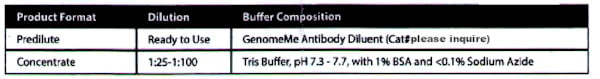
Reconstitution, Mixing, Dilution, and Titration
The prediluted antibody does not require any mixing, dilution, reconstitution, or titration; the antibody is ready-to-use and optimized for staining. Any further dilution may affect the quality of the staining signal or antibody-antigen interaction. The concentrated antibody requires dilution using an Antibody Diluent Buffer, to the recommended working dilution range listed in the table above, prior to use.
Specimen Collection and Preparation
Each tissue section should be fixed with 10% neutral buffered formalin, cut to the applicable thickness (4um), and placed on a glass slide that is positively charged. The prepared slide should then be baked for a minimum of 30 minutes in a 53-65°C oven (do not exceed 24 hours).
Note: Performance eva luation has been shown on human tissues only. Variable results may occur due to extended fixation time or variations in tissue preparation. Do not use alcohol containing fixatives as those may result in a loss of staining activity.
Note: Performance eva luation has been shown on human tissues only. Variable results may occur due to extended fixation time or variations in tissue preparation. Do not use alcohol containing fixatives as those may result in a loss of staining activity.
Material Required but not Provided
The following materials are required but are not provided:
a) Detection system (ie. BOND Polymer Refine Detection Kit or UltraView/OptiView Universal DAB Detection Kit)
b) Chromogen (ie. DAB Substrate Kit)
c) IHC wash buffer and blocking solution
d) Hematoxylin or other counterstaining reagents
e) Ethanol or reagent alcohol, xylene or xylene substitute and mounting medium
f) Antibody diluents
g) Positive and negative control tissue
Quality Control Procedures and Interpretation of Results
The immunohistochemical staining process results in a colorimetric reaction at the site of the antigen, localized by the primary antibody. The tissue specimen result should be interpreted only after the positive and negative control tissues have been analyzed. It is recommended to include a set of tissue controls with each staining run to monitor for antibody, tissue, and reagent performance. Tissue sections may contain both positive and negative staining elements. In these cases and where applicable, these sections may serve as both the positive and negative tissue control.
Positive Control Tissue
A positive control tissue should be processed in the same manner as the specimen and run with each test condition to provide control for variables such as tissue processing, fixation, and staining. It should function to provide validity to the specimen results obtained and can consist of fresh autopsy, biopsy, or surg ical tissue. Once stained, the positive control tissue should analyzed first to ensure that the antibody and all reagents are performing as intended. Counterstaining will result in a blue coloration, which may range from pale to dark depending on the length of the incubation time and potency of the hematoxylin. If positive staining is not observed, the positive control tissue must be deemed invalid and the results obtained with the tissue specimen must also be treated as such.
Negative Control Tissue
Some tissue sections can also function as an internal negative control due to the diversity of staining elements present. This, however, should first be confirmed by the user. Tissue components that do not stain should demonstrate an absence of specific staining. If specific staining is observed, the negative control tissue must be deemed invalid and the results obtained with the tissue specimen must also be treated as such.
Tissue Specimens
Tissue specimens should only be analyzed after the positive and negative control tissues have been deemed valid. Negative staining indicates that the antigen was not detected in the tissue while positive staining represents the presence of the antigen. A tissue section stained with hematoxylin and eosin should be used to analyze the morphology of the tissue specimen and verified by a qualified pathologist.
Peformance Characteristics
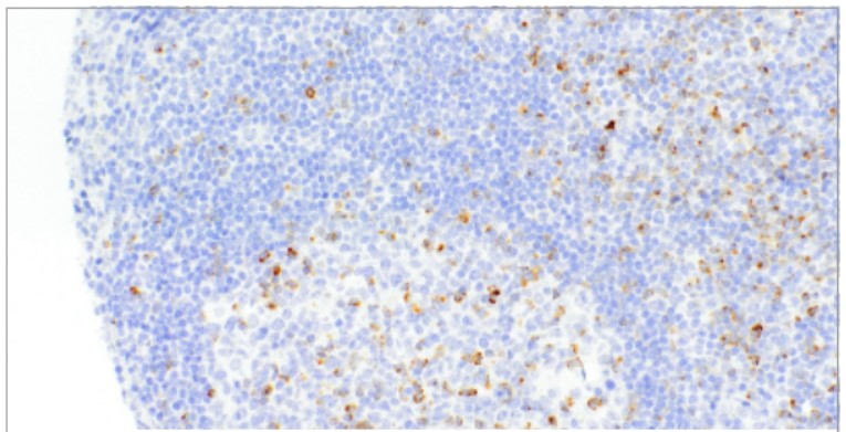
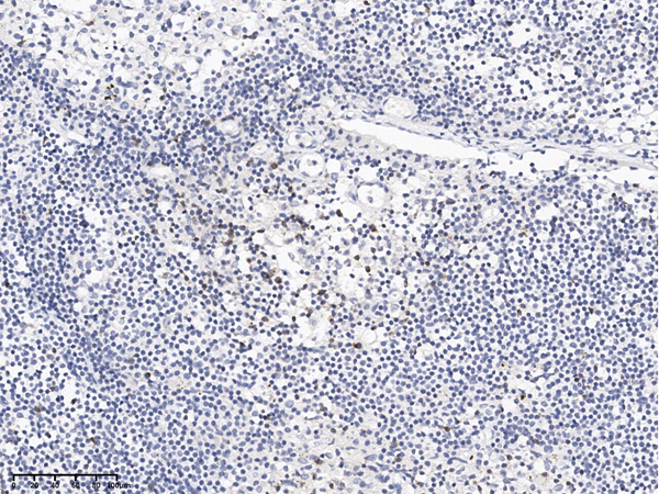
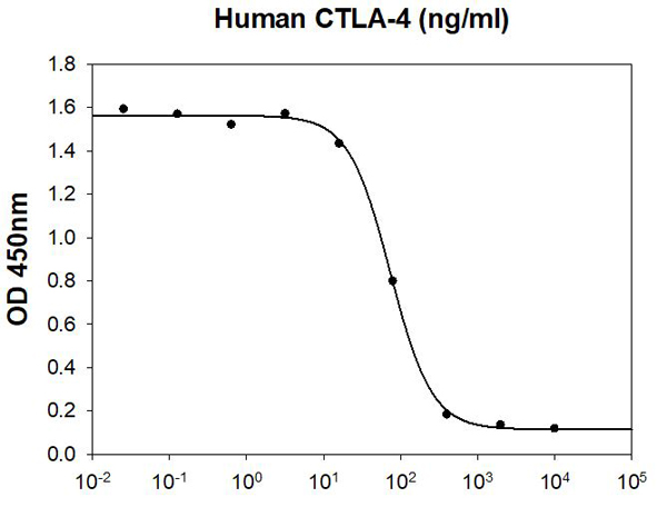
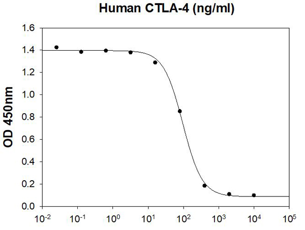
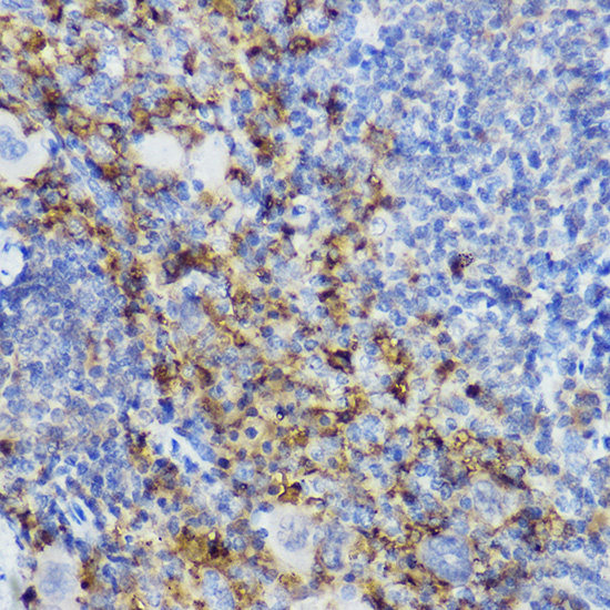
This antibody has been validated by immunohistochemistry using a FFPE human ti ssue microarray comprised of different types of normal and cancerous tissues. Positive staining was observed in tonsil, lymph nod and thymus tissues. No staining was seen in skeletal muscle and brain tissues. A representative positive staining image (see figure 1).
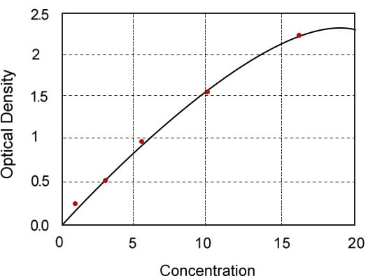
Analytical Performance
Trueness of Measure was analyzed using tissue samples from peer-reviewed, published literature known to be either positive or negative, and this study found no unexpected results. Precision of Measure analysis involved known positive tissue samples performed to assess the repeatability and reproducibility of the antibody product over time and in different lots; standard acceptance criteria were met in all studies. Analytical Specificity testing fou nd the only known interference comes from alcohol containing fixatives, which demonstrated a loss of stain ing activity and thus should not be used in conjunction with the processing of samples to be stained using this product. Analytica l Sensitivity was tested using randomly chosen tissues representing different levels of antigen expression, a range of staining intensities was observed illustrating the product is sensitive to staining a diverse range of expression levels. Limits of Detection, Measurement Range and Linearity of Measure are all unable to be defined for qualitative and non-linear products such as this.
Troubleshooting
1. If tissue sections wash off the slide, this may be caused by:
a) Slides are not positively charged.
b) Inadequate neutral-buffering of the formalin used for the fixation process.
c) A thick t issue section.
d) Inadequate drying of the tissue section prior to staining.
2. If the positive control tissue exhibits negative staining, this may be due to:
a) An issue with the primary antibody or one of the secondary reagents.
b) Improper collection, fixation or deparaffinization of the tissue section.
c) Errors in the IHC staining process.
3. If the positive control tissue exhibits weaker staining than expected, this may be due to sub-optimal lHC conditions, partial degradation of the primary antibody or improper storage of secondary reagents. Analysis of the positive and/or negative control tissues can help with determining the cause.
Limitations
1. Due to biological variability inherent to the expre ssion of certain antigens and immunohistochemical procedures, appropriate positive and negative controls should be used alongside the tissue specimen.
2. This antibody, when used with the appropriate detection systems and reagents. detects antigen(s) that remain intact through the tissue fixation, processing and sectioning as described. Any deviation from these recommended procedures or improper handling may compromise the validity andlor analysis of the results. Do not use alcohol containing fixatives as those may result in a loss of staining activity.
3. Provides prediluted antibodies in a ready-to-use, optimally diluted format for use as instructed. Due to the potential for variation in tissue processing and fixation, it may be necessary to adjust the incubation time of the primary antibody for different tissue specimens.
4. Provides concentrated antibodies in a format that requires dilution with Antibody Diluent. Use of a diluent different than that specified in the package insert must be validated by the user to ensure proper compatibility with the antibody.
5. False positive results may occur in tissue specimens due to the possibility of non-immmunological binding of substrate reaction products or proteins. False positive results may also occur subject to the type of immunostaining technique used, or due to the activity of pseudoperoxidase, endogenous peroxidase, or endogenous biotin.
6. Due to the effect of autoantibodies or natural antibodies, normal sera from an animal source that is the same as the secondary antisera may result in false negative or false positive results when used in blocking steps.
7. Non-specific staining with horseradish peroxidase may be observed when using tissues containing hepatitis B surface antigen due to the patient's infection with the hepatitis B virus.
Preparation and Storage
Store at 2-8°C. To ensure stability, immediately replace vial back in the refrigerator after each use. When stored correctly, the antibody is stable until the expiry date indicated on the label. Positive and negative controls should be concurrently run with tissue specimens, to enable identification of any inadequacies with the antibody or reagents.
Materials and Methods
Materials and Methods
Application Data
(CTLA-4 on Tonsil)
Application Data
(CTLA-4 on Tonsil)
Related Product Information for anti-CTLA-4 antibody
The CTLA-4 [IHC064] antibody is intended for qualified laboratories to qualitatively identify by light microscopy, the presence of associated antigens in formalin-fixed, paraffin-em~edded (FFPE) tissue sections using immunohistochemistry test methods. Use of this antibody is indicated as an aid in the identification of CTLA-4 strictly for research purposes.
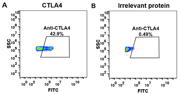
Background: Cytotoxic T-Lymp ocyte-AssOCiated-Protein 4 (CTLA-4jlSa receptor on T helper cells that functions as an immune checkpoint and downregulator of immune responses. Mutations In CTLA-4 are associated with insulin-dependent diabetes mellitus, Hashimoto's thyroiditis, Graves' disease, systemic lupus erythematosis, celiac disease, primary biliary cirrhosis, thyroid-associated orbitopathy, multiple sclerosis, and other autoimmune diseases. The spliced varient of CTLA-4 in SLE is present in the patient's serum. Haploinsufficiency of CTLA-4 causes the immune system disorder known as CTLA-4 deficiency or CHAI disease (CTLA-4 haploinsufficiency with autoimmune infiltration).
Background: Cytotoxic T-Lymp ocyte-AssOCiated-Protein 4 (CTLA-4jlSa receptor on T helper cells that functions as an immune checkpoint and downregulator of immune responses. Mutations In CTLA-4 are associated with insulin-dependent diabetes mellitus, Hashimoto's thyroiditis, Graves' disease, systemic lupus erythematosis, celiac disease, primary biliary cirrhosis, thyroid-associated orbitopathy, multiple sclerosis, and other autoimmune diseases. The spliced varient of CTLA-4 in SLE is present in the patient's serum. Haploinsufficiency of CTLA-4 causes the immune system disorder known as CTLA-4 deficiency or CHAI disease (CTLA-4 haploinsufficiency with autoimmune infiltration).
References
1. Denizot F, et al. Nature. 1987; 328:267-70. 2. Dariavach P, et al. Eur J Immunol. 1988; 18:1901-5. 3. Krummel MF, et al. J Exp Med. 1995; 182:459-65. 4. Kuehn HS, et al. Science. 2014; 345:1623-7. 5. Walunas TL, et al. J Exp Med. 1996; 183:2541-50.
Customer Reviews
Loading reviews...
Share Your Experience
Similar Products

Product Notes
The CTLA-4 (Catalog #AAA58783) is an Antibody produced from Rabbit and is intended for research purposes only. The product is available for immediate purchase. The CTLA-4 Antibody reacts with Human and may cross-react with other species as described in the data sheet. It is sometimes possible for the material contained within the vial of "CTLA-4, Monoclonal Antibody" to become dispersed throughout the inside of the vial, particularly around the seal of said vial, during shipment and storage. We always suggest centrifuging these vials to consolidate all of the liquid away from the lid and to the bottom of the vial prior to opening. Please be advised that certain products may require dry ice for shipping and that, if this is the case, an additional dry ice fee may also be required.Precautions
All products in the AAA Biotech catalog are strictly for research-use only, and are absolutely not suitable for use in any sort of medical, therapeutic, prophylactic, in-vivo, or diagnostic capacity. By purchasing a product from AAA Biotech, you are explicitly certifying that said products will be properly tested and used in line with industry standard. AAA Biotech and its authorized distribution partners reserve the right to refuse to fulfill any order if we have any indication that a purchaser may be intending to use a product outside of our accepted criteria.Disclaimer
Though we do strive to guarantee the information represented in this datasheet, AAA Biotech cannot be held responsible for any oversights or imprecisions. AAA Biotech reserves the right to adjust any aspect of this datasheet at any time and without notice. It is the responsibility of the customer to inform AAA Biotech of any product performance issues observed or experienced within 30 days of receipt of said product. To see additional details on this or any of our other policies, please see our Terms & Conditions page.Item has been added to Shopping Cart
If you are ready to order, navigate to Shopping Cart and get ready to checkout.