


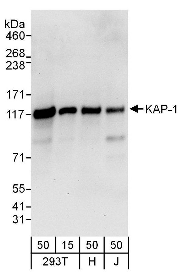
 WB (Western Blot)
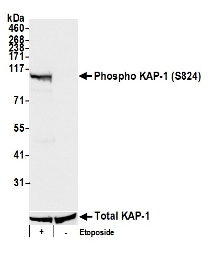
(Detection of human Phospho KAP-1 (S824) by western blot. Samples: Whole cell lysate (25 ug) from HEK293T cells treated with 100 uM etoposide (+) or mock treated (-) prepared using NETN lysis buffer. Antibody: Rabbit anti-Phospho KAP-1 (S824) recombinant monoclonal antibody [BL-246-7B5] (AAA23791 lot 4) used at 1:1000. Secondary: HRP-conjugated goat anti-rabbit IgG . Chemiluminescence with an exposure time of 3 seconds. Lower Panel: Rabbit anti-KAP1 recombinant monoclonal antibody [BL-248-2G6] .)
WB (Western Blot)
(Detection of human Phospho KAP-1 (S824) by western blot. Samples: Whole cell lysate (25 ug) from HEK293T cells treated with 100 uM etoposide (+) or mock treated (-) prepared using NETN lysis buffer. Antibody: Rabbit anti-Phospho KAP-1 (S824) recombinant monoclonal antibody [BL-246-7B5] (AAA23791 lot 4) used at 1:1000. Secondary: HRP-conjugated goat anti-rabbit IgG . Chemiluminescence with an exposure time of 3 seconds. Lower Panel: Rabbit anti-KAP1 recombinant monoclonal antibody [BL-248-2G6] .)
KAP-1 recombinant antibody
Rabbit anti-Phospho KAP-1 (S824) Recombinant Monoclonal Antibody [BL-246-7B5]
IP: 20 ul/mg lysate
IHC: 1:100-1:500. Epitope retrieval with citrate buffer pH 6.0 is recommended for FFPE tissue sections.
ICC-IF: 1:100-1:500. Formaldehyde fixation is recommended. Permeabilization with Triton-X 100 is recommended for formaldehyde-fixed cells.
FC/FACS: Fixed in 4% formaldehyde and permeabilized with 90% methanol. 1 ul per 1 x 10^6 cells.
Shelf Life: 1 year from date of receipt
WB (Western Blot)
(Detection of human Phospho KAP-1 (S824) by western blot. Samples: Whole cell lysate (25 ug) from HEK293T cells treated with 100 uM etoposide (+) or mock treated (-) prepared using NETN lysis buffer. Antibody: Rabbit anti-Phospho KAP-1 (S824) recombinant monoclonal antibody [BL-246-7B5] (AAA23791 lot 4) used at 1:1000. Secondary: HRP-conjugated goat anti-rabbit IgG . Chemiluminescence with an exposure time of 3 seconds. Lower Panel: Rabbit anti-KAP1 recombinant monoclonal antibody [BL-248-2G6] .)
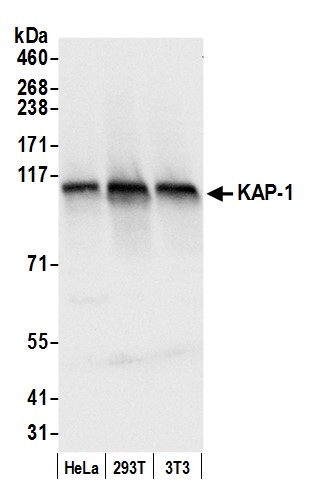
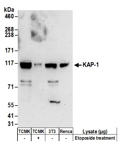
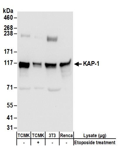
WB (Western Blot)
(Detection of mouse Phospho KAP-1 (S824) by western blot. Samples: Whole cell lysate (50 ug) from NIH3T3 cells treated with 100 uM etoposide (+) or mock treated (-) prepared using NETN lysis buffer. Antibody: Rabbit anti-Phospho KAP-1 (S824) recombinant monoclonal antibody [BL-246-7B5] (AAA23791 lot 4) used at 1:1000. Secondary: HRP-conjugated goat anti-rabbit IgG . Chemiluminescence with an exposure time of 3 seconds. Lower Panel: Rabbit anti-KAP1 recombinant monoclonal antibody [BL-248-2G6] .)
Simple Western
(Detection of human Phospho KAP-1 (S824) by Simple Western. Samples: Whole cell lysate (0.4 mg/mL) from HEK293T cells treated with 100 uM etoposide prepared using NETN lysis buffer. Antibody: Rabbit anti-Phospho KAP-1 (S824) recombinant monoclonal antibody [BL-246-7B5] (AAA23791) used at 1:10, 1:50, and 1:250. Separation and Detection: SallySue ProteinSimple instrument with the 12-230 kDa separation module and anti-Rabbit detection module. Left Panel: Virtual Lane View. Right Panel: Electropherogram.)
IP (Immunoprecipitation)
(Detection of human Phospho KAP-1 (S824) by western blot of immunoprecipitates. Samples: Whole cell lysate (1.0 mg per IP reaction; 5% of IP loaded) from HEK293T cells prepared using NETN lysis buffer that were treated with 100 uM etoposide (+) or mock treated (-). Antibodies: Rabbit anti-Phospho KAP-1 (S824) recombinant monoclonal antibody [BL-246-7B5] (AAA23791 lot 4) used for IP at 20 ul/mg lysate. Phospho KAP-1 (S824) was also immunoprecipitated by a previous lot of this antibody (AAA23791 lot 3) and rabbit anti-KAP-1 recombinant monoclonal antibody [BL-248-2G6] . For blotting immunoprecipitated Phospho KAP-1 (S824), AAA23791 was used at 1:1000. Chemiluminescence with an exposure time of 1 second. Lower Panel: Rabbit anti-KAP1 recombinant monoclonal antibody [BL-248-2G6] .)
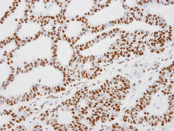
IHC (Immunohistochemistry)
(Detection of human Phospho KAP-1 (S824) in FFPE prostate carcinoma by IHC. Mock phosphatase treated section (left) and calf intestinal phosphatase-treated section (right). Antibody: Rabbit anti-Phospho KAP-1 (S824) recombinant monoclonal [BL-246-7B5] (AAA23791). Secondary: HRP-conjugated goat anti-rabbit IgG . Substrate: DAB.)
ICC (Immunocytochemistry)
(Detection of human Phospho KAP-1 (S824) by immunocytochemistry. Samples: Formaldehyde-fixed asynchronous HeLa cells grown in chambered microscope slides and treated with etoposide (right) or untreated (left). Antibody: Rabbit anti-Phospho KAP-1 (S824) recombinant monoclonal antibody [BL-246-7B5] (AAA23791-T lot 1) used at of 1:100. Secondary: DyLight 594-conjugated goat anti-rabbit IgG . Counterstain: Phalloidin conjugated Alexa Fluor 488 (green).)
ICC (Immunocytochemistry)
(Detection of human Phospho KAP-1 (S824) in FFPE etoposide treated HeLa cells by ICC. Mock phosphatase treated section (left) and calf intestinal phosphatase-treated section (right). Antibody: Rabbit anti-Phospho KAP-1 (S824) recombinant monoclonal [BL-246-7B5] (AAA23791). Secondary: HRP-conjugated goat anti-rabbit IgG . Substrate: DAB.)
FCM (Flow Cytometry)
(Detection of human phospho KAP-1 (shaded) in etoposide treated HEK293T cells (right) and untreated HEK293T cells (left) by flow cytometry. Antibody: Rabbit anti-phospho KAP-1 recombinant monoclonal [BL-246-7B5] (AAA23791) or isotype control (unshaded). Secondary: DyLight 488-conjugated goat anti-rabbit IgG .)
NCBI and Uniprot Product Information
Customer Reviews
Loading reviews...
Share Your Experience
Similar Products


Product Notes
The KAP-1 trim28 (Catalog #AAA23791) is a Recombinant Antibody produced from Rabbit and is intended for research purposes only. The product is available for immediate purchase. The Rabbit anti-Phospho KAP-1 (S824) Recombinant Monoclonal Antibody [BL-246-7B5] reacts with Human, Mouse and may cross-react with other species as described in the data sheet. AAA Biotech's KAP-1 can be used in a range of immunoassay formats including, but not limited to, WB (Western Blot), IP (Immunoprecipitation), IHC (Immunohistochemistry), ICC (Immunocytochemistry), FCM/FACS (Flow Cytometry), WB (Western Blot). WB: 1:1000 IP: 20 ul/mg lysate IHC: 1:100-1:500. Epitope retrieval with citrate buffer pH 6.0 is recommended for FFPE tissue sections. ICC-IF: 1:100-1:500. Formaldehyde fixation is recommended. Permeabilization with Triton-X 100 is recommended for formaldehyde-fixed cells. FC/FACS: Fixed in 4% formaldehyde and permeabilized with 90% methanol. 1 ul per 1 x 10^6 cells. Researchers should empirically determine the suitability of the KAP-1 trim28 for an application not listed in the data sheet. Researchers commonly develop new applications and it is an integral, important part of the investigative research process. It is sometimes possible for the material contained within the vial of "KAP-1, Monoclonal Recombinant Antibody" to become dispersed throughout the inside of the vial, particularly around the seal of said vial, during shipment and storage. We always suggest centrifuging these vials to consolidate all of the liquid away from the lid and to the bottom of the vial prior to opening. Please be advised that certain products may require dry ice for shipping and that, if this is the case, an additional dry ice fee may also be required.Precautions
All products in the AAA Biotech catalog are strictly for research-use only, and are absolutely not suitable for use in any sort of medical, therapeutic, prophylactic, in-vivo, or diagnostic capacity. By purchasing a product from AAA Biotech, you are explicitly certifying that said products will be properly tested and used in line with industry standard. AAA Biotech and its authorized distribution partners reserve the right to refuse to fulfill any order if we have any indication that a purchaser may be intending to use a product outside of our accepted criteria.Disclaimer
Though we do strive to guarantee the information represented in this datasheet, AAA Biotech cannot be held responsible for any oversights or imprecisions. AAA Biotech reserves the right to adjust any aspect of this datasheet at any time and without notice. It is the responsibility of the customer to inform AAA Biotech of any product performance issues observed or experienced within 30 days of receipt of said product. To see additional details on this or any of our other policies, please see our Terms & Conditions page.Item has been added to Shopping Cart
If you are ready to order, navigate to Shopping Cart and get ready to checkout.