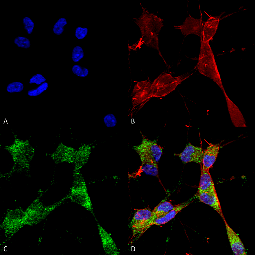
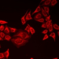
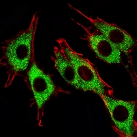
 ICC (Immunocytochemistry)
(1. Immunocytochemistry/Immunofluorescence analysis using Mouse Anti-PINK1 Monoclonal Antibody, Clone S4-15 (AAA102887). Tissue: Neuroblastoma cells. Species: Human. Fixation: 4% PFA for 15 min. Primary Antibody: Mouse Anti-PINK1 Monoclonal Antibody (AAA102887) at 1:50 for overnight at 4°C with slow rocking. Secondary Antibody: AlexaFluor 488 at 1:1000 for 1 hour at RT. Counterstain: Phalloidin-iFluor 647 (red) F-Actin stain; Hoechst (blue) nuclear stain at 1:800, 1.6mM for 20 min at RT. (A) Hoechst (blue) nuclear stain. (B) Phalloidin-iFluor 647 (red) F-Actin stain. (C) PINK1 Antibody (D) Composite.)
ICC (Immunocytochemistry)
(1. Immunocytochemistry/Immunofluorescence analysis using Mouse Anti-PINK1 Monoclonal Antibody, Clone S4-15 (AAA102887). Tissue: Neuroblastoma cells. Species: Human. Fixation: 4% PFA for 15 min. Primary Antibody: Mouse Anti-PINK1 Monoclonal Antibody (AAA102887) at 1:50 for overnight at 4°C with slow rocking. Secondary Antibody: AlexaFluor 488 at 1:1000 for 1 hour at RT. Counterstain: Phalloidin-iFluor 647 (red) F-Actin stain; Hoechst (blue) nuclear stain at 1:800, 1.6mM for 20 min at RT. (A) Hoechst (blue) nuclear stain. (B) Phalloidin-iFluor 647 (red) F-Actin stain. (C) PINK1 Antibody (D) Composite.)
Mouse PINK1 Monoclonal Antibody | anti-PINK1 antibody
PINK1 Antibody
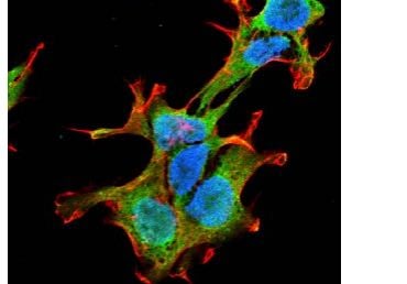
ICC (Immunocytochemistry)
(1. Immunocytochemistry/Immunofluorescence analysis using Mouse Anti-PINK1 Monoclonal Antibody, Clone S4-15 (AAA102887). Tissue: Neuroblastoma cells. Species: Human. Fixation: 4% PFA for 15 min. Primary Antibody: Mouse Anti-PINK1 Monoclonal Antibody (AAA102887) at 1:50 for overnight at 4°C with slow rocking. Secondary Antibody: AlexaFluor 488 at 1:1000 for 1 hour at RT. Counterstain: Phalloidin-iFluor 647 (red) F-Actin stain; Hoechst (blue) nuclear stain at 1:800, 1.6mM for 20 min at RT. (A) Hoechst (blue) nuclear stain. (B) Phalloidin-iFluor 647 (red) F-Actin stain. (C) PINK1 Antibody (D) Composite.)
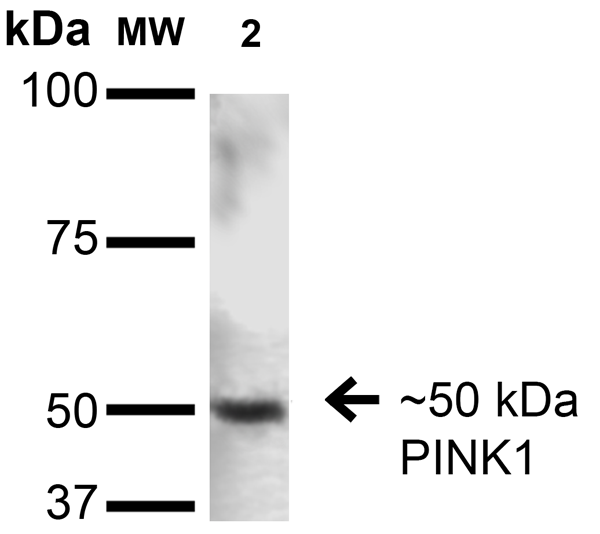
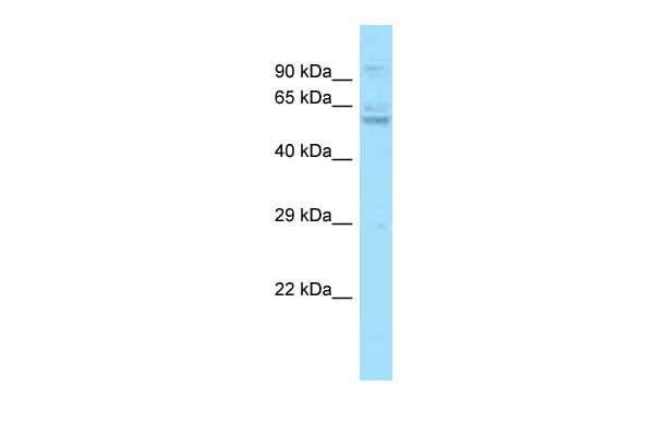
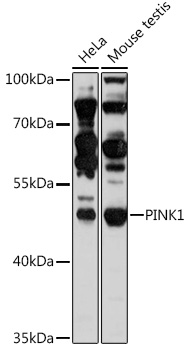
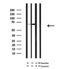
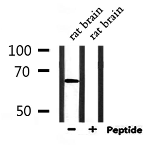
WB (Western Blot)
(Western Blot analysis of Rat Brain showing detection of ~50 kDa PINK1 protein using Mouse Anti-PINK1 Monoclonal Antibody, Clone S4-15. Lane 1: Molecular Weight Ladder. Lane 2: Rat Brain. Load: 15 ug. Block: 2% BSA and 2% Skim Milk in 1X TBST. Primary Antibody: Mouse Anti-PINK1 Monoclonal Antibody at 1:200 for 16 hours at 4 degree C. Secondary Antibody: Goat Anti-Mouse IgG: HRP at 1:1000 for 1 hour RT. Color Development: ECL solution for 6 min in RT. Predicted/Observed Size: ~50 kDa.)
NCBI and Uniprot Product Information
Customer Reviews
Loading reviews...
Share Your Experience
Similar Products

Product Notes
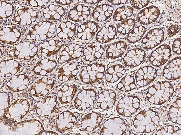
The PINK1 pink1 (Catalog #AAA102887) is an Antibody produced from Mouse and is intended for research purposes only. The product is available for immediate purchase. The PINK1 Antibody reacts with Human, Rat, Mouse and may cross-react with other species as described in the data sheet. AAA Biotech's PINK1 can be used in a range of immunoassay formats including, but not limited to, IF (Immunofluorescence), IHC (Immunohistochemistry), ICC (Immunocytochemistry), WB (Western Blot). Researchers should empirically determine the suitability of the PINK1 pink1 for an application not listed in the data sheet. Researchers commonly develop new applications and it is an integral, important part of the investigative research process. It is sometimes possible for the material contained within the vial of "PINK1, Monoclonal Antibody" to become dispersed throughout the inside of the vial, particularly around the seal of said vial, during shipment and storage. We always suggest centrifuging these vials to consolidate all of the liquid away from the lid and to the bottom of the vial prior to opening. Please be advised that certain products may require dry ice for shipping and that, if this is the case, an additional dry ice fee may also be required.Precautions
All products in the AAA Biotech catalog are strictly for research-use only, and are absolutely not suitable for use in any sort of medical, therapeutic, prophylactic, in-vivo, or diagnostic capacity. By purchasing a product from AAA Biotech, you are explicitly certifying that said products will be properly tested and used in line with industry standard. AAA Biotech and its authorized distribution partners reserve the right to refuse to fulfill any order if we have any indication that a purchaser may be intending to use a product outside of our accepted criteria.Disclaimer
Though we do strive to guarantee the information represented in this datasheet, AAA Biotech cannot be held responsible for any oversights or imprecisions. AAA Biotech reserves the right to adjust any aspect of this datasheet at any time and without notice. It is the responsibility of the customer to inform AAA Biotech of any product performance issues observed or experienced within 30 days of receipt of said product. To see additional details on this or any of our other policies, please see our Terms & Conditions page.Item has been added to Shopping Cart
If you are ready to order, navigate to Shopping Cart and get ready to checkout.