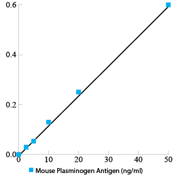
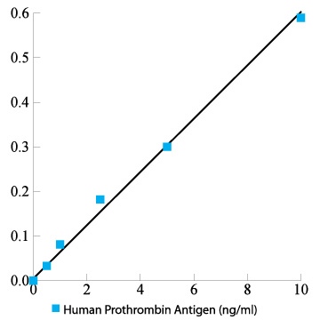
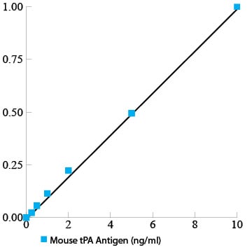
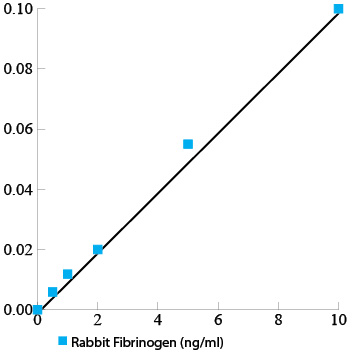
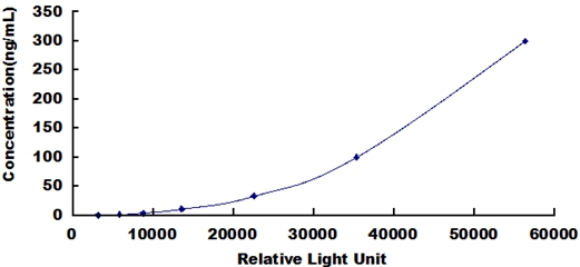
 Standard Curve (Sample)
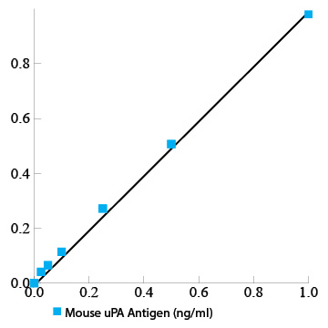
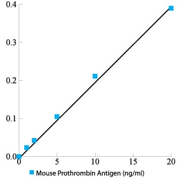
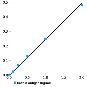
General plasminogen total antigen assay ELISA Kit | PLG elisa kit
Mouse plasminogen total antigen assay ELISA Kit
Gene Names
Plg; Pg; AI649309
Reactivity
General
Synonyms
plasminogen total antigen assay; N/A; Mouse plasminogen total antigen assay ELISA Kit; PLG elisa kit
Reactivity
General
Form/Format
Complete Kit
Sequence Length
812
Samples
Mouse Serum, Cell Culture Supernatant, And Other Biological Fluids
Assay Type
Quantitative Sandwich
Intra-assay Precision
To determine within-run precision, three different samples of known concentration were assayed by using 8 replicates in 1 assay
Inter-assay Precision
To determine between-run precision, three different samples of known concentration were assayed by using replicates on 18 different assays.
Preparation and Storage
Store at 4 degree C. Shelf Life: 12 months from manufacture, see label for expiration date
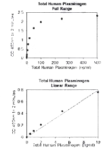
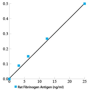
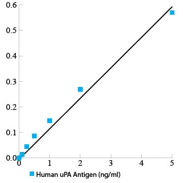
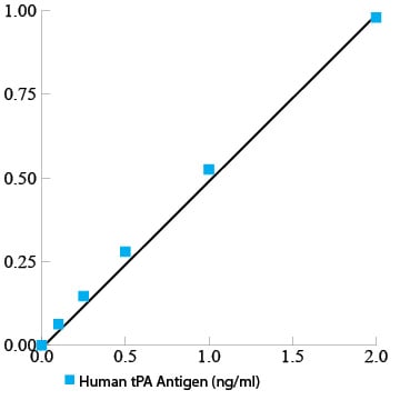
Standard Curve (Sample)
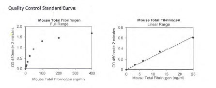
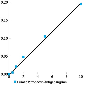
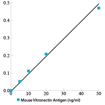
Standard Curve (Sample)
Related Product Information for PLG elisa kit
Intended Uses: This Mouse MCP-1 ELISA kit is to be used for the in vitro quantitative determination of mouse Monocyte Chemoattractant Protein-1 (MCP-1) concentrations in serum, cell culture supernatant, and other biological fluids. This kit is intended for LABORATORY RESEARCH USE ONLY.
Principle of the Assay: This mouse MCP-1 enzyme-linked immunosorbent assay (ELISA) applies a technique called a quantitative sandwich immunoassay. The microtiter plate provided in this kit has been pre-coated with a monoclonal antibody specific for mouse MCP-1. Standards or samples are then added to the appropriate microtiter plate wells and incubated. Mouse MCP-1, if present, will bind and become immobilized by the antibody pre-coated on the wells. A biotin conjugated antibody specific for mouse MCP-1 is added to each well. The biotin conjugated antibody will bind to the mouse MCP-1 on the plate. The microtiter plate wells are thoroughly washed to remove unbound biotin conjugate and other components of the sample. Avidin has a very high affinity to biotin. In order to quantify the amount of mouse MCP-1 present in the sample, a standardized preparation of avidin conjugated horseradish peroxidase (HRP) is added to each well. Avidin-HRP will bind to the biotin on plate during incubation. The wells are thoroughly washed to remove all unbound Avidin-HRP conjugate and a TMB (3,3'5,5' tetramethyl-benzidine) substrate solution is added to each well. The enzyme (HRP) and substrate are allowed to react over a short incubation period. Only wells to which mouse MCP-1, Biotin conjugate and Avidin-HRP are attached will exhibit a change in colour. The enzyme-substrate reaction is terminated by the addition of a sulphuric acid solution and the colour change is measured spectrophotometrically at a wavelength of 450 nm +/- 2 nm. In order to measure the concentration of mouse MCP-1 in the samples, this kit contains two calibration diluents (Calibrator Diluent I for serum/plasma testing and Calibrator Diluent II for cell culture supernatant/ urine testing). According to the testing system, the provided standard is diluted (2-fold) with the appropriate Calibrator Diluent and assayed at the same time as the samples. This allows the operator to produce a standard curve of Optical Density (O.D.) versus MCP-1 concentration (pg/mL). The concentration of MCP-1 in the samples is then determined by comparing the O.D. of the samples to the standard curve!!Background/Introduction: Monocyte chemotactic protein 1(MCP-1), also known as monocyte chemotactic and activating factor(MCAF), lymphocyte-derived chemotactic factor (LDCF) and glioma-derived chemotactic factor (GDF), is a chemotactic cytokine for monocytes. It is a CC chemokine with two adjacent cystines at its NH2 terminus. MCP1 is produced by a variety of cell types, including monocytes, lymphocytes, fibroblasts, endothelial cells, epithelial cells and smooth muscle cells. MCP-1 is up-regulated in response to infectious agents, oxidative radicals and pro-inflammatory mediators released by host cells. The major physiological function of MCP-1 is to mediate host defense. MCP-1 acts as a chemoattractant to recruit monocytes to the infected or injured area and activates the cells to secret cytokines and superoxide, enhances phagocytosis and antigen presentation by macrophages. The expression patterns of MCP-1 and IL-8 are similar. Both cytokines will increase upon stimulation with LPS and inflammatory cytokines such as IL-1, TNF-alpha, and IFN-gamma. In mouse fibroblasts, platelet derived growth factor (PDGF) is a strong inducer of MCP-1 mRNA expression but failed to induce IL-8 mRNA, suggesting different regulatory mechanisms. In addition to being chemotactic for monocytes, MCP-1 also activates mouse monocytes to become cytostatic for several mouse tumour cell lines, release lysosomal enzymes, and generate superoxide. Elevated MCP-1 levels are associated with autoimmune disease, allergic inflammation, atherosclerosis, glomerulonephritis, granuloma formation. This ELISA kit provides a tool for studying MCP-1 expression and its relationship with various diseases in animal model.
Principle of the Assay: This mouse MCP-1 enzyme-linked immunosorbent assay (ELISA) applies a technique called a quantitative sandwich immunoassay. The microtiter plate provided in this kit has been pre-coated with a monoclonal antibody specific for mouse MCP-1. Standards or samples are then added to the appropriate microtiter plate wells and incubated. Mouse MCP-1, if present, will bind and become immobilized by the antibody pre-coated on the wells. A biotin conjugated antibody specific for mouse MCP-1 is added to each well. The biotin conjugated antibody will bind to the mouse MCP-1 on the plate. The microtiter plate wells are thoroughly washed to remove unbound biotin conjugate and other components of the sample. Avidin has a very high affinity to biotin. In order to quantify the amount of mouse MCP-1 present in the sample, a standardized preparation of avidin conjugated horseradish peroxidase (HRP) is added to each well. Avidin-HRP will bind to the biotin on plate during incubation. The wells are thoroughly washed to remove all unbound Avidin-HRP conjugate and a TMB (3,3'5,5' tetramethyl-benzidine) substrate solution is added to each well. The enzyme (HRP) and substrate are allowed to react over a short incubation period. Only wells to which mouse MCP-1, Biotin conjugate and Avidin-HRP are attached will exhibit a change in colour. The enzyme-substrate reaction is terminated by the addition of a sulphuric acid solution and the colour change is measured spectrophotometrically at a wavelength of 450 nm +/- 2 nm. In order to measure the concentration of mouse MCP-1 in the samples, this kit contains two calibration diluents (Calibrator Diluent I for serum/plasma testing and Calibrator Diluent II for cell culture supernatant/ urine testing). According to the testing system, the provided standard is diluted (2-fold) with the appropriate Calibrator Diluent and assayed at the same time as the samples. This allows the operator to produce a standard curve of Optical Density (O.D.) versus MCP-1 concentration (pg/mL). The concentration of MCP-1 in the samples is then determined by comparing the O.D. of the samples to the standard curve!!Background/Introduction: Monocyte chemotactic protein 1(MCP-1), also known as monocyte chemotactic and activating factor(MCAF), lymphocyte-derived chemotactic factor (LDCF) and glioma-derived chemotactic factor (GDF), is a chemotactic cytokine for monocytes. It is a CC chemokine with two adjacent cystines at its NH2 terminus. MCP1 is produced by a variety of cell types, including monocytes, lymphocytes, fibroblasts, endothelial cells, epithelial cells and smooth muscle cells. MCP-1 is up-regulated in response to infectious agents, oxidative radicals and pro-inflammatory mediators released by host cells. The major physiological function of MCP-1 is to mediate host defense. MCP-1 acts as a chemoattractant to recruit monocytes to the infected or injured area and activates the cells to secret cytokines and superoxide, enhances phagocytosis and antigen presentation by macrophages. The expression patterns of MCP-1 and IL-8 are similar. Both cytokines will increase upon stimulation with LPS and inflammatory cytokines such as IL-1, TNF-alpha, and IFN-gamma. In mouse fibroblasts, platelet derived growth factor (PDGF) is a strong inducer of MCP-1 mRNA expression but failed to induce IL-8 mRNA, suggesting different regulatory mechanisms. In addition to being chemotactic for monocytes, MCP-1 also activates mouse monocytes to become cytostatic for several mouse tumour cell lines, release lysosomal enzymes, and generate superoxide. Elevated MCP-1 levels are associated with autoimmune disease, allergic inflammation, atherosclerosis, glomerulonephritis, granuloma formation. This ELISA kit provides a tool for studying MCP-1 expression and its relationship with various diseases in animal model.
Product Categories/Family for PLG elisa kit
NCBI and Uniprot Product Information
NCBI GI #
NCBI GeneID
NCBI Accession #
NCBI GenBank Nucleotide #
Molecular Weight
90,808 Da
NCBI Official Full Name
plasminogen
NCBI Official Synonym Full Names
plasminogen
NCBI Official Symbol
Plg
NCBI Official Synonym Symbols
Pg; AI649309
NCBI Protein Information
plasminogen; angiostatin; plasmin heavy chain A; plasmin light chain B
UniProt Protein Name
Plasminogen
UniProt Gene Name
Plg
UniProt Entry Name
PLMN_MOUSE
Customer Reviews
Loading reviews...
Share Your Experience
Similar Products


Product Notes
The General PLG plg (Catalog #AAA37729) is an ELISA Kit and is intended for research purposes only. The product is available for immediate purchase. The AAA37729 ELISA Kit recognizes General PLG. It is sometimes possible for the material contained within the vial of "plasminogen total antigen assay, ELISA Kit" to become dispersed throughout the inside of the vial, particularly around the seal of said vial, during shipment and storage. We always suggest centrifuging these vials to consolidate all of the liquid away from the lid and to the bottom of the vial prior to opening. Please be advised that certain products may require dry ice for shipping and that, if this is the case, an additional dry ice fee may also be required.Precautions
All products in the AAA Biotech catalog are strictly for research-use only, and are absolutely not suitable for use in any sort of medical, therapeutic, prophylactic, in-vivo, or diagnostic capacity. By purchasing a product from AAA Biotech, you are explicitly certifying that said products will be properly tested and used in line with industry standard. AAA Biotech and its authorized distribution partners reserve the right to refuse to fulfill any order if we have any indication that a purchaser may be intending to use a product outside of our accepted criteria.Disclaimer
Though we do strive to guarantee the information represented in this datasheet, AAA Biotech cannot be held responsible for any oversights or imprecisions. AAA Biotech reserves the right to adjust any aspect of this datasheet at any time and without notice. It is the responsibility of the customer to inform AAA Biotech of any product performance issues observed or experienced within 30 days of receipt of said product. To see additional details on this or any of our other policies, please see our Terms & Conditions page.Item has been added to Shopping Cart
If you are ready to order, navigate to Shopping Cart and get ready to checkout.