Filters
Clonality
Type
Reactivity
Gene Name
Isotype
Host
Application
Clone
30 results for "Goat Anti Human IgG chain specific " - showing 1-30
 Application Data
(ELISA plate was coated with purified human IgG, IgM, and IgA. Immunoglobulins were detected with serially diluted Goat Anti-Human IgG-HRP.)
Application Data
(ELISA plate was coated with purified human IgG, IgM, and IgA. Immunoglobulins were detected with serially diluted Goat Anti-Human IgG-HRP.)
Goat Anti-Human IgG (gamma chain specific), Polyclonal Secondary Antibody (Cat# AAA14904)
Full Name
Goat Anti-Human IgG-HRP
Applications
ELISA
Purity
Affinity chromatography on human IgG covalently linked to agarose
Pricing
Goat F(ab) Anti-Human IgG (gamma chain specific), Secondary Antibody (Cat# AAA14905)
Full Name
Goat Fab Anti-Human IgG-UNLB
Reactivity
Human
Pricing
 Application Data
(Human peripheral blood lymphocytes were stained with Goat F(ab')2 Anti-Human IgM-PE and Mouse Anti-Human CD19-FITC .)
Application Data
(Human peripheral blood lymphocytes were stained with Goat F(ab')2 Anti-Human IgM-PE and Mouse Anti-Human CD19-FITC .)
Goat F(ab')2 Anti-Human IgM (u chain specific), Polyclonal Secondary Antibody (Cat# AAA14901)
Full Name
Goat F(ab')2 Anti-Human IgM-FITC
Reactivity
Human
Applications
FC
Pricing
Goat Anti-Human IgG, Monkey ads, Secondary Antibody (Cat# AAA14913)
Full Name
Goat Anti-Human IgG, Monkey ads-UNLB
Applications
ELISA; FLISA
Purity
Affinity chromatography on human IgG covalently linked to agarose
Pricing
Goat F(ab')2 Anti-Human IgG (gamma chain specific), Polyclonal Secondary Antibody (Cat# AAA14907)
Full Name
Goat F(ab')2 Anti-Human IgG-FITC
Applications
FC
Pricing


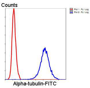 FCM (Flow Cytometry)
(Flow cytometric analysis of Hela cells with Alpha-tubulin antibody at 1/100 dilution (blue) compared with an unlabelled control (cells without incubation with primary antibody; red). Goat anti rabbit IgG (FITC) was used as the secondary antibody.)
FCM (Flow Cytometry)
(Flow cytometric analysis of Hela cells with Alpha-tubulin antibody at 1/100 dilution (blue) compared with an unlabelled control (cells without incubation with primary antibody; red). Goat anti rabbit IgG (FITC) was used as the secondary antibody.)
Alpha-Tubulin, Polyclonal Antibody (Cat# AAA29919)
Full Name
Alpha-Tubulin Antibody
Gene Names
TUBA4A; TUBA1; H2-ALPHA
Reactivity
Human, Mouse, Rat, Zebrafish
Applications
WB, IHC, ICC, FC/FACS, IF
Purity
Peptide affinity purified
Pricing


 WB (Western Blot)
(Western blot analysis of Tubulin alpha expression in various sample)
WB (Western Blot)
(Western blot analysis of Tubulin alpha expression in various sample)
Tubulin alpha, Polyclonal Antibody (Cat# AAA31146)
Full Name
Tubulin alpha Antibody
Gene Names
TUBA1B; K-ALPHA-1
Reactivity
Human, Mouse, Rat, Pig, Bovine, Rabbit, Chicken, Fish
Predicted: Horse, Sheep, Dog
Predicted: Horse, Sheep, Dog
Applications
WB, IHC, IF, ICC, EIA
Purity
The antiserum was purified by peptide affinity chromatography using SulfoLink Coupling Resin (Thermo Fisher Scientific).
Pricing


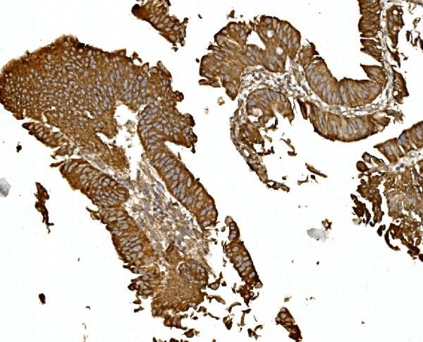
 IHC (Immunohistchemistry)
(Annexin II Antibody for IHC in human colon tissue)
IHC (Immunohistchemistry)
(Annexin II Antibody for IHC in human colon tissue)
Annexin II, Polyclonal Antibody (Cat# AAA31078)
Full Name
Annexin II Antibody
Gene Names
ANXA2; P36; ANX2; LIP2; LPC2; CAL1H; LPC2D; ANX2L4; PAP-IV; HEL-S-270
Reactivity
Human, Mouse, Rat
Applications
WB, IHC, EIA
Purity
The antiserum was purified by peptide affinity chromatography using SulfoLink Coupling Resin.
Pricing


 IHC (Immunohistchemistry)
(Figure 6. IHC analysis of Integrin alpha 5 using anti-Integrin alpha 5 antibody (AAA19156).Integrin alpha 5 was detected in paraffin-embedded section of human placenta tissue. Heat mediated antigen retrieval was performed in citrate buffer (pH6, epitope retrieval solution) for 20 mins. The tissue section was blocked with 10% goat serum. The tissue section was then incubated with 1ug/ml rabbit anti-Integrin alpha 5 Antibody (AAA19156) overnight at 4 degree C. Biotinylated goat anti-rabbit IgG was used as secondary antibody and incubated for 30 minutes at 37 degree C. The tissue section was developed using Strepavidin-Biotin-Complex (SABC) with DAB as the chromogen.)
IHC (Immunohistchemistry)
(Figure 6. IHC analysis of Integrin alpha 5 using anti-Integrin alpha 5 antibody (AAA19156).Integrin alpha 5 was detected in paraffin-embedded section of human placenta tissue. Heat mediated antigen retrieval was performed in citrate buffer (pH6, epitope retrieval solution) for 20 mins. The tissue section was blocked with 10% goat serum. The tissue section was then incubated with 1ug/ml rabbit anti-Integrin alpha 5 Antibody (AAA19156) overnight at 4 degree C. Biotinylated goat anti-rabbit IgG was used as secondary antibody and incubated for 30 minutes at 37 degree C. The tissue section was developed using Strepavidin-Biotin-Complex (SABC) with DAB as the chromogen.)
Integrin alpha 5, Polyclonal Antibody (Cat# AAA19156)
Full Name
Anti-Integrin alpha 5 Picoband antibody
Gene Names
ITGA5; FNRA; CD49e; VLA-5; VLA5A
Reactivity
Human, Mouse, Rat
No cross reactivity with other proteins.
No cross reactivity with other proteins.
Applications
EIA, IHC, WB
Pricing


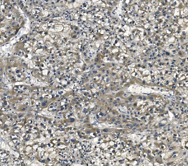
 IHC (Immunohistochemistry)
(Figure 8. IHC analysis of HBD using anti-HBD antibody (AAA19142).HBD was detected in paraffin-embedded section of rat liver tissue. Heat mediated antigen retrieval was performed in citrate buffer (pH6, epitope retrieval solution) for 20 mins. The tissue section was blocked with 10% goat serum. The tissue section was then incubated with 1ug/ml rabbit anti-HBD Antibody (AAA19142) overnight at 4 degree C. Biotinylated goat anti-rabbit IgG was used as secondary antibody and incubated for 30 minutes at 37 degree C. The tissue section was developed using Strepavidin-Biotin-Complex (SABC) with DAB as the chromogen.)
IHC (Immunohistochemistry)
(Figure 8. IHC analysis of HBD using anti-HBD antibody (AAA19142).HBD was detected in paraffin-embedded section of rat liver tissue. Heat mediated antigen retrieval was performed in citrate buffer (pH6, epitope retrieval solution) for 20 mins. The tissue section was blocked with 10% goat serum. The tissue section was then incubated with 1ug/ml rabbit anti-HBD Antibody (AAA19142) overnight at 4 degree C. Biotinylated goat anti-rabbit IgG was used as secondary antibody and incubated for 30 minutes at 37 degree C. The tissue section was developed using Strepavidin-Biotin-Complex (SABC) with DAB as the chromogen.)
HBD, Polyclonal Antibody (Cat# AAA19142)
Full Name
Anti-HBD Picoband Antibody
Reactivity
Human, Mouse, Rat
No cross reactivity with other proteins.
No cross reactivity with other proteins.
Applications
EIA, IHC, WB
Purity
Immunogen affinity purified
Pricing


 IHC (Immunohistchemistry)
(Figure 6. IHC analysis of S100A10 using anti-S100A10 antibody (AAA19162).S100A10 was detected in paraffin-embedded section of rat spleen tissue. Heat mediated antigen retrieval was performed in citrate buffer (pH6, epitope retrieval solution) for 20 mins. The tissue section was blocked with 10% goat serum. The tissue section was then incubated with 1ug/ml rabbit anti-S100A10 Antibody (AAA19162) overnight at 4 degree C. Biotinylated goat anti-rabbit IgG was used as secondary antibody and incubated for 30 minutes at 37 degree C. The tissue section was developed using Strepavidin-Biotin-Complex (SABC) with DAB as the chromogen.)
IHC (Immunohistchemistry)
(Figure 6. IHC analysis of S100A10 using anti-S100A10 antibody (AAA19162).S100A10 was detected in paraffin-embedded section of rat spleen tissue. Heat mediated antigen retrieval was performed in citrate buffer (pH6, epitope retrieval solution) for 20 mins. The tissue section was blocked with 10% goat serum. The tissue section was then incubated with 1ug/ml rabbit anti-S100A10 Antibody (AAA19162) overnight at 4 degree C. Biotinylated goat anti-rabbit IgG was used as secondary antibody and incubated for 30 minutes at 37 degree C. The tissue section was developed using Strepavidin-Biotin-Complex (SABC) with DAB as the chromogen.)
S100A10, Polyclonal Antibody (Cat# AAA19162)
Full Name
Anti-S100A10 Picoband antibody
Gene Names
S100A10; 42C; P11; p10; GP11; ANX2L; CAL1L; CLP11; Ca[1]; ANX2LG
Reactivity
Human, Mouse, Rat
No cross reactivity with other proteins.
No cross reactivity with other proteins.
Applications
EIA, IHC, WB
Pricing


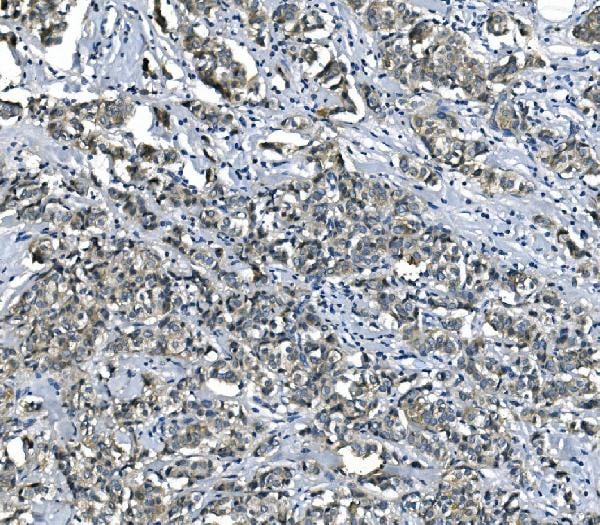
 IHC (Immunohistchemistry)
(Figure 6. IHC analysis of MAP1LC3A using anti-MAP1LC3A antibody (AAA19151).MAP1LC3A was detected in paraffin-embedded section of rat brain tissue. Heat mediated antigen retrieval was performed in citrate buffer (pH6, epitope retrieval solution) for 20 mins. The tissue section was blocked with 10% goat serum. The tissue section was then incubated with 1ug/ml rabbit anti-MAP1LC3A Antibody (AAA19151) overnight at 4 degree C. Biotinylated goat anti-rabbit IgG was used as secondary antibody and incubated for 30 minutes at 37 degree C. The tissue section was developed using Strepavidin-Biotin-Complex (SABC) with DAB as the chromogen.)
IHC (Immunohistchemistry)
(Figure 6. IHC analysis of MAP1LC3A using anti-MAP1LC3A antibody (AAA19151).MAP1LC3A was detected in paraffin-embedded section of rat brain tissue. Heat mediated antigen retrieval was performed in citrate buffer (pH6, epitope retrieval solution) for 20 mins. The tissue section was blocked with 10% goat serum. The tissue section was then incubated with 1ug/ml rabbit anti-MAP1LC3A Antibody (AAA19151) overnight at 4 degree C. Biotinylated goat anti-rabbit IgG was used as secondary antibody and incubated for 30 minutes at 37 degree C. The tissue section was developed using Strepavidin-Biotin-Complex (SABC) with DAB as the chromogen.)
MAP1LC3A, Polyclonal Antibody (Cat# AAA19151)
Full Name
Anti-MAP1LC3A Picoband antibody
Gene Names
MAP1LC3A; LC3; LC3A; ATG8E; MAP1ALC3; MAP1BLC3
Reactivity
Human, Mouse, Rat
No cross reactivity with other proteins.
No cross reactivity with other proteins.
Applications
EIA, IHC, WB
Pricing


 WB (Western Blot)
(Western Blot Analysis of Human Spleen cell lysate using HLA-Pan Mouse Monoclonal Antibody (CR3/43).)
WB (Western Blot)
(Western Blot Analysis of Human Spleen cell lysate using HLA-Pan Mouse Monoclonal Antibody (CR3/43).)
HLA-DP/-DQ/-DR, Monoclonal Antibody (Cat# AAA23889)
Full Name
HLA-DP/-DQ/-DR (MHC II)
Gene Names
HLA-DPB1; DPB1; HLA-DP; HLA-DPB; HLA-DP1B
Reactivity
Human
Applications
IF, IHC
Pricing


 IHC (Immunohistchemistry)
(Figure 6. IHC analysis of DYNLT1 using anti-DYNLT1 antibody (AAA19175).DYNLT1 was detected in paraffin-embedded section of rat lung tissue. Heat mediated antigen retrieval was performed in citrate buffer (pH6, epitope retrieval solution) for 20 mins. The tissue section was blocked with 10% goat serum. The tissue section was then incubated with 1ug/ml rabbit anti-DYNLT1 Antibody (AAA19175) overnight at 4 degree C. Biotinylated goat anti-rabbit IgG was used as secondary antibody and incubated for 30 minutes at 37 degree C. The tissue section was developed using Strepavidin-Biotin-Complex (SABC) with DAB as the chromogen.)
IHC (Immunohistchemistry)
(Figure 6. IHC analysis of DYNLT1 using anti-DYNLT1 antibody (AAA19175).DYNLT1 was detected in paraffin-embedded section of rat lung tissue. Heat mediated antigen retrieval was performed in citrate buffer (pH6, epitope retrieval solution) for 20 mins. The tissue section was blocked with 10% goat serum. The tissue section was then incubated with 1ug/ml rabbit anti-DYNLT1 Antibody (AAA19175) overnight at 4 degree C. Biotinylated goat anti-rabbit IgG was used as secondary antibody and incubated for 30 minutes at 37 degree C. The tissue section was developed using Strepavidin-Biotin-Complex (SABC) with DAB as the chromogen.)
DYNLT1, Polyclonal Antibody (Cat# AAA19175)
Full Name
Anti-DYNLT1 Picoband antibody
Gene Names
DYNLT1; CW-1; TCTEL1; tctex-1
Reactivity
Human, Mouse, Rat
No cross reactivity with other proteins.
No cross reactivity with other proteins.
Applications
EIA, IHC, WB
Pricing


 ICC (Immunocytochemistry)
(Figure 7. IHC analysis of ERAB using anti-ERAB antibody (AAA11668).ERAB was detected in immunocytochemical section of SMMC-7721 cell. Heat mediated antigen retrieval was performed in citrate buffer (pH6, epitope retrieval solution) for 20 mins. The tissue section was blocked with 10% goat serum. The tissue section was then incubated with 1ug/ml rabbit anti-ERAB Antibody (AAA11668) overnight at 4 degree C. Biotinylated goat anti-rabbit IgG was used as secondary antibody and incubated for 30 minutes at 37 degree C. The tissue section was developed using Strepavidin-Biotin-Complex (SABC) with DAB as the chromogen.)
ICC (Immunocytochemistry)
(Figure 7. IHC analysis of ERAB using anti-ERAB antibody (AAA11668).ERAB was detected in immunocytochemical section of SMMC-7721 cell. Heat mediated antigen retrieval was performed in citrate buffer (pH6, epitope retrieval solution) for 20 mins. The tissue section was blocked with 10% goat serum. The tissue section was then incubated with 1ug/ml rabbit anti-ERAB Antibody (AAA11668) overnight at 4 degree C. Biotinylated goat anti-rabbit IgG was used as secondary antibody and incubated for 30 minutes at 37 degree C. The tissue section was developed using Strepavidin-Biotin-Complex (SABC) with DAB as the chromogen.)
ERAB, Polyclonal Antibody (Cat# AAA11668)
Full Name
Anti-ERAB Antibody
Gene Names
HSD17B10; ABAD; CAMR; ERAB; HCD2; MHBD; HADH2; MRPP2; MRX17; MRX31; SCHAD; MRXS10; SDR5C1; 17b-HSD10; DUPXp11.22
Reactivity
Human, Mouse
Applications
WB, IHC
Purity
Immunogen Affinity Purified
Pricing


 IHC (Immunohistochemistry)
(Figure 7. IHC analysis of MYLK using anti-MYLK antibody (AAA19154).MYLK was detected in paraffin-embedded section of rat lung tissue. Heat mediated antigen retrieval was performed in citrate buffer (pH6, epitope retrieval solution) for 20 mins. The tissue section was blocked with 10% goat serum. The tissue section was then incubated with 1ug/ml rabbit anti-MYLK Antibody (AAA19154) overnight at 4 degree C. Biotinylated goat anti-rabbit IgG was used as secondary antibody and incubated for 30 minutes at 37 degree C. The tissue section was developed using Strepavidin-Biotin-Complex (SABC) with DAB as the chromogen. )
IHC (Immunohistochemistry)
(Figure 7. IHC analysis of MYLK using anti-MYLK antibody (AAA19154).MYLK was detected in paraffin-embedded section of rat lung tissue. Heat mediated antigen retrieval was performed in citrate buffer (pH6, epitope retrieval solution) for 20 mins. The tissue section was blocked with 10% goat serum. The tissue section was then incubated with 1ug/ml rabbit anti-MYLK Antibody (AAA19154) overnight at 4 degree C. Biotinylated goat anti-rabbit IgG was used as secondary antibody and incubated for 30 minutes at 37 degree C. The tissue section was developed using Strepavidin-Biotin-Complex (SABC) with DAB as the chromogen. )
MYLK, Polyclonal Antibody (Cat# AAA19154)
Full Name
Anti-MYLK Picoband antibody
Gene Names
MYLK; KRP; AAT7; MLCK; MLCK1; MYLK1; smMLCK; MLCK108; MLCK210; MSTP083
Reactivity
Human, Mouse, Rat
No cross reactivity with other proteins.
No cross reactivity with other proteins.
Applications
EIA, IHC, WB
Pricing


 FCM (Flow Cytometry)
(Figure 6. Flow Cytometry analysis of U937 cells using anti-BDH1 antibody (AAA19328).Overlay histogram showing U937 cells stained with AAA19328 (Blue line). The cells were blocked with 10% normal goat serum. And then incubated with rabbit anti-BDH1 Antibody (AAA19328,1μg/1x106 cells) for 30 min at 20 degree C. DyLight®488 conjugated goat anti-rabbit IgG (5-10μg/1x106 cells) was used as secondary antibody for 30 minutes at 20 degree C. Isotype control antibody (Green line) was rabbit IgG (1μg/1x106) used under the same conditions. Unlabelled sample (Red line) was also used as a control.)
FCM (Flow Cytometry)
(Figure 6. Flow Cytometry analysis of U937 cells using anti-BDH1 antibody (AAA19328).Overlay histogram showing U937 cells stained with AAA19328 (Blue line). The cells were blocked with 10% normal goat serum. And then incubated with rabbit anti-BDH1 Antibody (AAA19328,1μg/1x106 cells) for 30 min at 20 degree C. DyLight®488 conjugated goat anti-rabbit IgG (5-10μg/1x106 cells) was used as secondary antibody for 30 minutes at 20 degree C. Isotype control antibody (Green line) was rabbit IgG (1μg/1x106) used under the same conditions. Unlabelled sample (Red line) was also used as a control.)
BDH1, Polyclonal Antibody (Cat# AAA19328)
Full Name
Anti-BDH1 Antibody
Gene Names
BDH1; BDH; SDR9C1
Reactivity
Human, Rat
Applications
WB, IHC-P, ICC, IF, FC/FACS/FCM, EIA
Purity
Immunogen affinity purified.
Pricing


 FCM (Flow Cytometry)
(Figure 8. Flow Cytometry analysis of U87 cells using anti-PSMB5/MB1 antibody (AAA19274).Overlay histogram showing U87 cells stained with AAA19274 (Blue line). The cells were blocked with 10% normal goat serum. And then incubated with rabbit anti-PSMB5/MB1 Antibody (AAA19274, 1μg/1x106 cells) for 30 min at 20 degree C. DyLight®488 conjugated goat anti-rabbit IgG (5-10μg/1x106 cells) was used as secondary antibody for 30 minutes at 20 degree C. Isotype control antibody (Green line) was rabbit IgG (1μg/1x106) used under the same conditions. Unlabelled sample (Red line) was also used as a control.)
FCM (Flow Cytometry)
(Figure 8. Flow Cytometry analysis of U87 cells using anti-PSMB5/MB1 antibody (AAA19274).Overlay histogram showing U87 cells stained with AAA19274 (Blue line). The cells were blocked with 10% normal goat serum. And then incubated with rabbit anti-PSMB5/MB1 Antibody (AAA19274, 1μg/1x106 cells) for 30 min at 20 degree C. DyLight®488 conjugated goat anti-rabbit IgG (5-10μg/1x106 cells) was used as secondary antibody for 30 minutes at 20 degree C. Isotype control antibody (Green line) was rabbit IgG (1μg/1x106) used under the same conditions. Unlabelled sample (Red line) was also used as a control.)
PSMB5/MB1, Polyclonal Antibody (Cat# AAA19274)
Full Name
Anti-PSMB5/MB1 Antibody
Gene Names
PSMB5; X; MB1; LMPX
Reactivity
Human, Mouse, Rat
Applications
WB, IHC-P, ICC, IF, FC/FACS/FCM, EIA
Purity
Immunogen affinity purified.
Pricing
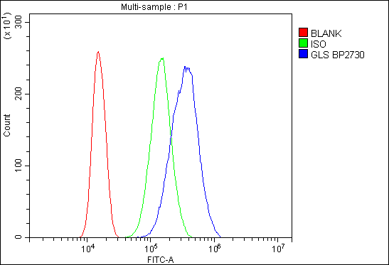


 FCM (Flow Cytometry)
(Figure 6. Flow Cytometry analysis of SiHa cells using anti-Glutaminase/GLS antibody (AAA19239).Overlay histogram showing SiHa cells stained with AAA19239 (Blue line). The cells were blocked with 10% normal goat serum. And then incubated with rabbit anti-Glutaminase/GLS Antibody (AAA19239,1μg/1x106 cells) for 30 min at 20 degree C. DyLight®488 conjugated goat anti-rabbit IgG (5-10μg/1x106 cells) was used as secondary antibody for 30 minutes at 20 degree C. Isotype control antibody (Green line) was rabbit IgG (1μg/1x106) used under the same conditions. Unlabelled sample (Red line) was also used as a control.)
FCM (Flow Cytometry)
(Figure 6. Flow Cytometry analysis of SiHa cells using anti-Glutaminase/GLS antibody (AAA19239).Overlay histogram showing SiHa cells stained with AAA19239 (Blue line). The cells were blocked with 10% normal goat serum. And then incubated with rabbit anti-Glutaminase/GLS Antibody (AAA19239,1μg/1x106 cells) for 30 min at 20 degree C. DyLight®488 conjugated goat anti-rabbit IgG (5-10μg/1x106 cells) was used as secondary antibody for 30 minutes at 20 degree C. Isotype control antibody (Green line) was rabbit IgG (1μg/1x106) used under the same conditions. Unlabelled sample (Red line) was also used as a control.)
Glutaminase/GLS, Polyclonal Antibody (Cat# AAA19239)
Full Name
Anti-Glutaminase/GLS Antibody
Gene Names
GLS; GAC; GAM; KGA; GLS1; AAD20
Reactivity
Human, Mouse, Rat, Monkey
Applications
WB, IHC-P, ICC, IF, FC/FACS/FCM, EIA
Purity
Immunogen affinity purified.
Pricing


 FCM (Flow Cytometry)
(Figure 8. Flow Cytometry analysis of C6 cells using anti-Clathrin heavy chain/CLTC antibody (AAA19270).Overlay histogram showing C6 cells stained with AAA19270 (Blue line). The cells were blocked with 10% normal goat serum. And then incubated with rabbit anti-Clathrin heavy chain/CLTC Antibody (AAA19270,1μg/1x106 cells) for 30 min at 20 degree C. DyLight®488 conjugated goat anti-rabbit IgG (5-10μg/1x106 cells) was used as secondary antibody for 30 minutes at 20 degree C. Isotype control antibody (Green line) was rabbit IgG (1μg/1x106) used under the same conditions. Unlabelled sample (Red line) was also used as a control.)
FCM (Flow Cytometry)
(Figure 8. Flow Cytometry analysis of C6 cells using anti-Clathrin heavy chain/CLTC antibody (AAA19270).Overlay histogram showing C6 cells stained with AAA19270 (Blue line). The cells were blocked with 10% normal goat serum. And then incubated with rabbit anti-Clathrin heavy chain/CLTC Antibody (AAA19270,1μg/1x106 cells) for 30 min at 20 degree C. DyLight®488 conjugated goat anti-rabbit IgG (5-10μg/1x106 cells) was used as secondary antibody for 30 minutes at 20 degree C. Isotype control antibody (Green line) was rabbit IgG (1μg/1x106) used under the same conditions. Unlabelled sample (Red line) was also used as a control.)
Clathrin heavy chain/CLTC, Polyclonal Antibody (Cat# AAA19270)
Full Name
Anti-Clathrin heavy chain/CLTC Antibody
Gene Names
CLTC; Hc; CHC; CHC17; CLH-17; CLTCL2
Reactivity
Human, Mouse, Rat
Applications
WB, IHC-P, ICC, IF, FC/FACS/FCM, EIA
Purity
Immunogen affinity purified.
Pricing


 FCM (Flow Cytometry)
(Figure 12. Flow Cytometry analysis of U937 cells using anti-DYNLL1/PIN antibody (AAA19276).Overlay histogram showing U937 cells stained with AAA19276 (Blue line). The cells were blocked with 10% normal goat serum. And then incubated with rabbit anti-DYNLL1/PIN Antibody (AAA19276, 1μg/1x106 cells) for 30 min at 20 degree C. DyLight®488 conjugated goat anti-rabbit IgG (5-10μg/1x106 cells) was used as secondary antibody for 30 minutes at 20 degree C. Isotype control antibody (Green line) was rabbit IgG (1μg/1x106) used under the same conditions. Unlabelled sample (Red line) was also used as a control.)
FCM (Flow Cytometry)
(Figure 12. Flow Cytometry analysis of U937 cells using anti-DYNLL1/PIN antibody (AAA19276).Overlay histogram showing U937 cells stained with AAA19276 (Blue line). The cells were blocked with 10% normal goat serum. And then incubated with rabbit anti-DYNLL1/PIN Antibody (AAA19276, 1μg/1x106 cells) for 30 min at 20 degree C. DyLight®488 conjugated goat anti-rabbit IgG (5-10μg/1x106 cells) was used as secondary antibody for 30 minutes at 20 degree C. Isotype control antibody (Green line) was rabbit IgG (1μg/1x106) used under the same conditions. Unlabelled sample (Red line) was also used as a control.)
DYNLL1/PIN, Polyclonal Antibody (Cat# AAA19276)
Full Name
Anti-DYNLL1/PIN Antibody
Gene Names
DYNLL1; LC8; PIN; DLC1; DLC8; LC8a; DNCL1; hdlc1; DNCLC1
Reactivity
Human, Mouse, Rat
Applications
WB, IHC-P, ICC, IF, FC/FACS/FCM, EIA
Purity
Immunogen affinity purified.
Pricing


 IF (Immunofluorescence)
(Figure 8. IF analysis of eRF1/ETF1 using anti- eRF1/ETF1 antibody (AAA19383).eRF1/ETF1 was detected in immunocytochemical section of A431 cells. Enzyme antigen retrieval was performed using IHC enzyme antigen retrieval reagent for 15 mins. The cells were blocked with 10% goat serum. And then incubated with 5μg/mL mouse anti- eRF1/ETF1 Antibody (AAA19383) overnight at 4 degree C. DyLight®488 Conjugated Goat Anti-Mouse IgG (BA1126) was used as secondary antibody at 1:100 dilution and incubated for 30 minutes at 37 degree C. The section was counterstained with DAPI. Visualize using a fluorescence microscope and filter sets appropriate for the label used.)
IF (Immunofluorescence)
(Figure 8. IF analysis of eRF1/ETF1 using anti- eRF1/ETF1 antibody (AAA19383).eRF1/ETF1 was detected in immunocytochemical section of A431 cells. Enzyme antigen retrieval was performed using IHC enzyme antigen retrieval reagent for 15 mins. The cells were blocked with 10% goat serum. And then incubated with 5μg/mL mouse anti- eRF1/ETF1 Antibody (AAA19383) overnight at 4 degree C. DyLight®488 Conjugated Goat Anti-Mouse IgG (BA1126) was used as secondary antibody at 1:100 dilution and incubated for 30 minutes at 37 degree C. The section was counterstained with DAPI. Visualize using a fluorescence microscope and filter sets appropriate for the label used.)
eRF1/ETF1, Monoclonal Antibody (Cat# AAA19383)
Full Name
Anti-eRF1/ETF1 Antibody (monoclonal, 3E5)
Gene Names
ETF1; ERF; RF1; ERF1; TB3-1; D5S1995; SUP45L1
Reactivity
Human, Mouse, Rat
Applications
WB, IHC-P, ICC, IF, FC/FACS/FCM
Purity
Immunogen affinity purified.
Pricing


 FCM (Flow Cytometry)
(Figure 8. Flow Cytometry analysis of C6 cells using anti-non-muscle Myosin IIB/MYH10 antibody (AAA19262).Overlay histogram showing C6 cells stained with AAA19262 (Blue line). The cells were blocked with 10% normal goat serum. And then incubated with rabbit anti-non-muscle Myosin IIB/MYH10 Antibody (AAA19262,1μg/1x106 cells) for 30 min at 20 degree C. DyLight®488 conjugated goat anti-rabbit IgG (5-10μg/1x106 cells) was used as secondary antibody for 30 minutes at 20 degree C. Isotype control antibody (Green line) was rabbit IgG (1μg/1x106) used under the same conditions. Unlabelled sample (Red line) was also used as a control.)
FCM (Flow Cytometry)
(Figure 8. Flow Cytometry analysis of C6 cells using anti-non-muscle Myosin IIB/MYH10 antibody (AAA19262).Overlay histogram showing C6 cells stained with AAA19262 (Blue line). The cells were blocked with 10% normal goat serum. And then incubated with rabbit anti-non-muscle Myosin IIB/MYH10 Antibody (AAA19262,1μg/1x106 cells) for 30 min at 20 degree C. DyLight®488 conjugated goat anti-rabbit IgG (5-10μg/1x106 cells) was used as secondary antibody for 30 minutes at 20 degree C. Isotype control antibody (Green line) was rabbit IgG (1μg/1x106) used under the same conditions. Unlabelled sample (Red line) was also used as a control.)
non-muscle Myosin IIB/MYH10, Polyclonal Antibody (Cat# AAA19262)
Full Name
Anti-non-muscle Myosin IIB/MYH10 Antibody
Gene Names
MYH10; NMMHCB; NMMHC-IIB
Reactivity
Human, Mouse, Rat
Applications
WB, IHC-P, ICC, IF, FC/FACS/FCM
Purity
Immunogen affinity purified.
Pricing


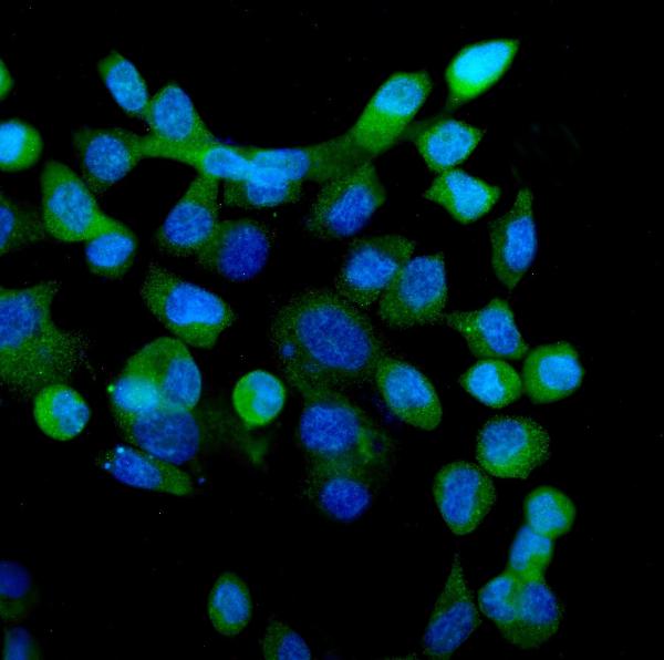
 IF (Immunofluorescence)
(Figure 11. IF analysis of Tubulin alpha using anti- Tubulin alpha antibody (AAA19381).Tubulin alpha was detected in immunocytochemical section of CACO-2 cells. Enzyme antigen retrieval was performed using IHC enzyme antigen retrieval reagent for 15 mins. The cells were blocked with 10% goat serum. And then incubated with 5μg/mL mouse anti- Tubulin alpha Antibody (AAA19381) overnight at 4 degree C. DyLight®594 Conjugated Goat Anti-Mouse IgG (BA1141) was used as secondary antibody at 1:100 dilution and incubated for 30 minutes at 37 degree C. The section was counterstained with DAPI. Visualize using a fluorescence microscope and filter sets appropriate for the label used.)
IF (Immunofluorescence)
(Figure 11. IF analysis of Tubulin alpha using anti- Tubulin alpha antibody (AAA19381).Tubulin alpha was detected in immunocytochemical section of CACO-2 cells. Enzyme antigen retrieval was performed using IHC enzyme antigen retrieval reagent for 15 mins. The cells were blocked with 10% goat serum. And then incubated with 5μg/mL mouse anti- Tubulin alpha Antibody (AAA19381) overnight at 4 degree C. DyLight®594 Conjugated Goat Anti-Mouse IgG (BA1141) was used as secondary antibody at 1:100 dilution and incubated for 30 minutes at 37 degree C. The section was counterstained with DAPI. Visualize using a fluorescence microscope and filter sets appropriate for the label used.)
Tubulin alpha, Monoclonal Antibody (Cat# AAA19381)
Full Name
Anti-Tubulin alpha Antibody (monoclonal, 7B12)
Gene Names
TUBA1A; LIS3; TUBA3; B-ALPHA-1
Reactivity
Human, Mouse, Rat
Applications
WB, IHC-P, ICC, IF, FC/FACS/FCM
Purity
Immunogen affinity purified.
Pricing
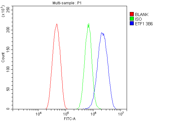


 FCM (Flow Cytometry)
(Figure 11. Flow Cytometry analysis of RH35 cells using anti- eRF1/ETF1 antibody (AAA19382).Overlay histogram showing RH35 cells stained with AAA19382 (Blue line). The cells were blocked with 10% normal goat serum. And then incubated with mouse anti-eRF1/ETF1 Antibody (AAA19382, 1μg/1x106 cells) for 30 min at 20 degree C. DyLight®488 conjugated goat anti-mouse IgG (BA1126, 5-10μg/1x106 cells) was used as secondary antibody for 30 minutes at 20 degree C. Isotype control antibody (Green line) was mouse IgG (1μg/1x106) used under the same conditions. Unlabelled sample (Red line) was also used as a control.)
FCM (Flow Cytometry)
(Figure 11. Flow Cytometry analysis of RH35 cells using anti- eRF1/ETF1 antibody (AAA19382).Overlay histogram showing RH35 cells stained with AAA19382 (Blue line). The cells were blocked with 10% normal goat serum. And then incubated with mouse anti-eRF1/ETF1 Antibody (AAA19382, 1μg/1x106 cells) for 30 min at 20 degree C. DyLight®488 conjugated goat anti-mouse IgG (BA1126, 5-10μg/1x106 cells) was used as secondary antibody for 30 minutes at 20 degree C. Isotype control antibody (Green line) was mouse IgG (1μg/1x106) used under the same conditions. Unlabelled sample (Red line) was also used as a control.)
eRF1/ETF1, Monoclonal Antibody (Cat# AAA19382)
Full Name
Anti-eRF1/ETF1 Antibody (monoclonal, 3B6)
Gene Names
ETF1; ERF; RF1; ERF1; TB3-1; D5S1995; SUP45L1
Reactivity
Human, Mouse, Rat
Applications
WB, IHC-P, ICC, IF, FC/FACS/FCM
Purity
Immunogen affinity purified.
Pricing


 FCM (Flow Cytometry)
(Figure 8. Flow Cytometry analysis of THP-1 cells using anti-NOX2/gp91phox/CYBB antibody (AAA19218).Overlay histogram showing THP-1 cells stained with AAA19218 (Blue line). The cells were blocked with 10% normal goat serum. And then incubated with rabbit anti-NOX2/gp91phox/CYBB Antibody (AAA19218, 1μg/1x106 cells) for 30 min at 20 degree C. DyLight®488 conjugated goat anti-rabbit IgG (5-10μg/1x106 cells) was used as secondary antibody for 30 minutes at 20 degree C. Isotype control antibody (Green line) was rabbit IgG (1μg/1x106) used under the same conditions. Unlabelled sample (Red line) was also used as a control.)
FCM (Flow Cytometry)
(Figure 8. Flow Cytometry analysis of THP-1 cells using anti-NOX2/gp91phox/CYBB antibody (AAA19218).Overlay histogram showing THP-1 cells stained with AAA19218 (Blue line). The cells were blocked with 10% normal goat serum. And then incubated with rabbit anti-NOX2/gp91phox/CYBB Antibody (AAA19218, 1μg/1x106 cells) for 30 min at 20 degree C. DyLight®488 conjugated goat anti-rabbit IgG (5-10μg/1x106 cells) was used as secondary antibody for 30 minutes at 20 degree C. Isotype control antibody (Green line) was rabbit IgG (1μg/1x106) used under the same conditions. Unlabelled sample (Red line) was also used as a control.)
NOX2/gp91phox/CYBB, Polyclonal Antibody (Cat# AAA19218)
Full Name
Anti-NOX2/gp91phox/CYBB Antibody
Gene Names
CYBB; CGD; NOX2; AMCBX2; GP91-1; GP91PHOX; p91-PHOX; GP91-PHOX
Reactivity
Human
Applications
WB, IHC-P, ICC, IF, FC/FACS/FCM, EIA
Purity
Immunogen affinity purified.
Pricing


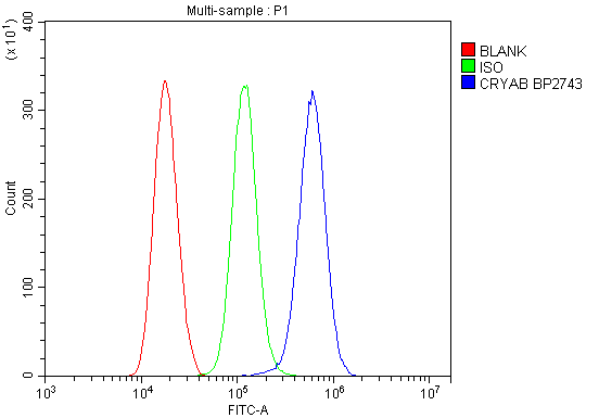 FCM (Flow Cytometry)
(Figure 13. Flow Cytometry analysis of THP-1 cells using anti-Alpha B Crystallin/CRYAB antibody (AAA19277).Overlay histogram showing THP-1 cells stained with AAA19277 (Blue line). The cells were blocked with 10% normal goat serum. And then incubated with rabbit anti-Alpha B Crystallin/CRYAB Antibody (AAA19277, 1μg/1x106 cells) for 30 min at 20 degree C. DyLight®488 conjugated goat anti-rabbit IgG (5-10μg/1x106 cells) was used as secondary antibody for 30 minutes at 20 degree C. Isotype control antibody (Green line) was rabbit IgG (1μg/1x106) used under the same conditions. Unlabelled sample (Red line) was also used as a control.)
FCM (Flow Cytometry)
(Figure 13. Flow Cytometry analysis of THP-1 cells using anti-Alpha B Crystallin/CRYAB antibody (AAA19277).Overlay histogram showing THP-1 cells stained with AAA19277 (Blue line). The cells were blocked with 10% normal goat serum. And then incubated with rabbit anti-Alpha B Crystallin/CRYAB Antibody (AAA19277, 1μg/1x106 cells) for 30 min at 20 degree C. DyLight®488 conjugated goat anti-rabbit IgG (5-10μg/1x106 cells) was used as secondary antibody for 30 minutes at 20 degree C. Isotype control antibody (Green line) was rabbit IgG (1μg/1x106) used under the same conditions. Unlabelled sample (Red line) was also used as a control.)
Alpha B Crystallin/CRYAB, Polyclonal Antibody (Cat# AAA19277)
Full Name
Anti-Alpha B Crystallin/CRYAB Antibody
Gene Names
CRYAB; CRYA2; CTPP2; HSPB5; CMD1II
Reactivity
Human, Mouse, Rat, Monkey
Applications
WB, IHC-P, ICC, IF, FC/FACS/FCM, EIA
Purity
Immunogen affinity purified.
Pricing


 Application Data
(Published customer image Infiltration of GFP+ BM-cells in infarct and peri-infarct regions. (A-B) Dot plots of viable macrophages/granulocytes (CD11b+CD45high, top right quadrants) and microglia (CD11b+CD45dim, bottom right quadrants) in cortex from BM-chimeric unmanipulated mice and mice exposed to pMCAO. (C) Bar graph showing mean numbers of CD11b+CD45dim microglia and CD11b+CD45high macrophages/granulocytes in BM-chimeric mice 24 hours after pMCAO, subdivided based on expression of GFP (n = 5). Approximately 92% of of the CD45high population were GFP+. (D) Estimation and comparison of mean numbers of CD11b+CD45dim microglia in non-chimeric (n = 10) versus BM-chimeric mice (n = 5) 24 hours after of pMCAO shows significantly fewer CD11b+CD45dim microglial cells in irradiated mice. (E) Overview, showing distribution of infiltrating GFP+ BM-derived cells into infarct (IF) and peri-infarct (P-IF) regions 24 hours after pMCAO. (E-G) By 24 hours, GFP+ single cells (F) and vessel-associated aggregates of GFP+ cells (arrows in G) were observed in infarct and peri-infarct regions. Some of the vessel-associated cells were round, leukocyte-like cells (arrows) while others were elongated cells lining the vasculature (arrow heads in G and in insert). (H) Bar graph showing mean numbers of single GFP+ cells and vessel-associated aggregates of GFP+ cells in ipsi- and contralateral cortex 24 hours after surgery (n = 10). (I-P) Immunohistochemical staining of CD45.1 (I, K), CD45.2 (J, L), IgG2a (M, O) and CD45 (N, P) in ischemic tissue in BM-chimeric (I, J, M, N) and non-chimeric mice (K, L, O, P) 24 hours after pMCAO. N.D, none detected. Scale bars: 200 um (A), 10 um (B, C). 50 um (I-P) *P < 0.05, **P < 0.01, and ***P < 0.001.From: Clausen BH, Lambertsen KL, Babcock AA, Holm TH, Dagnaes-Hansen F, Finsen B. Interleukin-1beta and tumor necrosis factor-alpha are expressed by different subsets of microglia and macrophages after ischemic stroke in mice. J Neuroinflammation. 2008 Oct 23;5:46.)
Application Data
(Published customer image Infiltration of GFP+ BM-cells in infarct and peri-infarct regions. (A-B) Dot plots of viable macrophages/granulocytes (CD11b+CD45high, top right quadrants) and microglia (CD11b+CD45dim, bottom right quadrants) in cortex from BM-chimeric unmanipulated mice and mice exposed to pMCAO. (C) Bar graph showing mean numbers of CD11b+CD45dim microglia and CD11b+CD45high macrophages/granulocytes in BM-chimeric mice 24 hours after pMCAO, subdivided based on expression of GFP (n = 5). Approximately 92% of of the CD45high population were GFP+. (D) Estimation and comparison of mean numbers of CD11b+CD45dim microglia in non-chimeric (n = 10) versus BM-chimeric mice (n = 5) 24 hours after of pMCAO shows significantly fewer CD11b+CD45dim microglial cells in irradiated mice. (E) Overview, showing distribution of infiltrating GFP+ BM-derived cells into infarct (IF) and peri-infarct (P-IF) regions 24 hours after pMCAO. (E-G) By 24 hours, GFP+ single cells (F) and vessel-associated aggregates of GFP+ cells (arrows in G) were observed in infarct and peri-infarct regions. Some of the vessel-associated cells were round, leukocyte-like cells (arrows) while others were elongated cells lining the vasculature (arrow heads in G and in insert). (H) Bar graph showing mean numbers of single GFP+ cells and vessel-associated aggregates of GFP+ cells in ipsi- and contralateral cortex 24 hours after surgery (n = 10). (I-P) Immunohistochemical staining of CD45.1 (I, K), CD45.2 (J, L), IgG2a (M, O) and CD45 (N, P) in ischemic tissue in BM-chimeric (I, J, M, N) and non-chimeric mice (K, L, O, P) 24 hours after pMCAO. N.D, none detected. Scale bars: 200 um (A), 10 um (B, C). 50 um (I-P) *P < 0.05, **P < 0.01, and ***P < 0.001.From: Clausen BH, Lambertsen KL, Babcock AA, Holm TH, Dagnaes-Hansen F, Finsen B. Interleukin-1beta and tumor necrosis factor-alpha are expressed by different subsets of microglia and macrophages after ischemic stroke in mice. J Neuroinflammation. 2008 Oct 23;5:46.)
CD11b, Monoclonal Antibody (Cat# AAA12181)
Full Name
RAT ANTI MOUSE CD11b
Gene Names
Itgam; CR3; CR3A; MAC1; Cd11b; Ly-40; Mac-1; Mac-1a; CD11b/CD18; F730045J24Rik
Reactivity
Human
Applications
FC/FACS, IF, IP
Pricing


 Application Data
(Published customer image Infiltration of GFP+ BM-cells in infarct and peri-infarct regions. (A-B) Dot plots of viable macrophages/granulocytes (CD11b+CD45high, top right quadrants) and microglia (CD11b+CD45dim, bottom right quadrants) in cortex from BM-chimeric unmanipulated mice and mice exposed to pMCAO. (C) Bar graph showing mean numbers of CD11b+CD45dim microglia and CD11b+CD45high macrophages/granulocytes in BM-chimeric mice 24 hours after pMCAO, subdivided based on expression of GFP (n = 5). Approximately 92% of of the CD45high population were GFP+. (D) Estimation and comparison of mean numbers of CD11b+CD45dim microglia in non-chimeric (n = 10) versus BM-chimeric mice (n = 5) 24 hours after of pMCAO shows significantly fewer CD11b+CD45dim microglial cells in irradiated mice. (E) Overview, showing distribution of infiltrating GFP+ BM-derived cells into infarct (IF) and peri-infarct (P-IF) regions 24 hours after pMCAO. (E-G) By 24 hours, GFP+ single cells (F) and vessel-associated aggregates of GFP+ cells (arrows in G) were observed in infarct and peri-infarct regions. Some of the vessel-associated cells were round, leukocyte-like cells (arrows) while others were elongated cells lining the vasculature (arrow heads in G and in insert). (H) Bar graph showing mean numbers of single GFP+ cells and vessel-associated aggregates of GFP+ cells in ipsi- and contralateral cortex 24 hours after surgery (n = 10). (I-P) Immunohistochemical staining of CD45.1 (I, K), CD45.2 (J, L), IgG2a (M, O) and CD45 (N, P) in ischemic tissue in BM-chimeric (I, J, M, N) and non-chimeric mice (K, L, O, P) 24 hours after pMCAO. N.D, none detected. Scale bars: 200 um (A), 10 um (B, C). 50 um (I-P) *P < 0.05, **P < 0.01, and ***P < 0.001.From: Clausen BH, Lambertsen KL, Babcock AA, Holm TH, Dagnaes-Hansen F, Finsen B. Interleukin-1beta and tumor necrosis factor-alpha are expressed by different subsets of microglia and macrophages after ischemic stroke in mice. J Neuroinflammation. 2008 Oct 23;5:46.)
Application Data
(Published customer image Infiltration of GFP+ BM-cells in infarct and peri-infarct regions. (A-B) Dot plots of viable macrophages/granulocytes (CD11b+CD45high, top right quadrants) and microglia (CD11b+CD45dim, bottom right quadrants) in cortex from BM-chimeric unmanipulated mice and mice exposed to pMCAO. (C) Bar graph showing mean numbers of CD11b+CD45dim microglia and CD11b+CD45high macrophages/granulocytes in BM-chimeric mice 24 hours after pMCAO, subdivided based on expression of GFP (n = 5). Approximately 92% of of the CD45high population were GFP+. (D) Estimation and comparison of mean numbers of CD11b+CD45dim microglia in non-chimeric (n = 10) versus BM-chimeric mice (n = 5) 24 hours after of pMCAO shows significantly fewer CD11b+CD45dim microglial cells in irradiated mice. (E) Overview, showing distribution of infiltrating GFP+ BM-derived cells into infarct (IF) and peri-infarct (P-IF) regions 24 hours after pMCAO. (E-G) By 24 hours, GFP+ single cells (F) and vessel-associated aggregates of GFP+ cells (arrows in G) were observed in infarct and peri-infarct regions. Some of the vessel-associated cells were round, leukocyte-like cells (arrows) while others were elongated cells lining the vasculature (arrow heads in G and in insert). (H) Bar graph showing mean numbers of single GFP+ cells and vessel-associated aggregates of GFP+ cells in ipsi- and contralateral cortex 24 hours after surgery (n = 10). (I-P) Immunohistochemical staining of CD45.1 (I, K), CD45.2 (J, L), IgG2a (M, O) and CD45 (N, P) in ischemic tissue in BM-chimeric (I, J, M, N) and non-chimeric mice (K, L, O, P) 24 hours after pMCAO. N.D, none detected. Scale bars: 200 um (A), 10 um (B, C). 50 um (I-P) *P < 0.05, **P < 0.01, and ***P < 0.001.From: Clausen BH, Lambertsen KL, Babcock AA, Holm TH, Dagnaes-Hansen F, Finsen B. Interleukin-1beta and tumor necrosis factor-alpha are expressed by different subsets of microglia and macrophages after ischemic stroke in mice. J Neuroinflammation. 2008 Oct 23;5:46.)
CD11b, Monoclonal Antibody (Cat# AAA12185)
Full Name
RAT ANTI MOUSE CD11b
Gene Names
Itgam; CR3; CR3A; MAC1; Cd11b; Ly-40; Mac-1; Mac-1a; CD11b/CD18; F730045J24Rik
Applications
FC/FACS, IF, IP
Pricing


 WB (Western Blot)
(FG Pancreatic Carcinoma Cell Lines stably expressing vector along (FG-V) the b3 integrin subunit (FG-b3) or a b3 truncation mutant (FG-759x). Src Mab (AAA28639) was diluted 1:500 in 1% BSA/TBST and incubated Overnight at 4 degree C. After washing 3x 5 min. with TBST the blots were incubated with 1:5000 Goat anti-mouse or Goat anti-rabbit secondary antibody for 1 hr at Room temperature. The blots were again washed 3x 5 min. with TBST and developed using ECL reagent.Data and protocol kindly provided by Dr. Weis of Cheresh Lab, UCSD.)
WB (Western Blot)
(FG Pancreatic Carcinoma Cell Lines stably expressing vector along (FG-V) the b3 integrin subunit (FG-b3) or a b3 truncation mutant (FG-759x). Src Mab (AAA28639) was diluted 1:500 in 1% BSA/TBST and incubated Overnight at 4 degree C. After washing 3x 5 min. with TBST the blots were incubated with 1:5000 Goat anti-mouse or Goat anti-rabbit secondary antibody for 1 hr at Room temperature. The blots were again washed 3x 5 min. with TBST and developed using ECL reagent.Data and protocol kindly provided by Dr. Weis of Cheresh Lab, UCSD.)
SRC, Monoclonal Antibody (Cat# AAA28639)
Full Name
SRC Antibody
Gene Names
SRC; ASV; SRC1; c-SRC; p60-Src
Reactivity
Human, mouse
Applications
WB, EIA, IF
Purity
This antibody is purified through a protein G column, followed by dialysis against PBS.
Pricing